Here, There, and Everywhere: The Wide Host Range and Geographic Distribution of Zoonotic Orthopoxviruses - MDPI
←
→
Page content transcription
If your browser does not render page correctly, please read the page content below
viruses
Review
Here, There, and Everywhere: The Wide Host Range and
Geographic Distribution of Zoonotic Orthopoxviruses
Natalia Ingrid Oliveira Silva, Jaqueline Silva de Oliveira, Erna Geessien Kroon                             , Giliane de Souza Trindade and
Betânia Paiva Drumond *

                                           Laboratório de Vírus, Departamento de Microbiologia, Instituto de Ciências Biológicas,
                                           Universidade Federal de Minas Gerais: Belo Horizonte, Minas Gerais 31270-901, Brazil;
                                           nios2402@ufmg.br (N.I.O.S.); jaquelinebmedica@hotmail.com (J.S.d.O.); ernagkroon@gmail.com (E.G.K.);
                                           giliane@icb.ufmg.br (G.d.S.T.)
                                           * Correspondence: betaniadrumond@ufmg.br

                                           Abstract: The global emergence of zoonotic viruses, including poxviruses, poses one of the greatest
                                           threats to human and animal health. Forty years after the eradication of smallpox, emerging zoonotic
                                           orthopoxviruses, such as monkeypox, cowpox, and vaccinia viruses continue to infect humans
                                           as well as wild and domestic animals. Currently, the geographical distribution of poxviruses in
                                           a broad range of hosts worldwide raises concerns regarding the possibility of outbreaks or viral
                                           dissemination to new geographical regions. Here, we review the global host ranges and current
                                           epidemiological understanding of zoonotic orthopoxviruses while focusing on orthopoxviruses with
                                           epidemic potential, including monkeypox, cowpox, and vaccinia viruses.

                                 Keywords: Orthopoxvirus; Poxviridae; zoonosis; Monkeypox virus; Cowpox virus; Vaccinia virus; host

                                           range; wild and domestic animals; emergent viruses; outbreak
Citation: Silva, N.I.O.; de Oliveira,
J.S.; Kroon, E.G.; Trindade, G.d.S.;
Drumond, B.P. Here, There, and
Everywhere: The Wide Host Range            1. Poxvirus and Emerging Diseases
and Geographic Distribution of
                                                Zoonotic diseases, defined as diseases or infections that are naturally transmissible
Zoonotic Orthopoxviruses. Viruses
                                           from vertebrate animals to humans, represent a significant threat to global health [1].
2021, 13, 43. https://doi.org/
10.3390/v13010043
                                           Among the species recognized as pathogenic to humans, more than half originated in
                                           animals, and some have been characterized as emerging or re-emerging pathogens [2,3].
Academic Editors: Jônatas Abrahão
                                           Most zoonotic pathogens originated in wild and domesticated mammalian hosts such as
and Hermann Meyer                          bats, rodents, and primates [4]. The analysis of global trends indicates that new zoonotic
Received: 14 November 2020                 threats will continue to emerge at an accelerating rate, and are mainly associated with an
Accepted: 24 December 2020                 growthing population, changes in land use, climate changes, increased intercontinental
Published: 30 December 2020                travel, and expanded trade networks [4,5].
                                                Poxviruses are of great veterinary and human importance and infect numerous
Publisher’s Note: MDPI stays neu-          vertebrate and invertebrate animals, including humans. The Poxviridae family is di-
tral with regard to jurisdictional clai-   vided into two subfamilies, namely: Chordopoxvirinae, which infect vertebrates, and Ento-
ms in published maps and institutio-       mopoxvirinae (A–C), which infect invertebrates. The Chordopoxvirinae subfamily is further
nal affiliations.                          divided into 18 genera (Avipoxvirus, Capripoxvirus, Centapoxvirus, Cervidpoxvirus, Crocodylid-
                                           poxvirus, Leporipoxvirus, Macropopoxvirus, Molluscipoxvirus, Mustelpoxvirus, Orthopoxvirus,
                                           Oryzopoxvirus, Parapoxvirus, Pteropopoxvirus, Salmonpoxvirus, Sciuripoxvirus, Suipoxvirus,
                                           Vespertilionpoxvirus, and Yatapoxvirus), distinguishable by their serological reactions [6,7].
Copyright: © 2020 by the authors. Li-
censee MDPI, Basel, Switzerland.
                                                The family Poxviridae comprises large, brick-shaped or ovoid enveloped viruses
This article is an open access article
                                           containing a linear, double-stranded DNA genome approximately 200 kilobase pairs in
distributed under the terms and con-       length [7,8]. Poxviruses are among mankind’s longest and best-known viruses mainly
ditions of the Creative Commons At-        because of their most feared and lethal representative, Variola virus (VARV), the causative
tribution (CC BY) license (https://        agent of smallpox. Before its remarkable eradication in 1980, VARV represented a centuries-
creativecommons.org/licenses/by/           old threat to humans worldwide and killed approximately 300–500 million people during
4.0/).                                     the 20th century [9]. The global eradication of smallpox marked the culmination of an

Viruses 2021, 13, 43. https://doi.org/10.3390/v13010043                                                     https://www.mdpi.com/journal/viruses
Viruses 2021, 13, 43 2 of 20
Viruses 2021, 13, x FOR PEER REVIEW 2 of 21
of an intensive
intensive vaccination
vaccination program
program and quarantine
and quarantine measures
measures promoted
promoted by the by the World
World Health
Health
Organization (WHO) [8,10,11]. Although VARV was eradicated 40 years ago, many ago,
Organization (WHO) [8,10,11]. Although VARV was eradicated 40 years chal-
many
lengeschallenges
regardingregarding poxvirus infections
poxvirus infections persist, the
persist, including including the worrisome
worrisome possibility possibil-
of VARV
ity of VARV reintroduction
reintroduction by accidentalby accidental
release, its userelease, its use as
as a biological a biological
weapon, or theweapon,
emergence or and
the
emergence
re-emergence andofre-emergence of zoonotic orthopoxviruses
zoonotic orthopoxviruses worldwide [12,13].worldwide [12,13].
Orthopoxviruses
Orthopoxvirusesare areremarkable
remarkablefor fortheir
theirwide
widehost
hostspectrum,
spectrum,ranging
rangingfrom
fromhumans
humans
to
todomestic
domesticand andwild
wild animals
animals(Figure
(Figure 1).
1). Orthopoxvirus
Orthopoxvirusisisthethemost
mostimportant
importantand andwell-
well-
characterized
characterizedpoxvirus
poxvirusgenus,
genus,mainly
mainlydueduetotoits
itsimpact
impacton onhuman
humanand andanimal
animalhealth
health[7,8].
[7,8].
Here, we
Here, we review
review the
the major
major aspects
aspects related
related toto the
the dynamics
dynamics and and emergence
emergence of of zoonotic
zoonotic
orthopoxvirusinfections
orthopoxvirus infectionsworldwide,
worldwide,focusing
focusingon onthe
thehost
hostrange
rangeandandcurrent
currentepidemio-
epidemio-
logicalsituation
logical situationrelating
relatingto
tomonkeypox
monkeypox(MPXV),
(MPXV),cowpox
cowpox(CPXV),
(CPXV),vaccinia
vaccinia(VACV),
(VACV), and
and
VACV-like viruses.
VACV-like
Theworldwide
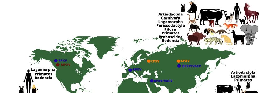
Figure1.1.The
Figure worldwidedistribution
distributionand
andhost
hostrange
rangeof
ofmonkeypox,
monkeypox,cowpox
cowpox and
and vaccinia
vaccinia viruses.
viruses.
Theimage
The imageshows
showsthetherange
rangeofof animal
animal hosts
hosts (represented
(representedby by orders)
orders) that
that have
have been
been
demonstrated to be naturally infected by some Orthopoxvirus species, according
demonstrated to be naturally infected by some Orthopoxvirus species, according to differ- to different
regions
ent of the
regions world
of the (except
world by Monkeypox
(except by Monkeypox virusvirus
in theinUnited StatesStates
the United of America, repre-
of America,
sented by imported cases). Orthopoxvirus infections have been demonstrated
represented by imported cases). Orthopoxvirus infections have been demonstrated in an- in animals
belonging
imals to different
belonging orders,orders,
to different using different methodsmethods
using different (virus isolation, molecularmolecular
(virus isolation, detection
of viral genomes or serological screening for antibodies against orthopoxviruses).
detection of viral genomes or serological screening for antibodies against orthopox- The oc-
currence of some zoonotic orthopoxviruses has already been confirmed
viruses). The occurrence of some zoonotic orthopoxviruses has already been confirmed(by virus isolation
or molecular
(by detection
virus isolation of the viral
or molecular genome)
detection in some
of the viral geographical
genome) in some regions (indicatedre-
geographical by
colored dots: blue: vaccinia virus (including buffalopox and rabbitpox viruses)
gions (indicated by colored dots: blue: vaccinia virus (including buffalopox and rabbitpox in South
America, Europe, Asia, and the Middle East; brown: monkeypox virus in Africa and North
viruses) in South America, Europe, Asia, and the Middle East; brown: monkeypox virus
America; orange: cowpox virus in Europe and Asia).
in Africa and North America; orange: cowpox virus in Europe and Asia).Viruses 2021, 13, 43 3 of 20
2. Orthopoxvirus
The Orthopoxvirus genus comprises VARV, VACV, CPXV, MPXV, camelpox virus, Akhmeta
virus, and other species with zoonotic potential. All orthopoxviruses share significant DNA
sequence similarity and are immunologically cross-reactive and cross-protective. Infection
with any orthopoxvirus is considered to generate protection against exposure or re- expo-
sure to any other member of the genus [14,15]. Orthopoxvirus species are named primarily
according to the hosts from which they were first isolated and identified; however, the
name does not necessarily represents its natural reservoir or complete host range [8,16–19].
Despite the large number of studies, little is known about the primary hosts and reservoirs
of zoonotic orthopoxviruses in nature, or their transmission and maintenance cycles [20].
Regarding the host range, orthopoxviruses can be both highly specialized and host re-
stricted or generalists with a broad host range. For instance, VARV is a highly specialized
virus that infects only humans, whereas MPXV, CPXV, and VACV are examples of gener-
alist zoonotic orthopoxviruses that can infect several mammalian host species and also
spillover into humans [20].
The evolution of generalists pathogens requires the successful crossing of host trans-
mission barriers [21]. These include geographical, ecological, and behavioral constraints
that separate a virus from its possible recipient hosts; virus-host cell incompatibility, such
as tissue tropism, differences in receptor binding, genome replication, production, and
shedding of infectious particles; and host immunity evasion, which includes cellular bar-
riers or responses that restrict the infection and/or evasion of a virus from the innate
immune system of its host [22]. To overcome these barriers, orthopoxviruses have dif-
ferent biological features that can synergistically contribute to the transmission to, and
exploitation of, a broad range of new hosts species as observed for CXPV, MPXV, and VACV.
Orthopoxviruses can cause both local lesions on the skin and systemic infections, resulting
in direct and indirect transmission routes. When accompanied by viral particle stability
in the environment, this can increase the likelihood of potential hosts being exposed to
the virus independently of direct contact with infected hosts. In addition, orthopoxviruses
can infect a variety of mammalian cells in a manner that is mostly independent of species-
specific receptors and have large genomes that carry the information essential for viral
replication, thereby increasing the possibility of successful infection in a new cell/host.
Although the double stranded DNA genomes of orthopoxviruses have low mutation rates
when compared with other viruses, such as RNA viruses, orthopoxviruses possess a genetic
arsenal comprising several immune-regulatory, virulence, and host range genes [20]. The
variety of host-genes among poxviruses enables them to express different viral proteins
with important roles in cell tropism, as well as in the modulation of host signaling pathways
and immunomodulatory responses, thereby establishing optimal cellular conditions for
viral replication [23]. Finally, many of the strategies employed by orthopoxviruses to evade
host immune defenses target conserved elements of the immune system in different poten-
tial hosts [20]. Combined, these features altogether are crucial for virus-cell and virus-host
interactions and can contribute to the success of viral replication and transmission.
Despite the eradication of smallpox, the possibility of its re-emergence or the emer-
gence of other orthopoxviruses in human and animal populations is a relevant global
health issue. As smallpox vaccination is no longer mandatory, most of the world’s popu-
lation that is under 40 years of age lacks immunity against orthopoxviruses [24,25]. This
scenario is highlighted by numerous reports in recent years of human diseases caused by
zoonotic orthopoxviruses such as MPXV [26–33], CPXV [34–41], VACV-like [42–49], and
Akhmeta virus [18]. To date, the circulation of orthopoxviruses among wild and domestic
animals has been recorded in different regions of the world, including South America,
Africa, Europe, the Middle East, and Asia [27,40,42,43,50–57]. These facts raise concerns
regarding the host ranges and distribution of orthopoxviruses, as well as their potential to
cause outbreaks in animals and human populations, thereby further impacting animal and
public health.Viruses 2021, 13, 43 4 of 20
2.1. Monkeypox Virus
Monkeypox virus isolates are subdivided into two clades, namely, the West African
and the Congo Basin clades, based on genetic and phenotypic (virulence) differences [58].
Notably, several studies have indicated that the clinical signs are similar between infections
caused by viruses from either clades [59]. The first observation of MPXV infection was
reported in 1958 during an outbreak of pustular rash illness in cynomolgus macaques
(Macaca fascicularis) arriving in Copenhagen, Denmark, from Singapore [60]. Despite
being named after the first described host, non-human primates are accidental hosts for
MPXV [61].
Further insights into the range of taxa susceptible to MPXV infection were obtained
by laboratory studies and field surveys. MPXV infections have been reported in a broad
range of rodents, such as mice (Mus musculus), rabbits (Oryctolagus cuniculus), hamsters,
woodchucks (Marmota monax), jerboas (Jaculus sp.), and porcupines (Atherurus africanus)
(Table 1). Similarly, based on methods such as viral isolation, molecular assay, or experimen-
tal infection, susceptibility to MPXV infection was reported in ant-eaters (Myrmecophaga
tridactyla), black-tailed prairie dogs (Cynomys ludovicianus), southern opossums (Didelphis
marsupialis), short-tailed opossums (Monodelphis domestica), African hedgehogs (Atelerix sp.),
and several non-human primate species. Additionally, serological surveys have implicated
several African rodents, including giant pouched rats (Cricetomys spp.), African dormices
(Graphiurus spp.), rope squirrels (Funisciurus spp.), and sun squirrels (Heliosciurus spp.) as
primary orthopoxvirus hosts in Africa [61–63].
Among Old World non-human primates, cynomolgus monkeys (Macaca fascicularis),
sooty mangabeys (Cercocebus atys), orangutans (Pongo pygmaeus), and chimpanzees (Pan
troglodytes) can be infected with MPXV. Among New World non-human primates [60,64–75],
the common marmosets (Callithrix jacchus) was shown to be susceptible to MPXV infection
through intravenous inoculation [76] (Table 1).
In 2003, a MPXV outbreak occurred in the United States of America (USA). Human
infection was associated with direct contact with ill pet prairie dogs that were kept near
to infected exotic animals imported from Ghana, West Africa [77]. This episode, as well
as the infection of rodents, heightened concerns regarding the introduction of MPXV into
the Americas. Meanwhile, the susceptibility of several African rodents to MPXV raised
worries about the transmission of the virus to humans, as these animals are sometimes
kept as pets [78,79].
Although humans are also accidental hosts [61], MPXV became the most significant
pathogenic zoonotic orthopoxvirus for humans since the eradication of smallpox, given its
associated morbidity (systemic infection) and lethality. The first human MPXV infection
was described in 1970 for a 9-month old child in the Democratic Republic of Congo who had
presented smallpox-like skin eruptions [70,80]. Several other human cases were reported
in the following years. From 1970 to 1999, the WHO reported at least 404 confirmed and
approximately 500 suspected human cases of monkeypox in different African countries
(Central African Republic, Cameroon, Nigeria, Côte d’Ivoire, Liberia, Sierra Leone, and
Gabon), but mainly in the Democratic Republic of Congo [52,81,82]. From the 2000s,
alongside outbreaks in the Democratic Republic of Congo, the Republic of Congo, and
South Sudan, the first human cases outside the Africa continent were also described.
During May and June of 2003, cases of people with febrile illness and skin eruptions were
notified to the Wisconsin Division of Public Health, but no deaths were reported and no
person-to-person transmission was proven [78]. The source of this outbreak was traced
back to imported infected exotic animals from Ghana [52,62,78]. Fortunately, the multi-state
episode of captive rodent infection in the USA was short-lived, and the transmission cycle
in the country was broken [83].Viruses 2021, 13, 43 5 of 20
Table 1. Hosts and susceptible animals to monkeypox virus infection.
Method of Association to
Order/Family Species
Investigation * Human Infection **
Humans (Homo sapiens) viral isolation yes
Primates/ Orangutans (Pongo pygmaeus) viral isolation yes
Hominidae Chimpanzees (Pan troglodytes) viral isolation no
PCR/
Primates/ Sooty mangabeys (Cercocebus atys) no
viral isolation
Cercopithecidae
Cynomolgus monkeys (Macaca fascicularis) viral isolation yes
Primates/
White-tufted marmosets (Callithrix jacchus) Lab. Infec. no
Callithrichidae
Rodentia/Chinchillidae Rabbits (Oryctolagus cuniculus) Lab. Infec. no
Rodentia/Muridae Inbred mouses (Mus musculus) Lab. Infec. no
Rodentia/Cricetidae hamsters Lab. Infec. no
PCR/
Rodentia/Nesomyidae Giant-pouched rats (Cricetomys sp.) no
viral isolation
PCR/
Rodentia/Gliridae African dormices (Graphiurus sp.) no
viral isolation
PCR/
Rope squirrels (Funisciurus sp.) yes
viral isolation
Rodentia/Sciuridae Black-tailed prairie dogs (Cynomys ludovicianus) PCR yes
PCR/
Woodchucks (Marmota monax) no
viral isolation
Rodentia/ PCR/
Jerboas (Jaculus sp.) no
Dipodidae viral isolation
PCR/
Rodentia/Hystricidae Porcupines (Atherurus africanus) no
viral isolation
Pilosa/Macroscelididae Ant-eaters (Myrmecophaga tridactyla) viral Isolation no
Southern opossums (Didelphis PCR/
no
Didelphimorphia/ marsupialis) viral isolation
Didelphidae Shot-tailed opossums (Monodelphis PCR/
no
domestica) viral isolation
Erinaceomorpha/ PCR/
African hedgehogs (Atelerix sp.) no
Erinaceidae viral isolation
* Method of investigation: viral infection demonstrated by molecular assay (PCR) or viral isolation using samples obtained from naturally
infected animals; Lab. Infec.: MPXV infection susceptibility was observed during experimental studies in laboratory. ** Transmission to
humans already reported in the literature.
Alarmingly, several outbreaks of monkeypox in humans have been reported in African
regions in the last decade. In 2010, two confirmed and eight suspected cases were described
in the Republic of Congo related to the migration of refugees, regional inter-ethnic conflicts,
or autochthonous cases. No deaths were reported among the confirmed cases, although
one individual with suspected infection died [84]. In the same year, two cases of MPXV
infection associated with hunting and the consumption of wild rodent meat were reported
in the Central African Republic, with no deaths [85]. Numerous suspected and confirmed
cases were reported in the Democratic Republic of Congo, from 2010 to 2016 [86,87], and
in Serra Leone in 2014 [88]. Several suspected and 12 confirmed cases, as well as three
deaths were reported in different provinces in the Central African Republic (Mbomou,
Basse-Kotto, and Haute-Kotto) [52,89,90]. In 2017, the Republic of Congo reported its
largest MPXV outbreak (88 suspected and seven confirmed cases, with six deaths), which
affected 18 villages in five districts. This outbreak presented risks of MPXV spreading to
neighboring countries given the extent of population mobility and refugee presence in the
region [30].Viruses 2021, 13, 43 6 of 20
Some African regions have continuously reported human cases of MPXV infection
in recent years (2017 to 2020), including the Central African Republican (27 confirmed
cases and two deaths) [91,92], Nigeria (181 confirmed cases and seven deaths) [31,93],
Sierra Leone (one confirmed case) [94], Liberia (two confirmed cases and two deaths) [95],
Cameroon (one confirmed case) [96], and the Democratic Republic of Congo (numerous
confirmed cases and 321 deaths) [33,97]. Recently (2018), three cases of monkeypox were
reported in the United Kingdom. Two were of people who had traveled to Nigeria, while
the third concerned a health care worker who had had contact with one infected patient.
One of the patients who had traveled to Nigeria reported having contact with a person
with a rash and the possible consumption of bushmeat, raising the possibility that this may
have been a case of secondary or even tertiary human-to-human transmission. Meanwhile,
the infection contracted by the British care worker confirms human-to-human MPXV
transmission [96]. Other cases of MPXV infections outside of Africa were reported in Israel
(2018) and Singapore (2019), for travelers who imported the disease from Nigeria [98,99].
The natural source of MPXV and its maintenance cycle in nature remains unknown as
the virus has only been isolated twice in nature (wild animals): once from the rope squirrel
(Funisciurus anerythrus), Zaire, in 1985 [62], and once from the sooty mangabey (Cercocebus
atys), Côte d’Ivoire, in 2012 [100]. To date, naturally occurring MPXV infections remain
confined to the forest regions of West and Central Africa [101,102]. Consequently, a higher
proportion of human MPXV cases are reported in regions (mainly African villages) where
humans and non-human primates live in close proximity. The consumption, hunting, and
handling of meat derived from non-human primates, rodents, and other small mammals
have also been associated with human cases of MPXV infection [71,85,86,103–105]. Close
contact with rodents has also been implicated as a source of human infection [67,106].
Human cases of monkeypox have been increasing even though they may have been
underreported. Notably, diagnostic capabilities in the affected countries are most often lim-
ited, while health care workers worldwide are generally not aware of monkeypox disease.
A lack of understanding about monkeypox disease associated with factors such as the in-
creasing encroachment of humans into wild habitats, the inter-continental travel of people
from endemic areas to MPXV-free regions, and the importation of animals both as pets and
for laboratory studies raises concern regarding MPXV emergence, surveillance, prevention,
and control [15]. Additionally, vaccination against smallpox was ceased decades ago,
resulting in an increasingly larger number of people that are vulnerable to infection by
MPXV or other orthopoxviruses. Although some animal species have been described as
being susceptible to MPXV infection, most of what is known about its pathogenesis and
clinical characteristics is derived from descriptions of animals in captivity or laboratory
facilities. As monkeypox is an emerging zoonotic disease with epidemic potential and
much of its host range and maintenance cycle in nature remains obscure, advances are
urgently needed to better understand natural cycle of MPXV.
2.2. Cowpox Virus
Edward Jenner was the first to document CPXV infection after observing local le-
sions on the teats of cows, which he called “cow-pox”. Then, in 1798, Jenner demonstrated
the efficacy of “true cow-pox” scarification in inducing immunity against smallpox [8,107].
There were frequent reports of bovine cowpox cases until the early 1970s in Europe, with
sporadic transmission to humans, mainly milkers, occurring via contact with infected
cows [108]. However, the number of bovine cowpox cases decreased, while reports of
“cowpox-like” infections in several animal species, such as cats and elephants, [109] in-
creased. “Cowpox-like” infections were described in a broad range of captive and domes-
tic animals like non-human primates [110–112], felines [108,111,113–117], dogs [118], ro-
dents [39,50,111,119–125], foxes [126,127], rhinoceroses [15,114,128], tapirs [129], okapis [130],
horses [131], anteaters [114], mongooses [129], stone martens [132], bearcats [133], and
farmed llamas [134,135] (Table 2). The viruses responsible for these infections induced
clinical signs similar to those of CPXV infection such as hemorrhagic pocks on the chorioal-Viruses 2021, 13, 43 7 of 20
lantoic membrane and A-type inclusions bodies, and were thus considered to be “true
cowpox” [136,137]. Most of these animals are thought to be accidental hosts for CPXV, and
not reservoirs. Rodents, particularly voles (Microtus spp. and Myodes spp.), are known to
be the primary CPXV reservoirs in nature [136,138].
Table 2. Hosts and susceptible animals to cowpox virus infection.
Method of Association to Human
Order/Family Species
Investigation * Infection **
Primates/Hominidae Humans (Homo sapiens) virus isolation no
Primates/Callithrichidae White-tufted marmosets (Callithrix jacchus) virus isolation no
Barbary macaques (Macaca sylvanus) virus isolation no
Primates/Cercopithecidae Cynomolgus macaques (Macaca fascicularis) Lab. Infec. no
Rhesus macaques (Macaca mulata) Lab. Infec. no
Domestic cats (Felis catus) virus isolation yes
Cheetahs (Acinonyx jubatus) virus isolation yes
Lions (Panthera leo) virus isolation no
Carnivora/Felidae Pumas (Felis concolor) virus isolation no
Black panthers (Panthera padus) virus isolation no
Jaguarundis (Herpailurus yaguarondi) virus isolation no
Jaguares (Felis onca) virus isolation no
Dogs (Canis lupus familiaris) virus isolation no
Carnivora/Canidae
Foxes (Vulpes vulpes) Lab. Infec. no
Carnivora/Herpestidae Banded mongooses (Mungos mungo) virus isolation no
Carnivora/Ailuridae Bearcats (Aiulurus fulgens) virus isolation no
Perissodactyla/ Black rhinoceros (Diceros bicornis) virus isolation no
Rhinocerotidae White rhinoceros (Ceratotherium s. simum) virus isolation no
Perissodactyla/Equidae Horses (Equus caballus) virus isolation no
Artiodactyla/Bovidae Cows (Bos taurus) virus isolation yes
Artiodactyla/Giraffidae Okapis (Okapia johnstoni) virus isolation no
Artiodactyla/Camelidae Lamas (Lama glama sp.) virus isolation no
Rodentia/Arvicolidae Field voles (Microtus agrestis.) virus isolation no
Brown rats (Rattus norvegicus) virus isolation yes
Rodentia/Muridae
Giant gerbils (Rhombomys opimus) virus isolation no
Rodentia/Cricetidae Root voles (Microtus oeconomus) virus isolation no
Rodentia/Caviidae Patagonian cavys (Dolichotis patagonum) PCR no
Rodentia/Castoridae Beavers (Castor fibor canadensis) virus isolation no
Rodentia/Sciuridae Ground squirrels (Citellus fulvus) virus isolation no
Proboscidea/ Asian elephants (Elephas maximus) virus isolation yes
Elephantidae African elephants (Loxodonta africana) virus isolation no
* Method of investigation: virus infection demonstrated by molecular assay (PCR) or viral isolation using samples obtained fromnaturally
infected animals; Lab. Infec.: MPXV infection susceptibility was observed during experimental studies in laboratory. ** Transmission to
humans already reported in the literature.
CPXV is currently mostly found in Europe and northern and central Asia where cases
of infections in rodents, cats, and humans continue to be reported. In Great Britain, CPXV
is endemic in rodents such as bank voles (Myodes glareolus) and wood mice (Apodemus
sylvaticus), while in Turkmenistan and Russia CPXV was isolated in the laboratory as well
as in wild rodents [119]. Furthermore, serological surveys have also detected orthopoxvirus
infections in France, Austria, and Norway in voles and wood mice [119]. Antibodies against
orthopoxviruses were also detected in red foxes (Vulpes vulpes) in Western Europe being
possibly related to CPXV infection, halthough red foxes are also known to be susceptibleViruses 2021, 13, 43 8 of 20
to ectromelia virus [119,139]. These reports of CPXV infection have occurred alongside
an increasing number of reported infections in different animal species, leading to the
designation of CPXV as an emerging health threat [140]. The first reported case of CPXV in a
domestic cat occurred in 1977 in the Netherlands, and the number of CPXV infections in cats
has since increased. According to Essbauer and collaborators (2010), more than 400 cases of
CPXV infections in domestic cats were described in Western Eurasia until 2004 [15]. In cats,
CPXV infection causes multiple skin lesions on the head, neck, forelimb, paws, and eyes
(conjunctivitis), and the appearance of vesicles in the oral cavity and tongue. In the most
severe cases, the disease can be systemic, affecting inner organs (mainly the lungs), with
fatal outcomes being mostly associated with secondary bacterial infection [141]. Cats are
the most affected domestic animals, mainly due to their predatory behavior against rodents,
which are the CPXV reservoir in domestic and peridomestic environments [15,141–144].
However, the exact prevalence of feline cowpox is uncertain. CPXV infections in cats are
mostly observed after increases in the rat population density [15,144].
The infection of pet rats and domestic cats by CPXV brings a higher risk of exposure
to humans in the domestic environment, but rural or wild areas may be important as the
source of infection [36]. Cowpox in humans is mainly caused by contact with infected
domestic cats or rodents (such as Rattus norvegicus) that are kept as pets [15,34,37,38,121].
Even though human cowpox cases are usually self-limiting and not lethal, most people are
susceptible to the disease, particularly children who are more often in close contact with
pet animals [37,121]. The zoonotic potential of CPXV and its capacity to cause infection in
wild and domestic environments are well established; however, many aspects of its natural
maintenance cycle remains unknown. Besides the domestic animals, CPXV has a vast
range of hosts and the increase in the breeding and commercialization of exotic animals
raises concerns among health authorities regarding the emergence of cowpox, including in
new geographical regions.
2.3. Vaccinia Virus and Related Viruses
Although VACV is the most extensively studied orthopoxvirus, its origin remains un-
known [145]. Vaccinia virus, the prototype species of the Orthopoxvirus genus, is best known
as the live attenuated virus used worldwide by the WHO in the smallpox vaccine [146–148].
Despite the successful use of VACV as a vaccine, several vaccine strain-dependent compli-
cations have been reported, including progressive vaccinia, eczema vaccinatum, vaccinia
gangraenosum, and neurological complications [145,149]. During smallpox eradication
campaigns, various VACV strains with different degrees of virulence were used. The highly
attenuated and modified VACV Ankara is a well-stablished third generation smallpox
vaccine [150,151]. For a long time, VACV vaccine strains were assumed to be incapable of
establishing a natural cycle due to their attenuation in the laboratory. However, several
VACVs have been isolated from different host species, and in different locations around
the world [42,152–154]. Similarly, sub-lineages of VACV (as buffalopox virus (BPXV) and
rabbitpox virus (RPXV)) have been consistently isolated in different countries and from a
wide range of hosts [14–16,43,53,155].
In India, BPXV was first described in 1934, and was responsible for infections that
mainly affected domestic buffaloes (Bubalus bubalis), but also cows and humans [155]. BPXV
resembles VACV in terms of its properties (size, shape, structure, and physicochemical
properties) [156], pathogenesis, and pathology. Phylogenetic analyses confirmed the
monophyly of the BPXV and its likely origin from the VACV Lister vaccine [155–159].
Since its first description, outbreaks of BPXV have been reported in India, Pakistan, Nepal,
Egypt, Bangladesh, Indonesia, Russia, and Italy [15,43,53,160–162].
Humans become infected with BPXV through close contact with infected animals and
no human-to-human transmission has been reported to date. In 2004–2005, a nosocomial
outbreak in humans occurred in Pakistan, and the source of infection was traced to buffalo
fat used as a first-aid medication for covering skin burns. This unusual source of infection
was indicative of indirect BPXV transmission [14,163]. Additionally, a variety of animalViruses 2021, 13, 43 9 of 20
species, such guinea pigs, BALB and white Swiss mice, cows, buffalo calves, rabbits, and
chickens have been experimentally demonstrated to be susceptible to BPXV. Neverthe-
less, the role of these species in BPXV transmission and maintenance in nature remains
unknown [44,164] and requires clarification.
RPXV is another VACV described as affecting different animal species worldwide
(Table 3). RPXV was first described between 1930–1933 after outbreaks in laboratory rabbits
in the USA. Additional outbreaks were later reported in 1941 in the Netherlands, while
several other cases were also reported in Europe and the USA [165,166]. To date, no human
transmission has been described for RPXV [165,167].
Table 3. Hosts and susceptible animals to vaccinia and vaccinia-like viruses infection.
Method of Association to
Order/Family Species
Investigation * Human Infection **
PCR/
Artiodactyla/Bovidae domestic buffaloes (Bubalus bubalis) yes
Viral isolation
PCR/
Artiodactyla/Bovidae cattle/cows (Bos taurus) yes
Viral isolation
PCR/
Primates/Hominidae Humans (Homo sapiens) yes
Viral isolation
Primates/Cebidae Capuchin monkeys (Sapajus apella) PCR no
Primates/Atelidae Black-howler monkeys (Alouatta caraya) PCR no
Black-eared possums (Didelphis aurita) PCR no
Didelphimorphia/Didelphidae White-eared possums (Didelphis albiventris) PCR no
Wooly-cuycas (Caluromys philander) PCR no
Carnivora/Procyonidae Ring-tailed coatis (Nasua nasua) PCR no
Carnivora/Felidae Domestic cats (Felis catus) PCR no
Carnivora/Canidae Domestic dogs (Canis familiaris) PCR no
Cingulata/Chlamyphoridae Armadillos (Euphractus sexcintus) PCR no
PCR/
Horses (Equus ferus caballus) yes
Viral isolation
Perissodactyla/Equidae
Donkeys (Equus africanus sp.) PCR yes
Mules (Equus mulus) PCR yes
Black-molossus bats (Molossus rufus) PCR no
Chiroptera/Molossidae
Broad-eared bats (Eumops perotis) PCR no
Lagomorpha/Leporidae Rabbits + PCR yes
PCR/
(Oryzomys spp.) no
Viral isolation
Black-footed colilargos (Oligoryzomys nigripes) PCR no
Yellow pygmy rice rats (Oligoryzomys flavenscens) PCR no
Rodentia/Cricetidae Rat-headed rice rats (Sooretamys angouya) PCR no
Vesper mouses (Calomys spp.) PCR no
Grass mouses (Akodon spp.) PCR no
Hairy-tailed Bolo Mouses (Necromys Lasiurus) PCR no
Bush mouses (Cerradomys subflavus) PCR no
Rodentia/Echimyidae Hairy Atlantic spiny rats (Trinomys setosus) PCR no
PCR/
Inbred-mouses (Mus musculus) yes
Rodentia/Muridae Viral isolation
Black-mouses (Rattus rattus) PCR no
Rodentia/Caviidae Capybaras (Hydrochoerus hydrochaeris) PCR no
* Method of investigation: viral infection demonstrated by molecular assay (PCR) or viral isolation using samples obtained from naturally
infected animals; Lab. Infec.: VACV infection susceptibility was observed during experimental studies in laboratory. ** Transmission to
humans already reported in the literature. + Human infection from occupational exposure to rabbit skins inoculated with VACV.Viruses 2021, 13, 43 10 of 20
Different VACV isolates also circulate in South American countries, including Uruguay,
Argentina, Colombia, and Brazil [54–56,168]. In the last few decades, several outbreaks of
VACV infection have occurred in Brazil where the disease caused by VACV is popularly
known as “bovine vaccinia”, due to its association with dairy cattle [42,168]. Bovine vaccinia
is characterized by vesiculopustular exanthematous disease in cattle, anddairy workers
who have direct contact with infected animals [169–171].
Since the detection of VACV in rural areas in Southeast Brazil, in 1999, several Brazilian-
VACV (Br-VACV) isolates have been identified in the country (Araçatuba virus, Belo Hori-
zonte virus, Cantagalo virus, Carangola eye virus 1, Carangola eye virus 2, Guarani P1virus,
Guarani P2 virus, Mariana virus, Passatempo virus, Pelotas 1 virus, Pelotas 2 virus, and
Serro virus) [148,169,172–175]. One hypothesis for the origin of the Br-VACVs assumes that
they are derived from the spillback of a vaccine strain to the sylvatic environment [154,172],
while another postulates that they may represent natural genetically and phenotypically
diverse VACV populations, circulating in an unknown natural reservoir [148,152,173]. In
particular, the presence or absence of an 18 nucleotide sequence within gene A56R gene
(viral hemagglutinin) was proposed to be a molecular marker that can separate Br-VACVs
into two distinct clades (group 1 and group 2) [176–178]. The existence of at least two clades
was further confirmed through genetic and evolutionary analyses, of Br-VACVs, causing in-
fection or co-infections in diversity of hosts in Brazil. [47,48,54–56,153,169,174,175,179–188].
In addition to the genetic diversity, some studies have also shown distinct biological profiles
between the two Br-VACV groups [189,190]. The biological implications of this diversity in
the context of the epidemiology and clinical evolution of the disease in humans should be
further investigated.
Initially, VACV outbreaks were described as affecting dairy cattle and humans in rural
environments. Consequently, the epidemiology of bovine vaccine in Brazil is associated
with economic losses resulting from compromised milking herds [42,171,191,192]. In
Brazil, bovine vaccinia have been mainly reported in the Southeast (Minas Gerais State),
which has the largest dairy cattle herds in the country [42,148]. Nevertheless, VACV
circulation in Brazil has already been documented for all the regions, affecting farm animals
other than cattle, as well as wild animals [42]. Consistent with its wide geographical
occurrence in Brazil, VACV has been detected in different biomes and related fauna.
VACV genomes and antibodies against orthopoxviruses have been detected in a broad
range of animalsincluding non-human primates (Sapajus apella and Alouatta caraya) [193];
procynoides (Didelphis aurita, Didelphis albiventris, and, Nasua nasua) [188,194]; cingulates
(Euphractus sexcintus) [185]; marsupials (Didelphis sp. and Caluromys philander) [153,194];
bats (Molossus rufus and Eumops perotis) [185]; and wild rodents (Oligoryzomys nigripes,
Oligoryzomys flavescens, Sooretamys angouya, Calomys sp., Akodon sp., Necromys lasiurus,
Necromys squamipes, Trinomys setosu, Cerradomys subflavus, Mus musculus, Rattus rattus, and
Hydrochoerus hydrochaeris) [153,180,185,195,196]. Furthermore, VACV has been detected
in diverse peridomestic and domestic animals, including buffaloes [183,197,198], horses,
donkeys [174,181,182,195], pigs [195], cows [195], dogs [188], cats [179], and mice [184]
(Table 2).
Although direct VACV transmission between wild and domestic animals and between
wild animals and humans has not been documented to date, these possibilities cannot be
excluded. Several studies have indicated that cattle have a role as amplifiers in the bovine
vaccinia cycle and have also demonstrated that VACV excretion in feces may favor viral
transmission and its maintenance in the environment [170,199–201]. Subsequently, it was
proposed that other farm animals could also be implicated in the VACV transmission chain,
although direct transmission to humans has yet to be documented. Lastly, wild rodents
could be VACV reservoirs, while peridomestic rodents could act as the link for VACV
spread between wild and rural environments, promoting the transmission among wild
mammals and farm animals [183,184].
Although VACV is known to have a broad range of hosts, many aspects of its natural
history remain unknown. Bovine vaccinia is mainly caused by contact with infected cattleViruses 2021, 13, 43 11 of 20
and is associated with economic losses to the dairy industry in Asia and South Amer-
ica [42,43,45,53,148]; however, the epidemic potential of VACV is a reality. Although VACV
infection is usually self-limiting and not lethal, the disease profile in immunocompromised
individuals may be differentially affected, presenting with severe and generalized manifes-
tation [202], similar to that observed for cowpox. As currently documented for VACV, until
the 1970s, CPXV mainly infected cattle and milkers. However, when cattle were replaced
by cats and other animals as the primary hosts of CPXV infection, the number of human
cases of CPXV infection increased. Given the similarities with CPXV, its plausible that
VACV could follow similar path. Although farm animals are important sources of infection,
the commercialization and consumption of dairy products could be alternative routes of
zoonotic VACV transmission. In addition, VACV circulation in domestic animals such as
cats and dogs bring the risk of viral transmission to humans in the domestic environment.
The urban emergence of VACV could be an important health burden due to the unpre-
paredness of healthcare professionals to correctly identify and handle emerging cases [203].
Moreover, VACV infection presents a high attack rate, and VACV emerging cases in an
urban area, where agglomeration of people is more frequent, could favor transmission, and
trigger a public health emergency [148,204].
3. What Is Next for Monkeypox, Cowpox, and Vaccinia Viruses?
The history of poxviruses and orthopoxviruses has frequently been related to hu-
man cultural behavior. The establishment of agricultural settlements is considered one
of the factors that favored the emergence of smallpox approximately 10,000 years ago.
Orthopoxviruses continue to emerge and re-emerge due to the increasing proximity of
humans to wild and rural habitats. Following smallpox eradication, the global scenario
is marked by a vast naïve human population and the wide circulation of different or-
thopoxviruses. These facts raise concerns on the possible epidemic potential of these
viruses in animals and humans. In fact, zoonotic orthopoxviruses already represent an
important issue for animal health and economics. An example is the case of VACV and
BPXV that have been associated with significant economic losses resulting from dairy cattle
and livestock infection in several Asian and South American countries [42,43,53,191,192].
Currently, MPXV is mostly observed in Africa; CPXV in Europe and Asia; BPXV in
Asia and the Middle East; and VACV in South America [53–56,119,140,160,162,168,205]. Al-
though the factors that restrict the geographic distribution of some zoonotic orthopoxviruses
are still unknown, their distribution range has been increasing as MPXV has been exported
to parts of the USA [77], United Kingdom [96], Israel [98], and Singapore [99]. Legal or
illegal trade of animals or animal-derived products, migration of animal populations, and
traveling and migration of people are some factors that can contribute to the geographic
dissemination of orthopoxviruses on local or global scales. Indeed, animal trade leading
to the MPXV importation into the USA illustrated how globalization can favor the spatial
spread of viruses [77]. On a local scale, migration of refugees within Africa is another
example related to MPXV dissemination [84].
Because orthopoxviruses such as VACV, CPXV, and MPXV have genetic and pheno-
typic traits that allow them to possess a variety of mammals hosts [23], one cannot exclude
the possibility of the virus infecting susceptible hosts in new geographical areas. These
viruses are more prevalent in certain animal species, such as VACV in cattle and BPXV
in buffalos. Molecular and immunological factors may be associated with productive
infections in these animals while ecological factors may be linked to transmission between
individuals of the same species. On the other hand, to cross host barriers and infect a
new host, a virus must be able to infect and replicate in the new host, evade the immune
system, and be efficiently transmitted [22]. Regardless of the remote possibility of an
orthopoxvirus infecting new hosts, the current host plasticity is already notable, especially
for MPXV, CPXV, and VACV. In addition, the emergence of a virus in a new host does not
necessarily require evolutionary changes (mutations, rearrangements, etc.). One exampleViruses 2021, 13, 43 12 of 20
of this process is the canine distemper virus, which has a very wide host range in mammals
and its emergence in these species appears to be limited primarily by contact [22].
Orthopoxvirus outbreaks are usually related to populations living in rural areas or
small villages. However, factors such as a high population density, increased urbanization,
agriculture activities, deforestation, approximation to wild habitats, and inter-continental
travel of people from endemic to pox-free regions may introduce poxviruses into different
zones including urban environments [3,206]. A primary concern related to infected ani-
mals in periurban and urbanized environments is associated with a possible increase in
orthopoxvirus transmission in a naïve population and even human-to-human transmission.
The epidemic potential of a virus is related to several factors, including geographical
distribution, route of transmission, pathogenicity, and host range, among others. The
epidemic potential may be lower for orthopoxviruses than for other RNA viruses or viruses
transmitted by airway routes. Nevertheless, orthopoxviruses are remarkable regarding
their transmission and dissemination among several hosts and environments. Wild and
domestic animals could act as intermediate hosts for the emergence or re-emergence of or-
thopoxviruses in the human population. For instance, CPXV is transmitted from wild to do-
mestic animals and then to humans, MPXV can be transmitted directly from wild animals to
humans, and VACV is transmitted from domestic animals to humans [15,42,85,86,104,144].
Zoonotic orthopoxviruses may be transmitted either directly or indirectly and new forms of
viral transmission have been described, which is a concern for public health. Milk and dairy
products might be a potential source of VACV exposure or transmission [191]. Even under
a low transmission rate, human-to-human transmission has already been demonstrated for
zoonotic orthopoxviruses (MPXV and VACV) [96,207]. These are significant findings that
should be further evaluated and closely monitored.
The cessation of routine vaccination against smallpox decades ago has resulted in a
large contingent of people that are susceptible to orthopoxvirus infections, which have high
morbidity rates. Moreover, in immunosuppressed individuals, exposure to orthopoxvirus
infection can result in severe forms of the disease, or even death [208]. To date, there have
been no reports of fatalities resulting from CXPV or VACV infection; however, MPXV
infection in humans can progress to a lethality of up to 10% [209]. These facts indicate that
VACV, MPXV, and CPXV pose a potential threat not only for humans, but for animals in
different regions of the world. Together, these factors highlight the need for continuous
epidemiological surveillance and the need to better understand the natural cycles and
evolution of orthopoxviruses, their host range and reservoirs, the burden of outbreaks
and dissemination of orthopoxvirus-associated diseases. This information is crucial for
the development and application of control measures such as sanitary barriers and public
policies aimed at controlling these viruses.
Author Contributions: Conceptualization, N.I.O.S. and B.P.D.; writing—original draft preparation,
N.I.O.S. and J.S.d.O.; writing—review and editing, N.I.O.S., J.S.d.O., E.G.K., G.d.S.T. and B.P.D.;
visualization, N.I.O.S. and J.S.d.O.; supervision, G.d.S.T. and B.P.D.; project administration, G.d.S.T.
and B.P.D.; funding acquisition, E.G.K., G.d.S.T. and B.P.D. All authors have read and agreed to the
published version of the manuscript.
Funding: This study was developed in the context of projects supported by different agencies.
B.P.D., E.G.K., and G.d.S.T. were supported by the National Council for Scientific and Technological
Development (CNPq) (grant: Research fellowship). The authors acknowledge Fundação de Amparo à
Pesquisa no Estado de Minas Gerais (FAPEMIG)—Finance codes CDS-APQ-01574-17 and APQ-04039-
17, Coordenação de Aperfeiçoamento de Pessoal de Nível Superior (CAPES) and CNPq. G.D.S.T.
acknowledges Centers for Disease, Control and Prevention (USA)—Finance Zoonotic Poxviruses.
N.I.O.S. acknowledges CNPq (grant graduate scholarship). J.S.d.O. acknowledges CAPES—Finance
code 001 (grant graduate scholarship). The funders had no role in study design, data collection, and
analysis, decision to publish, or preparation of the manuscript.
Acknowledgments: We thank the support of our colleagues from Laboratório de Virus-UFMG.
Conflicts of Interest: The authors declare that they have no competing interests.Viruses 2021, 13, 43 13 of 20
References
1. World Health Organization (WHO). Zoonoses. Available online: https://www.who.int/topics/zoonoses/en/#:~{}:text=
Azoonosisisanydisease,zoonoticinfectionsinnature (accessed on 24 October 2020).
2. Woolhouse, M.E.J.; Gowtage-Sequeria, S. Host range and emerging and reemerging pathogens. Emerg. Infect. Dis. 2005. [CrossRef]
[PubMed]
3. Bird, B.H.; Mazet, J.A.K. Detection of Emerging Zoonotic Pathogens: An Integrated One Health Approach. Annu. Rev. Anim.
Biosci. 2018. [CrossRef]
4. Woolhouse, M.; Gaunt, E. Ecological origins of novel human pathogens. Crit. Rev. Microbiol. 2007, 33, 231–242. [CrossRef]
[PubMed]
5. Taylor, L.H.; Latham, S.M.; Woolhouse, M.E.J. Risk factors for human disease emergence. Philos. Trans. R. Soc. B Biol. Sci. 2001.
[CrossRef]
6. Fenner, F. Adventures with poxviruses of vertebrates. FEMS Microbiol. Rev. 2000, 24, 123–133. [CrossRef] [PubMed]
7. Moss, B. Poxviridae: The viruses and their replication. In Fields Virology; Fields, B.N., Knipe, D.M., Howley, P.M., Griffin, D.E.,
Eds.; Lippincott Williams & Wilkins: Philadelphia, PA, USA, 2013; Volume 2, pp. 2126–2159. ISBN 9781451105636.
8. Fenner, F. The Poxviruses. In Portraits of Viruses—A History of Virology; Gibbs, A., Ed.; Karger: Basil, Switzerland, 1988; pp. 1–23.
9. Thèves, C.; Biagini, P.; Crubézy, E. The rediscovery of smallpox. Clin. Microbiol. Infect. 2014, 20, 210–218. [CrossRef] [PubMed]
10. Ladnyi, I.D.; Breman, J.G. Smallpox eradication: Progress and problems. Dev. Biol. Stand. 1978, 41, 281–290.
11. Damon, I.K. Poxviruses. In Manual of Clinical Microbiology, 10th ed.; Versalovic, J., Carroll, K., Funke, G., Jorgensen, J., Landry, M.,
Warnock, D., Eds.; American Society of Microbiology: Washington, DC, USA, 2011; pp. 1647–1658.
12. Lefkowitz, E.J.; Wang, C.; Upton, C. Poxviruses: Past, present and future. Virus Res. 2006, 117, 105–118. [CrossRef]
13. Mahy, B.W.J. An overview on the use of a viral pathogen as a bioterrorism agent: Why smallpox? Antiviral Res. 2003, 57, 1–5.
[CrossRef]
14. Gubser, C.; Hué, S.; Kellam, P.; Smith, G.L. Poxvirus genomes: A phylogenetic analysis. J. Gen. Virol. 2004, 85, 105–117. [CrossRef]
15. Essbauer, S.; Pfeffer, M.; Meyer, H. Zoonotic poxviruses. Vet. Microbiol. 2010, 140, 229–236. [CrossRef] [PubMed]
16. Shchelkunov, S.N. An Increasing Danger of Zoonotic Orthopoxvirus Infections. PLoS Pathog. 2013, 9, e1003756. [CrossRef]
[PubMed]
17. Khalafalla, A.I.; Abdelazim, F. Human and Dromedary Camel Infection. Vector-Borne Zoonotic Dis. 2017, 17, 281–284. [CrossRef]
18. Vora, N.M.; Li, Y.; Ph, D.; Geleishvili, M.; Emerson, G.L.; Ph, D.; Khmaladze, E.; Maghlakelidze, G.; Navdarashvili, A. Human
Infection with a Zoonotic Orthopoxvirus in the Country of Georgia. N. Engl. J. Med. 2016, 372, 1223–1230. [CrossRef] [PubMed]
19. Doty, J.B.; Maghlakelidze, G.; Sikharulidze, I.; Tu, S.-L.; Morgan, C.N.; Mauldin, M.R.; Parkadze, O.; Kartskhia, N.; Turmanidze,
M.; Matheny, A.M.; et al. Isolation and Characterization of Akhmeta Virus from Wild-Caught Rodents (Apodemus spp.) in Georgia.
J. Virol. 2019, 93. [CrossRef] [PubMed]
20. Reynolds, M.G.; Guagliardo, S.A.J.; Nakazawa, Y.J.; Doty, J.B.; Mauldin, M.R. Understanding orthopoxvirus host range and
evolution: From the enigmatic to the usual suspects. Curr. Opin. Virol. 2018, 28, 108–115. [CrossRef]
21. Woolhouse, M.E.J.; Taylor, L.H.; Haydon, D.T. Population biology of multihost pathogens. Science 2001, 292, 1109–1112. [CrossRef]
22. Parrish, C.R.; Holmes, E.C.; Morens, D.M.; Park, E.-C.; Burke, D.S.; Calisher, C.H.; Laughlin, C.A.; Saif, L.J.; Daszak, P. Cross-
Species Virus Transmission and the Emergence of New Epidemic Diseases. Microbiol. Mol. Biol. Rev. 2008. [CrossRef]
23. Werden, S.J.; Rahman, M.M.; McFadden, G. Chapter 3 Poxvirus Host Range Genes. Adv. Virus Res. 2008, 71, 135–171. [CrossRef]
24. Gallwitz, S.; Schutzbank, T.; Heberling, R.L.; Kalter, S.S.; Galpin, J.E. Smallpox: Residual antibody after vaccination. J. Clin.
Microbiol. 2003, 41, 4068–4070. [CrossRef]
25. Shchelkunov, S.N. Emergence and reemergence of smallpox: The need for development of a new generation smallpox vaccine.
Vaccine 2011, 29, D49–D53. [CrossRef] [PubMed]
26. Dubois, M.E.; Slifka, M.K. Retrospective analysis of monkeypox infection. Emerg. Infect. Dis. 2008, 14, 592. [CrossRef] [PubMed]
27. Doshi, R.H.; Guagliardo, S.A.J.; Doty, J.B.; Babeaux, A.D.; Matheny, A.; Burgado, J.; Townsend, M.B.; Morgan, C.N.; Satheshkumar,
P.S.; Ndakala, N. Epidemiologic and ecologic investigations of monkeypox, Likouala Department, Republic of the Congo, 2017.
Emerg. Infect. Dis. 2019, 25, 273. [CrossRef] [PubMed]
28. Huhn, G.D.; Bauer, A.M.; Yorita, K.; Graham, M.B.; Sejvar, J.; Likos, A.; Damon, I.K.; Reynolds, M.G.; Kuehnert, M.J. Clinical
characteristics of human monkeypox, and risk factors for severe disease. Clin. Infect. Dis. 2005, 41, 1742–1751. [CrossRef]
[PubMed]
29. Nakoune, E.; Lampaert, E.; Ndjapou, S.G.; Janssens, C.; Zuniga, I.; Van Herp, M.; Fongbia, J.P.; Koyazegbe, T.D.; Selekon, B.;
Komoyo, G.F. A nosocomial outbreak of human monkeypox in the Central African Republic. Open forum Infect. Dis. 2017, 4,
ofx168. [CrossRef]
30. World Health Organization (WHO). Weekly Bulletin on Outbreaks and Other Emergencies, Week 48. 2017. Available online: https:
//apps.who.int/iris/bitstream/handle/10665/259557/OEW482504122017.pdf?sequence=1 (accessed on 25 September 2020).
31. World Health Organization (WHO). Weekly Bulletin on Outbreaks and Other Emergencies, Week 52. 2017. Available online: https:
//apps.who.int/iris/bitstream/handle/10665/259794/OEW52-2329122017.pdf?sequence=1 (accessed on 25 September 2020).
32. World Health Organization (WHO). Weekly Bulletin on Outbreaks and Other Emergencies, Week 01. 2020. Available online:
https://apps.who.int/iris/bitstream/handle/10665/330353/OEW01-05012020.pdf (accessed on 26 September 2020).Viruses 2021, 13, 43 14 of 20
33. World Health Organization (WHO). Weekly Bulletin on Outbreaks and Other Emergencies, Week 37. 2020. Available online:
https://apps.who.int/iris/bitstream/handle/10665/334303/OEW37-0713092020.pdf (accessed on 26 September 2020).
34. Coras, B.; Eßbauer, S.; Pfeffer, M.; Meyer, H.; Schröder, J.; Stolz, W.; Landthaler, M.; Vogt, T. Cowpox and a cat. Lancet 2005, 365,
446. [CrossRef]
35. Ninove, L.; Domart, Y.; Vervel, C.; Voinot, C.; Salez, N.; Raoult, D.; Meyer, H.; Capek, I.; Zandotti, C.; Charrel, R.N. Cowpox virus
transmission from pet rats to humans, France. Emerg. Infect. Dis. 2009, 15, 781–784. [CrossRef]
36. Popova, A.Y.; Maksyutov, R.A.; Taranov, O.S.; Tregubchak, T.V.; Zaikovskaya, A.V.; Sergeev, A.A.; Vlashchenko, I.V.; Bodnev, S.A.;
Ternovoi, V.A.; Alexandrova, N.S.; et al. Cowpox in a human, Russia, 2015. Epidemiol. Infect. 2017, 145, 755–759. [CrossRef]
37. Nardin, C.; Dupond, A.S.; Pelletier, F.; Puzenat, E.; Aubin, F. Skin Lesions in a Child after Contact with a Domestic Rat. Clin.
Infect. Dis. 2019, 68, 1063–1064. [CrossRef]
38. Haddadeen, C.; Van Ouwerkerk, M.; Vicek, T.; Fityan, A. A case of cowpox virus infection in the UK occurring in a domestic cat
and transmitted to the adult male owner. Br. J. Dermatol. 2020, 19319, 19319. [CrossRef]
39. Wolfs, T.F.W.; Wagenaar, J.A.; Niesters, H.G.M.; Osterhaus, A.D.M.E. Rat-to-human transmission of cowpox infection. Emerg.
Infect. Dis. 2002, 8, 1495. [CrossRef]
40. Ducournau, C.; Ferrier-Rembert, A.; Ferraris, O.; Joffre, A.; Favier, A.-L.; Flusin, O.; Van Cauteren, D.; Kecir, K.; Auburtin, B.;
Védy, S. Concomitant human infections with 2 cowpox virus strains in related cases, France, 2011. Emerg. Infect. Dis. 2013, 19,
1996. [CrossRef] [PubMed]
41. Vogel, S.; Sárdy, M.; Glos, K.; Korting, H.C.; Ruzicka, T.; Wollenberg, A. The Munich outbreak of cutaneous cowpox infection:
Transmission by infected pet rats. Acta Derm. Venereol. 2012, 92, 126–131. [CrossRef] [PubMed]
42. de Oliveira, J.S.; Figueiredo, P.d.O.; Costa, G.B.; De Assis, F.L.; Drumond, B.P.; Da Fonseca, F.G.; Nogueira, M.L.; Kroon, E.G.;
de Souza Trindade, G. Vaccinia virus natural infections in Brazil: The good, the bad, and the ugly. Viruses 2017, 9. [CrossRef]
[PubMed]
43. Venkatesan, G.; Balamurugan, V.; Prabhu, M.; Yogisharadhya, R.; Bora, D.P.; Gandhale, P.N.; Sankar, M.S.; Kulkarni, A.M.; Singh,
R.K.; Bhanuprakash, V. Emerging and re-emerging zoonotic buffalopox infection: A severe outbreak in Kolhapur (Maharashtra),
India. Vet Ital 2010, 46, 439–448. [PubMed]
44. Singh, R.K.; Hosamani, M.; Balamurugan, V.; Bhanuprakash, V.; Rasool, T.J.; Yadav, M.P. Buffalopox: An emerging and re-
emerging zoonosis. Anim. Heal. Res. Rev. 2007, 8, 105. [CrossRef] [PubMed]
45. Gurav, Y.K.; Raut, C.G.; Yadav, P.D.; Tandale, B.V.; Sivaram, A.; Pore, M.D.; Basu, A.; Mourya, D.T.; Mishra, A.C. Buffalopox
outbreak in humans and animals in Western Maharashtra, India. Prev. Vet. Med. 2011, 100, 242–247. [CrossRef]
46. Silva-Fernandes, A.T.; Travassos, C.E.P.F.; Ferreira, J.M.S.; Abrahão, J.S.; de Oliveira Rocha, E.S.; Viana-Ferreira, F.; dos Santos, J.R.;
Bonjardim, C.A.; Ferreira, P.C.P.; Kroon, E.G. Natural human infections with Vaccinia virus during bovine vaccinia outbreaks. J.
Clin. Virol. 2009, 44, 308–313. [CrossRef]
47. Nagasse-Sugahara, T.K.; Kisielius, J.J.; Ueda-Ito, M.; Curti, S.P.; Figueiredo, C.A.; Cruz, Á.S.; Silva, M.M.J.; Ramos, C.H.;
Silva, M.C.C.; Sakurai, T. Human vaccinia-like virus outbreaks in Sao Paulo and Goias States, Brazil: Virus detection, isolation
and identification. Rev. Inst. Med. Trop. Sao Paulo 2004, 46, 315–322. [CrossRef]
48. Abrahão, J.S.; Campos, R.K.; Trindade, G.S.; da Fonseca, F.G.; Ferreira, P.C.P.; Kroon, E.G. Outbreak of severe zoonotic vaccinia
virus infection, Southeastern Brazil. Emerg. Infect. Dis. 2015, 21, 695–698. [CrossRef]
49. Oliveira, D.B.; Assis, F.L.; Ferreira, P.C.P.; Bonjardim, C.A.; de Souza Trindade, G.; Kroon, E.G.; Abrahão, J.S. Group 1 vaccinia
virus zoonotic outbreak in Maranhao State, Brazil. Am. J. Trop. Med. Hyg. 2013, 89, 1142–1145. [CrossRef] [PubMed]
50. Becker, C.; Kurth, A.; Hessler, F.; Kramp, H.; Gokel, M.; Hoffmann, R.; Kuczka, A.; Nitsche, A. Cowpox virus infection in pet rat
owners: Not always immediately recognized. Dtsch. Arztebl. Int. 2009, 106, 329. [CrossRef]
51. Lu, B.; Cui, L.-B.; Gu, M.-H.; Shi, C.; Sun, C.-W.; Zhao, K.-C.; Bi, J.; Tan, Z.-M.; Guo, X.-L.; Huo, X. Outbreak of Vaccinia Virus
Infection from Occupational Exposure, China, 2017. Emerg. Infect. Dis. 2019, 25, 1192. [CrossRef] [PubMed]
52. Sklenovská, N.; Van Ranst, M. Emergence of Monkeypox as the Most Important Orthopoxvirus Infection in Humans. Front.
Public Heal. 2018, 6, 1–12. [CrossRef] [PubMed]
53. Singh, R.K.; Balamurugan, V.; Bhanuprakash, V.; Venkatesan, G.; Hosamani, M. Emergence and reemergence of vaccinia-like
viruses: Global scenario and perspectives. Indian J. Virol. 2012, 23, 1–11. [CrossRef] [PubMed]
54. Franco-Luiz, A.P.M.; Fagundes-Pereira, A.; Costa, G.B.; Alves, P.A.; Oliveira, D.B.; Bonjardim, C.A.; Ferreira, P.C.P.;
de Souza Trindade, G.; Panei, C.J.; Galosi, C.M. Spread of vaccinia virus to cattle herds, Argentina, 2011. Emerg. Infect. Dis. 2014,
20, 1576. [CrossRef] [PubMed]
55. Franco-Luiz, A.P.M.; Oliveira, D.B.; Pereira, A.F.; Gasparini, M.C.S.; Bonjardim, C.A.; Ferreira, P.C.P.; de Souza Trindade, G.;
Puentes, R.; Furtado, A.; Abrahão, J.S. Detection of vaccinia virus in dairy cattle serum samples from 2009, Uruguay. Emerg. Infect.
Dis. 2016, 22, 2174. [CrossRef]
56. Usme-Ciro, J.A.; Paredes, A.; Walteros, D.M.; Tolosa-Pérez, E.N.; Laiton-Donato, K.; del Carmen Pinzón, M.; Petersen, B.W.;
Gallardo-Romero, N.F.; Li, Y.; Wilkins, K. Detection and molecular characterization of zoonotic poxviruses circulating in the
Amazon region of Colombia, 2014. Emerg. Infect. Dis. 2017, 23, 649. [CrossRef]
57. Eder, I.; Vollmar, P.; Pfeffer, M.; Naether, P.; Rodloff, A.C.; Meyer, H. Two distinct clinical courses of human cowpox, Germany,
2015. Viruses 2017, 9, 375. [CrossRef]You can also read