Circadian Rhythm: Potential Therapeutic Target for Atherosclerosis and Thrombosis - MDPI
←
→
Page content transcription
If your browser does not render page correctly, please read the page content below
International Journal of
               Molecular Sciences

Review
Circadian Rhythm: Potential Therapeutic Target for
Atherosclerosis and Thrombosis
Andy W. C. Man                , Huige Li       and Ning Xia *

                                           Department of Pharmacology, Johannes Gutenberg University Medical Center, 55131 Mainz, Germany;
                                           wingcman@uni-mainz.de (A.W.C.M.); huigeli@uni-mainz.de (H.L.)
                                           * Correspondence: xianing@uni-mainz.de

                                           Abstract: Every organism has an intrinsic biological rhythm that orchestrates biological processes
                                           in adjusting to daily environmental changes. Circadian rhythms are maintained by networks of
                                           molecular clocks throughout the core and peripheral tissues, including immune cells, blood vessels,
                                           and perivascular adipose tissues. Recent findings have suggested strong correlations between the
                                           circadian clock and cardiovascular diseases. Desynchronization between the circadian rhythm and
                                           body metabolism contributes to the development of cardiovascular diseases including arterioscle-
                                           rosis and thrombosis. Circadian rhythms are involved in controlling inflammatory processes and
                                           metabolisms, which can influence the pathology of arteriosclerosis and thrombosis. Circadian clock
                                           genes are critical in maintaining the robust relationship between diurnal variation and the cardio-
                                           vascular system. The circadian machinery in the vascular system may be a novel therapeutic target
                                           for the prevention and treatment of cardiovascular diseases. The research on circadian rhythms in
                                           cardiovascular diseases is still progressing. In this review, we briefly summarize recent studies on
                                           circadian rhythms and cardiovascular homeostasis, focusing on the circadian control of inflammatory
                                           processes and metabolisms. Based on the recent findings, we discuss the potential target molecules
                                           for future therapeutic strategies against cardiovascular diseases by targeting the circadian clock.

                                 Keywords: clock genes; inflammation; oxidative stress; circadian disruption; cardiovascular diseases


Citation: Man, A.W.C.; Li, H.; Xia, N.
Circadian Rhythm: Potential
Therapeutic Target for Atherosclerosis
                                           1. Introduction
and Thrombosis. Int. J. Mol. Sci. 2021,
22, 676. https://doi.org/10.3390/
                                                The behavioral patterns of human activities in modern society have changed dramati-
ijms22020676                               cally in terms of day–night rhythms. Longitudinal studies have shown that shift workers
                                           are at higher risk for metabolic and cardiovascular complications [1,2]. Shift workers may
Received: 29 October 2020                  have higher chances of getting adverse health outcomes via multifactorial pathways, in-
Accepted: 8 January 2021                   cluding psycho-social factors, insomnia, reduced physical activity, altered nutrition quality,
Published: 12 January 2021                 and reduced light exposure [3]. Night shift workers and individuals with sleep disorders
                                           exhibit exacerbated blood vessel stiffening and increased chance of getting coronary artery
Publisher’s Note: MDPI stays neu-          diseases [4–7]. Studies have reported that night shift workers have worse metabolic profiles
tral with regard to jurisdictional clai-   and electrocardiographic changes than normal workers [6,8]. A “non-dipping” systolic
ms in published maps and institutio-       blood pressure exhibits a night/day ratio of >0.9 [9], and is associated with various vascular
nal affiliations.                          and metabolic dysfunctions [10]. Chronically, shift workers may develop a “non-dipper”
                                           status which increases the risk for hypertension [11]. The increased risk for cardiovascular
                                           complications in shift workers has raised concerns about the misalignment between the
Copyright: © 2021 by the authors. Li-
                                           body metabolism and the circadian rhythm [12].
censee MDPI, Basel, Switzerland.
                                                The intrinsic mechanism that responds to the environmental light–dark cycle, is
This article is an open access article
                                           called circadian (derived from Latin “circa diem”, meaning “about a day”). The circadian
distributed under the terms and con-       clock has intimate relationships with many important physiologies and pathways. The
ditions of the Creative Commons At-        intrinsic circadian clock has an approximately 24-h oscillation cycle that responses to abi-
tribution (CC BY) license (https://        otic/biotic factors and orchestrates biological processes in adjusting to daily environmental
creativecommons.org/licenses/by/           changes [13,14]. The circadian clock components include networks of genes and molecules
4.0/).                                     in the core and peripheral tissues. The intrinsic circadian clock is self-sustaining, through

Int. J. Mol. Sci. 2021, 22, 676. https://doi.org/10.3390/ijms22020676                                        https://www.mdpi.com/journal/ijms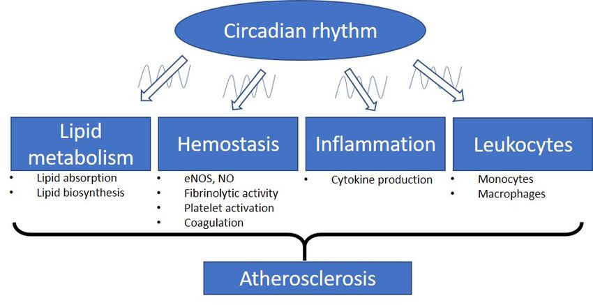
Int. J. Mol. Sci. 2021, 22, 676                                                                                           2 of 19

                                  the control of negative feedback loops of the molecular clock. Zeitgeber (German for
                                  time-giver) refers to the internal or external factors, which can cue the circadian clock and
                                  modulate the circadian rhythm from the molecular to the behavioral level [15,16].
                                        The core circadian clock is located in the hypothalamic suprachiasmatic nucleus
                                  (SCN) [17]. The SCN is the master clock that synchronizes neurons and coordinates circa-
                                  dian outputs. The circadian outputs are triggered by the photic information transmitted
                                  from the retina [18]. Aberrant light exposure disturbs the function of SCN and causes
                                  circadian disruption [19]. The central clock in the SCN regulates peripheral clocks and
                                  coordinates circadian gene expression. This process can be regulated directly by neuronal
                                  and hormonal signaling. It can also be regulated by driving appetite, blood pressure and
                                  body temperature indirectly [20,21].
                                        Although SCN is the master clock regulator, peripheral tissues are capable of local and
                                  autonomous clock regulation [22]. Peripheral clocks are found in almost all the peripheral
                                  tissues including immune cells, adipose tissues, kidney, liver and also the tissues of the
                                  vascular system [17]. The circadian gene expression and function in these peripheral tissues
                                  can be affected by the peripheral clocks [23]. In mice, 6% and 4% transcripts of protein-
                                  coding genes show circadian oscillations in the heart and aorta, respectively [24]. Ex vivo
                                  study has revealed that around 8% of macrophage transcriptomes are under local circadian
                                  regulation. The macrophages are isolated from spleen, lymph node, and peritoneum in
                                  this study [25]. These genes include those related to the molecular clock, glucose and lipid
                                  metabolism, and vascular integrity. These results suggest the importance of the peripheral
                                  clock regulation.
                                        Atherosclerosis results from the progressive accumulation of lipids and fibrous ele-
                                  ments in large arteries. Atherosclerosis is the primary cause of cardiovascular diseases,
                                  stroke and myocardial infarction [26]. Obesity, diabetes mellitus, dyslipidemia, hyperten-
                                  sion, and smoking are well-known risk factors for atherosclerosis and other cardiovascular
                                  diseases. Accumulating evidence suggests, that the circadian disruption is also a critical
                                  factor leading to atherosclerosis [27–29].
                                        Physiological parameters of the cardiovascular system, such as blood pressure, heart
                                  rate, and endothelial function, exhibit diurnal variations within a day [30–34]. A normal
                                  day–night difference in blood pressure is essential in maintaining cardiovascular health.
                                  Clinical studies have suggested the attribution of diurnal variations of cardiovascular pa-
                                  rameters in the pathophysiology and pathogenesis of cardiovascular complications [30–34].
                                  In humans, frequencies of thromboembolic and cardiovascular events exhibit clear diurnal
                                  variations, which peak during the morning-to-noon period [35,36]. Plasma levels of lipids
                                  display circadian oscillations independent of food intake [37]. It suggests that the intrinsic
                                  biological clock is an important regulator of the body lipid metabolism. Levels of immune
                                  cells and pro-inflammatory cytokines show circadian fluctuations [38], while the activity
                                  of the immune system is strongly linked to the circadian rhythm [39,40]. Therefore, the
                                  desynchronization of the clock and the misalignment between the circadian rhythm, lipid
                                  metabolism, and immune system could result in the development of dyslipidemia and
                                  inflammation and contribute to the risk of atherosclerosis. This suggests that the circadian
                                  rhythm could be a novel therapeutic target for and cardiovascular diseases, especially
                                  arteriosclerosis and thrombosis. However, the underlying mechanisms remain elusive.
                                        In this review, we summarize the recent findings on the role of the circadian rhythm in
                                  the progression of arteriosclerosis and thrombosis. The possible treatment of atherosclerosis
                                  and thrombosis through targeting circadian clocks is discussed.

                                  2. Circadian Rhythm and Arteriosclerosis
                                        The blood clotting is a protective mechanism against bleeding events. However,
                                  the formation of blood clots in vasculatures can also lead to severe cardiovascular events
                                  including ischemic stroke, myocardial infarction, and sudden cardiac arrest. Atherosclerosis
                                  is a chronic inflammatory condition that is initiated by endothelial dysfunction and the
                                  upregulation of adhesion molecules [41]. These promote the recruitment of leukocytes to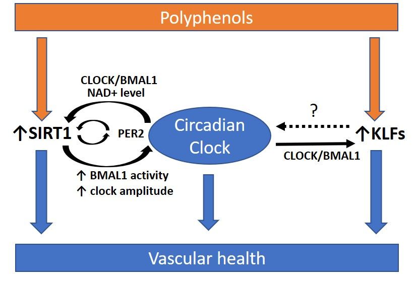
Int. J. Mol. Sci. 2021, 22, 676                                                                                          3 of 19

                                  the inflamed endothelium and the formation of atherosclerotic plaques. Atherosclerosis
                                  is the underlying pathology of most cardiovascular diseases. Thrombosis can be caused
                                  by atherosclerosis and occlude the blood vessel. Thrombolysis can break down clots to
                                  maintain normal blood flow in the blood vessels [42]. The balance between clotting and
                                  thrombolysis is regulated in a circadian manner [43].
                                       The ability of the vascular endothelium to cause vasodilation is important in protect-
                                  ing against cardiovascular disease. The vascular endothelial nitric oxide synthase (eNOS)
                                  has anti-atherosclerotic functions. NO produced by eNOS can inhibit platelet aggregation
                                  and regulate the patency of vessels [44,45]. Dysfunction of eNOS causes NO imbalance
                                  in the endothelium and leads to endothelial dysfunction [46]. There is evidence showing
                                  that the peripheral circadian clock can regulate eNOS expression, which in turn, modu-
                                  lates the diurnal variation of blood pressure [47–49]. Normally, endothelium-dependent
                                  vasorelaxation is reduced during the light cycle, due to the lowered NO production in the
                                  morning [50]. The deterioration of NO production might contribute to the morning peak
                                  of incidence of cardiovascular diseases [44,45,51].
                                       Monocytes and macrophages are the key players in inflammatory response and athero-
                                  genesis [51]. When the blood cholesterol level is high, inflammatory Ly6chi monocytes
                                  adhere to the inflamed endothelium and differentiate into lesion macrophages [51]. This
                                  is the critical step for the initiation and exacerbation of atherosclerotic plaque formation.
                                  The functions of the macrophages in atherosclerotic lesions, such as proliferation, M1,
                                  and M2 polarization, apoptosis and cholesterol efflux are important for the progression of
                                  atherosclerosis [52]. In addition, the numbers of hematopoietic cells and the production
                                  of cytokines have been shown to oscillate in diurnal rhythm and are orchestrated by the
                                  molecular clock [40,53].
                                       Recent study reveals that circulating leukocyte counts peak during the inactive phase
                                  in the murine blood [54]. By contrast, leukocyte counts in other tissues, such as bone
                                  marrow, skeletal muscle, and the heart, peak during of the active phase [54,55]. The
                                  detailed mechanism of the leukocyte counts oscillation is not well-known, it is likely
                                  regulated by networks of chemokines and endothelial adhesion molecules [54].
                                       Expression of many hemostasis-related molecules has been shown to align with the
                                  circadian rhythm. The fibrinolytic activity is lowest at night and starts to increase before
                                  morning. This circadian rhythm has first been described since the 1950s [56]. In human,
                                  the number of platelets peaks in the afternoon and is most active in the morning [57,58].
                                  Markers of platelet activation, including β-thromboglobulin (β-TG) and platelet factor 4
                                  (CXCL4), are expressed in a circadian rhythm and peak in the afternoon [59]. Activities of
                                  coagulation factors, including factor VII, factor VIII, factor IX, and von Willebrand factor
                                  (vWF) oscillate in circadian rhythms [57,59–61]. Factor X activity (Xa) and D-dimers, the
                                  markers of fibrinolysis, peak in the morning [61].
                                       There is a correlation between the circadian oscillations of interleukin-6 (IL-6) and
                                  fibrinogen. The IL-6 level peaks in the early hours of the night, while the fibrinogen level
                                  peaks later in the morning [61,62].
                                       The activities of plasminogen activator inhibitor-1 (PAI-1) and tissue plasminogen acti-
                                  vator (t-PA) oscillate in phase opposition. The activity of PAI-1 peaks in the morning, while
                                  the activity of t-PA peaks in the afternoon [63,64]. Protein C, protein S, and antithrombin
                                  (AT) are also expressed in a circadian rhythm [65]. The expression of thrombomodulin
                                  is controlled by the peripheral clock in endothelial cells [66]. Matrix metalloproteinase
                                  (MMP-1 and MMP-3), collagen IIIA1, transgelin, and calponin are involved in the stability
                                  of atherosclerotic plaques. In mouse smooth muscle cells, these molecules have been shown
                                  to express in a circadian pattern [67].
                                       These findings suggest the significant involvement of the circadian clock in hemostasis.
                                  The misalignment between the circadian and the fibrinolytic system may increase the risk
                                  of cardiovascular events [68] (Figure 1).
the stability of atherosclerotic plaques. In mouse smooth muscle cells, these molecules
have been shown to express in a circadian pattern [67].
Int. J. Mol. Sci. 2021, 22, 676 These findings suggest the significant involvement of the circadian clock in hemosta-
4 of 19
sis. The misalignment between the circadian and the fibrinolytic system may increase the
risk of cardiovascular events [68] (Figure 1).
Involvementofofthe
Figure1.1.Involvement
Figure thecircadian
circadianrhythm
rhythmininatherosclerosis.
atherosclerosis.TheTheintrinsic
intrinsicbiological
biologicalclock
clockhas
hasan
anoscillation
oscillationcycle
cycleofof
approximately 24 h. The circadian clock controls different physiological parameters of the cardiovascular
approximately 24 h. The circadian clock controls different physiological parameters of the cardiovascular system such system suchasas
bloodpressure,
blood pressure,heart
heartrate,
rate,and
andendothelial
endothelialfunction.
function.Circadian
Circadiandisruption
disruptionisisaacritical
criticalfactor
factorleading
leadingtotoatherosclerosis
atherosclerosisand and
cardiovascular
cardiovasculardiseases.
diseases.Plasma
Plasmalipid
lipidlevels
levelsare
aremediated
mediatedby bythe
thebalance
balanceofoflipid
lipidabsorption
absorptionand andbiosynthesis.
biosynthesis.The Thelipid
lipid
plasma
plasmalevels
levelsdisplay
displaycircadian
circadianoscillations
oscillationsand
andare
areindependent
independentofoffood
foodintake.
intake.Vascular
Vascularfunctions,
functions,especially
especiallythetherelated
related
endothelial
endothelialnitric
nitricoxide
oxidesynthase
synthase(eNOS)
(eNOS)expression
expressionand andnitric
nitricoxide
oxide(NO)
(NO)production,
production,are areregulated
regulatedby bythe
theperipheral
peripheral
circadian
circadian clock.
clock. Many
Many of ofthe
theimportant
important molecules
molecules involved
involved in hemostasis
in hemostasis havehavebeen been
shownshownto aligntowith
align with circadian
circadian rhythms,
rhythms, including molecules responsible for fibrinolytic activity, platelet activation, and coagulation. Dysregulation of
including molecules responsible for fibrinolytic activity, platelet activation, and coagulation. Dysregulation of the circadian
the circadian rhythm leads to inflammation. Proinflammatory cytokines are expressed in a circadian manner. In response
rhythm leads to inflammation. Proinflammatory cytokines are expressed in a circadian manner. In response to inflammatory
to inflammatory stimuli, circulating counts of leukocytes and the function of monocytes/macrophages are modulated by
stimuli,
the circulating
circadian counts of leukocytes
clock. Therefore, and theof
the misalignment function of monocytes/macrophages
the circadian clock with these parametersare modulated
could leadbyto
the circadian
the clock.
progression
Therefore, the misalignment
of atherosclerosis. of the circadian clock with these parameters could lead to the progression of atherosclerosis.
3.3.Circadian
CircadianDisruption
Disruptionand andVascular
VascularComplications
Complications
Theintrinsic
The intrinsiccircadian
circadianclock
clockworsen
worsenduring
duringaging
aging[69]
[69]and
andobesity
obesity[70].
[70].The
Theampli-
ampli-
tudes of circadian rhythms are dampened, or the peaks of the rhythms are shifted ininthe
tudes of circadian rhythms are dampened, or the peaks of the rhythms are shifted the
worsened clocks [71]. Recent evidence indicates that the circadian rhythm
worsened clocks [71]. Recent evidence indicates that the circadian rhythm in the vascula- in the vascu-
lature
ture is important
is important for for vascular
vascular function
function andand health
health [72–74].
[72–74]. The The misalignment
misalignment betweenbetween
the
the clock and metabolism can cause cardiovascular complications, including pathological
clock and metabolism can cause cardiovascular complications, including pathological vas-
vascular remodeling, vascular senescence, hypertension, stenotic atherosclerotic lesions,
cular remodeling, vascular senescence, hypertension, stenotic atherosclerotic lesions, vas-
vascular graft failure, and diabetic vasculopathies [44,75–78]. Compromised circadian clock
cular graft failure, and diabetic vasculopathies [44,75–78]. Compromised circadian clock
machineries in the vasculature, including mutations or polymorphisms in clock genes and
machineries in the vasculature, including mutations or polymorphisms in clock genes and
the reduction in the oscillation amplitude of the clock genes, are observed in models of
the reduction in the oscillation amplitude of the clock genes, are observed in models of
obesity and cellular senescence [70,79].
obesity and cellular senescence [70,79].
Proinflammatory stimuli can disrupt circadian rhythms and suppress the oscillation
Proinflammatory stimuli can disrupt circadian rhythms and suppress the oscillation
amplitude of clock components with negative feedback in isolated macrophages, whereas
amplitude of clock components with negative feedback in isolated macrophages, whereas
anti-inflammatory signals can improve circadian rhythms [80,81]. In mice, long-term sleep
anti-inflammatory signals can improve circadian rhythms [80,81]. In mice, long-term sleep
fragmentation increases circulating levels of inflammatory cytokines, including IL-1β,
fragmentation increases circulating levels of inflammatory cytokines, including IL-1β, IL-
IL-6, and TNF-α [82]. Long-term sleep fragmentation can reduce the phosphorylation
6,and
andtranscriptional
TNF-α [82]. Long-term
activity of sleep
cyclicfragmentation
AMP responsecan reduce the phosphorylation
element-binding protein (CREB)and [82],
transcriptional
whereas the downregulation of CREB may contribute to the pathological(CREB)
activity of cyclic AMP response element-binding protein [82],to
responses
vascular injury and plaque progression [83].
Apolipoprotein E (ApoE)−/− mouse is a widely used murine model for atherosclero-
sis [84]. ApoE−/− mice fed with Western diet for four weeks have accelerated atherosclerosis
and exhibit an altered circadian expression profile of cardiac clock genes and apoptosis-
related genes (c-Myc and p53) [85]. This suggests an interrelationship between the circadian
clock and lipid metabolism. Severe disruption of circadian rhythms by exposing to con-Int. J. Mol. Sci. 2021, 22, 676 5 of 19
stant light exacerbates atherosclerosis in male, but not in female ApoE−/− mice. When the
circadian rhythm is disrupted, male ApoE−/− mice have increased serum LDL level [86].
In hyperlipidemic female APOE*3-Leiden.CETP mice, exposure to 12-h shift of light-dark
cycles for 15 weeks causes a significant increase in atherosclerosis, while male mice do
not. Higher lesion macrophage contents, increased inflammation and oxidative stress are
observed in these hyperlipidemic mice. These suggest the involvement of the immune
system in disrupted circadian-induced atherosclerosis development [87]. The underlying
mechanisms of the observed gender difference are not yet studied, but it is hypothesized
that these could be due to the differences in circulating sex hormones.
Interestingly, time of surgery can affect the thrombus formation and resolution re-
sponse in mice. In a recent study on thrombus formation, mice ligated at 12:00 p.m.
have lower survival rate than the mice ligated at 7:00 a.m. [88]. The thrombi sizes of the
12:00 p.m.-ligated mice are larger than that of the 7:00 a.m.-ligated mice, while the gene
expression of MMP9 is significantly reduced in the 12:00 p.m.-ligated mice. These suggest
that treatment time may affect the response of mice to thrombosis [88].
4. Clock Components
The intrinsic molecular clock consists of interlocked transcription–translation feedback
loops of clock genes and proteins [89]. The master clock regulators include brain and muscle
aryl hydrocarbon receptor nuclear translocator-like protein 1 (BMAL1 or ARNTL), circadian
locomotor output cycles kaput (CLOCK), Period 1/2/3 (PER1/2/3), and Cryptochrome
1/2 (CRY1/2). BMAL and CLOCK are important transcription factors. PER1/2/3 and
CRY1/2 are transcriptional modulators [22,89]. In mouse aorta, PER1/2 and CRY1/2
mRNA levels peak at early night cycle and trough at day cycle [90], while the protein
expression of BMAL1 peaks at the beginning of the day cycle and troughs at the night cycle.
Similar phase difference relationships of the clock genes are reported in both human and
mouse smooth muscle cell model in vitro [90].
During the light phase, BMAL1 can dimerize with CLOCK and bind to the E-box
regulatory sites (50 -CACGTG-30 ) in the promoter regions. This binding can activate the
transcription of many circadian proteins including PER1/2/3 and CRY1/2 [17]. When
PERs and CRYs proteins accumulate in the cytoplasm and reach certain levels, they can
dimerize to form repressor complex and translocate into the nucleus, where they inhibit
the CLOCK:BMAL1-mediated transcription, forming a negative feedback loop [17,91].
The reinforcing loops of the molecular clock are composed by the circadian nuclear
receptors, reverse ERB (REV-ERB α/β) and retinoic acid receptor-related orphan receptors
(RORα/β/γ). These loops are important in controlling the rhythmic gene transcriptions of
BMAL1 and CLOCK. Both REV-ERB and ROR interact with the ROR response elements at
the promotor regions of BMAL1 and CLOCK. REV-ERBα negatively regulates the gene
expression of BMAL1 and CLOCK [92], while RORα and RORγ positively regulate the
gene expression of BMAL1 and CLOCK [93,94].
The molecular circadian clock can modulate the rhythmic expression of clock-controlled
genes (CCGs) by activating different circadian promoter elements. These regulatory pro-
moter elements include D-boxes, E-boxes, and ROR response elements [93]. CCGs en-
code different important proteins involved in cellular metabolisms and inflammatory
responses [95,96] (Figure 2). Although identical clock machineries are found in most
cells, the circadian expression pattern of CCGs are highly tissue-specific or even cell-type-
specific [14]. Post-translational modifications also contribute to the regulation of circadian
clock gene expression [14,97].Int. J. Mol. Sci. 2021, 22, x FOR PEER REVIEW 6 of 20
Int. J. Mol. Sci. 2021, 22, 676 the circadian expression pattern of CCGs are highly tissue-specific or even cell-type-spe-
6 of 19
cific [14]. Post-translational modifications also contribute to the regulation of circadian
clock gene expression [14,97].
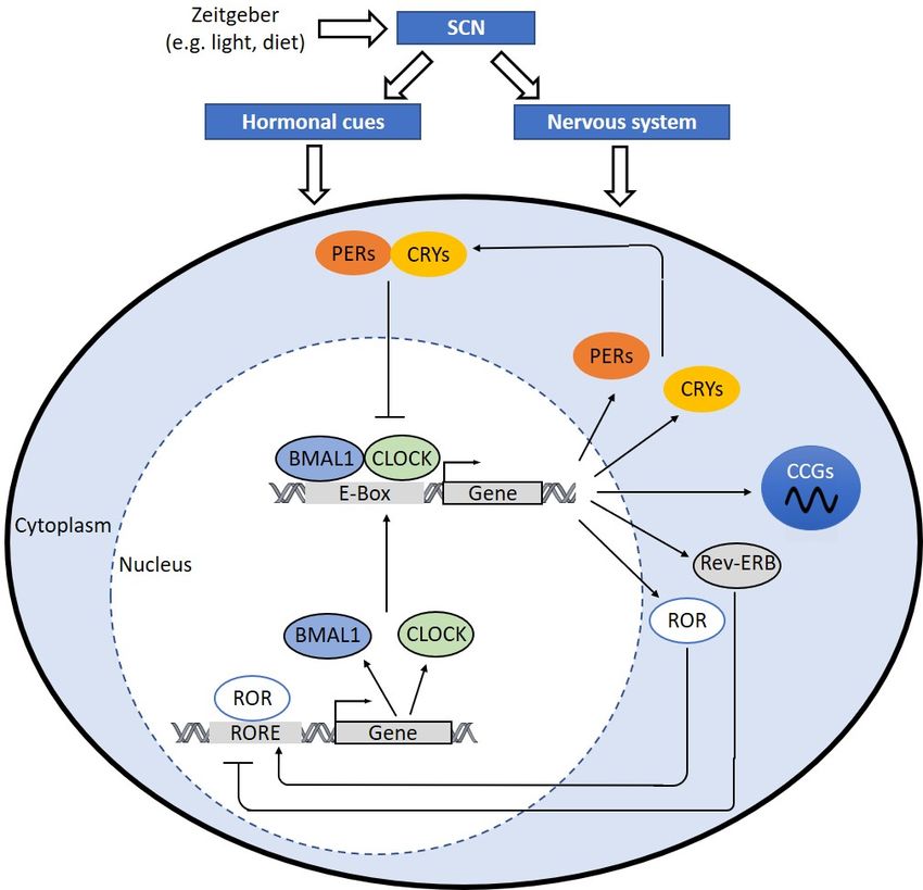
Figure 2. The molecular clock mechanism. When the hypothalamic suprachiasmatic nucleus (SCN) is triggered, the SCN
Figure 2. The molecular clock mechanism. When the hypothalamic suprachiasmatic nucleus (SCN) is triggered, the
translates signals into hormonal cues and nerve impulses, which can regulate the peripheral clock. In cells, the heterodimer
SCN translates signals into hormonal cues and nerve impulses, which can regulate the peripheral clock. In cells, the
of circadian locomotor output cycles kaput (CLOCK) and brain and muscle aryl hydrocarbon receptor nuclear transloca-
heterodimer of circadian locomotor output cycles kaput (CLOCK) and brain and muscle aryl hydrocarbon receptor nuclear
tor-like protein 1 (BMAL1) binds to E-box sequence (5′-CACGTG-3′) in the promoter and activates the transcription of
translocator-like
Period protein
(PER)1/2/3, 1 (BMAL1)(CRY)1/2,
Cryptochrome binds to E-box sequence
retinoic (50 -CACGTG-3orphan
acid receptor-related
0 ) in the promoter and activates the transcription
receptors (ROR), and reverse ERB (Rev-
of Period
ERB). (PER)1/2/3,
Dimerized Cryptochrome
PERs and (CRY)1/2,
CRYs translocate retinoicand
into nucleus acidinterfere
receptor-related orphan receptors (ROR),
CLOCK:BMAL1-mediated and reverse
transcription. ERB
REV-ERB
(Rev-ERB).regulates
negatively Dimerized PERs and
BMAL1 and CLOCK
CRYs translocate
expression. into
RORsnucleus and interfere
positively regulate CLOCK:BMAL1-mediated
BMAL and CLOCK expression transcription.
via ROR
response
REV-ERB elements
negatively(RORE) at BMAL1
regulates their promotor
and CLOCK regions. The clock
expression. RORs drives rhythmic
positively expression
regulate BMAL andof clock-controlled
CLOCK expression genes
via
(CCGs) through
ROR response CLOCK:BMAL1-mediated
elements activation
(RORE) at their promotor of circadian
regions. The clockpromoter elements,
drives rhythmic including
expression of E-boxes, D-boxes,genes
clock-controlled and
ROR
(CCGs)response
throughelements. CCGs encode important
CLOCK:BMAL1-mediated proteins
activation involved
of circadian in processes
promoter elements, of including
atherosclerosis development,
E-boxes, D-boxes, andhemo-
ROR
stasis,
responseinflammation, lipid metabolism,
elements. CCGs and macrophage
encode important trafficking.
proteins involved in processes of atherosclerosis development, hemostasis,
inflammation, lipid metabolism, and macrophage trafficking.
5. Clock Components and Vascular Complications
5.1. BMAL1
5. Clock and CLOCKand Vascular Complications
Components
Bmal1-deficient
5.1. BMAL1 and CLOCK mice exhibit reduced lifespans and premature aging with related pa-
thologies. Sarcopenia,mice
Bmal1-deficient loss of visceral
exhibit and subcutaneous
reduced lifespans and adipose tissues,
premature osteoporosis,
aging or-
with related
gan shrinkage, changes in blood cell composition, and loss of pressor response
pathologies. Sarcopenia, loss of visceral and subcutaneous adipose tissues, osteoporosis, to stress
are observed
organ [98].changes
shrinkage, Young inBmal1-deficient mice haveand
blood cell composition, increased pathological
loss of pressor remodeling
response to stress
and vascular injuries with reduced blood flow. Arteries from Bmal1-deficient
are observed [98]. Young Bmal1-deficient mice have increased pathological remodeling mice loss the
ability to narrow (inward remodeling), which is reminiscent of the similar
and vascular injuries with reduced blood flow. Arteries from Bmal1-deficient mice loss response in
the ability to narrow (inward remodeling), which is reminiscent of the similar response in
eNOS knockout mice. Arteries from Bmal1-deficient mice also have significant increase in
collagen deposition in the medial layer, which leads to wall thickening [78].Int. J. Mol. Sci. 2021, 22, 676 7 of 19
Transplantation of arteries from Bmal1-knockout mice to wild type mice leads to the
development of atherosclerosis in the transplanted blood vessels without affecting the
systemic hemodynamics. This suggests a critical role for autonomous peripheral circadian
clocks [99]. Most downstream target genes of BMAL1 appear to be tissue specific and play
differential pathophysiological roles in atherosclerosis [100,101]. BMAL1 and CLOCK can
directly regulate the expression of the prothrombotic mediator von Willebrand factor (vWF).
The expression levels of vWF, fibrinogen, and plasminogen activation inhibitor-1 (PAI-1) are
increased in Bmal1−/− mice, which lead to accelerated arterial thrombus formation [102].
BMAL1 can regulate the expression of inflammatory marker CCL2. CCL2-CCR2
chemokine axis is involved in the early lesion development in mice [95]. The circadian
expression of monocyte chemoattractant protein-1 (MCP-1) in macrophages is regulated
by BMAL1-mediated activation of nuclear factor kappa-light-chain-enhancer of activated
B cells (NF-κB) [103]. In Bmal1−/− mice, both basal and tumor necrosis factor-α (TNF-α)-
induced NF-κB activations are upregulated in macrophages [104,105]. BMAL1 is required
to maintain the diurnal oscillation of inflammatory Ly6chi monocytes and their trafficking
to sites of acute inflammation [95].
Mice with monocytes- and macrophages-specific Bmal1-deficiency have enhanced
atherosclerosis in carotid arteries. These mice also have increased total number of macrophages
and Ly6chi infiltrating monocyte-macrophages in atherosclerotic lesions. These suggest the
importance of BMAL1 in maintaining normal macrophage functions [101]. Endothelial-
specific Bmal1−/− mice maintain the circadian rhythm of blood pressure, but their blood
pressure in the active phase is lower than the blood pressure of control mice [106]. Specific
deletions of Bmal1 in endothelial and hematopoietic cells result in accentuated vascular
injuries [107]. Smooth muscle-specific Bmal1−/− mice have reduced amplitude of blood
pressure oscillation without affecting locomotor activity. These suggest that the vascular
BMAL1 can regulate blood pressure master clock independently [108]. However, further
studies on the atherosclerotic development on these mice are needed to dissect the role of
BMAL1 in peripheral tissues.
Clock-deficient mice have a significantly reduced lifespan, which is about 15% shorter
than that of wild type littermates [109]. Clock mutant mice show phenotypes that are
reminiscent of accelerated aging [110], obesity, and hypertension [111,112]. Macrophages
isolated from Clock mutant mice have higher intracellular levels of total, free, and esterified
cholesterol. The macrophages from these mice also have reduced expression of the ATP-
binding cassette transporter (ABCA1 and ABCG1) and blunted abilities to efflux cholesterol
to ApoA1 [113]. Both Bmal1−/− and Clockmut animals loss the circadian variation in glucose
and triglycerides [111]. Either deletion or knockdown of Clock or Bmal1 abolishes the
rhythmic oscillation of genes involved in lipid metabolism in the liver, including acetyl co-
A carboxylase (ACC), acetyl-CoA synthetase (ACS), and sterol regulatory element-binding
protein-1c (SREBP-1c). These suggest important roles of CLOCK and BMAL in modulating
glucose homeostasis and lipid profiles in vivo [114,115].
5.2. CRY1/2
Atherosclerotic patients have lower serum CRY1 mRNA level [116]. In mice, deletion
of Cry1 and Cry2 leads to constant elevation of proinflammatory cytokines including IL-
6, TNF-α and inducible nitric oxide synthase (iNOS) [117]. CRY1 and CRY2 have been
shown to interact with the glucocorticoid receptor in a ligand-dependent fashion [118].
Both Cry1- and Cry2-deficient mice exhibit glucose intolerance and have elevated plasma
glucose levels in response to acute feeding after a 12 h overnight fasting [118]. In ApoE−/−
mice, overexpression of CRY1 by adenovirus-mediated gene transfer significantly reduces
the expression of proinflammatory markers, including IL-1 and 6, TNF-α, NF-κB, and
macrophage inflammatory protein-1α (MIP-1α). These mice also have reduced plasma
total cholesterol (TC), triglyceride (TG), and low-density lipoprotein cholesterol (LDL-C)
levels. The mice are protected against plaque development [116].Int. J. Mol. Sci. 2021, 22, 676 8 of 19
5.3. PER1/2
Deficiency of Per1 and Per2 in mice results in altered circadian rhythms [119,120].
Although Per1 and Per2 mutant mice cannot be distinguished morphologically from wild
type mice at birth, phenotypes of premature aging are observed from the age of 12 months,
including faster decline in fertility and loss of soft tissues [121]. Per2 mutant mice have
impaired clock resetting ability and lose circadian rhythms in constant darkness [122].
PER2 is a major regulator of lipid metabolism by controlling the proadipogenic activity
of peroxisome proliferating activated receptor (PPARγ) [123]. Both PER1/2-null mice and
PER2-null mice have lower hepatic TG levels [124].
The transplantation of arteries from Per1/2−/− mice to wild type mice leads to the
development of atherosclerosis in the transplanted graft, which suggests the important
role of peripheral PER1/2 in the vascular system [99].
During the transition from resting to active phase, endothelium-dependent relaxation
response is increased in the aortae of wild-type mice, but not in PER2 mutant mice, con-
firming a circadian control of endothelial function [125]. PER2 mutant mice also show
increased vascular senescence and endothelial dysfunctions [126]. The blood vessels
from Per2 mutant mice have reduced production of endothelial-derived relaxation factors
(EDRF), including prostaglandins and nitric oxide (NO). The expression of vasoconstrictor
cyclooxygenase-1 (COX1) is increased in Per2 mutant mice. The aortae from Per2 mutant
mice show a significant reduction of NO dependent endothelial function and enhanced
lesion development [126]. Angiogenic response to hind limb ischemia is blunted in Per2
mutant mice [125]. Per2 mutant mice show diabetes-like vascular phenotypes such as
retinal vascular damage and neuronal loss [127].
5.4. REV-ERB
Rev-erbα-mutant mice have increased adiposity and mild hyperglycemia without in-
sulin resistance after high-fat diet (HFD) [128]. Liver-specific Rev-erbα-knockout mice have
increased serum levels of cholesterol, TGs, and free fatty acids [129]. REV-ERBα modulates
the infiltration of inflammatory macrophages by inhibiting the expression of Ccl2 [130].
Knocking-down of Rev-erbα in hematopoietic cells enhances atherosclerotic lesion for-
mation in mouse aorta and increases the inflammatory phenotype of macrophages both
in vitro and in vivo [131]. Pharmacological activation of REV-ERBα reduces atherosclerotic
lesion formation and promotes anti-inflammatory M2 markers expression [131].
A synthetic REV-ERB agonist, SR9009, has been shown to activate REV-ERB activity
and leads to reduced size of atherosclerotic plaque in atherosclerosis-prone LDL receptor
(Ldlr)-deficient mice [132]. SR9009 administration can normalize cardiac gene expression
and function by mediating the circadian clock-controlled processes in the heart [133].
REV-ERB agonist treatment can reduce the polarization of bone marrow-derived mouse
macrophages (BMDMs) to proinflammatory M1 macrophages and increase the polarization
of BMDMs to anti-inflammatory M2 macrophages [132]. These indicate the possibility
of targeting REV-ERBs for the treatment of atherosclerosis. However, the outcome of
SR9009 treatments may not be solely attributed to its effect on the circadian rhythms.
A recent study reported REV-ERB-independent effects of SR9009 on cell proliferation
and metabolism [134].
In short summary, disruption of the circadian clock at different nodes (BMAL1,
CLOCK, CRYs, PERs, and REV-ERB) promotes atherosclerosis. These clock components are
involved in the homeostasis of glucose and lipid metabolism by controlling the circadian
expression and activities of key regulatory enzymes. Maintaining the high amplitude
oscillation of these clock components may be a potential strategy for prevention against
atherosclerosis and other cardiovascular complications.Int. J. Mol. Sci. 2021, 22, 676 9 of 19
6. Targeting the Circadian Clock for the Treatment of Atherosclerosis
6.1. The Role of SIRT1 in Regulating the Circadian Rhythm
SIRT1 is a nicotinamide adenine dinucleotide (NAD+)-dependent protein deacetylase.
SIRT1 is well-known for its vascular protective effects, including enhancing endothelium-
dependent vasodilatation, promoting endothelial angiogenesis and migration, suppressing
vascular inflammation, preventing endothelial senescence and adverse arterial remodeling,
and suppressing foam cell formation. These effects of SIRT1 made it an important player
in protecting against atherosclerosis [135]. SIRT1 can suppress vascular inflammation by
regulating NF-κB activity through deacetylating K310 on the p65 subunit [136]. Reduced
SIRT1 level has been shown to upregulate NF-κB and increase inflammatory responses
in monocytes/macrophages, myeloid cells, and endothelial cells [137–140]. SIRT1 can
regulate the expression of liver X receptors (LXRs), which confers beneficial effects in lipid
metabolism and suppresses foam cell formation [141].
SIRT1 is highly involved in the crosstalk between the circadian clock and energy
metabolism [142]. SIRT1 is required for the high-magnitude circadian transcription of
circadian clock genes including Clock, Bmal1, Crys, and Pers [143]. Sirt1-deficienct mice have
disrupted circadian rhythms and altered amplitudes of Per1/2 and Cry1/2 expression [144].
SIRT1 directly activates the transcription of Bmal1, and increases the oscillating amplitude
of other clock genes via peroxisome proliferator-activated receptor gamma coactivator
1-alpha (PGC-1α) [145]. SIRT1 can directly deacetylate BMAL1 and PER2 to affect their
transcriptional activities [146]. The deacetylation of PER2 by SIRT1 can lead to PER2
degradation [143]. A negative reciprocal relationship exists between SIRT1 and PER2 [144].
PER2 negatively regulates Sirt1 transcription through competing CLOCK/BMAL1 binding
sites at SIRT1 promotor [144].
One of the transcriptional targets of the CLOCK:BMAL1 dimer is nicotinamide phos-
phoribosyltransferase (NAMPT), which is an enzyme required for the biosynthesis of
NAD+ [147,148]. The circadian clock can exert a rhythmic regulation on SIRT1 activity via
NAMPT [147,148]. While rhythmic NAD+ level affects SIRT1 activity, SIRT1 may in turn
affect the circadian levels of metabolites including NAD+ and acetyl-CoA. The intracellular
acetyl-CoA level is controlled by SIRT1-mediated deacetylation of acetyl-coenzyme A
synthetase 1 (ACS1) [149]. SIRT1 can be expressed in a circadian manner. The expression
of SIRT1 is in high oscillating rhythms in young animals, whereas the rhythmic oscillations
of SIRT1 expressions are nearly flattened in aged animals. The components of this ampli-
fying loop, including SIRT1, PGC-1α, and NAMPT, are critical in the intrinsic circadian
regulation [145].
In ApoE−/− mice, abnormal exposure to light exacerbates atherosclerotic plaque
formation and circadian disruption, which are associated with altered expression of clock
genes, lipid metabolism genes and SIRT1 [150].
Collectively, SIRT1 may serve as an important link between the circadian clock and
lipid-related gene oscillation. SIRT1 has many beneficial effects in protection against
atherosclerosis. These findings raise the potential use of SIRT1 activators in modulating
the circadian rhythm and preventing atherosclerosis.
6.2. Krüppel-Like Factors (KLFs), Circadian Rhythms and Atherosclerosis
Krüppel-like factors (KLFs) belong to an evolutionarily conserved zinc-finger tran-
scription factors family, which bind to CACCC elements and GC-rich regions of DNA.
Members of the KLF family are key regulators of important biological processes, including
cell differentiation, proliferation, apoptosis, metabolism, and anti-polymicrobial activ-
ity [151,152]. The transcriptions of the KLFs are regulated by direct promoter binding of
CLOCK and BMAL1 [153,154].
In endothelial cells, overexpression of KLF2 leads to the secretion of vascular protective
miRs-143/145 in microvesicles [155]. miRs-143/145 can reduce atherosclerosis by targeting
critical genes for vascular smooth muscle cells dedifferentiation, including Mmp3, ETS
like-1 protein (Elk1) and calcium/calmodulin-dependent protein kinase type II delta chainInt. J. Mol. Sci. 2021, 22, 676 10 of 19
(Camk2d) [155]. KLF2 represses endothelial inflammation and modulates anti-thrombotic
transcription. KLF2 directly binds to the promoter of thombomodulin-1 and increases the
expression of this potent anti-thrombotic and anti-inflammatory factor [156]. KLF2 inhibits
thrombin-mediated endothelial activation by preventing the transcription of protease-
activated receptor (PAR-1). PAR-1 is a thrombin receptor [157]. In vivo, Klf2+/− ApoE−/−
mice are more susceptible to the development of atherosclerotic lesion compared with
Klf2+/+ ApoE−/− mice [158]. Post-natal deletion of KLF2 leads to a thrombotic phenotype
in mice, while overexpression of KLF2 protects mice from thrombus formation. Over-
expression of KLF2 decreases the expression of thrombotic genes coding for iNOS and
MCP-1 in peritoneal macrophages. The expression levels of PAR-1 and thrombomodulin
in endothelial cells are also reduced by the KLF2 overexpression [159]. Myeloid-specific
KLF2 deletion in mice with the ApoE−/− background promotes vascular oxidative stress
and atherosclerosis [160]. In human, monocytes from atherosclerotic patients have reduced
Klf2 expression [161].
KLF4 regulates the reverse cholesterol transport out of the vascular wall and inhibits
the inflammation by inducing the expression of cholesterol-25-hydroxylase (Ch25h) and
LXR in endothelial cells [162]. Overexpression of KLF4 in endothelial cell protects against
the pathogenesis of atherosclerosis and thrombosis [163]. Loss of myeloid KLF4 pro-
motes atherosclerosis, whereas macrophages specific Klf4-deficient mice have increased
inflammation in response to oxidized phospholipids [164].
KLF10 is a regulator of bone physiology. KLF10 also regulates glucose and lipid
metabolism in liver [165]. 36% and 23.4% of KLF10- regulated genes are involved in
lipid and carbohydrate metabolisms respectively [165]. Klf10− /− male mice had 20%
higher blood glucose levels than wild-type mice, while Klf10− /− female mice exhibit a 20%
increase of plasma TG level compared to wild-type mice [166].
In ApoE−/− mice, Klf14 expression is increased in the aorta compared to wild-type
mice [167]. Overexpression of KLF14 in macrophages increases the production of inflamma-
tory cytokine, TC and cholesteryl ester content, reminiscent of the phenotype of atherogenic
foam cells [168].
Both rat aortic vascular smoother muscle cells exposed to oxidized phospholipids and
human atherosclerotic tissues have markedly reduced KLF15 expression [169]. Both sys-
temic and smooth muscle-specific Klf15-deficient mice exhibit an aggressive inflammatory
vasculopathy in diet-induced atherosclerosis [169]. Recently, KLF15 has been shown to
regulate circadian susceptibility to ischemia reperfusion injury in the heart, while KLF15
expression is reduced in the heart of patients with cardiomyopathies [170]. These suggest
that KLF15 may play an important role in atherosclerosis.
Collectively, circadian oscillation of KLFs contributes to the rhythmic regulation of
their target genes. Dysregulation of KLFs may promote atherosclerosis. To clarify the
pharmacological potential for arteriosclerosis treatment with KLFs as target, further studies
of the detailed association between KLFs, circadian clock, and atherosclerosis are needed.
6.3. Polyphenols and the Circadian Clock
Polyphenols are secondary metabolites of plants, which have been widely studied
on their beneficial effects as antioxidants [171,172]. Polyphenols can affect cholesterol
metabolism via bile acid biosynthesis [173]. In addition to their antioxidative and anti-
inflammatory properties, polyphenols may prevent atherosclerosis by modulating the
circadian clock. Polyphenols interact with circadian clock by modulating the amplitude
and period of the clock gene oscillations [174–176]. The use of polyphenols in entraining
the circadian clock has been widely studied and reviewed [171,177–179].
Resveratrol is a well-known activator of SIRT1 [180]. Resveratrol has been shown to atten-
uate HFD-induced obesity in mice by normalizing the nearly flattened circadian expression
of PER2, CLOCK, and BMAL1 [181]. In ApoE−/− mice, resveratrol can inhibit either TMAO-
induced atherosclerosis or HFD- and lipopolysaccharides (LPS)-induced atherosclerosis [182].Int. J. Mol. Sci. 2021, 22, x FOR PEER REVIEW 11 of 20
Resveratrol is a well-known activator of SIRT1 [180]. Resveratrol has been shown to
Int. J. Mol. Sci. 2021, 22, 676 attenuate HFD-induced obesity in mice by normalizing the nearly flattened circadian 11 ofex-
19
pression of PER2, CLOCK, and BMAL1 [181]. In ApoE−/− mice, resveratrol can inhibit either
TMAO-induced atherosclerosis or HFD- and lipopolysaccharides (LPS)-induced athero-
sclerosis [182].
Equol,
Equol, aasoysoybean-based
bean-basedisoflavone-derived
isoflavone-derived metabolite,
metabolite, can
cansignificantly
significantlyreduce
reducethethe
atherosclerotic lesions, serum TG, TC and LDL-cholesterol levels, and
atherosclerotic lesions, serum TG, TC and LDL-cholesterol levels, and increase HDL-cho- increase HDL-
cholesterol level in ApoE −/−
−/− mice mice fed with
lesterol level in ApoE fed with HFD HFD
[183].[183].
Polyphenols
Polyphenols have been shown to enhance the
have been shown to enhance theexpression
expressionofofKLFs
KLFs[184–186].
[184–186].These
These
suggest
suggest the beneficial effect of polyphenols against circadian disruption ispossibly
the beneficial effect of polyphenols against circadian disruption is possiblyacting
acting
through
throughSIRT1
SIRT1and andKLFs
KLFs (Figure
(Figure3). 3).
Since most
Since studies
most about
studies polyphenols
about focused
polyphenols on their
focused on
antioxidant and anti-inflammatory properties, future study direction may
their antioxidant and anti-inflammatory properties, future study direction may include include their
effects on modulating
their effects circadian
on modulating rhythms
circadian and atherosclerosis
rhythms treatment.
and atherosclerosis treatment.
Figure3.3.Polyphenols
Figure Polyphenolsandandthe
thecircadian
circadianrhythm
rhythmfor forpromoting
promotingvascular
vascularhealth.
health.Polyphenols
Polyphenolsare aresecondary
secondarymetabolites
metabolitesofof
plants,
plants,which
whichhave
havebeen
beenwidely
widelystudied
studiedfor
fortheir
theirbeneficial
beneficialeffects
effectson
oncardiovascular
cardiovascularhealth.
health.Polyphenols
Polyphenolsmay maybe bebeneficial
beneficial
in
inpreventing
preventingatherosclerosis
atherosclerosisvia
viamodulating
modulatingthe thecircadian
circadianclock.
clock.Polyphenols
Polyphenolshave
havebeen
beenshown
shownto toenhance
enhancethe theexpression
expression
ofKrüppel-like
of Krüppel-likefactors
factors(KLFs)
(KLFs)and
andSirtuin
Sirtuin11(SIRT1),
(SIRT1),which
whichare
arehighly
highlyassociated
associatedwith
withthe
thecircadian
circadianclock.
clock.SIRT1
SIRT1and andKLFs
KLFs
areimportant
are importantplayers
playersininprotecting
protectingagainst
againstatherosclerosis.
atherosclerosis. SIRT1
SIRT1 interacts
interacts with
with the
the circadian
circadian clock
clock and
and isisrequired
requiredfor
for
high-magnitudecircadian
high-magnitude circadiantranscription
transcription ofofcircadian
circadianclockclockgenes.
genes. The
The circadian
circadian clock
clock can
canregulate
regulateSIRT1
SIRT1activity
activityvia
viathe
the
oscillatingNAD+
oscillating NAD+ level.
level. The
The transcription
transcription ofof KLFs
KLFs is is regulated
regulated by
by direct
directpromoter
promoterbinding
bindingof ofCLOCK:BMAL1.
CLOCK:BMAL1. However, However,
whether KLFs have a feedback control to the circadian clock remains unclear (dotted arrow and ?). A functional circadian
whether KLFs have a feedback control to the circadian clock remains unclear (dotted arrow and ?). A functional circadian
clock is required for preventing atherosclerosis. PER2, period 2. BMAL1, brain and muscle aryl hydrocarbon receptor
clock is required for preventing atherosclerosis. PER2, period 2. BMAL1, brain and muscle aryl hydrocarbon receptor
nuclear translocator-like protein 1; CLOCK, circadian locomotor output cycles kaput. NAD+, oxidized nicotinamide ade-
nuclear translocator-like protein 1; CLOCK, circadian locomotor output cycles kaput. NAD+, oxidized nicotinamide
nine dinucleotide.
adenine dinucleotide.
7. Summary and Future Directions
7. Summary and Future Directions
In summary, a large body of evidence suggests that the intrinsic circadian clock plays
In summary, a large body of evidence suggests that the intrinsic circadian clock plays
an important role in atherosclerosis in many aspects. The combinations of the central clock
an important role in atherosclerosis in many aspects. The combinations of the central
and peripheral clocks in the blood vessels, leukocytes and monocytes/macrophages or-
clock and peripheral clocks in the blood vessels, leukocytes and monocytes/macrophages
chestrate the normal hemodynamics and inflammatory responses. When the circadian
orchestrate the normal hemodynamics and inflammatory responses. When the circadian
rhythm is disrupted, the sequential inflammatory processes, endothelial dysfunction and
lipid imbalance could promote the development of atherosclerotic lesions.
However, most of the study results are based on global clock gene knockout mice
rather than tissue-specific knockout mice. Although SCN is the master clock, peripheral
tissues are able to regulate the clock locally and autonomously, and the peripheral clocks areInt. J. Mol. Sci. 2021, 22, 676 12 of 19
also important for regulating the function of local tissues [22]. Local homeostatic signaling
pathways can affect circadian genes expression and function in the peripheral tissues [23].
We have mentioned a few reports demonstrated that the implantation of circadian disrupted
tissues can cause atherosclerosis in normal mice. These results demonstrate and highlight
the importance of peripheral clocks. The effects of the core clock and peripheral clocks must
be clearly discriminated in studying their effects in cardiovascular diseases. Therefore, it
can be more informative for in vivo studies to use cell type (i.e., endothelium, macrophages,
or smooth muscle cells)-specific knockout mice in the future.
Ex vivo experiments have reported varied functions depending on the tissue collecting
time in mouse heart and aorta, which suggested detailed records on the time of experiments
should be included in future circadian studies [187].
SIRT1 and KLFs are important players in protecting against atherosclerosis. SIRT1
and KLFs are involved in the circadian regulation of cellular metabolisms. We propose the
use of polyphenols as the potential supplement targeting SIRT1 and KLFs. It would be
interesting to research other potential substances that regulate and link the circadian clock
and atherosclerosis.
Based on current evidence, the circadian clock and its influence on cardiovascular
diseases should be considered in the future studies for looking for therapeutic strategies
of atherosclerosis and thrombosis. The pharmacokinetics of anti-atherosclerosis drugs
may also be influenced by the circadian rhythm. Therefore, further studies on circadian
rhythms could be needed to improve the effectiveness of medicines. Novel therapeutic
targets entraining circadian clocks should be fully investigated.
Author Contributions: A.W.C.M. wrote the initial draft of the manuscript. H.L. and N.X., critically
reviewed and edited the manuscript. All authors have read and agreed to the published version of
the manuscript.
Funding: Original works from the authors’ laboratory contributing to this review were supported by
grants LI-1042/1-1, LI-1042/3-1, LI-1042/5-1, and XI 139/2-1 from the Deutsche Forschungsgemein-
schaft (DFG), Bonn, Germany. H.L. and N.X. were supported by a research grant from the Boehringer
Ingelheim Foundation for the collaborative research consortium “Novel and neglected cardiovascular
risk factors: molecular mechanisms and therapeutic implications.”
Conflicts of Interest: The authors declare no conflict of interest.
References
1. Vetter, C.; Devore, E.E.; Wegrzyn, L.R.; Massa, J.; Speizer, F.E.; Kawachi, I.; Rosner, B.; Stampfer, M.J.; Schernhammer, E.S.
Association Between Rotating Night Shift Work and Risk of Coronary Heart Disease Among Women. JAMA 2016, 315, 1726–1734.
[CrossRef] [PubMed]
2. Brown, D.L.; Feskanich, D.; Sanchez, B.N.; Rexrode, K.M.; Schernhammer, E.S.; Lisabeth, L.D. Rotating night shift work and the
risk of ischemic stroke. Am. J. Epidemiol. 2009, 169, 1370–1377. [CrossRef]
3. Deng, N.; Kohn, T.P.; Lipshultz, L.I.; Pastuszak, A.W. The Relationship Between Shift Work and Men’s Health. Sex. Med. Rev.
2018, 6, 446–456. [CrossRef] [PubMed]
4. Oishi, M.; Suwazono, Y.; Sakata, K.; Okubo, Y.; Harada, H.; Kobayashi, E.; Uetani, M.; Nogawa, K. A longitudinal study on the
relationship between shift work and the progression of hypertension in male Japanese workers. J. Hypertens. 2005, 23, 2173–2178.
[CrossRef]
5. Nazri, S.M.; Tengku, M.A.; Winn, T. The association of shift work and hypertension among male factory workers in Kota Bharu,
Kelantan, Malaysia. Southeast Asian J. Trop. Med. Public Health 2008, 39, 176–183.
6. Esquirol, Y.; Perret, B.; Ruidavets, J.B.; Marquie, J.C.; Dienne, E.; Niezborala, M.; Ferrieres, J. Shift work and cardiovascular risk
factors: New knowledge from the past decade. Arch. Cardiovasc. Dis. 2011, 104, 636–668. [CrossRef]
7. Haus, E.; Smolensky, M. Biological clocks and shift work: Circadian dysregulation and potential long-term effects. Cancer Causes
Control 2006, 17, 489–500. [CrossRef]
8. Brum, M.C.; Filho, F.F.; Schnorr, C.C.; Bottega, G.B.; Rodrigues, T.C. Shift work and its association with metabolic disorders.
Diabetol. Metab. Syndr. 2015, 7, 45. [CrossRef]
9. Muxfeldt, E.S.; Cardoso, C.R.; Salles, G.F. Prognostic value of nocturnal blood pressure reduction in resistant hypertension. Arch.
Intern. Med. 2009, 169, 874–880. [CrossRef]Int. J. Mol. Sci. 2021, 22, 676 13 of 19
10. Ayala, D.E.; Moya, A.; Crespo, J.J.; Castineira, C.; Dominguez-Sardina, M.; Gomara, S.; Sineiro, E.; Mojon, A.; Fontao, M.J.;
Hermida, R.C.; et al. Circadian pattern of ambulatory blood pressure in hypertensive patients with and without type 2 diabetes.
Chronobiol. Int. 2013, 30, 99–115. [CrossRef]
11. Ohlander, J.; Keskin, M.C.; Stork, J.; Radon, K. Shift work and hypertension: Prevalence and analysis of disease pathways in a
German car manufacturing company. Am. J. Ind. Med. 2015, 58, 549–560. [CrossRef] [PubMed]
12. Mukherji, A.; Kobiita, A.; Damara, M.; Misra, N.; Meziane, H.; Champy, M.F.; Chambon, P. Shifting eating to the circadian rest
phase misaligns the peripheral clocks with the master SCN clock and leads to a metabolic syndrome. Proc. Natl. Acad. Sci. USA
2015, 112, E6691–E6698. [CrossRef]
13. Bass, J.; Takahashi, J.S. Circadian integration of metabolism and energetics. Science 2010, 330, 1349–1354. [CrossRef] [PubMed]
14. Mohawk, J.A.; Green, C.B.; Takahashi, J.S. Central and peripheral circadian clocks in mammals. Annu. Rev. Neurosci. 2012,
35, 445–462. [CrossRef] [PubMed]
15. Potter, G.D.; Cade, J.E.; Grant, P.J.; Hardie, L.J. Nutrition and the circadian system. Br. J. Nutr. 2016, 116, 434–442. [CrossRef]
16. Monk, T.H. Enhancing circadian zeitgebers. Sleep 2010, 33, 421–422. [CrossRef] [PubMed]
17. Reppert, S.M.; Weaver, D.R. Coordination of circadian timing in mammals. Nature 2002, 418, 935. [CrossRef]
18. Reppert, S.M.; Weaver, D.R. Molecular analysis of mammalian circadian rhythms. Annu. Rev. Physiol. 2001, 63, 647–676.
[CrossRef]
19. Coomans, C.P.; van den Berg, S.A.; Houben, T.; van Klinken, J.B.; van den Berg, R.; Pronk, A.C.; Havekes, L.M.; Romijn, J.A.; van
Dijk, K.W.; Biermasz, N.R.; et al. Detrimental effects of constant light exposure and high-fat diet on circadian energy metabolism
and insulin sensitivity. FASEB J. 2013, 27, 1721–1732. [CrossRef]
20. Kraves, S.; Weitz, C.J. A role for cardiotrophin-like cytokine in the circadian control of mammalian locomotor activity. Nat.
Neurosci. 2006, 9, 212–219. [CrossRef]
21. Cheng, M.Y.; Bullock, C.M.; Li, C.; Lee, A.G.; Bermak, J.C.; Belluzzi, J.; Weaver, D.R.; Leslie, F.M.; Zhou, Q.Y. Prokineticin 2
transmits the behavioural circadian rhythm of the suprachiasmatic nucleus. Nature 2002, 417, 405–410. [CrossRef] [PubMed]
22. Schibler, U.; Ripperger, J.; Brown, S.A. Peripheral circadian oscillators in mammals: Time and food. J. Biol. Rhythm. 2003,
18, 250–260. [CrossRef] [PubMed]
23. Brown, S.A.; Azzi, A. Peripheral circadian oscillators in mammals. In Circadian Clocks; Springer: Berlin/Heidelberg, Germany,
2013; pp. 45–66.
24. Zhang, R.; Lahens, N.F.; Ballance, H.I.; Hughes, M.E.; Hogenesch, J.B. A circadian gene expression atlas in mammals: Implications
for biology and medicine. Proc. Natl. Acad. Sci. USA 2014, 111, 16219–16224. [CrossRef] [PubMed]
25. Keller, M.; Mazuch, J.; Abraham, U.; Eom, G.D.; Herzog, E.D.; Volk, H.D.; Kramer, A.; Maier, B. A circadian clock in macrophages
controls inflammatory immune responses. Proc. Natl. Acad. Sci. USA 2009, 106, 21407–21412. [CrossRef]
26. Lusis, A.J. Atherosclerosis. Nature 2000, 407, 233–241. [CrossRef]
27. Pan, A.; Schernhammer, E.S.; Sun, Q.; Hu, F.B. Rotating night shift work and risk of type 2 diabetes: Two prospective cohort
studies in women. Plos Med. 2011, 8, e1001141. [CrossRef]
28. Parkes, K.R. Shift work and age as interactive predictors of body mass index among offshore workers. Scand. J. Work Environ.
Health 2002, 28, 64–71. [CrossRef]
29. Reutrakul, S.; Van Cauter, E. Interactions between sleep, circadian function, and glucose metabolism: Implications for risk and
severity of diabetes. Ann. N. Y. Acad Sci. 2014, 1311, 151–173. [CrossRef]
30. Kawano, H.; Motoyama, T.; Yasue, H.; Hirai, N.; Waly, H.M.; Kugiyama, K.; Ogawa, H. Endothelial function fluctuates with
diurnal variation in the frequency of ischemic episodes in patients with variant angina. J. Am. Coll. Cardiol. 2002, 40, 266–270.
[CrossRef]
31. Otto, M.E.; Svatikova, A.; Barretto, R.B.; Santos, S.; Hoffmann, M.; Khandheria, B.; Somers, V. Early morning attenuation of
endothelial function in healthy humans. Circulation 2004, 109, 2507–2510. [CrossRef]
32. Walters, J.; Skene, D.; Hampton, S.M.; Ferns, G.A. Biological rhythms, endothelial health and cardiovascular disease. Med. Sci.
Monit. Int. Med. J. Exp. Clin. Res. 2003, 9, RA1-8.
33. Singh, R.B.; Cornelissen, G.; Weydahl, A.; Schwartzkopff, O.; Katinas, G.; Otsuka, K.; Watanabe, Y.; Yano, S.; Mori, H.; Ichimaru,
Y.; et al. Circadian heart rate and blood pressure variability considered for research and patient care. Int. J. Cardiol. 2003, 87, 9–28.
[CrossRef]
34. Panza, J.A.; Epstein, S.E.; Quyyumi, A.A. Circadian variation in vascular tone and its relation to alpha-sympathetic vasoconstrictor
activity. N. Engl. J. Med. 1991, 325, 986–990. [CrossRef] [PubMed]
35. Marler, J.R.; Price, T.R.; Clark, G.L.; Muller, J.E.; Robertson, T.; Mohr, J.P.; Hier, D.B.; Wolf, P.A.; Caplan, L.R.; Foulkes, M.A.
Morning increase in onset of ischemic stroke. Stroke 1989, 20, 473–476. [CrossRef]
36. Kumar, S.; Kumar, N.; Kumar, H.; Niazi, R.A.; Rashid, M.F. Circadian Variation In The Onset Of Acute Myocardial Infarction In
Diabetics. J. Ayub Med. Coll. Abbottabad Jamc 2018, 30, 71–73.
37. Chua, E.C.; Shui, G.; Lee, I.T.; Lau, P.; Tan, L.C.; Yeo, S.C.; Lam, B.D.; Bulchand, S.; Summers, S.A.; Puvanendran, K.; et al.
Extensive diversity in circadian regulation of plasma lipids and evidence for different circadian metabolic phenotypes in humans.
Proc. Natl. Acad. Sci. USA 2013, 110, 14468–14473. [CrossRef]
38. Lange, T.; Dimitrov, S.; Born, J. Effects of sleep and circadian rhythm on the human immune system. Ann. N. Y. Acad Sci. 2010,
1193, 48–59. [CrossRef]You can also read