A pivotal role for Interferon- α receptor-1 in neuronal injury induced by HIV-1
←
→
Page content transcription
If your browser does not render page correctly, please read the page content below
Singh et al. Journal of Neuroinflammation             (2020) 17:226
https://doi.org/10.1186/s12974-020-01894-2

 RESEARCH                                                                                                                                             Open Access

A pivotal role for Interferon-α receptor-1 in
neuronal injury induced by HIV-1
Hina Singh1,2, Daniel Ojeda-Juárez1,2, Ricky Maung1,2, Rohan Shah1,2, Amanda J. Roberts3 and Marcus Kaul1,2*

  Abstract
  Background: HIV-1 infection remains a major public health concern despite effective combination antiretroviral
  therapy (cART). The virus enters the central nervous system (CNS) early in infection and continues to cause HIV-
  associated neurocognitive disorders (HAND). The pathogenic mechanisms of HIV-associated brain injury remain
  incompletely understood. Since HIV-1 activates the type I interferon system, which signals via interferon-α receptor
  (IFNAR) 1 and 2, this study investigated the potential role of IFNAR1 in HIV-induced neurotoxicity.
  Methods: We cross-bred HIVgp120-transgenic (tg) and IFNAR1 knockout (IFNAR1KO) mice. At 11–14 months of age,
  we performed a behavioral assessment and subsequently analyzed neuropathological alterations using deconvolution
  and quantitative immunofluorescence microscopy, quantitative RT-PCR, and bioinformatics. Western blotting of brain
  lysates and an in vitro neurotoxicity assay were employed for analysis of cellular signaling pathways.
  Results: We show that IFNAR1KO results in partial, sex-dependent protection from neuronal injury and behavioral
  deficits in a transgenic model of HIV-induced brain injury. The IFNAR1KO rescues spatial memory and ameliorates loss
  of presynaptic terminals preferentially in female HIVgp120tg mice. Similarly, expression of genes involved in
  neurotransmission reveals sex-dependent effects of IFNAR1KO and HIVgp120. In contrast, IFNAR1-deficiency,
  independent of sex, limits damage to neuronal dendrites, microgliosis, and activation of p38 MAPK and restores ERK
  activity in the HIVgp120tg brain. In vitro, inhibition of p38 MAPK abrogates neurotoxicity caused similarly by blockade
  of ERK kinase and HIVgp120.
  Conclusion: Our findings indicate that IFNAR1 plays a pivotal role in both sex-dependent and independent processes
  of neuronal injury and behavioral impairment triggered by HIV-1.

Background                                                                               associated dementia (HAD), the most severe neurological
HIV-1 infection remains a major public health concern                                    complication of HIV/AIDS, remains incompletely under-
despite effective combination antiretroviral therapy                                     stood, and there is no specific treatment available [6, 7].
(cART) [1–3]. HIV-1 enters the central nervous system                                      HIV primarily infects CD4+ cells of the immune sys-
(CNS) early on and continues to cause HIV-associated                                     tem, such CD4+ T cells and monocytes/macrophages
neurocognitive disorders (HAND) which remain one of the                                  and microglia in the CNS [8]. Immune-activated,
independent risk factors for death due to HIV infection [2,                              infiltrating macrophages and resident microglia can
4, 5]. The pathogenic mechanism of HAND and HIV-                                         harbor HIV in the CNS and start producing neurotoxins,
                                                                                         such as excitatory amino acids, arachidonic acid
* Correspondence: marcus.kaul@medsch.ucr.edu
1
                                                                                         derivatives, free radicals, and pro-inflammatory cytokines
 Division of Biomedical Sciences, School of Medicine, University of California,
                                                                                         [9, 10]. These factors induce neuronal injury, including
Riverside, CA 92521, USA
2
 Infectious and Inflammatory Disease Center, Sanford Burnham Prebys                      dendritic and synaptic damage, and eventually apoptosis
Medical Discovery Institute, 10901 North Torrey Pines Road, La Jolla, CA                 in the frontal cortex, hippocampus, substantia nigra,
92037, USA
                                                                                         putamen, basal ganglia, and cerebellum [8, 11–14]. The
Full list of author information is available at the end of the article

                                          © The Author(s). 2020 Open Access This article is licensed under a Creative Commons Attribution 4.0 International License,
                                          which permits use, sharing, adaptation, distribution and reproduction in any medium or format, as long as you give
                                          appropriate credit to the original author(s) and the source, provide a link to the Creative Commons licence, and indicate if
                                          changes were made. The images or other third party material in this article are included in the article's Creative Commons
                                          licence, unless indicated otherwise in a credit line to the material. If material is not included in the article's Creative Commons
                                          licence and your intended use is not permitted by statutory regulation or exceeds the permitted use, you will need to obtain
                                          permission directly from the copyright holder. To view a copy of this licence, visit http://creativecommons.org/licenses/by/4.0/.
                                          The Creative Commons Public Domain Dedication waiver (http://creativecommons.org/publicdomain/zero/1.0/) applies to the
                                          data made available in this article, unless otherwise stated in a credit line to the data.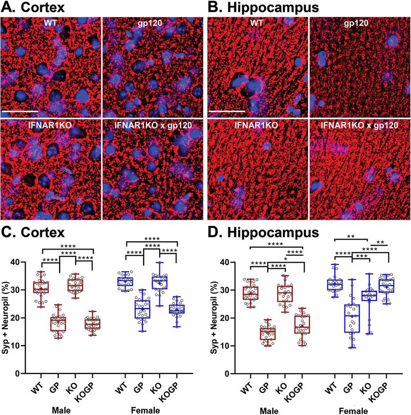
Singh et al. Journal of Neuroinflammation   (2020) 17:226                                                        Page 2 of 23

key neuropathological features observed in NeuroHIV             and independent processes of neuronal injury and be-
are astrocytosis, microgliosis, multinucleated giant            havioral impairment triggered by HIV-1.
cells, infiltration of macrophages, decreased synaptic
and dendritic density, and frank loss of neuronal cells         Methods
[8, 11–18].                                                     Mice
  One model that shares key features of neuropathology,         HIVgp120tg mice were kindly provided by Dr. Lennart
differential gene expression, and impaired learning and         Mucke (Gladstone Institute of Neurological Disease,
memory with people living with HIV (PLWH) and                   University of California, San Francisco, CA) [19], and mice
HAND/HAD is a transgenic (tg) mouse expressing the              deficient in functional IFNAR1 (B6.129S2-Ifnar1tm1Agt)
HIV envelope glycoprotein gp120 (HIVgp120) in the               [31] were provided by Dr. Carl Ware (Sanford Burnham
CNS [15, 18–20]. HIVgp120tg mice express the soluble            Prebys Medical Discovery Institute, La Jolla, CA). IFNA
viral envelope gp120 of HIV-1 LAV under the control of          R1KO and HIV gp120tg mice were crossbred, and the F3
the glial fibrillary acidic protein (GFAP) promoter in as-      generation of the HIVgp120tghet-IFNAR1het mice was
trocytes [19], and recent studies conducted in our lab re-      used to obtain the following four genotypes: (1) IFNA
vealed that the brains of HIVgp120tg mice show increased        R1KO-gp120 (IFNAR1-/-gp120+), (2) HIVgp120tg (IFNA
expression of interferon-stimulated genes (ISGs) [15, 16].      R1+/+gp120+), (3) IFNAR1KO (IFNAR1-/-gp120-; control),
Interferons (IFNs) are signaling proteins produced and          and (4) WT (IFNAR1+/+gp120-; wildtype control). All
released by host cells in response to pathogens, including      animals were maintained in a mixed C57BL/6.129/SJL
viruses, bacteria, parasites, and tumor cells [21]. Since the   genetic background. Genotyping of mice was performed
blood-brain barrier (BBB) restricts access of T and B cells     using genomic DNA isolated from tail clippings as
to the brain, the burden of HIV control largely rests with      previously published in the literature [15, 19] and by
local innate immune defense mechanisms [22]. As a result,       The Jackson Laboratory. In the present study, both
IFNs play a major role as the first line of host defense        male and female mice of all four genotypes were ana-
against HIV in the brain [23].                                  lyzed at the age of 11–14 months.
  The type I IFNs (IFNα/β) bind to a specific IFN-α/β re-         The preparation of cerebrocortical cell cultures was
ceptor (IFNAR) that consists of IFNAR1 and IFNAR2               performed as described earlier [17, 32–34]. Briefly, cere-
chains [24]. Both IFNα and IFNβ exert their effects by sig-     brocortical cells isolated from embryos of E16 Sprague
naling in an autocrine and paracrine manner through the         Dawley rats (Harlan Sprague Dawley Inc., San Diego,
JAK/STAT pathway to activate ISGs [25, 26]. While higher        CA) were plated on Poly-L-lysine-coated glass coverslips
levels of IFNα in the HIV-infected brain correlate with cog-    in 35-mm plastic dishes and generally used for experi-
nitive problems, IFNβ has been reported to control HIV          ments between 17 and 23 days in vitro when the cultures
and simian immunodeficiency virus (SIV) infection in the        contained ~30% neurons, ~70% astrocytes, and ~0.1 to
CNS [27–30]. Moreover, we recently demonstrated that            1% microglia [17, 32–34].
IFNβ treatment prevented in vitro and in vivo neuronal in-        All experimental procedures and protocols involving
jury induced by HIVgp120, depending on the presence of          animals were performed in compliance with the National
IFNAR1 and CCL 4[16]. However, without the application          Institute of Health (NIH) guidelines and approved by
of exogenous IFNβ, HIVgp120tg mice develop neuropath-           the Institutional Animal Care and Use Committees
ology, raising the question of whether or not IFNAR1 can        (IACUC) of the Sanford Burnham Prebys Medical Dis-
contribute to brain injury in the presence of baseline          covery Institute (SBP, The Scripps Research Institute
amounts of type I IFNs. Therefore, we investigated in the       (TSRI) and the University of California Riverside (UCR).
present study the potential role of type 1 IFN receptor
(IFNAR1 subunit) in HIV-induced neurotoxicity.                  Behavioral assessments
  Here, we report that genetic knock-out of IFNAR1              Eleven to 14-month-old WT (male: 11, female: 11),
provides partial protection against HIVgp120-induced            HIVgp120tg (male: 9, female: 8), IFNAR1KO (male: 9,
neuropathology and behavioral impairment in a sex-              female: 11), and IFNAR1KO × gp120 mice (male: 13, fe-
dependent fashion. In contrast, the associated cellular         male: 14) were behaviorally tested by the Animal Models
signaling via mitogen-activated protein kinases (MAPK)          Core Facility of TSRI. Mice were housed in standard
and signal transducer and activator of transcription-1          plastic cages on a reversed 12-h light/dark period (lights
(STAT1) shows sex-independent effects of HIVgp120               on at 8:00 PM), with food and water available ad libitum.
and genetic knockout of IFNAR1 (IFNAR1KO). How-                 The behavioral test battery was designed to examine
ever, expression of genes involved in neurotransmission         cognitive abilities as well as general activity, anxiety-like
and bioinformatics analysis provides further evidence of        behavior, and visual ability. The order of assessments
sexual dimorphism. Overall, our findings suggest that           was light/dark transfer (LDT), locomotor activity (LMA),
IFNAR1 plays a pivotal role in distinct sex-dependent           optomotor (OM), novel object recognition (NOR), and
Singh et al. Journal of Neuroinflammation   (2020) 17:226                                                      Page 3 of 23

Barnes maze test (BM) and was designed to reduce the           images were deconvolved using a constrained iterative
potential for confounds due to previous experience. Be-        algorithm, and threshold segmentation was applied to
havioral testing occurred between 9:00 AM and 12:00            estimate the percentage of the neuropil occupied by
PM (active phase) with 5–7 days between tests. LDT,            microtubule-associated protein 2 (MAP2+) neuronal
LMA, OM, and BM were performed as previously pub-              dendrites and synaptophysin (SYP+) presynaptic termi-
lished with a minor modification in the BM tests [15,          nals and compared between the different genotypes. For
18]. The NOR test assays recognition memory while              direct quantitative analysis of fluorescence intensities of
leaving the spatial location of the objects intact and is      GFAP in the frontal cerebral cortex or the hippocampus
believed to involve the hippocampus, perirhinal cortex,        (CA1), we recorded 2D images and the sum of fluores-
and raphe nuclei [35–37]. Mice were tested with two            cence intensities (SFLI) was quantified using the area of
identical objects placed in the field for 5 min. After three   interest. The SFLI values were normalized to the con-
such trials (each separated by 1 min in a holding cage),       trols for the measured area and adjusted for background
one of the familiar objects was changed for a novel ob-        by subtracting values obtained from sections incubated
ject. Habituation to the objects across the familiarization    only with secondary antibodies. For quantification of
trials (decreased contacts) was an initial measure of          Iba1+ microglia, cell bodies were counted in the cerebral
learning and then renewed interest (increased contacts)        cortex and hippocampus (CA1) on one side of three
in the new object indicated successful object memory.          sagittal brain sections spaced 320 μm apart medial to
                                                               lateral. The total cell bodies counts were normalized
Immunofluorescence staining, microscopy, and analysis          to the area.
The harvest of the brain tissue, immunofluorescence
staining, deconvolution and quantitative fluorescence          RNA isolation and quantitative RT-PCR
microscopy, and cell counting was performed as recently        Total RNA from mice hippocampus was extracted and
described [15, 16, 18]. In brief, mice were anesthetized       qRT-PCR was performed and analyzed as described pre-
using isoflurane and immediately transcardially perfused       viously with minor modifications [15, 16, 18]. Briefly,
with 0.9% saline. The brains were quickly removed and          RNA was extracted using a RNeasy Mini kit (Qiagen,
fixed for 72 h at 4 °C in 4% paraformaldehyde. For             Valencia, CA, cat# 74104), according to the manufac-
neuropathological analysis, 40-μm-thick sagittal brain         turer’s instructions. The RNA quality and quantity for
sections were obtained using a vibratome (Leica VT             all the samples were determined using a NanoDropTM
1000S, Leica Biosystems, Buffalo Grove, IL) and stained        DS-11 spectrophotometer (De Novix Inc., USA). Purified
with primary anti-microtubule-associated protein 2             RNA (500 ng) was used to generate a cDNA template.
monoclonal antibody (mAb; mouse anti-MAP2; 1:200;              qRT-PCR was carried out using 10 μL of Power PCR
Sigma) or a mouse anti-synaptophysin mAb (1:50; Dako)          SYBR green master mix (Applied Biosystems, Thermo
for neuronal markers, or rabbit anti-ionized calcium-          Fisher Scientific, Life Technologies LTD., Warrington,
binding adaptor molecule 1 (Iba1) IgG (1:125; Wako) for        UK), 1 μL of cDNA, 8 μL of PCR grade water, and 0.5 μL
microglia or rabbit anti-glial fibrillary acidic protein       of PCR primer pair (primer concentration: 20 μM except
(GFAP) IgG (1:250; Dako) for astrocytes. Rhodamine-            CXCL10: 10 μM). The primer sequences used for ampli-
conjugated goat anti-mouse (1:50 Jackson ImmunoRe-             fication are listed in Table 1. The RT-PCR was per-
search) and Alexa Fluor 488-labeled goat anti-rabbit (1:       formed in a QuantStudio™ 6 flex system Real-Time PCR
200; Invitrogen) secondary antibodies (Abs) were               System (Applied Biosystems/Life Technologies) using
employed to visualize primary Abs. Nuclei were counter-        the following settings: 95 °C for 10 min, 95 °C for 30 s,
stained with Hoechst 33342 (Invitrogen). Separate sections     59 °C for 1 min, 72 °C for 1 min, and for 40 cycles and
were incubated with mouse IgG1 (MOPC21, Sigma, M-              denaturation steps were added at the end of the amplifi-
9269) as primary isotype control or primary Ab was omit-       cation reaction for Tm analysis. RNA samples corre-
ted. Immunolabeled brain slices were mounted on glass          sponding to three to four biological replicates were
slides with fluorescence-protecting mounting medium            analyzed. The signal for internal control (glyceralde-
(VectaShield, Vector laboratories, Burlingame, CA, cat#        hyde-3-phosphate dehydrogenase, GAPDH) was used to
H1000) and overlaid by a coverslip.                            normalize the data. The relative amount of mRNA of
   Images were acquired using a Zeiss 200 M fluores-           every gene normalized to GAPDH was calculated follow-
cence deconvolution microscope equipped with a                 ing the 2−ΔΔCt method.
computer-controlled 3D stage and the appropriate filters
for DAPI, FITC, CY3, and CY5 (Carl Zeiss Microscopy            GABA glutamate and dopamine serotonin RT2 Profiler™
GmbH, Jena, Germany). Slidebook software (version 6,           PCR array
Intelligent Imaging Innovations, Inc., Denver, CO) was         The RNA extracted from mice hippocampus was used
used for all image acquisition and analysis. The Z-stack       to run GABA/glutamate (GG) and dopamine/serotonin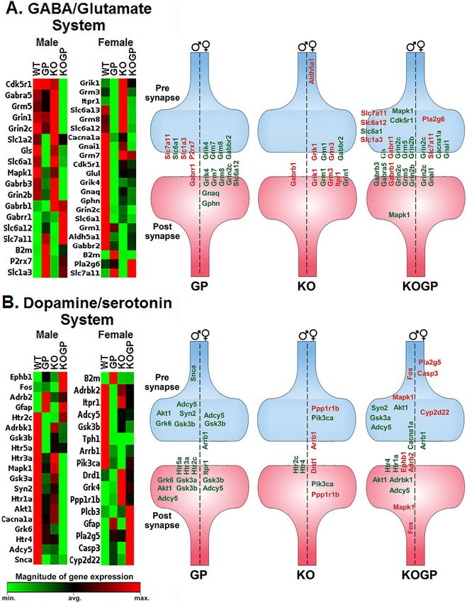
Singh et al. Journal of Neuroinflammation      (2020) 17:226                                                       Page 4 of 23

Table 1 Primers for qRT-PCR                                        Ingenuity® Systems, www.ingenuity.com; build version:
Gene            Genebank         Primer sequence (5′-3′)           486617 M; content version: 46901286; release date:
HIV-1 gp120     M19921           Fwd: TGAGCCAATTCCCATACATTATTG     2018-11-21). The core analysis function was employed
                                 Rev: CCTGTTCCATTGAACGTCTTATTATT   for identification of functional and biological gene net-
                                 AC                                works, upstream regulators as described previously with
mCcl2           NM_011333.3      Fwd: CCCAATGAGTAGGCTGGAGA         some modifications [15, 16, 18]. The following settings
                                 Rev: TCTGGACCCATTCCTTCTTG         were used: reference set, Ingenuity Knowledge Base
mCcl5/Rantes    NM_013653.3      Fwd: ACACCACTCCCTGCTGCTTT         (genes + endogenous chemicals), and relationship to in-
                                                                   clude direct; optional analyses: My Pathways My List;
                                 Rev: TGCTGCTGGTGTAGAAATACTCCTT
                                                                   Network Interaction and Causal, interaction network in-
mCcl3/Mip1α     NM_011337.2 15 Fwd: GCGCCATATGGAGCTGACA
                                                                   clude endogenous chemical molecules per network (35)
                                 Rev: GATGAATTGGCGTGGAATCTTC
                                                                   network per analysis (25), causal networks score using
mCcl4           NM_013652.2      Fwd: AGGGTTCTCAGCACCAATGG         causal path only; node type: all; data sources: all; confi-
                                 Rev: AGCTGCCGGGAGGTGTAAG          dence: experimentally observed and high predicted; spe-
mCcr5/CD195     NM_009917.5      Fwd: CGAAAACACATGGTCAAACG         cies: all; tissues and cell lines (adipocytes, astrocytes,
                                 Rev: GTTCTCCTGTGGATCGGGTA 20      immune cells, neurons, nervous system); mutation: all;
mCxcr4/CD184    NM_009911.3      Fwd: CTGGCTGAAAAGGCAGTCTATGT      fold change: − 1.1 to 1.1. Right-tailed Fisher’s exact test
                                                                   was performed for gene enrichment analysis.
                                 Rev: CGTCGGCAAAGATGAAGTCA
mSdf-1/         NM_              Fwd: TGCCCCTGCCGGTTCT 16
mCxcl12         001012477.2                                        Western blot analysis
                                 Rev: GAGTGTTGAGGATTTTCAGATGCTT
                                                                   Mouse brain tissue lysates were prepared as previously
mMx1            NM_010846.1      Fwd: AGAGCAAGTCTTCTTCAAGGATCAC    described with modifications [15, 39]. Briefly, the dis-
                                 Rev: GTGGCCTTCCCATCTTCCA          sected cerebral cortex or hippocampus was lysed on ice
mCxcl10         NM_021274.2      Fwd: GCCGTCATTTTCTGCCTCAT         using 600 μL of lysis buffer (a mixture of 10 mL of 1 ×
                                 Rev: GGCCCGTCATCGATATGG           RIPA buffer, 100 μL of phosphatase inhibitor (Calbio-
mCxcl11         NM_019594.1      Fwd: GGCTTCCTTATGTTCAAACAGGG      chem), and one tablet of complete protease inhibitor
                                                                   cocktail (Roche; Indianapolis, IN). The tissue samples
                                 Rev: GCCGTTACTCGGGTAAATTACA
                                                                   were homogenized using an electric pestle and 3 mL syr-
Gapdh           NM_008084.2      Fwd: AGGTCGGTGTGAACGGATTTG
                                                                   inge in 1.5 mL Eppendorf tubes. The lysed samples were
                                 Rev: TGTAGACCATGTAGTTGAGGTCA
                                                                   cleared by centrifugation (14,000×g, 15 min) at 4 °C, and
                                                                   the supernatant was collected as lysate. Total protein
(DS) RT2 PCR arrays (Qiagen). Each sample was reverse              concentrations were determined using the bicinchoninic
transcribed using the RT2 First-Strand kit (Qiagen),               acid (BCA) protein assay kit (Pierce/Thermo Fisher Sci-
mixed with RT2 qPCR Master Mix containing SYBR                     entific; Rockford, IL). Equal amount of protein samples
Green (Qiagen), and aliquoted (10 μL) into each well of            (50 μg) was mixed with 4× LDS sample buffer (Life
the RT2 Profiler™ PCR Arrays (Qiagen). Each sample was             Technologies, CA, USA) and 10× reducing agents (Invi-
used to assess the expression of 168 genes related to              trogen; Carlsbad, CA) and heated for 5 min at 100 °C.
neurotransmission. Samples were analyzed in the mouse              The samples were then electrophoretically separated on
GG (PAMM-152) and DS (PAMM-158) neurotransmit-                     SDS-PAGE gels (NUPAGETM; Invitrogen) and subse-
ter systems by RT2 Profiler™ PCR arrays following the              quently electro-transferred to polyvinyl difluoride
supplier’s instructions (SABioscience/Qiagen). The ar-             (PVDF) membranes (Invitrogen, USA). The following
rays were run on a QuantStudio 6 flex Real-Time PCR                antibodies were employed: phospho-p38 (1°Ab- 1:1000;
System (Applied Biosystems by Thermo Fisher). The                  anti-rabbit 2°Ab- 1:5000) (Cell Signaling; 9212), total
RT2 Profiler™ PCR Array Data Analysis software package             p38 (1°Ab- 1:2000; anti-rabbit 2°Ab- 1:25,000) (Cell Sig-
(version 3.5, Qiagen) used 2−(ΔΔCT)-based fold change              naling; 9211S), phospho-ERK1 (1°Ab- 1:1000; anti-rabbit
calculations [38] and a modified Student’s t test to com-          2°Ab- 1:5000) (Cell Signaling; 9101S), total ERK1 (1°Ab-
pute two-tail, equal variance P values. Data were nor-             1:2000; anti-rabbit 2°Ab- 1:5000) (Cell Signaling; 9102),
malized to the housekeeping genes actin beta (Actβ),               active JNK (1°Ab- 1:1000; anti-rabbit 2°Ab- 1:3000) (Pro-
glucuronidase beta (Gusβ), heat shock protein 90 alpha             mega; V93A 20542917), total JNK (1°Ab- 1:1000; anti-
(cytosolic) class B member 1 (Hsp90ab1), and Gapdh.                rabbit 2°Ab- 1:3000) (Cell Signaling; 9252), phospho-
                                                                   STAT1 serine 727 (1°Ab- 1:1000; anti-rabbit 2°Ab- 1:
Ingenuity Pathway Analysis                                         3000) (Cell Signaling; D3B7), total STAT1 (1°Ab- 1:
The results of the RT2 Profiler arrays were further inter-         1000; anti-rabbit 2°Ab- 1:3000) (Cell Signaling, 14994S),
rogated using Ingenuity Pathway Analysis software (IPA;            and housekeeping genes β-tubulin (1°Ab- 1:2000; anti-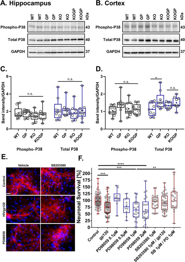
Singh et al. Journal of Neuroinflammation   (2020) 17:226                                                    Page 5 of 23

mouse 2°Ab- 1:10,000) (Sigma T9026) or GAPDH                  enrichment analysis right-tailed Fisher’s exact test was
(1°Ab- 1:20,000; anti-mouse 2°Ab- 1:25,000) (Ambion;          performed. Statistical significance was set at P ≤ 0.05.
4300). The secondary antibodies anti-mouse-HRP
(AP128P) and anti-rabbit-HRP (111-036-045) were pur-
chased from Millipore Sigma and Jackson Immuno Re-            Results
search. Western blots were imaged with a Bio-Rad              IFNAR1 and HIVgp120 affect memory in a sex-dependent
ChemiDoc system (Bio-Rad, Hercules, CA) and analyzed          fashion
using ImageJ software (NIH). Densitometric measure-           Mice of the following four genotypes were behaviorally
ments were normalized against housekeeping β-tubulin          assessed at 11 to 14 months of age: (1) IFNAR1KO ×
or GAPDH expression levels.                                   gp120 (IFNAR1-/-gp120+), (2) HIVgp120 (IFNA
                                                              R1+/+gp120+), (3) IFNAR1KO (IFNAR1-/-gp120-; con-
In vitro neurotoxicity experiments                            trol), and (4) WT (IFNAR1+/+gp120-; control).
Neurotoxicity experiments were performed as described            HIVgp120tg mice, but not WT, spent more time in
earlier [17, 32–34]. Recombinant gp120 of HIV-1 strain        the light compared to IFNAR1KO regardless of viral en-
SF2 was obtained from NIH AIDS Research and Reference         velope expression (Fig. 1a). However, neither IFNA
Reagent Program and was reconstituted in 0.1% bovine          R1KO nor HIVgp120 expression, nor any interaction in-
serum albumin (BSA) at 100× the final concentration.          volving the two factors, affected the number of transi-
Controls received BSA vehicle alone (0.001% final concen-     tions into the light compartment. Time spent in the
tration). The specific inhibitors of ERK kinase (MEK),        light compartment or dark-to-light transitions did not
PD98059, and p38 MAPK, SB203580, were obtained from           indicate any sex difference, regardless of genotype.
Calbiochem (San Diego, CA). Kinase inhibitors were dis-          The locomotor test showed significant effects of time
solved in dimethyl sulfoxide (DMSO) at 1000× final con-       on each activity measure (counts went down across time
centration (0.1, − 2 μM), and the p38 MAPK inhibitor was      as the mice habituated to the novel environment), but
added to the cerebrocortical cell cultures for 15 min prior   there were no significant interactions involving time,
to treatment with 200 pM HIVgp120 or PD98059. After           and therefore, the 2 h totals are shown in Fig. 1b. No sex
24-h incubation, surviving neurons were quantified by im-     differences or any interactions involving sex were de-
munostaining for the neuron-specific marker MAP-2, and        tected. Significant effects of IFNAR1KO and significant
apoptotic nuclei were identified morphologically after        IFNAR1KO × gp120 interactions, respectively, were
staining nuclei with the DNA dye Hoechst 33342 [32–34].       found in ambulation (P < 0.05), but not on activity in
Neuronal survival was calculated from the percentage of       the center of the cages and rearing (Fig. 1b). Also, all
neurons remaining after subtraction of those that had         mice showed head tracking in the optomotor test, sug-
undergone apoptosis. Two to six independent experiments       gesting that vision was intact (Additional file 1A).
were performed for each treatment.                               The novel object recognition test examined the differ-
                                                              ence between familiar vs. novel object contact time for
Statistical analysis                                          each group separately. Males in all groups showed sig-
Experimental results are presented in combined box-dot        nificantly more interest in the novel object than in the
plots with the 25th and 75th percentiles. The middle line     familiar object suggesting that recognition memory is
of the box shows the median, and the mean is indicated        not affected by IFNAR1KO and/or HIVgp120 expres-
by a “+.” The whiskers show the full range of values.         sion. In contrast and revealing sexual dimorphism, fe-
Values in Additional file 1, B and C, are shown as line       male HIVgp120tg mice failed to distinguish familiar and
graphs. GraphPad Prism 7.03 (GraphPad Software, Inc.,         novel objects, and this impairment was reversed by the
CA, USA) was employed for the generation of graphs            IFNAR1KO (P < 0.01) (Fig. 1c).
and a comparison of more than two experimental groups            The Barnes maze test indicated no significant effects
used ANOVA followed by Tukey’s honestly significant           of sex, nor any interaction involving sex, on the latency
difference (HSD) post hoc test. For behavioral outcomes       to escape and the number of errors made before escap-
in Barnes maze probe test and novel object recognition,       ing (Additional file 1B and C). While IFNAR1KO was
ANOVA analysis was performed followed by Fisher’s             associated with increased latencies to escape, there were
protected least significant difference post hoc test using    no effects of either IFNAR1KO or HIVgp120 on errors
StatView software package (version 5.0.1; SAS Institute,      made before escaping. In contrast, the probe test re-
Cary, NC). For the RT2 Profiler™ PCR Array Data ana-          vealed differences in spatial memory based on sex and
lysis, QIAGEN data analysis center was used for               presence of IFNAR1 and HIVgp120. HIVgp120 compro-
2–(ΔΔCT)-based fold change calculations and a modified        mised spatial memory in both sexes, but IFNARKO itself
Student’s t test to compute two tails, equal variance P       disrupted spatial memory in males while abrogating the
values (http://www.qiagen.com/geneglobe). For gene            impairment in females (Fig. 1d).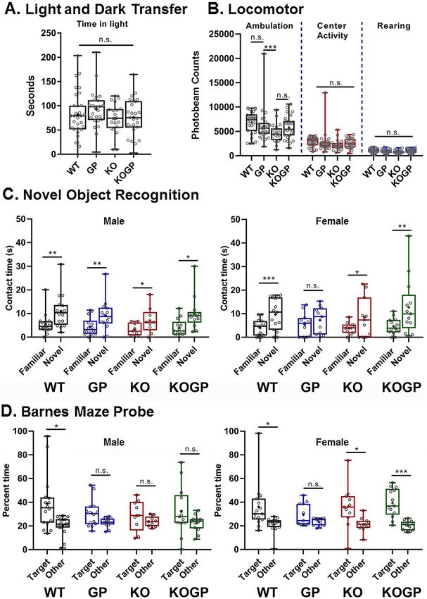
Singh et al. Journal of Neuroinflammation        (2020) 17:226                                                                               Page 6 of 23

 Fig. 1 IFNAR1 deficiency rescues recognition and spatial memory of HIVgp120tg females. Eleven to 14-month-old mice were behaviorally
 assessed (genotype: n for males/n for females—WT (WT): 11/11, HIVgp120tg (GP): 9/8, IFNAR1KO (KO): 9/11, and IFNAR1KO × gp120 (KOGP): 13/
 14). Light and dark transfer test for anxiety-like behavior (a); Locomotor (ambulation, center activity, and rearing (b); novel object recognition (c;
 left panel: males, right panel females); Barnes maze probe test for spatial learning and memory (d; left panel: males, right panel females). Data is
 presented in combined box-dot plots with the 25th and 75th percentiles. The middle line of the box shows the median, and the mean is
 indicated by a “+.” Statistical analysis was performed as described in the methods section. ***P < 0.001, **P < 0.01, *P ≤ 0.05; ANOVAs Tukey’s
 HSD (a, b) and Fisher’s PLSD post hoc tests (c, d); n = 17–27 (males and females) per group/genotype (total n = 86 animals); n.s., not significant.

IFNAR1KO partially prevents neuronal damage in                                 sections for synaptophysin (SYP) in presynaptic termi-
HIVgp120tg mice                                                                nals and for microtubule-associated protein 2 (MAP2) in
Following behavioral assessment and thus at an age of                          neuronal dendrites, respectively. Quantitative analysis
11–14 months, we analyzed the effect of IFNAR1-defi-                           using deconvolution microscopy showed significant loss
ciency on presynaptic terminals and neuronal dendrites.                        of SYP-positive neuropil (presynaptic terminals; P <
First, we immunofluorescence-labeled sagittal brain                            0.0001) in HIVgp120tg mice compared to WT in both
Singh et al. Journal of Neuroinflammation     (2020) 17:226                                                                          Page 7 of 23

frontal cortex (layer III; Fig. 2a, c) and hippocampus                     neuronal dendrites in both the cortex and hippocampus,
(CA1, stratum radiatum; Fig. 2b, d). IFNAR1KO did                          and this effect on MAP2 was sex-independent.
not affect presynaptic terminals in the cortex of both
males and females. In the male hippocampus, there
was no significant change between the WT and IFNA                          IFNAR1 deficiency regulates microglia activation but not
R1KO mice but the absence of IFNAR1 resulted in                            astrocytosis
protection against the loss of SYP+ neuropil otherwise                     Quantitative immunofluorescence microscopy of
seen in HIVgp120 expressing mice (Fig. 2d, left                            immunofluorescence-labeled GFAP of astrocytes showed
panel). In contrast, in the female hippocampus, IFNA                       an increase in association with HIVgp120 expression re-
R1 deficiency resulted in a loss of SYP+ presynaptic                       gardless of IFNAR1 genotype (Fig. 3a middle panels,
terminals in the absence of HIVgp120 but paradoxic-                        GFAP in the cortex, Fig. 3c quantification for the cortex
ally a more pronounced protection in IFNAR1KO ×                            and hippocampus). In the cortex, IFNAR1KO × gp120
gp120 mice (Fig. 2d, right panel). Overall, the results                    mice showed a significant decrease (P < 0.01) in the ex-
indicated a protective effect of IFNAR1 deficiency                         pression of GFAP compared to HIVgp120tg mice, while
against HIVgp120 induced loss for presynaptic termi-                       in the hippocampus GFAP expression was unaffected by
nals which was specific to the hippocampus and influ-                      IFNAR1 deficiency in the HIVgp120tg mice. However,
enced by sex in that females were more protected                           GFAP immunoreactivity in the hippocampus was de-
than males.                                                                creased in IFNAR1KO compared to the WT mice (P <
  The analysis of MAP2+ neurites showed a significant                      0.01) suggesting that IFNAR1 is necessary to maintain
reduction in HIVgp120tg mice in comparison to all the                      baseline levels of the astrocytic protein GFAP in this re-
other three genotypes (P < 0.0001; Fig. 3a, b). The IFNA                   gion of the brain. No sex-dependent differences were ob-
R1KO rescued HIVgp120tg mice from the loss of                              served for GFAP and astrocytosis.

 Fig. 2 IFNAR1 deficiency partially ameliorates the loss of presynaptic terminals preferentially in the hippocampus of females. Representative
 images of the cortex (a; layer 3) and hippocampus (b; CA1) immunolabeled for neuronal synaptophysin (SYP); deconvolution microscopy; scale
 bar, 40 μm. c, d Quantification of microscopy data obtained in the cortex and hippocampus of sagittal brain sections of 12–14-month-old mice.
 Genotypes: WT (WT), HIVgp120tg (GP), IFNAR1KO (KO), and IFNAR1KO × gp120 (KOGP). Values are presented in combined box-dot plots with the
 25th and 75th percentiles. The middle line of the box shows the median, and the mean is indicated by a “+”; ****P < 0.0001, ***P < 0.001, **P <
 0.01, *P < 0.05; ANOVA and Tukey’s HSD post hoc test; n = 6 animals (3 males and 3 females) per group/genotype (total n = 24 animals)
Singh et al. Journal of Neuroinflammation      (2020) 17:226                                                                           Page 8 of 23

 Fig. 3 IFNAR1 deficiency prevents damage to neuronal dendrites and microgliosis but not astrocytosis in HIVgp120tg mice. Representative
 images of the frontal cerebral cortex (a) immunolabeled for neuronal MAP2 (MAP2; layer 3; deconvolution microscopy; scale bar, 40 μm);
 astrocytic GFAP (cortex, all layers; fluorescence microscopy; scale bar, 40 μm), and Iba+ microglia numbers (cortex, all layers; fluorescence
 microscopy; scale bar, 100 μm). b–d Quantification of microscopy data obtained in the cortex and hippocampus of sagittal brain sections of 12–
 14-month-old mice. Neuropil positive for neuronal MAP2 (b); Fluorescence signal for astrocytic GFAP (c); Counts of Iba1+ microglia (d). Genotypes:
 WT (WT), HIVgp120tg (GP), IFNAR1KO (KO), and IFNAR1KO × gp120 (KOGP). Values are presented in combined box-dot plots with the 25th and
 75th percentiles. The middle line of the box shows the median, and the mean is indicated by a “+”; ****P < 0.0001, ***P < 0.001, **P < 0.01, *P <
 0.05; ANOVA and Tukey’s HSD post hoc test; n = 6 animals (3 males and 3 females) per group/genotype (total n = 24 animals)

  Iba1 was used as the marker for labeling microglia,                       significantly higher in HIVgp120tg mice compared to
and positive cell bodies were counted in the cerebral                       the other three genotypes in both cortex and hippocam-
cortex (layer III) and hippocampus (CA1) on one side of                     pus (Fig. 3a bottom panels, Iba1 in the cortex, Fig. 3d
three sagittal brain sections spaced 320 μm apart medial                    quantification for the cortex and hippocampus; P <
to lateral for each animal. Iba1+ microglia counts were                     0.05–0.0001). Similar to the astrocyte data, no sex
Singh et al. Journal of Neuroinflammation (2020) 17:226 Page 9 of 23
differences were observed. IFNAR1KO mice had lower CXCL12 mRNA expression because this natural ligand
counts of Iba1+ cells compared to all other genotypes in could compete with viral HIVgp120 for the interaction
both the cortex and hippocampus (P < 0.05–0.0001). In with CXCR4 [15, 40]. However, no significant effect was
the cortex, IFNAR1KO × gp120 mice had counts of detected of HIVgp120 or IFNAR1 deficiency on the ex-
microglia similar to WT mice and significantly less than pression level of CXCL12 (Fig. 4f, left panel).
the HIVgp120tg mice (P < 0.0001). In the hippocampus, Expression of MX1, an interferon-induced GTP-
IFNAR1KO × gp120 mice showed a significant increase binding protein with activity against DNA and RNA vi-
in the number of microglia compared to WT (P < 0.05) ruses [42], was significantly upregulated in the presence
and IFNAR1KO (P < 0.0001) mice, but still, the counts of HIVgp120 (P < 0.01; Fig. 4f, right panel). This upregu-
were lower than in HIVgp120tg mice (P < 0.05) (Fig. lation of MX1 was absent in IFNAR1KO and IFNA
3d). Thus, IFNAR1 deficiency appeared to overall R1KO × gp120 mice confirming the blockade of type I
dampen microgliosis. IFN signaling.
The chemokines CXCL10 and CXCL11 were assessed
IFNAR1KO affects the expression of the viral transgene because we found their levels to be elevated in the brain
and host genes of HIVgp120tg mice and CXCL10 has been linked to
We next investigated if the neuroprotection conveyed by neuronal dysfunction and found to be overexpressed in
IFNAR1 deficiency in the hippocampus was due to a the CNS of HIV encephalitis (HIVE) patients [8, 15, 16].
change in the expression of the viral envelope protein Results confirmed that the RNA expression of both che-
HIVgp120. The analysis by qRT-PCR revealed that mokines was significantly upregulated in the presence of
IFNAR1KO results in differential RNA expression for HIVgp120 (P < 0.0001 and P < 0.001, respectively; Fig.
HIVgp120 in males but not females (Fig. 4a). Expression 4g). The upregulation of CXCL10 mRNA in the pres-
of HIVgp120 was 2.5-fold higher in IFNAR1KO × gp120 ence of HIVgp120 occurred regardless of the IFNAR1
males compared to their respective HIVgp120tg controls genotype (Fig. 4g, left panel). In contrast, CXCL11 ex-
(P < 0.001). pression was significantly decreased in IFNAR1KO ×
We observed earlier that HIVgp120 expression in the gp120 compared to the HIVgp120tg mice hippocampus
brain induces ISGs, cytokines, and chemokines [15, 16]. (P < 0.05; Fig. 4g, right panel). IFNAR1KO did not sig-
In order to deduce the potential role of IFNAR1 in this nificantly affect the expression of CXCL10 and CXCL11
innate immune response, we analyzed the effect of in the absence of HIVgp120.
HIVgp120 and IFNAR1 deficiency on cytokine mRNA
expression in the hippocampus. The results of qRT-PCR Sex-dependent effects of IFNAR1KO and HIVgp120 on
showed that the expression of CCL2 and CCL5 was sig- neurotransmission-related gene networks
nificantly upregulated in HIVgp120tg compared to WT We recently showed that GABAergic, glutamatergic, dopa-
mice (P < 0.01 for CCL2 in females only, and P < 0.0001 minergic, and serotoninergic neurotransmission systems all
and 0.01 in males and females, respectively, for CCL5). are altered at the RNA level in HIVgp120tg mice [15, 18].
In males, CCL2 expression was highest for IFNAR1KO To test whether IFNAR1KO ameliorated the disturbance of
× gp120 mice and significantly increased compared to neurotransmission, we used RT2 PCR arrays to analyze the
WT and IFNAR1KO mice (P < 0.05 and 0.01). In con- expression of 168 genes associated with neurotransmission
trast, CCL5 expression in males was significantly in- by GABA and glutamate (GG array: 84 genes) and dopa-
creased in IFNAR1KO × gp120 mice compared to WT mine and serotonin (DS array: 84 genes) in the hippocam-
and IFNAR1KO controls (P < 0.05), but less than in pus. Compared to WT all other genotypes displayed
HIVgp120tg mice (P < 0.01; Fig. 4c, left panel). In fe- significant sex-dependent changes in the expression of
males, CCL5 expression was increased due to HIVgp120 genes related to neurotransmission. The differentially
irrespective of IFNAR1 (P < 0.05; Fig. 4c, right panel). expressed genes provided evidence for sex-dependent alter-
CCL3 and CCL4 were not significantly upregulated in ations of pre- and post-synaptic components and related
HIVgp120tg mice compared to the WT mice, but the signaling factors in HIVgp120tg mice with and without
knockout of IFNAR1 resulted in significant increases in IFNAR1, and IFNAR1-deficient females (Fig. 5 and
IFNAR1KO × gp120 mice compared to the other three Table 2). IFNAR1KO males only displayed alterations
genotypes (Fig. 4d). Sex differences were found only for in RNA for subunits of postsynaptic neurotransmitter
CCL2 and CCL5, but not CCL3 and CCL4. receptors. Notably, HIVgp120 reduced expression of
The chemokine receptors CCR5 and CXCR4 are the co- MAPK1/extracellularly regulated kinase (ERK), which
receptors for HIV infections alongside its primary receptor has been implicated in HAND [43, 44], and is a
CD4 [40, 41]. The qRT-PCR indicated that the mRNA ex- major upstream regulator of cAMP-response element-
pression of CCR5 and CXCR4 is not significantly different binding protein (CREB) which in turn affects multiple
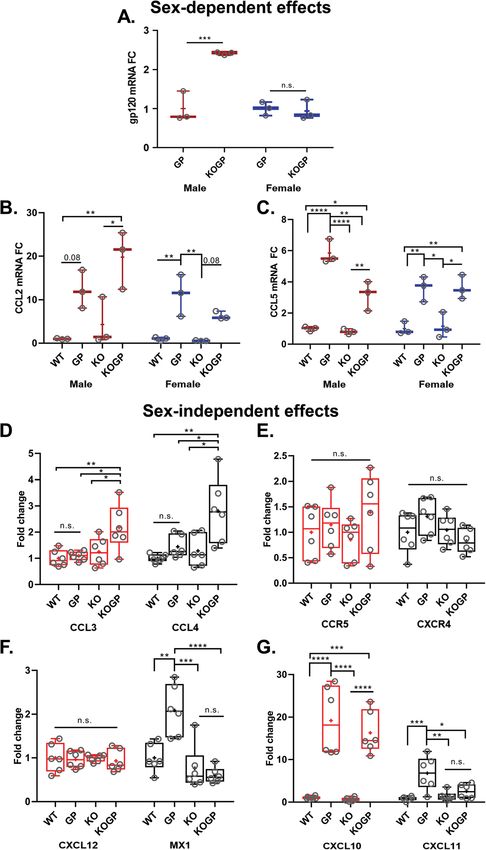
between the four genotypes (Fig. 4e). We also assayed components of neurotransmission.Singh et al. Journal of Neuroinflammation (2020) 17:226 Page 10 of 23 Fig. 4 Sex-dependent and independent effects of IFNAR1 deficiency on RNA expression of viral HIVgp120 and host genes. The mRNA expression in the hippocampus of 11-14 months-old mice, were detected by quantitative RT-PCR as described in the material and method section. HIVgp120 (a); CCL2 (b) and CCL5 (c), CCL3 and CCL4 (d); CCR5 and CXCR4 (e); CXCL12 and MX1 (f); CXCL10 and CXCL11 (g). The obtained CT values were normalized to the level of GAPDH mRNA. Genotypes: WT (WT), HIVgp120tg (GP), IFNAR1KO (KO), and IFNAR1KO × gp120 (KOGP). Values are presented in combined box-dot plots with the 25th and 75th percentiles. The middle line of the box shows the median, and the mean is indicated by a “+”; ****P < 0.0001, ***P < 0.001, **P < 0.01, *P < 0.05; ANOVA and Tukey’s HSD post hoc test; n = 3 per sex/experimental group/genotype or 6 animals (3 males and 3 females combined) per group/genotype (total n = 24 animals); n.s., not significant
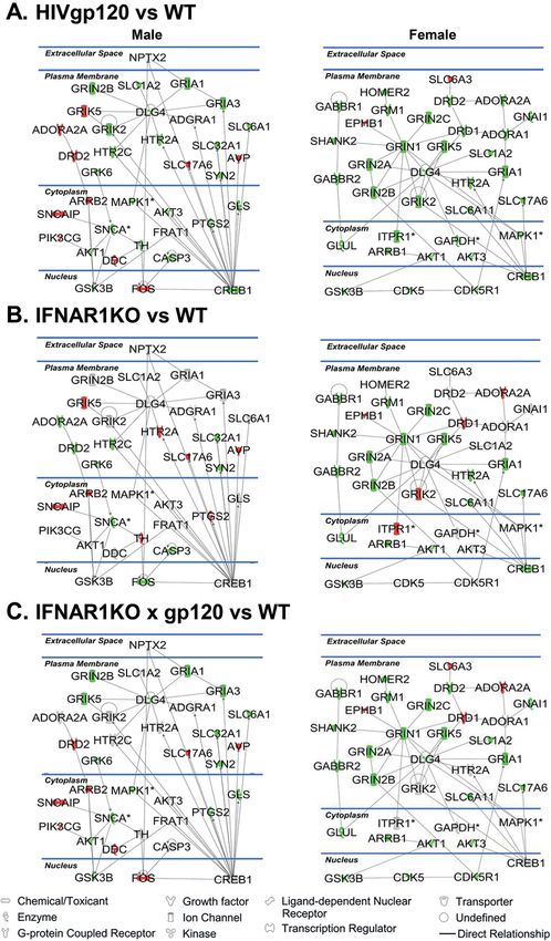
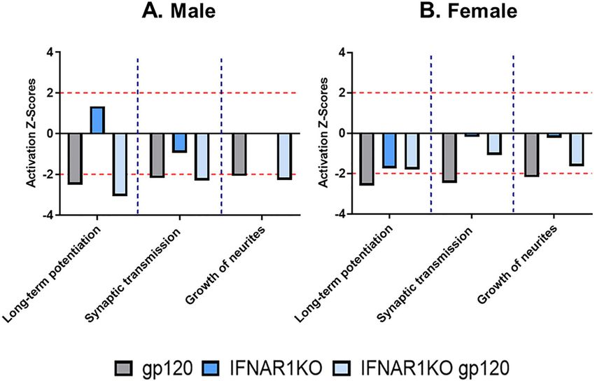
Singh et al. Journal of Neuroinflammation      (2020) 17:226                                                                           Page 11 of 23

 Fig. 5 HIVgp120 and IFNAR1 cause sex-dependent, differential expression of genes related to neurotransmission systems in the hippocampus.
 Clustergram heat map of GABA/glutamate system (a) and dopamine/serotonin system (b) showing gene expression profile of WT, HIVgp120tg,
 IFNAR1-deficient mice with or without HIVgp120. Red indicated the higher gene expression while green indicates the lower gene expression in
 the sample set. Hippocampus RNA was analyzed using RT2 Profiler PCR Array and the associated Qiagen data analysis software. The heat maps
 represent significantly changed genes as the averages of three biological replicates. The schematic figure to the right represents pre- and post-
 synaptic distribution of the differentially expressed genes in neurons of each genotype. Genotypes: WT (WT), HIVgp120tg (GP), IFNAR1KO (KO),
 and IFNAR1KO × gp120 (KOGP); n = 6 animals (3 males and 3 females) per group/genotype (total n = 24 animals)

  For further bioinformatics analysis, the data were                         potentiation (z-score = − 2.51 (M), − 2.59 (F)), (ii)
uploaded into the Ingenuity Pathway Analysis (IPA)                           synaptic transmission of nervous tissue (z-score = −
software application (Qiagen). The core analysis func-                       2.18 (M), − 2.47 (F)), and (iii) growth of neurites (z-
tion in IPA was used to interrogate the data in the                          score = − 2.9 (M), − 2.18 (F)). For IFNAR1KO ×
context of biological processes, canonical pathways,                         gp120 mice IPA indicated a slight mitigation of the
and networks. IPA predicted the diminished activation                        HIVgp120-associated effects in females but not in
of several functional gene networks in both males and                        males (Fig. 6a, b). In IFNAR1KO mice IPA showed
females of HIVgp120tg mice: (i) reduced long-term                            increased long-term potentiation in males but the
Singh et al. Journal of Neuroinflammation    (2020) 17:226                                                            Page 12 of 23

Table 2 Genes differentially regulated in males and females in the GABA/glutamate and dopamine/serotonin neurotransmission
systems
GABAergic & glutamatergic neurotransmission systems (male)
HIVgp120                                       IFNAR1KO                                  IFNAR1KO gp120
Gene         Fold regulation       P value     Gene          Fold regulation   P value   Gene       Fold Regulation        P value
Gabrr1       4.00                  0.041       Gabrb1        1.16              0.003     Cdk5r1     −1.21                  0.020
P2rx7        1.46                  0.002                                                 Gabra5     −1.32                  0.040
Slc1a2       −1.17                 0.044                                                 Gabrb1     1.13                   0.046
Slc1a3       1.21                  0.003                                                 Gabrb3     −1.14                  0.035
Slc6a1       −1.21                 0.023                                                 Gabrr1     13.52                  0.006
Slc7a11      2.52                  0.000                                                 Gls        −1.15                  0.027
B2m          1.90                  0.017                                                 Grin1      −1.29                  0.027
                                                                                         Grin2b     −1.35                  0.036
                                                                                         Grin2c     −1.91                  0.041
                                                                                         Grm5       −1.21                  0.026
                                                                                         Mapk1      −1.24                  0.026
                                                                                         Slc1a3     1.15                   0.033
                                                                                         Slc6a1     −1.18                  0.045
                                                                                         Slc6a12    3.50                   0.024
                                                                                         Slc7a11    2.77                   0.000
GABAergic & glutamatergic neurotransmission systems (female)
HIVgp120                                       IFNAR1KO                                  IFNAR1KO gp120
Gene         Fold regulation       P value     Gene          Fold regulation   P value   Gene       Fold regulation        P value
Cdk5r1       −1.25                 0.047       Aldh5a1       −1.30             0.039     Cacna1a    −1.60                  0.011
Gabbr2       −1.36                 0.031       Gabbr2        −1.74             0.005     Gnai1      −1.26                  0.010
Glul         −1.39                 0.014       Grik1         1.36              0.040     Grin2c     −1.77                  0.029
Gnai1        −1.27                 0.046       Grm1          −1.47             0.030     Pla2g6     1.16                   0.015
Gnaq         −1.18                 0.005       Grm3          1.21              0.015     Slc7a11    1.85                   0.044
Gphn         −1.38                 0.025       Itpr1         1.16              0.043
Grik4        −1.66                 0.048
Grin2c       −1.71                 0.030
Grm7         −1.25                 0.021
Grm8         −1.36                 0.032
Slc6a1       −1.33                 0.023
Slc6a12      −2.50                 0.039
Slc6a13      −2.11                 0.017
Slc7a11      1.84                  0.002
B2m          1.73                  0.024
Dopaminergic & serotonergic neurotransmission systems (male)
HIVgp120                                       IFNAR1KO                                  IFNAR1KO gp120
Gene         Fold regulation       P value     Gene          Fold regulation   P value   Gene       Fold regulation        P value
Adcy5        −1.27                 0.018       Htr2c         −1.27             0.0449    Adcy5      −1.42                  0.002
Adrbk1       −1.21                 0.045       Htr4          −1.17             0.0391    Adrb2      1.39                   0.009
Akt1         −1.14                 0.014                                                 Adrbk1     −1.14                  0.008
Gfap         14.54                 0.000                                                 Akt1       −1.30                  0.001
Grk6         −1.15                 0.036                                                 Cacna1a    −1.63                  0.022
Singh et al. Journal of Neuroinflammation (2020) 17:226 Page 13 of 23
Table 2 Genes differentially regulated in males and females in the GABA/glutamate and dopamine/serotonin neurotransmission
systems (Continued)
Gsk3a −1.18 0.023 Ephb1 1.77 0.004
Gsk3b −1.18 0.029 Fos 1.90 0.014
Htr2c −1.29 0.023 Gfap 13.82 0.000
Htr3a −1.21 0.027 Grk6 −1.46 0.003
Htr5a −1.55 0.032 Gsk3a −1.27 0.006
Snca −1.25 0.037 Htr1a −1.65 0.024
Syn2 −1.20 0.047 Htr4 −1.49 0.008
Mapk1 −1.27 0.046
Syn2 −1.34 0.018
Dopaminergic & serotonergic neurotransmission systems (female)
HIVgp120 IFNAR1KO IFNAR1KO gp120
Gene Fold regulation P value Gene Fold regulation P value Gene Fold regulation P value
Adcy5 −1.45 0.020 Arrb1 −1.59 0.018 Arrb1 −1.32 0.033
Adrbk2 −1.26 0.010 Drd1 1.56 0.046 Casp3 1.23 0.024
Arrb1 −1.43 0.014 Grk4 1.45 0.046 Cyp2d22 1.34 0.050
Gfap 10.43 0.001 Pik3ca −1.37 0.045 Gfap 12.41 0.000
Gsk3b −1.26 0.016 Ppp1r1b 1.51 0.048 Gsk3b −1.19 0.036
Itpr1 −1.13 0.040 Pla2g5 4.07 0.025
Tph1 −2.24 0.047 Plcb3 1.31 0.033
B2m 1.89 0.012
reduction in females, with a decline of overall synap- R1KO females (Fig. 7b) while its expression was un-
tic transmission in males. altered compared to WT control in IFNAR1KO × gp120
IPA also deduced several upstream regulators affected mice (Fig. 7c).
by HIVgp120 that explain the observed expression
changes [45], such as for IFNAR1KO × gp120 males and
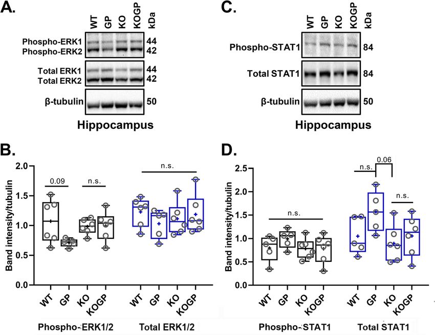
HIVgp120tg females an inhibition of CREB binding pro- IFNAR1 contributes to HIVgp120 effects on MAPK activity
tein (CREBBP; z-score = − 2.326 and − 2.00, respect- ERK1/2 is a major upstream regulator of CREB1 that
ively). The IFNAR1KO alone was not predicted to affect has also been implicated in HAND [43, 44]. We found
CREBBP in females. However, IPA implicated huntingtin here that the hippocampus of HIVgp120tg mice dis-
(HTT) as an activated upstream regulator (z-score = played a reduction in phospho-ERK1/2 (p-ERK1/2) com-
2.129) in HIVgp120tg females. The complete list of up- pared to WT controls (P = 0.091), and IFNAR1
stream regulators and their z-score values are shown in deficiency abrogated this effect (Fig. 8a, b). In contrast to
Table 3. the RNA expression of neurotransmission-related genes,
IPA generated for males and females high scoring the Western blotting experiments using 6 mice per
functional gene networks that were altered in the hippo- genotype (3 males and 3 females) showed no indication
campus of HIVgp120, IFNAR1KO, and IFNAR1KO × of sexual dimorphism.
gp120 in comparison to the WT. The high scoring net- Since IFNα/β bind IFNAR1/2 receptors and signal
work for HIVgp120tg males (score: 57; focus molecules: through the JAK/STAT pathway [25, 26], we also inves-
32; Table 4) is implicated in behavior, neurological dis- tigated the level of phospho-STAT1 (p-STAT1) and
ease, and cell to cell signaling interaction. The network STAT1. Western blotting indicated that p-STAT1 was
for HIVgp120tg females (score: 66; focus molecule: 35; slightly elevated above the baseline level by HIVgp120 in
Table 4) is linked to cell-to-cell signaling, nervous sys- the presence of IFNAR1 but without reaching signifi-
tem development, and behavior. These networks were cance. Expression of STAT1 protein was higher in the
affected by IFNAR1-deficiency as shown in Fig. 7. Not- hippocampus of HIVgp120tg mice compared to the
ably, CREB1, an important regulator of neuronal pre- other genotypes with a reduction in IFNAR1KO (P =
and postsynaptic function [44], was downregulated in 0.06; Fig. 8c, d) confirming the functional disruption of
both HIVgp120tg males and females (Fig. 7a) and IFNA IFNα/β signaling.Singh et al. Journal of Neuroinflammation (2020) 17:226 Page 14 of 23
Fig. 6 IFNAR1KO modifies the activity of functional, biological pathways affected by HIVgp120. Gene expression data obtained from RT2 Profiler
PCR Arrays were interrogated using Ingenuity Pathway Analysis (IPA) software to analyze neurotransmission-related biological pathways affected
by HIVgp120 and IFNAR1 deficiency. Note that most pathways were predicted to be reduced in activity except for long-term potentiation in IFNA
R1KO males. The dashed lines at 2 and − 2 on the axis for activation Z-score indicate the significance threshold for a predicted effect on the
activity of the given pathway (*P < 0.05); n = 6 animals (3 males (a) and 3 females (b) per group/genotype (total n = 24 animals)
Another regulator of CREB1 is the stress-activated p38 difference in phospho-p38 levels, suggesting IFNAR1 con-
MAPK [44], which is also essential for neuronal death tributes to HIVgp120-induced phosphorylation of p38
and microglia/macrophage activation in HIVgp120- MAPK. Overall, the data showed that IFNAR1 contrib-
induced neurotoxicity [32–34]. To quantify active p38 uted to the effects of HIVgp120 on p38 MAPK.
MAPK, we performed Western blots for the phosphory- Our previous studies have shown that neurons display
lated kinase and its total protein. Probing hippocampal the highest baseline levels of phosphorylated p38 MAPK
and cerebrocortical tissues, we found that phospho-p38 in the mixed neuro-glial cell populations [17, 34]. In an
MAPK was not significantly altered in any of the four in vitro approach using cerebrocortical cell cultures, we
tested genotypes; however, expression in the HIVgp120tg found that inhibition of ERK1/2 activation with increas-
mice trended higher compared to WT (P < 0.21, Tukey ing concentrations of the ERK kinase (MEK) inhibitor
HSD). The total-p38 (p38) protein levels were increased in PD98059 caused neurotoxicity to a similar extent as
the HIVgp120-expressing cortex (P < 0.05 for p38 protein; HIVgp120 (Fig. 9e). Moreover, both the toxicity induced
10 mice per genotype, 5 males, and 5 females) but not by the viral protein and the MEK inhibitor were com-
hippocampus compared to WT control (Fig. 9a–d). The pletely abrogated by blocking the activity of p38 MAPK
IFNAR1KO mice with or without HIVgp120 lacked a with SB203580. These findings were in line with the
Table 3 Ingenuity Pathway Analysis (IPA) predicted upstream regulators in male vs female
Upstream regulator Molecule type Predicted activation state Activation z-score P value
Males
HIVgp120 None scored
IFNAR1KO None scored
IFNAR1KOgp120 CREBBP Transcription regulator Inhibited − 2.236 0.0000532
Females
HIVgp120 HTT Transcription regulator Activated 2.129 2.3E−18
CREBBP Transcription regulator Inhibited − 2.0 0.0000145
IFNAR1KO CREBBP Transcription regulator Inhibited − 2.0 0.000054
IFNAR1KOgp120 CREBBP Transcription regulator Inhibited − 2.0 0.0000209Singh et al. Journal of Neuroinflammation (2020) 17:226 Page 15 of 23
Table 4 List of molecules comprising the top-scoring network in male vs female identified by Ingenuity Pathway Analysis (IPA)
Sex Molecules in network Score Focus molecules Top diseases and functions
Males ADGRA1,ADORA2A,AKT1,AKT3,ARRB2,AVP,CASP3,CREB1,DDC,DLG4, 57 32 Behavior, cell-to-cell signaling and
DRD2,FOS,FRAT1,GLS,GRIA1,GRIA3,GRIK2,GRIK5,GRIN2B,GRK6,GSK3B interaction, neurological disease
,HTR2A,HTR2C,MAPK1,NPTX2,PIK3CG,PTGS2,SLC17A6,SLC1A2,SLC32A1,
SLC6A1,SNCA,SNCAIP,SYN2,TH
Females ADORA1,ADORA2A,AKT1,AKT3,ARRB1,CDK5,CDK5R1,CREB1,DLG4,DRD1, 66 35 Behavior, cell-to-cell signaling and
DRD2,EPHB1,GABBR1,GABBR2,GAPDH,GLUL,GNAI1,GRIA1,GRIK2,GRIK5, interaction, nervous system
GRIN1,GRIN2A,GRIN2B,GRIN2C,GRM1,GSK3B,HOMER2,HTR2A,ITPR1, development and function
MAPK1,SHANK2,SLC17A6,SLC1A2,SLC6A11,SLC6A3
observations in the HIVgp120-expressing mouse brains neurocognitive and memory impairment. In addition, we
and suggested that both the reduction of active ERK1/2 find that the effect of IFNAR1 is, at least in part, sex-
and the activity of p38 MAPK are mechanistic compo- dependent.
nents of HIVgp120 neurotoxicity in the presence of Gender-specific studies in HIV-positive patients re-
IFNAR1. vealed that HIV-positive women display higher immune
In contrast to p38 MAPK, the stress-related cJun N- activation and have a higher risk of developing AIDS
terminal kinase (JNK) did not display any significant [56]. Additionally, a recent report of the Women’s In-
changes in terms of phosphorylation or total protein level teragency HIV study found evidence that women
in the cortex and hippocampus of HIVgp120tg, IFNA maybe more vulnerable to cognitive impairment [57].
R1KO, or IFNAR1KO × gp120 mice compared to WT Murine models of HIV-brain injury show some data to
controls, indicating that JNK activity was not significantly support this point as female HIV-1 transgenic rats
changed by chronic exposure to the viral envelope protein showed worse sustained attention than male rats [58].
regardless of IFNAR1 (Additional file 2A-D). While our behavioral assessment found that recogni-
tion memory was only impaired in female HIVgp120tg
Discussion mice, it was rescued by IFNAR1 deficiency. Interest-
HIV infection of the CNS often leads to brain injury, ingly, not all memory types are affected in a sex-
and HAND persists despite the use of cART, but the dependent manner as the Barnes maze paradigm re-
neuropathological mechanisms are still not completely vealed spatial learning and memory was impaired in
understood [1, 46, 47]. However, infection with HIV both sexes of HIVgp120tg mice but the knockout of
triggers an IFN response [23], and IFNα/β and IFNα/γ IFNAR1 rescued memory function in females while
all can inhibit HIV-1 infection in the periphery and in compromising it independently of HIVgp120tg in
the brain [48–50]. We recently found that similar to males. The Barnes maze probe test is sensitive to im-
SIV-infected rhesus macaques and mice intracerebrally paired hippocampal function [59], and this result fits
injected with HIV-1 infected human macrophages, well with the observation that pre-synaptic terminals in
HIVgp120tg mice mount a type I IFN response in the the hippocampus of males were diminished while the
CNS [15, 16, 27, 28, 30]. IFNα is produced by a wide pre-synaptic terminals in the females of IFNAR1KO ×
variety of nucleated cells that exist in or can enter the gp120 mice were indistinguishable from WT control.
CNS, including astrocytes, microglia, neurons, macro- Of note, the reduction of SYP+ presynaptic terminals in
phages, and T-lymphocytes [28, 51]. IFNα is an effective the female hippocampus and spatial memory impair-
anti-viral host factor [52, 53], but prolonged expression ment of male IFNAR1KO mice indicates an important
of IFNα causes cognitive dysfunction in the HIV-1 ex- role of the type I IFN receptor in normal brain function
posed CNS [28, 30, 54]. Conversely, IFNβ, which can be via mechanisms that warrant further investigation [55].
produced by parenchymal and immune cells [25, 26, 55], Others reported recently in a mouse model that lack of
has been reported to control of HIV and SIV infection IFNβ signaling via IFNAR1 caused Lewy body forma-
in the brain [27, 29], and we have shown that intranasal tion, motor and cognitive learning impairments, reduc-
IFNβ treatment confers in vivo neuronal protection tion in dopaminergic neurons, defective dopamine
against toxicity of viral HIVgp120 [16]. These same signaling, and a Parkinson’s disease phenotype [60].
treatment experiments indicated that endogenous levels However, beyond the sex-dependent spatial memory
of IFNβ were insufficient to provide neuroprotection impairment and reduced ambulatory locomotor activ-
over time and therefore raised the possibility that IFNA ity, we did not observe a comparably severe phenotype
R1 signaling could contribute to CNS injury because of in the absence of IFNAR1.
the presence of endogenous IFNα [16]. Indeed, the However, in HIVgp120tg mice, damage to MAP2+
present study provides evidence that IFNAR1 contrib- neurites seems to be more dependent on IFNAR1 than
utes to HIVgp120-induced neuronal injury and injury to SYP+ presynaptic terminals, and effects on theSingh et al. Journal of Neuroinflammation (2020) 17:226 Page 16 of 23 Fig. 7 Functional neural gene networks of neurotransmission affected by HIVgp120 and IFNAR1 deficiency. RNA expression data obtained with the GABA/glutamate and dopamine/serotonin RT2 Profiler PCR Array were analyzed using IPA software. Green indicates downregulated while red reflects upregulated genes respectively. The components without color represent genes that were included by IPA without experimentally determined expression levels. IPA identified for each sex a different highest scoring gene direct interaction network in which alterations on expression levels were driven by HIVgp120 in the absence or presence of IFNAR1. The solid lines represent a direct relationship and the arrow indicates the direction of action. *Indicates genes for which differential regulation reached significance in the RT2 Profiler PCR Array (*P < 0.05; modified t test). RNA expression data were derived from n = 6 animals (3 males and 3 females) per group/genotype (total n = 24 animals) latter are sex-dependent. Genetic ablation of IFNAR1 occurred only in the hippocampus. SYP+ presynaptic ter- ameliorated the loss of MAP2+ neurites in the cortex minals in the cerebral cortex were more reduced in and hippocampus with no difference between the sexes. HIVgp120tg males than females but unaffected by IFNA In contrast, the protection of SYP+ presynaptic terminals R1 deficiency in both sexes. In contrast, presynaptic ter- was sex-dependent and specific to the brain region as it minals were more protected in female than male
You can also read