Optimizing Nutritional Strategies to Prevent Necrotizing Enterocolitis and Growth Failure after Bowel Resection - MDPI
←
→
Page content transcription
If your browser does not render page correctly, please read the page content below
nutrients
Review
Optimizing Nutritional Strategies to Prevent Necrotizing
Enterocolitis and Growth Failure after Bowel Resection
Laura Moschino 1 , Miriam Duci 2 , Francesco Fascetti Leon 2 , Luca Bonadies 1 , Elena Priante 1 ,
Eugenio Baraldi 1 and Giovanna Verlato 1, *

                                            1   Neonatal Intensive Care Unit, Department of Women’s and Children’s Health, University Hospital of Padova,
                                                35128 Padova, Italy; lauramoschino13@gmail.com (L.M.); luca.bonadies@aopd.veneto.it (L.B.);
                                                elena.priante@aopd.veneto.it (E.P.); eugenio.baraldi@unipd.it (E.B.)
                                            2   Pediatric Surgery Unit, Department of Women’s and Children’s Health, University Hospital of Padova,
                                                35128 Padova, Italy; ducimiriam@gmail.com (M.D.); francesco.fascettileon@unipd.it (F.F.L.)
                                            *   Correspondence: verlatogiovanna@gmail.com; Tel.: +39-0498211428

                                            Abstract: Necrotizing enterocolitis (NEC), the first cause of short bowel syndrome (SBS) in the
                                            neonate, is a serious neonatal gastrointestinal disease with an incidence of up to 11% in preterm
                                            newborns less than 1500 g of birth weight. The rate of severe NEC requiring surgery remains
                                            high, and it is estimated between 20–50%. Newborns who develop SBS need prolonged parenteral
                                            nutrition (PN), experience nutrient deficiency, failure to thrive and are at risk of neurodevelopmental
                                            impairment. Prevention of NEC is therefore mandatory to avoid SBS and its associated morbidities.
                                            In this regard, nutritional practices seem to play a key role in early life. Individualized medical and
                                            surgical therapies, as well as intestinal rehabilitation programs, are fundamental in the achievement
                                            of enteral autonomy in infants with acquired SBS. In this descriptive review, we describe the most

                                     recent evidence on nutritional practices to prevent NEC, the available tools to early detect it, the
                                            surgical management to limit bowel resection and the best nutrition to sustain growth and intestinal
Citation: Moschino, L.; Duci, M.;
Fascetti Leon, F.; Bonadies, L.; Priante,
                                            function.
E.; Baraldi, E.; Verlato, G. Optimizing
Nutritional Strategies to Prevent           Keywords: necrotizing enterocolitis; short bowel syndrome; human milk; nutrition; surgical man-
Necrotizing Enterocolitis and Growth        agement; bowel sparing
Failure after Bowel Resection.
Nutrients 2021, 13, 340. https://
doi.org/10.3390/nu13020340
                                            1. Background
Academic Editor: Paola Roggero
                                                  Short bowel syndrome (SBS) is a state of malabsorption defined as the need for
Received: 30 November 2020
                                            parenteral nutrition (PN) for >60 days after bowel resection or as a bowel length of less
Accepted: 19 January 2021
                                            than 25% of expected [1]. SBS is the principal cause of intestinal failure (IF) in the pediatric
Published: 24 January 2021
                                            age [2]. The incidence of SBS has been estimated to be 24.5/100.000 births per year [3], but it
                                            may reach 7/1000 births in preterm newborns with birth weight (BW)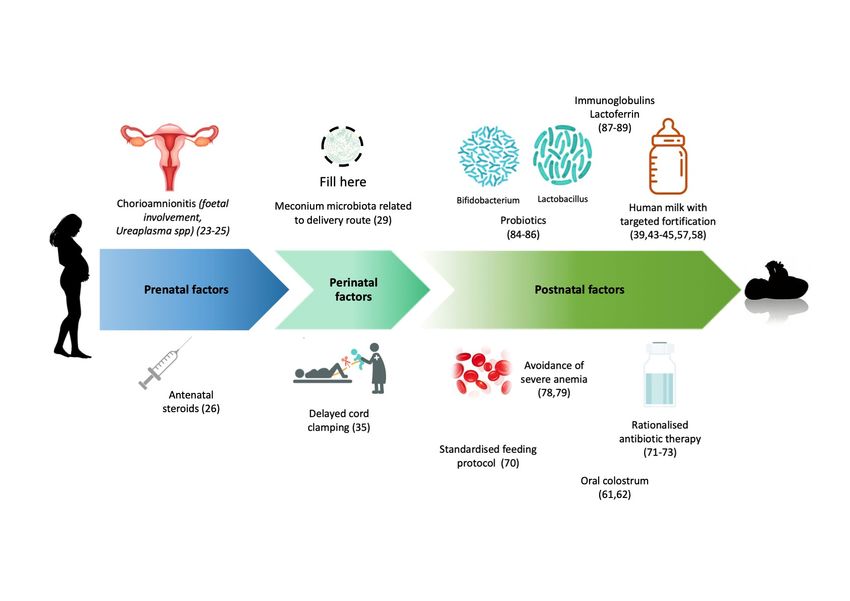
Nutrients 2021, 13, 340                                                                                            2 of 22

                          have a mild presentation (abdominal distention only) or a typical association of signs
                          and symptoms (emesis, bloody stools, intestinal pneumatosis, abdominal tenderness) as
                          described according to Bell’s staging and its modification by Walsh et al. [14,15]. Ultimately,
                          NEC can progress to full intestinal necrosis [16] with a high associated mortality (around
                          50% in those born extremely preterm) [17] and several comorbidities in those who survive,
                          with SBS developing in 42% of those requiring surgery [18].
                               Due to the lack of a specific therapy against NEC-related bowel damage, rationalized,
                          targeted, and prolonged prevention programs and early recognition are fundamental to
                          limit bowel loss. In fact, the length of the residual bowel, as well as the presence of the
                          ileo-caecal valve and of the colon are the main factors influencing the chances of weaning
                          from PN [19–22].
                               In this descriptive review, we report the most recent evidence to prevent and early
                          detect NEC, the surgical management to limit bowel resection and SBS development, and
                          the best nutrition to sustain growth and enhance intestinal function.

                          2. Factors to Prevent NEC Development
                               There are several measures which were demonstrated to be promising in reducing the
                          incidence of NEC in premature infants, although with variable evidence.

                          2.1. Prenatal and Perinatal Factors
                               Starting from the womb, clinical maternal chorioamnionitis seems to be significantly
                          associated with NEC, while this does not appear to be true for histological chorioamnionitis
                          without fetal involvement (funisitis, fetal surface vessel angiitis, increased inflammatory
                          markers in umbilical cord or fetal blood). Despite the data are still preliminary, there is
                          a good available evidence that supports a role of antenatal inflammation in NEC patho-
                          physiology [23,24]. It is possible that as in bronchopulmonary dysplasia (BPD), maternal
                          chorioamnionitis plays a different role in NEC pathophysiology depending on its onset
                          (acute or chronic), its association with severe inflammatory response syndrome (SIRS)
                          of the fetus, and the involved pathogen. Recently, Ureaplasma species have been ac-
                          knowledged as major causative pathogens of both BPD and NEC, most likely by inducing
                          pro-inflammatory factors and down-regulating the immune system [25].
                               These data could partially explain why antenatal corticosteroids appear to be effective
                          in NEC prevention. From randomized clinical trials (RCT), a decreased risk of NEC is
                          seen with antenatal corticosteroids in pregnant women at risk of preterm birth [26]. A
                          recent review and meta-analysis of nine observational studies, however, demonstrated that
                          antenatal corticosteroid use before 25 weeks’ gestation (which is controversial), does not
                          influence the rate of NEC ≥stage II of Bell [27].
                               Mode of delivery is one of the first determinants of gut microbiota, together with
                          gestational age, antibiotic treatment, and diet [28]. Compared to infants born vaginally,
                          those born via cesarean section show decreased intestinal population of Bifidobacteria and
                          Bacteroides and increased population of Clostridium difficile [29]. However, in a secondary
                          analysis of data from a randomized controlled trial, mode of delivery was not significantly
                          associated with development of NEC in neonates of women who were at imminent risk of
                          delivery at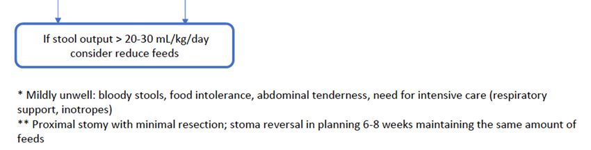
Nutrients 2021, 13, 340                                                                                          3 of 22

                               Finally, a lower birth weight at delivery increases the risk of NEC, with placental dis-
                          ease predisposing the severely growth-restricted neonate to the disease [36]. Additionally,
                          in antenatally identified pregnancies at risk of fetal growth restriction, abnormal Doppler
                          velocimetry in the umbilical artery (absent/reverse end-diastolic flow) is a useful guide to
                          predict NEC and mortality in the early neonatal period [37,38].

                          2.2. Post-Natal Factors
                               When it comes to post-natal life, other protective factors have come into focus, and
                          the importance of an optimized nutrition has been highlighted.

                          2.2.1. Feeding Management
                                Starting from the feeding type, since the 1990s, human milk (maternal or donor)
                          has proven to lower the risk of NEC compared with bovine protein-based formula [39].
                          Maternal breast milk is recommended for preterm and low birth weight infants as it
                          has been demonstrated to attenuate the toll-like receptor 4 mediated pro-inflammatory
                          response, typical hallmark in NEC pathogenesis, by activating the receptor for epidermal
                          growth factor (EGFR) and thus resulting in enhanced mucosal healing, intestinal stem
                          cell proliferation and decreased enterocyte apoptosis [28,40]. In the case of insufficient
                          supply, maternal breast milk can be replaced by donor human milk, despite pasteurization
                          and freezing of the latter reduce some of the protective benefits of the former [41,42].
                          The incidence of NEC, indeed, has been described as 6–10 times higher in exclusively
                          formula-fed infants compared to the exclusively breastfed ones [43–45]. Human breast
                          milk, which has an osmolarity of around 300 mOsm/L, acts by increasing proteolytic
                          enzymes and decreasing gastric pH, thus determining less pathogenic bacterial flora and
                          improving epithelial membrane and tight junctions. In addition, in preterm infants it
                          stimulates peristalsis and gut motility, together with the immune system through secretary
                          IgA, lactoferrin, growth hormones and oligosaccharides, thereby lowering the extent of
                          microbial dysbiosis [33]. By contrast, preterm infant formula appears to alter the intestinal
                          flora selecting potential pathogenic bacteria such as Clostridia and Proteobacteria [46],
                          despite the relatively safe osmolarity of most products (from 210 up to 270 mOsm/L) [47].
                          Interestingly, the positive effects of maternal milk appear to be dose-dependent, with
                          higher intake of human milk leading to higher protection from NEC [45,48].
                                Multi-nutrient fortification adds protein, vitamins, and other minerals to human milk,
                          therefore preventing nutrient deficits and extra-uterine growth restriction in exclusively
                          breast milk-fed preterm infants [49,50]. A Cochrane review published in 2016 concluded
                          that there is only low-quality evidence that multi-nutrient fortified breast milk compared
                          with unfortified breast milk does not increase the risk of NEC (RR 1.57, 95% CI 0.76 to 3.23;
                          11 studies, 882 infants) [51]. Similar findings have emerged from a recent RCT in South
                          India, where standard fortification of pasteurized donor human milk did not increase the
                          incidence of NEC compared to the unfortified one [52]. Commonly, multi-nutrient fortifiers
                          to breast milk derive from bovine milk, but fortification of breast milk feeds with human
                          milk-derived fortifier is available. Nevertheless, a Cochrane review of one randomized
                          trial showed that the latter does not seem to decrease the risk of NEC, feeding intolerance,
                          late-onset sepsis or death, compared to bovine milk-derived fortifier [53,54].
                                In recent years two new fortification strategies have gained popularity to optimize
                          macronutrient intake, improve growth and minimize feeding intolerance and NEC [55,56].
                          The first is adjustable fortification based on blood urea nitrogen levels to adjust fortifier
                          strength. The second is target and customized fortification through human milk analyzers
                          that fortifies macronutrients individually to achieve the desired intake [57]. Composition
                          of native breast milk, indeed, has individual inter- and intra-sample variation. Targeting
                          components of fortification ensures that current osmolarity recommendations are followed,
                          as fortification could increase the osmolarity of breast milk [58]. The addition of 1 g of
                          carbohydrates (glucose polymer), 1 g of hydrolyzed protein, or 1 g of whey protein per
                          100 mL breast milk, seem to determine an average increase in osmolality of 20, 38, and
Nutrients 2021, 13, 340 4 of 22
4 mOsm/kg respectively. Recently, prediction models to estimate osmolality values after
fortification have been published [59,60].
Oral colostrum, both of bovine or maternal origin, is rich in nutrients and bioactive
factors. Although intact bovine colostrum added to donor human milk appeared superior
to formula-based fortifiers to support gut function, nutrient absorption, and bacterial
defense in preterm pigs [61], the oral administration of colostrum does not seem to reduce
NEC onset from recent meta-analyses [62].
Regarding initiation and advancement of enteral feeds, recent systematic reviews
demonstrated that trophic feeds can be started within 96 h from birth and at higher volumes
without affecting the risk of NEC in VLBWI [63,64]. However, limited data exist for infants
bornNutrients 2021, 13, 340 5 of 22
Compared to placebo, probiotics seemed to provide positive results in several small
sample-sized studies [81,82]. Nevertheless, the optimal strain, dosing and timing of their
administration still need to be established [83], and concerns arouse in the past due to the
reported cases of sepsis related to their administration [84]. Recently a position paper has
been released by the ESPGHAN Committee on Nutrition and the ESPGHAN Working
Group for Probiotics and Prebiotics on the probiotic strains with greatest efficacy regarding
relevant clinical outcomes for preterm neonates. This paper favors the use of Lactobacillus
rhamnosus GG ATCC 53,103 or of a combination of Bacillus infantis Bb-02, Bacillus lactis
Bb-12 and Streptococcus thermophilus TH-4 to reduce NEC Bell’s stage II and III, although
with low certainty of evidence [85]. As concluded by a recent Cochrane review of the
literature, further, large, high-quality trials are needed to provide evidence of sufficient
quality and applicability to inform policy and practice [86].
Similarly, no recommendation can be made regarding the use of oral immunoglobulin
(IgG alone or IgG plus IgA) [87] nor of enteral lactoferrin as an adjunct to antibiotic therapy
Nutrients 2021, 13, x FOR PEER REVIEW 6 of 24
for the prevention or treatment of NEC [88]. In fact, the former did not reduce the incidence
of definite NEC, suspected NEC, need for surgery, or death from NEC in a meta-analysis of
five RCTs, while the latter did not improve the rates of Bell stage II and III (proven and
advanced) NEC nor that of late-onset sepsis compared to placebo in the largest RCT [89].
A summary of antenatal and postnatal factors involved in NEC pathogenesis is
reported in Figure 1.
Figure 1. Prenatal, perinatal and postnatal factors which have been found, with stronger or weaker recommendation, to
reduce the subsequent risk of NEC development. The role of certain factors, like chorioamnionitis, meconium at birth,
delayed cord clamping, oral colostrum, is still debated. Icons from Freepik and Pch.vector, downloaded from freepik.com.
3. Improving NEC Diagnosis: Indicators of Suspected NEC
Figure 1. Prenatal, perinatal and postnatal factors which have been found, with stronger or weaker recommendation, to
reduce the subsequent risk of NEC Given the frequently
development. sudden
The role onsetfactors,
of certain and thelike
potential devastating
chorioamnionitis, effects, itat
meconium is birth,
of extreme
importance to apply a combination of imaging and monitoring tools to early
delayed cord clamping, oral colostrum, is still debated. Icons from Freepik and Pch.vector, downloaded from freepik.com. recognise
patients at risk and act before development of the disease. Studies conducted so far have
predominantly explored laboratory features and thresholds that could reveal the incipient
onset of NEC, and their results have been extendedly described elsewhere [90,91].
Relatively new interesting approaches, such as metabolomic and microbiota analysis,
have been applied on serum, urine and fecal samples to investigate prognostic factorsNutrients 2021, 13, 340 6 of 22
of NEC onset. Studies using these techniques have been mostly prospective and have
included small sample sizes of matched NEC infants and controls. Some of them have
revealed that a NEC-associated gut microbiota can be identified in meconium or pre-NEC
stool samples [92], with an increased relative abundance of Proteobacteria and Firmicutes,
and decreased relative abundances of Bacteroidetes prior to NEC onset [93]. Among the
Preoteobacteria, Escherichia coli and Klebsiella pneumoniae seem the most pathogenic,
while Clostridia are the most abundant among Firmicutes. Bifidobacteria, instead, are often
lacking in pre-NEC stools of affected patients [94].
Non-invasive parameters have been advocated to continually monitor premature
neonates in order to early detect predictive changes. The monitoring of transcutaneous
PO2 (tcPO2 ) has proven to be safe and accurate in very sick infants [95,96] with a good
linear correlation between tcPO2 and Partial Pressure of Oxygen (PaO2 ) [97]. In one study,
drops of the tcPO2 /PaO2 ratio could be the spy of NEC requiring surgical intervention,
with appropriate response to fluid resuscitation in survivors [98].
Near Infrared Spectroscopy (NIRS) assessment of neonatal splanchnic oxygenation
(SrSO2 ) has gained increasing interest over the last decade. The infraumbilical abdomen
is considered the most reliable area for sensor placement [99]. In preterm neonates, a
reliable correlation between SrSO2 and mesenteric Doppler has been reported [100], with
evidence supporting the feasibility of NIRS in the monitoring of enteral feeding [101] and
NEC development [102,103]. Lower infraumbilical SrSO2 and higher Fractional Oxygen
Extraction within twenty-four hours after onset of symptoms suspicious of NEC can predict
subsequent gastrointestinal complication (Bell’s stage IIIB or death) [103]. Interestingly,
Doppler and NIRS techniques have been combined in the study of transfusion-associated
NEC and confirmed a possible pathogenic role of the pre-existing severe anemia rather
than of RBC transfusion in the onset of NEC [104]. The same authors found that feeding
during RBC transfusion, instead, was related to a post-prandial decline in the postprandial
mesenteric oxygenation as measured by SrSO2 [105]. The splanchnic–cerebral oxygenation
ratio has been proposed as an index to predict splanchnic ischemia [106].
Abdominal ultrasound has become more and more popular in the diagnostics of
necrotizing enterocolitis thanks to its non-invasiveness, quick use, and good performance
also in equivocal cases at abdominal radiography. With good accuracy typical signs of NEC
can be recognized, such as intestinal wall thickness and pneumatosis (hyperecogenic foci),
intestinal loops’ peristalsis and dilation, ascites, pneumobilia and pneumoperitoneum.
Color Doppler may reveal perfusion of the intestinal wall and flows in the abdominal aorta
and mesenteric vessels, with recognition of subtle hyperaemia of bowel loops or lack of
flow [107]. Interestingly, a recent single-center study in 104 preterm neonates showed a
promising value of Doppler ultrasound of the superior mesenteric artery as additional
prediction surrogate to predict NEC. In particular, a higher peak systolic velocity and
differential velocity measured in the superior mesenteric artery within the first 12 h of life
were significantly related to the risk of NEC [108].
Further studies are needed on the use of laboratory values, perfusion indices and
imaging techniques for the early recognition of NEC.
4. Surgical Treatment: Control of Long-Term Consequences
4.1. Surgery Aims
For Patients with NEC refractory to maximal medical treatment (multi-organ failure,
progressive clinical deterioration) or with perforated NEC, surgery is indicated. In the
current literature, the percentage of patients requiring surgical treatment is consistently
between 20% and 50% [109,110].
Several different operative management are described which vary between teams
from minimalistic strategies (e.g., peritoneal drainage) to demolitive laparotomy.
The surgery leading principles are:
− to prevent the short bowel syndrome (limit the resection)
− to limit bacterial translocation by diverting fecesNutrients 2021, 13, 340 7 of 22
− to reduce the risk of sepsis and control the inflammatory cascade (by resecting necrotic
bowel), reducing the consequent risk of multi-organ failure.
4.2. Surgery Options
Deciding the type of operation to perform depends on the extension of the disease but
also on the surgeons’ experience.
4.2.1. Stoma Versus Primary Anastomosis
The most traditional surgical approach for NEC is to perform a laparotomy with
diverting stoma proximal to most diseased bowel. This procedure has some advantages,
mainly of allowing the heal of downstream bowel without translocation of stool/bacteria.
Some centers consider refeeding the proximal stoma effluent through the distal mucous
fistula in order to stimulate mucosal growth and minimize fluid and electrolyte losses.
However, there is little evidence to support the efficacy of this practice [111,112]. Stomal
complications including fluid losses, electrolyte abnormalities, poor growth, prolapse or
retraction of stoma led some groups to consider primary anastomosis as the first option
to treat severe NEC. Guelfand et al. [113] reported primary anastomosis as a safe proce-
dure in the treatment of complicated NEC with low morbidity and mortality (11.6%). A
recent systematic review [114] of 12 studies compared these two approaches and found no
significant difference in terms of complications or mortality rate, although the spectrum
of complications was slightly different in each group, potentially due to selection bias
for treatment options and heterogeneity of included studies. Similarly, a survey of the
European Pediatric Surgeons’ Association (EUPSA) reported that the majority of surgeons
(67%) opted for bowel resection and primary anastomosis in the case of focal NEC, while
75% would perform a stoma in case of multi-focal disease [115].
4.2.2. Peritoneal Drainage
In VLBWI, a minimalistic strategy is often advocated, consisting of in the placement
of a peritoneal drainage (PD) without intestinal exploration in case of free air at the X-ray.
This procedure was firstly described by Ein et al. in 1977 [116] as a way to stabilize neonates
until they are in better conditions to undergo an explorative laparotomy. Over time, PD
has become popular and some pediatric surgeons consider PD not only as a temporizing
measure but also as a definitive treatment. Tashiro et al. showed that in premature (Nutrients 2021, 13, 340 8 of 22
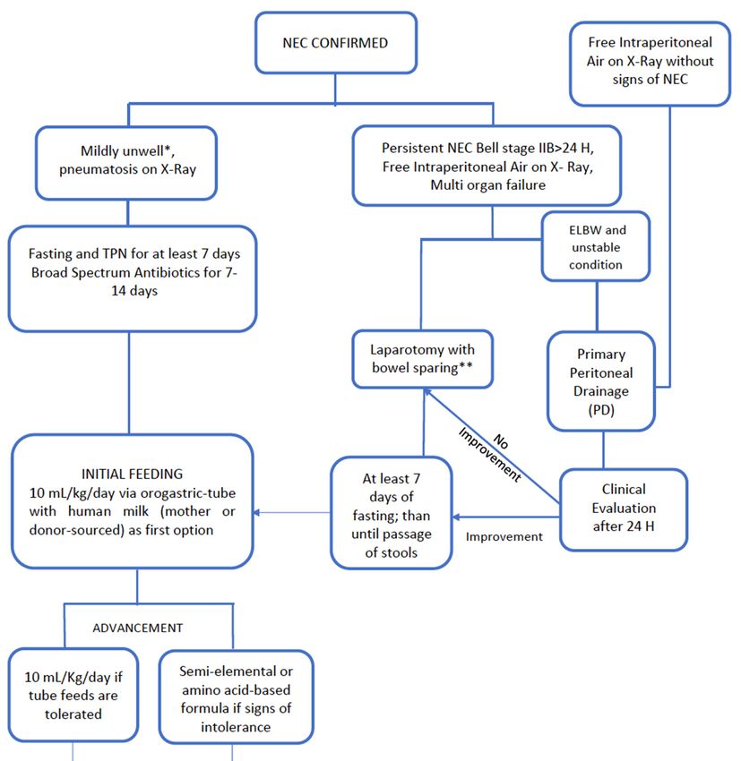
delayed laparotomy (Figure 2). As previously described, this policy based on the concept
Nutrients 2021, 13, x FOR PEER REVIEW 10 of 24
of “sparing surgery” is safe and it seems to be associated with a lower mortality rate than
the one reported in the literature (6.4%) [121].
Figure
Figure 2. Our center 2. Our
Policy forcenter
infantsPolicy
with for infants with NEC.
NEC.
4.3. NEC Totalis Management
A clinician’s challenge in the management of NEC is the NEC totalis (NEC-T), where
the risk of SBS is very high. No univocal definition is reported in the current literature, as
according to some groups NEC-T occurs when the entire small intestine is involved,
whereas for others when the necrosis involves the small and the large bowel [122]. In ad-
dition, defining the bowel vitality in a pan-intestinal necrosis is not always straightfor-
ward and it may result in unnecessary extensive resection. Furthermore, defining the realNutrients 2021, 13, 340 9 of 22
4.3. NEC Totalis Management
A clinician’s challenge in the management of NEC is the NEC totalis (NEC-T), where
the risk of SBS is very high. No univocal definition is reported in the current literature, as ac-
cording to some groups NEC-T occurs when the entire small intestine is involved, whereas
for others when the necrosis involves the small and the large bowel [122]. In addition,
defining the bowel vitality in a pan-intestinal necrosis is not always straightforward and it
may result in unnecessary extensive resection. Furthermore, defining the real extension of
the disease requires the surgical exposition of the whole bowel, with the aforementioned
danger. In 2004, Pierro et al. proposed the use of gasless laparoscopy to define the length
of intestine involved by necrosis [123]. Many different surgical approaches have been
described. In 1989, Moore et al. introduced the “patch, drain and wait technique” [124].
This approach consists of covering the perforation with sutures (patch), positioning two
drains (drain) and then waiting with long-term parenteral nutrition (wait). The critical
aspects of this type of treatment is the uncontrolled clearance of the abdomen from necrotic
tissue and feces. In 1996, Vaughan et al. proposed the “clip and drop technique” that also
aims to avoid ostomies and to preserve intestine length [125]. This technique involves
resection of necrotic bowel and tiding off the ends of healthy bowel tracts. A second-look
laparotomy after 48–72 h is used to determine the true extent of the disease and intestinal
continuity is restored. Arnold et al. reported their experience using this strategy and found
lower incidence of SBS (9.1%) compared to 16% in the published data [126]. Therefore, they
suggested the “clip and drop” technique in selected patients with NEC-T to help bowel
conservation in survivors.
Alternatively, intraluminal stenting could be used to preserve the length of the intes-
tine as much as possible in these sick infants [127]. The management of NEC-T remains
very controversial and significant practice variability persists. Nevertheless, it raises ethical
issues. In fact, the total-length extension of necrosis gives scarce chances of bowel function
recovery. In the pre-parenteral nutrition era, palliation was the choice rather than surgical
techniques to keep the patient alive. Presently, in developed countries tailored PN and
later in life the bowel transplant option, impose to consider and treat these patients. If no
healthy small bowel can be found at the laparotomy, tube duodenostomy can be used as a
temporizing maneuver in these neonates.
4.4. Complications
Intestinal strictures after NEC, firstly described by Rabinowitz 1968 [128] is a well-
known common complication of NEC, affecting about 9–36% of patients [129,130]. Dif-
ferently from the onset of NEC which is most frequent in the terminal ileum/cecum, the
most common site of post-NEC-stricture is the left colon (80%). In the current literature,
some studies focused on predictive factors of this complication in order to avoid intesti-
nal resection. A retrospective study by Phad et al. [131] found that leucocytosis during
NEC and length of resected bowel at surgery may be associated with increased risk of
developing post-NEC intestinal stricture. In their multicenter study, Zhang et al. showed
that the late onset of NEC (>10 days of life) and the higher level of procalcitonin at the
onset of NEC could be consider independent factors for post-NEC strictures [132]. A
recent meta-analysis showed that earlier feedingNutrients 2021, 13, 340 10 of 22
stoma in 6 weeks (if the management of ostomy’s losses allows it), and when the patient is
growing properly.
A recent systematic review demonstrated that there were no significant postoperative
complications after stoma reversal between early and late stoma closure [138]. However,
this review mainly included retrospective studies, with differences in the management of
NEC and small number of patients. Again, a RCT should be planned to demonstrate the
superiority of late versus early closure of stoma. In addition, further prospective trials are
needed to evaluate the outcomes of existing approaches.
5. Best Nutrition Strategies to Enhance Intestinal Adaptation and Sustain Growth
The origin, development, and treatment of NEC are still being debated. Nonetheless,
while certain feeding practices are recognized preventive factors, there is no consensus on
when and how reintroduce enteral feeding after NEC and in SBS patients after surgery.
5.1. When to Start Nutrition
After NEC diagnosis, bowel rest is suggested for 7–10 days [139,140]. However, the
reason for a long period of fasting is unclear since the presence of macro and micronutrients
in the intestinal lumen enhance intestinal adaptation [141–143].
Since feeding, together with prematurity and altered gut colonization, is one of the
key factors that triggers the inflammatory cascade leading to NEC, clinicians are afraid
of restarting enteral nutrition because of the possibility of recurrence of NEC. Recurrent
NEC was reported with an incidence of 4–6% [144,145], with a higher incidence in those
refed before 10 days after an episode of NEC [146]. However, other studies reported no
complications associated with an earlier refeeding practice after NEC [147–149].
In a retrospective study, Bonhorst et al. [148] compared patients subjected to a new
protocol of feeding practice with an historical group. The more recent group of newborns
was fed with a median of four days after three consecutive days without evidence of gas
bubbles in the portal vein studied with the use of abdominal ultrasound. This group
was compared with a cohort group fed on the basis of the neonatologist discretion (me-
dian 10 days). The early feeding group reached full enteral feedings in significantly less
days (10 days vs. 19 days), had a reduced duration of central venous access (13.5 days
vs. 26.0 days) and had a lower rate of catheter-related septicemia (18% vs. 29%,) with
consequent shorter length of hospital stay (63 days vs. 69 days).
Other studies, though considering only non-surgical NEC, retrospectively analyzed
the outcomes of patients receiving early (5 or ≥7 days) [147,149].
Brotschi et al. [149] found that neonates refed in less than 5 days developed less catheter-
related sepsis and required less surgery for early post-NEC stricture. Arbra et al. [147]
retrospectively reviewed the ten-years data in a single center and analyzed outcomes in
patients fed at < vs. ≥7 days from NEC diagnosis. After adjusting for NEC stage, the
composite outcome for stricture, recurrence of NEC or death was not significantly different
between the early and late refeeding groups.
Two Metanalyses have been performed on refeeding practices after NEC [133,150].
These found that early enteral feeding (within 5 days of NEC diagnosis) did not seem to be
associated with adverse outcomes, including NEC recurrence.
Early enteral feeding is important to prevent gut atrophy and to improve intestinal
growth in parenterally fed preterm newborns [151]. In a rat model of NEC, a 25% reduction
of enteral nutrients resulted in a reduced villus height and gut mass [152] while early
feeding after surgery was associated with improved intestinal adaptation in piglets [153].
Given this evidence, several experts report that enteral feeding should start as soon as
possible in newborns after surgery to stimulate gut adaptation [7,154–156], though no clear
guidelines have been established yet [115]. Other experts, in contrast, state that enteral
feeding should start when bowel sounds are present, enteral drainage is no longer bilious,
the abdomen is soft and there is no vomiting [157].Nutrients 2021, 13, 340 11 of 22
5.2. Type of Enteral Feeding
To our knowledge, there is no trial aimed at verifying the best type of feeding after
NEC resection. There is a large consensus reporting human milk as best choice of feed-
ing for newborns and infants with SBS [44,156,158–161]. Breast milk is rich in proteins
(immunoglobulin A), nucleotides, live cells, and growth factors together with other compo-
nents such as lactoferrin and more than 100 different oligosaccarydes that can reinforce the
immune system and enhance intestinal growth and adaptation [162–165].
Human milk is recommended by the Enhanced Recovery After Surgery Society as
first choice of nutrition in the newborn after surgery [166]. In the absence of human milk,
it is still debated the best type of formula milk to use. Several NICUs use extensively
hydrolyzed protein formula in the absence of human milk [167] as recommended by
some experts [168]. The use of Extensively Hydrolyzed Formula (EHF) is justified by the
increased risk of protein allergy characterizing newborns after intestinal surgery [169,170]
and by the possible relationship between protein’s allergy and NEC [171,172]. EHFs do not
contain lactose and usually have an increased concentration of medium-chain triglycerides
(MCT). It seems therefore to be preferable if it is taken into account that undigested lactose
is a contributing factor to NEC development in animals [173] and that preterm newborns
have a reduced lactase activity compared to their term counterparts [174]. In addition,
components such as MCT can be better absorbed in the case of rapid transit, bacterial
overgrowth, and bile acid depletion [175] as it is after surgical NEC. Nonetheless, the
possible disadvantages of EHF should be considered, since it does not satisfy the elevated
requirements in preterm infants [176]. As far as we know, the only randomized study
comparing hydrolyzed vs. non-hydrolyzed formula in children with SBS did not detect
differences in tolerance and weight gain [177]. Furthermore, breast milk together with
elemental formula (aminoacid-based) resulted in shorter parenteral nutrition dependence
compared to the hydrolyzed formula [20,178–182].
Finally, macronutrients in a complex form were found to better promote bowel adapta-
tion in animal models [183–186], outlining the importance of bowel workload in enhancing
adaptation [187].
For the aforementioned reasons, some authors suggested to feed premature infants
with preterm formula, in the absence of human milk, and then verify its tolerance [187].
More recently, the use of cow’s preterm formula has been recommended in the absence
of human milk [158], since it is characterized by higher caloric density and a composi-
tion based on lower lactose, higher MCT and long-chain triglycerides contents with the
well-known advantages of these lipids [188]. The use of semi-elemental or elemental
formula is suggested in those patients who are intolerant to conventional preterm/term
formula [159,160].
5.3. How to Increase Feeds
After an episode of NEC requiring surgery, most of the studies suggest starting
feeds with 10 mL/kg/day initially [148,149,157] and then advance by increasing from
10 to 20 mL/kg/day [188] or 20 mL/kg/day [148,149]. Others, however, suggest a
more cautious approach with smaller advancements (1–2 mL every 3 h for 24–48 h or
0.5–1 mL/kg/day) [141,143]. Especially in ELBWI who were never previously fed, some
authors report to increase 1 mL every 4 h for 5 days [189].
There is a quite uniform consensus to limit advancing of feeds when stool/stoma out-
put is more than 30–50 mL/kg/day [7,157,188–190], or when it is more than 20 mL/kg/day
or with a stool production of >6–10 times/day [157,191]. It is, therefore, mandatory for the
clinician to carefully quantify stool number/volume [157] and to observe possible clinical
changes (vomit, bowel distention, irritability) [168] before increasing enteral nutrition.
There is no clear preference in the literature for the feeding method (continuous vs.
bolus) in preterm [192] infants after bowel resection [193]. Despite of the fact that bolus
feeding increases splanchnic perfusion with an improvement in digestion [194], growth of
VLBWI could benefit from continuous feeding [195]. Continuous feeding is suggested inNutrients 2021, 13, 340 12 of 22
newborns and infants with SBS [7,155,158,160] at the beginning of the refeeding process, at
least during nighttime [196] or for the first 24 h [160]. It is reported that continuous feeding
may enhance absorption and improve growth in selected groups of patients [197].
On the other hand, both animal [198] and human studies [199] demonstrate that bolus
feeding is more physiological, increases mucosal mass and enzyme content and bowel
adaptation [190].
A practical approach could be to start with continuous feeding followed by bolus feed-
ing, and as soon as possible introduce small volume of feeding orally administered [159,193]
to stimulate swallow reflexes and avoid later full aversion disease [200,201].
In general, the evidence shows that the use of a standardized refeeding protocol
results in fewer days to achieve the 50% of enteral nutrition and in less Intestinal Failure
Associated Liver Disease (67% vs. 42%) [202].
5.4. Optimizing Parenteral Nutrition and Laboratory Controls during Refeeding
During the refeeding process, it is fundamental to maintain the potential growth of
the newborn, despite SBS avoiding both over- and underfeeding. The new 21st Century
Preterm Postnatal Growth Standards Charts from birth to 6 months of corrected age can
help the clinician to monitor growth and provide the right macro and micronutrient
requirements after bowel resection [203].
Although energy expenditure seems to be unaltered in surgical neonates [204], there
is a paucity of data in preterm infants. In addition, these subjects face a very rapid cerebral
and organ growth which requires high nutrient and caloric intakes compared to their term
counterparts [205,206].
Macro and micronutrients must be guaranteed through the parenteral route when
enteral nutrition is insufficient to meet required intakes. PN is important in all the phases
after bowel resection: In the first phase soon after bowel surgery, when the newborn is
completely dependent and aggressive fluid and electrolytes replacement are warranted; in
the second phase, when enteral nutrition is started; in the third phase when all the efforts
are directed toward PN weaning [158]. The necessity of maintaining growth has to be
balanced with the excess of macronutrient intakes by PN.
The excess of w6 Long Chain Tryglycerides, for instance, can adversely affect the
liver due to the pro-inflammatory and pro-oxidative actions [207]. Soy emulsion are
rich in phytosterols that by the enteral route would be only poorly absorbed, but by the
parenteral route may lead to liver damage. In preterm infants, especially in those with
cholestasis, phytosterols have longer half-life [208–210], therefore exposing these subjects
to an increased risk of liver injury. For this reason, recent guidelines on lipid intakes report
that for “PN lasting longer than a few days, pure soybean oil based intravenous lipid
emulsions should no longer be used, and composite Intravenous Lipid Emulsions with or
without fish oil should be the first choice” [211].
Higher dextrose and amminoacids intakes, as well, may cause increased prevalence
and earlier onset of PN -Related Cholestasis [212]. On the other hand, it should be kept
in mind that some non-essential amminoacids, such as taurine and cysteine, may become
conditionally essential in preterm newborns, who therefore could benefit from their sup-
plementation [213,214] together with the fact that in some cases an amminoacids’ amount
up to 3.5 g/kg/day should be administered to prevent catabolism [215].
It is, therefore, recommended to adjust energy intakes on patient’s condition and to
avoid excess energy intakes by PN maintaining a nonprotein carbohydrates/lipids ratio of
75/25 with an upper triglyceride level in the newborn of 2.5 g/L during lipid infusion [201].
Preterm infants with SBS need to receive macronutrient but also micronutrients (such
as iron, zinc, copper, selenium, Vitamins) and electrolytes (calcium magnesium, sodium) to
prevent their deficiencies [216–219].
For this purpose, a strict monitoring of laboratory values reflecting liver function, ni-
trogen content, renal function, and electrolytes and vitamin levels is of extreme importance
for infants [220] and newborns on long-term PN [155] keeping in mind that electrolytesNutrients 2021, 13, 340 13 of 22
serum levels could seem appropriate despite a low total body content. Levels of elec-
trolytes in urine (potassium, sodium, magnesium), should be routinely performed as well.
In particular, urinary sodium should be kept >20–30 mEq/L [220,221] to meet the elevated
newborn’s requirements for growth.
To conclude, nutrient intakes must be adapted according to the newborn’s nutritional
requirements and through frequent anthropometric and biochemical assessments [155,156].
Table 1 describes the feeding protocols adopted to initiate or advance after surgery
for NEC.
Table 1. Summarizes the Proposed Refeeding protocols after NEC in the current literature.
Type of Feeding in
Authors and Journal Year Initial Feeding Advancement Type of Feeding Absence of
Human Milk
Preterm/term formula
Christian V.J. et al.
Continuous feeds: Human milk - If patient is intolerant:
(Nutrition in Clinical 2018 10–20 mL/kg/day
20 mL/Kg/day (Mother or donor) semi elemental or amino
Practice) [158]
acid-based formula
Bolus:
Shores D.R. et al.
20 mL/Kg/day or 15–20 mL every Human milk
(Journal of 2015 Elemental Formula
15 mL/kg/day in 12–24 hours (Mother or donor)
Perinatology) [189]
VLBWI
Brotschi B. et al.
Bolus: 20 mL/Kg/day to Human Milk
(Journal of Perinatal 2009 Formula milk
10 mL/kg/day 140–150 mL/kg/day (Mother or donor)
Medicine ) [149]
Preterm/term formula
Parks P. et al. (Practical Continuous feeds: Human Milk - If severe NEC: Semi
2008 /
Gastroenterology) [160] 10–35 mL/kg/day (Mother or donor) elemental or amino
acid-based formula
Distilled water
Bohnhorst B. et al. 20 mL/kg/day to followed by Distilled water followed
2003 20 mL/kg/day
(Journal Pediatric) [148] 150 mL/Kg/day Human Milk by Full-strength formula
(Mother or donor)
6. Conclusions
Necrotizing enterocolitis is still an emerging disease in preterm newborn infants
carrying a high morbidity and mortality rate. There are several factors that appear to be
promising in preventing its onset, such as antenatal steroids, human maternal or donor milk,
and targeted fortification of feeds. At the same time, prediction tools such as abdominal
NIRS or abdominal ultrasound should be implemented to detect patients at risk early. After
diagnosis, it is fundamental to customize the medical and surgical management in order to
limit and treat NEC complications, especially short bowel syndrome. An efficacious and
customized parenteral nutrition and early refeeding with human milk play a key role in
these patients. An individualized follow-up based on growth and focused on avoiding
nutrients deficiencies is mandatory. Nutritional strategies with standardized protocols for
refeeding after surgery play a key role in this sense.
Author Contributions: Conceptualization: G.V., E.B.; Methodology: G.V., L.M., M.D., F.F.L.; In-
vestigation: G.V., L.M., M.D., L.B., E.P.; Writing—Original Draft Preparation: G.V., L.M., M.D.;
Writing—Review & Editing: G.V., L.M., L.B., E.B., F.F.L.; Visualization: G.V., L.M., M.D., F.F.L.; Super-
vision: G.V., F.F.L., E.B. All authors have read and agreed to the published version of the manuscript.
Funding: This research received no external funding.
Institutional Review Board Statement: Not applicable
Informed Consent Statement: Not applicable.Nutrients 2021, 13, 340 14 of 22
Conflicts of Interest: The authors declare no conflict of interest.
References
1. Merritt, R.J.; Cohran, V.; Raphael, B.P.; Sentongo, T.; Volpert, D.; Warner, B.W.; Goday, P.S. Nutrition Committee of the North
American Society for Pediatric Gastroenterology, Hepatology and Nutrition. Intestinal Rehabilitation Programs in the Man-
agement of Pediatric Intestinal Failure and Short Bowel Syndrome. J. Pediatr. Gastroenterol. Nutr. 2017, 65, 588–596. [CrossRef]
[PubMed]
2. Barclay, A.R.; Beattie, L.M.; Weaver, L.T.; Wilson, D.C. Systematic review: Medical and nutritional interventions for the
management of intestinal failure and its resultant complications in children. Aliment. Pharmacol. Ther. 2011, 33, 175–184.
[CrossRef]
3. Wales, P.W.; Christison-Lagay, E.R. Short bowel syndrome: Epidemiology and etiology. Semin. Pediatr. Surg. 2010, 19, 3–9.
[CrossRef] [PubMed]
4. Cole, C.R.; Hansen, N.I.; Higgins, R.D.; Ziegler, T.R.; Stoll, B.J. Eunice Kennedy Shriver NICHD Neonatal Research Network.
Very low birth weight preterm infants with surgical short bowel syndrome: Incidence, morbidity and mortality, and growth
outcomes at 18 to 22 months. Pediatrics 2008, 122, e573–e582. [CrossRef] [PubMed]
5. Wales, P.W.; de Silva, N.; Kim, J.; Lecce, L.; To, T.; Moore, A. Neonatal short bowel syndrome: Population-based estimates of
incidence and mortality rates. J. Pediatr. Surg. 2004, 39, 690–695. [CrossRef] [PubMed]
6. Goulet, O.; Ruemmele, F. Causes and management of intestinal failure in children. Gastroenterology 2006, 130, S16–S28. [CrossRef]
[PubMed]
7. Amin, S.C.; Pappas, C.; Iyengar, H.; Maheshwari, A. Short bowel syndrome in the NICU. Clin. Perinatol. 2013, 40, 53–68.
[CrossRef] [PubMed]
8. Holman, R.C.; Stoll, B.J.; Curns, A.T.; Yorita, K.L.; Steiner, C.A.; Schonberger, L.B. Necrotising enterocolitis hospitalisations among
neonates in the United States. Paediatr. Perinat. Epidemiol. 2006, 20, 498–506. [CrossRef] [PubMed]
9. Stoll, B.J.; Hansen, N.I.; Bell, E.F.; Shankaran, S.; Laptook, A.R.; Walsh, M.C.; Hale, E.C.; Newman, N.S.; Schibler, K.; Carlo,
W.A.; et al. Eunice Kennedy Shriver National Institute of Child Health and Human Development Neonatal Research Network.
Neonatal outcomes of extremely preterm infants from the NICHD Neonatal Research Network. Pediatrics 2010, 126, 443–456.
[CrossRef]
10. Battersby, C.; Santhalingam, T.; Costeloe, K.; Modi, N. Incidence of neonatal necrotising enterocolitis in high-income countries: A
systematic review. Arch. Dis. Child. Fetal Neonatal Ed. 2018, 103, F182–F189. [CrossRef]
11. Lin, H.C.; Wu, S.F.; Underwood, M. Necrotizing enterocolitis. NEJM 2011, 364, 1878–1879. [PubMed]
12. Ellsbury, D.L.; Clark, R.H.; Ursprung, R.; Handler, D.L.; Dodd, E.D.; Spitzer, A.R. A Multifaceted Approach to Improving
Outcomes in the NICU: The Pediatrix 100,000 Babies Campaign. Pediatrics 2016, 137, e20150389. [CrossRef] [PubMed]
13. Blencowe, H.; Cousens, S.; Oestergaard, M.Z.; Chou, D.; Moller, A.B.; Narwal, R.; Adler, A.; Vera Garcia, C.; Rohde, S.; Say, L.;
et al. National, regional, and worldwide estimates of preterm birth rates in the year 2010 with time trends since 1990 for selected
countries: A systematic analysis and implications. Lancet 2012, 379, 2162–2172. [CrossRef]
14. Bell, M.J.; Ternberg, J.L.; Feigin, R.D.; Keating, J.P.; Marshall, R.; Barton, L.; Brotherton, T. Neonatal necrotizing enterocolitis.
Therapeutic decisions based upon clinical staging. Annu. Surg. 1978, 187, 1–7. [CrossRef] [PubMed]
15. Walsh, M.C.; Kliegman, R.M. Necrotizing enterocolitis: Treatment based on staging criteria. Pediatr. Clin. N. Am. 1986, 33, 179–201.
[CrossRef]
16. Neu, J.; Walker, W.A. Necrotizing enterocolitis. NEJM 2011, 364, 255–264. [CrossRef]
17. Blakely, M.L.; Tyson, J.E.; Lally, K.P.; McDonald, S.; Stoll, B.J.; Stevenson, D.K.; Poole, W.K.; Jobe, A.H.; Wright, L.L.; Higgins, R.D.
NICHD Neonatal Research Network. Laparotomy versus peritoneal drainage for necrotizing enterocolitis or isolated intestinal
perforation in extremely low birth weight infants: Outcomes through 18 months adjusted age. Pediatrics 2006, 117, e680–e687.
[CrossRef]
18. Duro, D.; Kalish, L.A.; Johnston, P.; Jaksic, T.; McCarthy, M.; Martin, C.; Dunn, J.C.; Brandt, M.; Nobuhara, K.K.; Sylvester, K.G.;
et al. Risk factors for intestinal failure in infants with necrotizing enterocolitis: A Glaser Pediatric Research Network study. J.
Pediatr. 2010, 157, 203–208.e1. [CrossRef]
19. Belza, C.; Fitzgerald, K.; de Silva, N.; Avitzur, Y.; Steinberg, K.; Courtney-Martin, G.; Wales, P.W. Predicting Intestinal Adaptation
in Pediatric Intestinal Failure: A Retrospective Cohort Study. Annu. Surg. 2019, 269, 988–993. [CrossRef]
20. Andorsky, D.J.; Lund, D.P.; Lillehei, C.W.; Jaksic, T.; Dicanzio, J.; Richardson, D.S.; Collier, S.B.; Lo, C.; Duggan, C. Nutritional and
other postoperative management of neonates with short bowel syndrome correlates with clinical outcomes. J. Pediatr. 2001, 139,
27–33. [CrossRef]
21. Quirós-Tejeira, R.E.; Ament, M.E.; Reyen, L.; Herzog, F.; Merjanian, M.; Olivares-Serrano, N.; Vargas, J.H. Long-term parenteral
nutritional support and intestinal adaptation in children with short bowel syndrome: A 25-year experience. J. Pediatr. 2004, 145,
157–163. [CrossRef] [PubMed]
22. Khan, F.A.; Squires, R.H.; Litman, H.J.; Balint, J.; Carter, B.A.; Fisher, J.G.; Horslen, S.P.; Jaksic, T.; Kocoshis, S.; Martinez, J.A.;
et al. Pediatric Intestinal Failure Consortium. Predictors of Enteral. Autonomy in Children with Intestinal Failure: A Multicenter
Cohort Study. J. Pediatr. 2015, 167, 29–34.e1. [CrossRef] [PubMed]Nutrients 2021, 13, 340 15 of 22
23. Been, J.V.; Lievense, S.; Zimmermann, L.J.I.; Kramer, B.W.; Wolfs, T.G.A.M. Chorioamnionitis as a Risk Factor for Necrotizing
Enterocolitis: A Systematic Review and Meta-Analysis. J. Pediatr. 2013, 162, 236–242.e2. [CrossRef] [PubMed]
24. Duci, M.; Frigo, A.C.; Visentin, S.; Verlato, G.; Gamba, P.; Fascetti-Leon, F. Maternal and Placental Risk Factors Associated with
the Development of Necrotizing Enterocolitis (NEC) and Its Severity. J. Pediatr. Surg. 2019, 54, 2099–2102. [CrossRef]
25. Silwedel, C.; Speer, C.P.; Glaser, K. Ureaplasma-Associated Prenatal, Perinatal, and Neonatal Morbidities. Expert Rev. Clin.
Immunol. 2017, 13, 1073–1087. [CrossRef]
26. Xiong, T.; Maheshwari, A.; Neu, J.; EI-Saie, A.; Pammi, M. An Overview of Systematic Reviews of Randomized-Controlled Trials
for Preventing Necrotizing Enterocolitis in Preterm Infants. Neonatology 2020, 117, 46–56. [CrossRef]
27. Deshmukh, M.; Patole, S. Antenatal Corticosteroids in Impending Preterm Deliveries before 25 Weeks’ Gestation. Arch. Dis. Child.
Fetal. Neonatal Ed. 2018, 103, F173–F176. [CrossRef]
28. Hodzic, Z.; Bolock, A.M.; Good, M. The Role of Mucosal Immunity in the Pathogenesis of Necrotizing Enterocolitis. Front Pediatr.
2017, 3, 40. [CrossRef]
29. Penders, J.; Thijs, C.; Vink, C.; Stelma, F.F.; Snijders, B.; Kummeling, I.; van den Brandt, P.A.; Stobberingh, E.E. Factors Influencing
the Composition of the Intestinal Microbiota in Early Infancy. Pediatrics 2006, 118, 511–521. [CrossRef]
30. Son, M.; Grobman, W.A.; Miller, E.S. Is Mode of Delivery Associated with the Risk of Necrotizing Enterocolitis? Am J. Obstet.
Gynecol. 2016, 215, 389.e1–389.e4. [CrossRef]
31. Riskin, A.; Riskin-Mashiah, S.; Itzchaki, O.; Bader, D.; Zaslavsky-Paltiel, I.; Lerner-Geva, L.; Reichman, B. Mode of Delivery and
Necrotizing Enterocolitis in Very Preterm Very-Low-Birth-Weight Infants. J. Matern. Fetal Neonatal Med. 2019, 17, 1–7. [CrossRef]
[PubMed]
32. Hallstrom, M.; Eerola, E.; Vuento, R.; Janas, M.; Tammela, O. Effects of Mode of Delivery and Necrotising Enterocolitis on the
Intestinal Microflora in Preterm Infants. Eur. J. Clin. Microbiol. Infect 2004, 23, 463–470. [CrossRef] [PubMed]
33. Hunter, C.J.; Upperman, J.S.; Ford, H.R.; Camerini, V. Understanding the Susceptibility of the Premature Infant to Necrotizing
Enterocolitis (NEC). Pediatr. Res. 2008, 63, 117–123. [CrossRef] [PubMed]
34. Patel, R.M.; Denning, P.W. Intestinal Microbiota and Its Relationship with Necrotizing Enterocolitis. Pediatr. Res. 2015, 78, 232–238.
[CrossRef] [PubMed]
35. Rabe, H.; Diaz-Rossello, J.L.; Duley, L.; Dowswell, T. Effect of timing of umbilical cord clamping and other strategies to influence
placental transfusion at preterm birth on maternal and infant outcomes. Cochrane Database Syst. Rev. 2012. [CrossRef] [PubMed]
36. Manogura, A.C.; Turan, O.; Kush, M.L.; Berg, C.; Bhide, A.; Turan, S.; Moyano, D.; Bower, S.; Nicolaides, K.H.; Galan, H.L.;
et al. Predictors of Necrotizing Enterocolitis in Preterm Growth-Restricted Neonates. Am. J. Obstet. Gynecol. 2008, 198, e1–e638.
[CrossRef]
37. Bhatt, A.B.; Tank, P.D.; Barmade, K.B.; Damania, K.R. Abnormal Doppler flow velocimetry in the growth restricted foetus as a
predictor for necrotising enterocolitis. J. Postgrad Med. 2002, 48, 182–185, discussion 185.
38. Baschat, A.A.; Gembruch, U.; Reiss, I.; Gortner, L.; Weiner, C.P.; Harman, C.R. Relationship between Arterial and Venous Doppler
and Perinatal Outcome in Fetal Growth Restriction. Ultrasound Obstet. Gynecol. 2000, 16, 407–413. [CrossRef]
39. Gephart, S.M.; Hanson, C.; Wetzel, C.M.; Fleiner, M.; Umberger, E.; Martin, L.; Rao, S.; Agrawal, A.; Marin, T.; Kirmani, K.;
et al. NEC-Zero Recommendations from Scoping Review of Evidence to Prevent and Foster Timely Recognition of Necrotizing
Enterocolitis. Matern. Health Neonatol. Perinatol. 2017, 3, 1–4. [CrossRef]
40. Good, M.; Sodhi, C.P.; Egan, C.E.; Afrazi, A.; Jia, H.; Yamaguchi, Y.; Lu, P.; Branca, M.F.; Ma, C.; Prindle, T.; et al. Breast
Milk Protects against the Development of Necrotizing Enterocolitis through Inhibition of Toll-like Receptor 4 in the Intestinal
Epithelium via Activation of the Epidermal Growth Factor Receptor. Mucosal Immunol. 2015, 8, 1166–1179. [CrossRef]
41. Quigley, M.; Embleton, N.D.; McGuire, W. Formula versus donor breast milk for feeding preterm or low birth weight infants.
Cochrane Database Syst. Rev. 2019, 7, CD002971. [CrossRef] [PubMed]
42. Trang, S.; Zupancic, J.A.F.; Unger, S.; Kiss, A.; Bando, N.; Wong, S.; Gibbins, S.; O’Connor, D.L. Cost-Effectiveness of Supplemental
Donor Milk Versus Formula for Very Low Birth Weight Infants. Pediatrics 2018, 141, e20170737. [CrossRef] [PubMed]
43. Lucas, A.; Cole, T.J. Breast Milk and Neonatal Necrotising Enterocolitis. Lancet 1990, 336, 1519–1523. [CrossRef]
44. Ou, J.; Courtney, C.M.; Steinberger, A.E.; Tecos, M.E.; Warner, B.W. Nutrition in Necrotizing Enterocolitis and Following Intestinal
resection. Nutrients 2020, 12, 520. [CrossRef] [PubMed]
45. Meinzen-Derr, J.; Poindexter, B.; Wrage, L.; Morrow, A.L.; Stoll, B.; Donovan, E.F. Role of Human Milk in Extremely Low Birth
Weight Infants’ Risk of Necrotizing Enterocolitis or Death. J. Perinatol. 2008, 29, 57–62. [CrossRef] [PubMed]
46. Underwood, M.A.; Mukhopadhyay, S.; Lakshminrusimha, S.; Bevins, C.L. Neonatal Intestinal Dysbiosis. J. Perinatol. 2020, 40,
1597–1608. [CrossRef] [PubMed]
47. Hay, W.W., Jr.; Kendra, C. Preterm formula use in the preterm very low birth weight infant. Semin Fetal Neonatal Med. 2017, 22,
15–22. [CrossRef]
48. Zhang, B.; Xiu, W.; Dai, Y.; Yang, C. Protective Effects of Different Doses of Human Milk on Neonatal Necrotizing Enterocolitis.
Medicine 2020, 99, e22166. [CrossRef]
49. Pearson, F.; Johnson, M.J.; Leaf, A.A. Milk Osmolality: Does It Matter? Arch Dis. Child Fetal Neonatal Ed. 2013, 98, F166–F169.
[CrossRef]
50. Koo, W.; Tice, H. Human Milk Fortifiers Do Not Meet the Current Recommendation for Nutrients in Very Low Birth Weight
Infants. JPEN J. Parenter. Enteral. Nutr. 2018, 42, 813–820. [CrossRef]Nutrients 2021, 13, 340 16 of 22
51. Brown, J.V.; Embleton, N.D.; Harding, J.E.; McGuire, W. Multi-Nutrient Fortification of Human Milk for Preterm Infants. Cochrane
Database Syst. Rev. 2016, 8, CD000. [CrossRef] [PubMed]
52. Adhisivam, B.; Kohat, D.; Tanigasalam, V.; Bhat, V.; Plakkal, N.; Palanivel, C. Does Fortification of Pasteurized Donor Human
Milk Increase the Incidence of Necrotizing Enterocolitis among Preterm Neonates? A Randomized Controlled Trial. J. Mater. Fetal
Neonatal Med. 2019, 32, 3232–3237. [CrossRef] [PubMed]
53. Premkumar, M.H.; Pammi, M.; Suresh, G. Human milk-derived fortifier versus bovine milk-derived fortifier for prevention of
mortality and morbidity in preterm neonates. Cochrane Database Syst. Rev. 2019, 2019, CD013145. [CrossRef] [PubMed]
54. O’Connor, D.L.; Kiss, A.; Tomlinson, C.; Bando, N.; Bayliss, A.; Campbell, D.M.; Daneman, A.; Francis, J.; Kotsopoulos, K.; Shah,
P.S.; et al. Nutrient enrichment of human milk with human and bovine milk-based fortifiers for infants born weighingNutrients 2021, 13, 340 17 of 22
77. Guillet, R.; Stoll, B.J.; Cotten, C.M.; Gantz, M.; McDonald, S.M.; Poole, W.K.; Phelps, D.L. National Institute of Child Health
and Human Development Neonatal Research Network. Association of H2-blocker therapy and higher incidence of necrotizing
enterocolitis in very low birth weight infants. Pediatrics 2006, 117, e137–e142. [CrossRef]
78. Patel, R.M.; Knezevic, A.; Shenvi, N.; Hinkes, M.; Keene, S.; Roback, J.D.; Easley, K.A.; Josephson, C.D. Association of Red Blood
Cell Transfusion, Anemia, and Necrotizing Enterocolitis in Very Low-Birth-Weight Infants. JAMA 2016, 315, 889–897. [CrossRef]
79. Hay, S.; Zupancic, J.A.; Flannery, D.D.; Kirpalani, H.; Dukhovny, D. Should we believe in transfusion-associated enterocolitis?
Applying a GRADE to the literature. Sem Perinatol. 2017, 41, 80–91. [CrossRef]
80. Yeo, K.T.; Kong, J.Y.; Sasi, A.; Tan, K.; Lai, N.M.; Schindler, T. Stopping enteral feeds for prevention of transfusion-associated
necrotising enterocolitis in preterm infants. Cochrane Database Syst. Rev. 2019, 2019, CD012888. [CrossRef]
81. Morgan, R.L.; Preidis, G.A.; Kashyap, P.C.; Weizman, A.V.; Sadeghirad, B. McMaster Probiotic, Prebiotic, and Synbiotic Work
Group. Probiotics Reduce Mortality and Morbidity in Preterm, Low-Birth-Weight Infants: A Systematic Review and Network
Meta-analysis of Randomized Trials. Gastroenterology 2020, 159, 467–480. [CrossRef] [PubMed]
82. Al Faleh, K.; Anabrees, J. Probiotics for prevention of necrotizing enterocolitis in preterm infants. Cochrane Database Syst. Rev.
2014. [CrossRef] [PubMed]
83. van den Akker, C.; van Goudoever, J.B.; Szajewska, H.; Embleton, N.D.; Hojsak, I.; Reid, D.; Shamir, R.; ESPGHAN Working
Group for Probiotics, Prebiotics & Committee on Nutrition. Probiotics for Preterm Infants: A Strain-Specific Systematic Review
and Network Meta-analysis. JPEN J. Parenter. Enteral. Nutr. 2018, 67, 103–122. [CrossRef] [PubMed]
84. Bertelli, C.; Pillonel, T.; Torregrossa, A.; Prod’hom, G.; Fischer, C.J.M.; Greub, G.; Giannoni, E. Bifidobacterium longum bacteremia
in preterm infants receiving probiotics. Clin. Infect. Dis. An Off. Publ. Infect. Dis. Soc. Am. 2015, 60, 924–927. [CrossRef] [PubMed]
85. van den Akker, C.; van Goudoever, J.B.; Shamir, R.; Domellöf, M.; Embleton, N.D.; Hojsak, I.; Lapillonne, A.; Mihatsch, W.A.;
Berni Canani, R.; Bronsky, J.; et al. Probiotics and Preterm Infants: A Position Paper by the European Society for Paediatric
Gastroenterology Hepatology and Nutrition Committee on Nutrition and the European Society for Paediatric Gastroenterology
Hepatology and Nutrition Working Group for Probiotics and Prebiotics. JPEN J. Parenter. Enteral. Nutr. 2020, 70, 664–680.
86. Sharif, S.; Meader, N.; Oddie, S.J.; Rojas-Reyes, M.X.; McGuire, W. Probiotics to prevent necrotising enterocolitis in very preterm
or very low birth weight infants. Cochrane Database Syst. Rev. 2020, 10, CD005496. [CrossRef] [PubMed]
87. Foster, J.P.; Seth, R.; Cole, M.J. Oral immunoglobulin for preventing necrotizing enterocolitis in preterm and low birth weight
neonates. Cochrane Database Syst. Rev. 2016, 4, CD001816. [CrossRef] [PubMed]
88. Pammi, M.; Abrams, S.A. Enteral. lactoferrin for the treatment of sepsis and necrotizing enterocolitis in neonates. Cochrane
Database Syst. Rev. 2019, 5, CD007138. [CrossRef]
89. ELFIN trial investigators group. Enteral. lactoferrin supplementation for very preterm infants: A randomised placebo-controlled
trial. Lancet 2019, 393, 423–433. [CrossRef]
90. Rusconi, B.; Jiang, X.; Sidhu, R.; Ory, D.S.; Warner, B.B.; Tarr, P.I. Gut Sphingolipid Composition as a Prelude to Necrotizing
Enterocolitis. Scient. Rep. 2018, 8, 10984. [CrossRef]
91. Neu, J.; Pammi, M. Necrotizing enterocolitis: The intestinal microbiome, metabolome and inflammatory mediators. Sem Fet.
Neonat. Med. 2018, 23, 400–405. [CrossRef] [PubMed]
92. Heida, F.H.; van Zoonen, A.; Hulscher, J.; Te Kiefte, B.; Wessels, R.; Kooi, E.; Bos, A.F.; Harmsen, H.; de Goffau, M.C. A Necrotizing
Enterocolitis-Associated Gut Microbiota Is Present in the Meconium: Results of a Prospective Study. Clin. Infect Dis. 2016, 62,
863–870. [CrossRef] [PubMed]
93. Pammi, M.; Cope, J.; Tarr, P.I.; Warner, B.B.; Morrow, A.L.; Mai, V.; Gregory, K.E.; Kroll, J.S.; McMurtry, V.; Ferris, M.J.; et al.
Intestinal dysbiosis in preterm infants preceding necrotizing enterocolitis: A systematic review and meta-analysis. Microbiome
2017, 5, 31. [CrossRef] [PubMed]
94. Stewart, C.J.; Nelson, A.; Treumann, A.; Skeath, T.; Cummings, S.P.; Embleton, N.D.; Berrington, J.E. Metabolomic and proteomic
analysis of serum from preterm infants with necrotising entercolitis and late-onset sepsis. Ped. Res. 2016, 79, 425–431. [CrossRef]
[PubMed]
95. Cresi, F.; Pelle, E.; Calabrese, R.; Costa, L.; Farinasso, D.; Silvestro, L. Perfusion index variations in clinically and hemodynamically
stable preterm newborns in the first week of life. Ital. J. Pediatr. 2010, 36, 6. [CrossRef] [PubMed]
96. Piasek, C.Z.; Van Bel, F.; Sola, A. Perfusion index in newborn infants: A noninvasive tool for neonatal monitoring. Acta Paed 2014,
103, 468–473. [CrossRef]
97. Hu, X.J.; Ding, J.X.; Wang, Y.; Niu, C.; Zhang, Y.; Zhao, Q.M.; Yan, W.L.; Cao, Y.; Huang, G.Y. Peripheral perfusion index
percentiles for healthy newborns by gestational age and sex in China. Sci. Rep. 2020, 10, 4213. [CrossRef]
98. Buntain, W.L.; Conner, E.; Emrico, J.; Cassady, G. Transcutaneous oxygen (tcPO2 ) measurements as an aid to fluid therapy in
necrotizing enterocolitis. J. Ped. Surg. 1979, 14, 728–732. [CrossRef]
99. Martini, S.; Corvaglia, L. Splanchnic NIRS monitoring in neonatal care: Rationale, current applications and future perspectives. J.
Perinatol. 2018, 38, 431–443. [CrossRef]
100. Gillam-Krakauer, M.; Cochran, C.M.; Slaughter, J.C.; Polavarapu, S.; McElroy, S.J.; Hernanz-Schulman, M.; Engelhardt, B.
Correlation of abdominal rSO2 with superior mesenteric artery velocities in preterm infants. J. Perinatol. 2013, 33, 609–612.
[CrossRef]You can also read