Mycoplasmas-Host Interaction: Mechanisms of Inflammation and Association with Cellular Transformation - MDPI
←
→
Page content transcription
If your browser does not render page correctly, please read the page content below
microorganisms
Review
Mycoplasmas–Host Interaction: Mechanisms of
Inflammation and Association with
Cellular Transformation
Francesca Benedetti 1 , Sabrina Curreli 2 and Davide Zella 1, *
 1   Department of Biochemistry and Molecular Biology, Institute of Human Virology, School of Medicine,
     University of Maryland, Baltimore, MD 21201, USA; fbenedetti@ihv.umaryland.edu
 2   Department of Medicine, Institute of Human Virology, School of Medicine, University of Maryland,
     Baltimore, MD 21201, USA; SCurreli@ihv.umaryland.edu
 *   Correspondence: DZella@ihv.umaryland.edu

 Received: 17 August 2020; Accepted: 3 September 2020; Published: 4 September 2020

 Abstract: Mycoplasmas are the smallest and simplest self-replicating prokaryotes. Located everywhere
 in nature, they are widespread as parasites of humans, mammals, reptiles, fish, arthropods, and
 plants. They usually exhibiting organ and tissue specificity. Mycoplasmas belong to the class named
 Mollicutes (mollis = soft and cutis = skin, in Latin), and their small size and absence of a cell wall
 contribute to distinguish them from other bacteria. Mycoplasma species are found both outside the cells
 as membrane surface parasites and inside the cells, where they become intracellular residents as “silent
 parasites”. In humans, some Mycoplasma species are found as commensal inhabitants, while others
 have a significant impact on the cellular metabolism and physiology. Mollicutes lack typical
 bacterial PAMPs (e.g., lipoteichoic acid, flagellin, and some lipopolysaccharides) and consequently
 the exact molecular mechanisms of Mycoplasmas’ recognition by the cells of the immune system
 is the subjects of several researches for its pathogenic implications. It is well known that several
 strains of Mycoplasma suppress the transcriptional activity of p53, resulting in reduced apoptosis of
 damaged cells. In addition, some Mycoplasmas were reported to have oncogenic potential since they
 demonstrated not just accumulation of abnormalities but also phenotypic changes of the cells. Aim of
 this review is to provide an update of the current literature that implicates Mycoplasmas in triggering
 inflammation and altering critical cellular pathways, thus providing a better insight into potential
 mechanisms of cellular transformation.

 Keywords: Mycoplasma; cancer; inflammation; molecular pathways; p53; PARP

1. Mycoplasmas: Classification, Morphology, Genome Structure, and Organization
     Mycoplasmas range from 0.1–0.3 µm in diameter and up to 98 µm in length and are the smallest
and simplest self-replicating prokaryotes. Located everywhere in nature, they are widespread in
humans, mammals, reptiles, fish, arthropods, and plants. They live on the mucous surface of the
respiratory and urogenital tracts, in the eyes, in the alimentary canal, in the mammary glands and in
the joints, usually exhibiting organ and tissue specificity [1]. Mycoplasmas belong to the class named
Mollicutes (mollis = soft and cutis = skin, in Latin), and their small size and absence of a cell wall
contribute to distinguish them from other bacteria [2].
     One hypothesis (reductive or degenerative evolution) states that Mycoplasmas lost the cell wall
and other biosynthetic pathways by adopting a parasitic lifestyle. According to this hypothesis,
the parasitic way of life made disposable the presence of a cell wall. Consequently, Mycoplasmas
progressively lost the genes necessary for the synthesis of the polymers necessary to build the cell
wall. By living as parasites in the environment of their host, this development did not result in an

Microorganisms 2020, 8, 1351; doi:10.3390/microorganisms8091351         www.mdpi.com/journal/microorganisms
Microorganisms 2020, 8, 1351                                                                         2 of 21

evolutionary disadvantage. As a tradeoff, Mycoplasmas depends on their host for a number of essential
nutritional requirements, and this has hampered their growth in culture and consequently a detailed
study of their pathogenic determinants.
      Mycoplasmas have a small circular double-stranded genome, variable among strains of the same
species, ranging from less than 600 kb to 2200 kb, and they synthesize a relatively small number of
proteins; thus, having limited metabolic capabilities. In fact, Mycoplasmas’ membrane is very simple,
rigid, thin and resistant, composed of sterols (fatty acids, cholesterol, or complex lipids). The molecules
are taken up from the surrounding environment and not synthesized by these microorganisms; their
replication and survival depend on factors produced by the host or taken up by the growth medium [3].
      Mycoplasma genome has a low guanine–cytosine (G + C) content and its variability is due to
repetitive elements, consisting of segments of genes, different in size and number, or insertion sequence
elements (IS) [4]. Their shape is controlled by the presence of a cytoskeleton that contributes also
to the cell division (the reproduction occurs by binary fission) and to the motility of Mycoplasma.
Mycoplasmas dominating shape is a sphere, but they can have small coccid bodies, swollen ring like
forms, and filamentous-branched forms of variable length [2].
      Mycoplasma species are found both outside the cells as membrane surface parasites and inside
the cells, where they become intracellular residents as “silent parasites” [5]. Additional data showed
the intracellular localization of Mycoplasma fermentans in cellular samples of AIDS patients [6] and
that a Mycoplasma (named Mycoplasma penetrans) is capable of entry into many different human cells
both in vivo and in vitro [7]. Recently, confocal micrographs demonstrated the ability of Mycoplasma
pneumonia to bind and to internalize, depending on the cellular types [8,9].
      Recognized as pathogens and co-factors in several diseases, Mycoplasmas generally cause chronic
infections, and the identity and mechanisms of actions of most of their pathogenic determinants
are not completely understood [10]. Mycoplasmas tend to colonize, damage, and invade the deep
tissues as a result of mucosal surface disruption, local trauma, surgery, tissue necrosis, and impaired
clearance of a sterile site. As they can grow in anaerobic environments, this may result in localized
infections [11]. In fact, in a number of cases Mycoplasmas are considered causative agents for these
localized infections, and the difficulty in their isolation and identification through laboratory practices
likely renders these associations underestimated [12–16]. The recent addition of real time polymerase
chain reaction (RT-PCR) with specific primers allowed the specific determination of the presence of
Mycoplasmas in the site(s) of interest. For example, RT-PCR is the diagnostic method of choice for
Mycoplasma genitalium, which is not detected on routine culture due to extremely slow growth. In other
cases, for example with Ureaplasma urealyticum and Mycoplasma hominis, their fast growth allows the use
of routine culture to determine their presence and in this case RT-PCR could be used as a confirmatory
and faster assay [17].
      The advancement in protein sequences techniques [18] have allowed the identification of potential
determinants of Mycoplasmas’ pathogenicity both in humans (for example Mycoplasma pneumoniae [19,20],
Mycoplasma genitalium [21,22], and Mycoplasma fermentans [23,24]) and in animals (Mycoplasma mobile in
fish [25], Mycoplasma hypopneumoniae, and Mycoplasma. flocculare in swine [26,27]).
      Consequently, several data indicate that the interactions of lipid proteins present on the membrane
of Mycoplasmas interact with monocyte/macrophages modulating the immune response and sometimes
resulting in immune system evasion [28–30].

2. Mycoplasmas and Inflammation

2.1. Mycoplasmas Causing Diseases in Humans
     Whether attached to the surface of eukaryotic cells or upon invasion, some Mycoplasmas interfere
and alter cellular pathways of the host cell, both at the regulation and/or functional level [28] (Table 1).
To protect itself from such detrimental consequences, the host organism engages upon infection a series
of responses that involves a number of signaling pathways, eventually resulting in the activation of
Microorganisms 2020, 8, 1351 3 of 21
both innate and acquired immunity, which elicit processes stimulating acute and chronic inflammation,
respectively. In turn Mycoplasmas developed mechanisms to escape immune control, in such a way
that they are able to colonize mucosal surfaces and invade different areas of the body. The outcome of
this race between the host and the pathogen is determined by the efficiency and effective cooperation
of the immune response, involving both components of the immune system, the humoral one and
the cell-mediated one. Nonetheless, due to the delay between initial triggering and development of a
full-scale response, Mycoplasmas often are able to adapt [31,32].
As mentioned above, Mycoplasma attaches to the outside cellular membrane, resulting in the
interaction between certain bacterial proteins (lipoproteins (LPs)/lipopeptides or specific attachment
organelles) on one hand, with specific cellular receptors on the surface of the target cells on the other
hand. To this regard, a number of studies have identified several Mycoplasmas’ LPs that can interact
with epithelial cells and leukocytes of the host organism [31,33,34]. When these bacterial proteins
engage particular receptors expressed in immune cells (pattern-recognition receptors (PRR)), an
inflammatory reaction ensues. More in detail, the pathogen-associated molecular pattern (PAMPs) are
recognized by cells of the innate immune system through interaction with specialized PRRs—Toll-like
receptors (TLRs) and nucleotide-binding oligomerization domain-containing protein (NOD)-like
receptors. In general, TLRs are the first molecules to interact with PAMPs and subsequent to this
event, the specificity of the immune response against a certain infectious agent is determined by
the specific signaling pathway engaged by the interaction [35]. Moreover, some PRR can recognize
certain endogenous signals (including of bacterial origin) originating upon tissue or cell damage
events, and for this reason are named danger-associated molecular patterns (DAMPs) [34]. It is worth
noting that the exact molecular mechanisms of recognition of Mollicutes by the immune system is the
focus of active studies, because many classical bacterial PAMPs are indeed not expressed in certain
Mycoplasmas (for example, lipoteichoic acid, flagellin, some lipopolysaccharides (LPS)).
Bacterial LPs bind TLRs 1, 2, 4, and 6 [36,37], and, the first lipopeptide expressed in Mycoplasmas
demonstrated to bind TLRs was the macrophage-activating lipopeptide-2 (MALP-2) of Mycoplasma
fermentans. Subsequently, triacylated or diacylated lipopeptides were shown to bind heterodimers of
TLR 1/2 or TLR 2/6, respectively [38,39]. An in vivo confirmation of this set of events came with the
observation that TLR2-knockout mice could not induce signaling mediated by MALP-2. Additionally,
to underline the importance of this signaling pathway, binding of lipoprotein/lipopeptide with TLRs
results in cellular activation and in the downstream expression of NF-κB. Some of the inflammatory
diseases linked to infections by Mycoplasmas are mastitis, salpingitis, urethritis, arthritis atypical
pneumonia, and bronchopulmonary dysplasia, which is particularly dangerous for newborns [40].
Such inflammation is elicited by the presence of specific immune mediators, released by target cells
(epithelial cells and leukocytes) upon infection by Mycoplasmas. This event, in turn, promotes the
expression of proinflammatory cytokines and chemokines, a subsequent activation of NF-κB, and
migration of certain cells including granulocytes, macrophages, and lymphocytes, ultimately leading to
their recruitment to the site of infection [41]. Among the most important pro-inflammatory cytokines and
chemokines we note tumor necrosis factor alpha(TNF-α), interleukin-6 (IL-6), macrophage inflammatory
protein-1β (MIP-1β), growth-regulated alpha protein (GRO-α), monocyte chemoattractant protein
1 (MCP-1), MIP-1α [42], C-X-C motif chemokine 13 (CXCL13), C-X-C motif chemokine 14 (CXL14),
regulated on activation, normal T cell expressed and secreted (RANTES) [43], and MIP-2 [44].
Interestingly, individual lipopeptides (e.g., triacylated lipopeptides) isolated and purified from
Mycoplasmas can promote leukocyte infiltration in the respiratory tract, indicating a putative action of
these factors also in the absence of the whole Mycoplasma organism [45]. Finally, it is worth mentioning
that TLRs biding leading to the activation of NF-κB may engage the antiapoptotic program in the cell,
which may eventually result in a pro-cancer activity [46].
Several mechanisms are being employed by Mycoplasmas to escape the inflammatory immune
response. For example, Mycoplasma genitalium is able establish chronic urogenital infections by (a)
expression of two antigenic proteins associated with attachment (MgpB and MgpC variants) withMicroorganisms 2020, 8, 1351 4 of 21
different amino acid sequences, and (b) phase variation, during which Mycoplasma lose the ability to
adhere to cultured cells and instead acquires the ability to bind to red blood cells (hemadsorption) [47].
In another example, resistance of Mycoplasma pneumoniae, a causative agent of respiratory infection,
to in vitro killing by neutrophils has been demonstrated, For this purpose, Mycoplasma pneumoniae
employs Mpn491, a secreted nuclease, as a mean for evading the killing mechanism of infiltrated
neutrophils, production of inflammatory mediators, and induction of local and systemic antibodies [48].
In humans, some Mycoplasma species are found as commensal inhabitants, while others have
a significant impact on the cellular metabolism and physiology. Consequently, infection of the host
cells results in the production of reactive oxygen species (ROS). For example, Mycoplasma pneumoniae
induces production of ROS following infection, and the over-expression of some cellular proteins,
in particular, glucose-6-phosphate 1-dehydrogenase (G6PD), NADH dehydrogenase (ubiquinone)
Fe-S protein 2, and ubiquinol-cytochrome c reductase complex core protein I mitochondrial precursor
indicated their involvement in regulating cellular oxidative status. This in turn caused an increase in
DNA damage [49].
Mycoplasmas can also be associated with infectious diseases and post-infection pathologies,
and frequently persist as chronic, asymptomatic infections both in humans and animals [28]. In fact
they can cause a wide variety of diseases, including genitourinary tract [50], joint infections [51,52],
neurologic disorders [53,54], and acute respiratory illness [55].
Mycoplasma species relevant to the urogenital tract include Mycoplasma hominis, Mycoplasma
genitalium, and Ureaplasma urealyticum. Thought once classified as Mycoplasmas, Ureaplasmas are now
defined and differentiated from Mycoplasma species by their characteristic lysis of urea. The persistence
of irritable bladder symptoms following a urinary tract infection is a challenging situation for clinicians,
because of the need to identify the presence of possible pathogens for a correct diagnosis. While most
uropathogenic organisms—especially those originating from feces—can be demonstrated on standard
culture, Mycoplasma and Ureaplasma species present technical challenges for their isolation, as mentioned
earlier. In addition, these organisms may be found in both asymptomatic [56] and symptomatic patients
with sterile leukocyturia [57]. In women, pathological significance is differentiated from harmless
colonization by the presence of clinical symptoms, though bacterial count in urine does not necessarily
correlate with the amount of bacteria in the bladder wall. In fact, the presence of a significant
number of these intracellular organisms may be demonstrated in the bladder wall in the absence of
bacteriuria. Pyelonephritis is another condition associated with Mycoplasma hominis and Ureaplasma
urealyticum [58]. It is hypothesized that these pathogens reached the renal pelvis from the lower urinary
tract. Consequently, bacterial colonization of the upper urinary tract is not completely demonstrated in
catheter urine from the bladder [59]. The so-called “non-chlamydial non-gonococcal urethritis due to
Mycoplasma and Ureaplasma infection,” is frequently described in men. Urethritis in women have been
associated with Mycoplasma hominis, Ureaplasma urealyticum [60], and Mycoplasma genitalium [61–63],
but clear evidence of causative effect by these microorganisms is still lacking.
In addition to being associated to respiratory diseases, Mycoplasma pneumonia is also found in
several extra-respiratory conditions without a previous, clinically evident respiratory episode [64].
Among these conditions, the most difficult to be diagnosed and treated are diseases affecting the
nervous system, both the peripheral (PNS) and the central nervous system (CNS). Mycoplasma pneumonia
positivity can be detected in 5–10% of patients presenting with acute, febrile CNS disease, and represent
a medical emergency [65–68]. In some cases, Mycoplasma pneumoniae-related neuropathies can lead
to death or to persistent neurologic problems [64,69]. Though several studies have tried to shed
light to the precise pathogenic mechanism(s), the results are still not definitive [70–72] except for
aseptic meningitis, a disease that seems to be directly caused by Mycoplasma pneumoniae. In several
cases, CSF (cerebrospinal fluid) analysis has led to the identification of Mycoplasma pneumoniae DNA
and to increased IL-6 and IL-8 concentrations. However, so far Mycoplasma pneumoniae antigens
have never been detected in the CNS [73,74]. Similar pathogenetic mechanisms can be supposed for
early-onset CNS disease related to Mycoplasma pneumonia, since concentrations of interleukin IL-6, IL-8,Microorganisms 2020, 8, x 5 of 22
Microorganisms 2020, 8, 1351 5 of 21
can be supposed for early-onset CNS disease related to Mycoplasma pneumonia, since concentrations
of interleukin IL-6, IL-8, IL-18, interferon (INF)-g, tumor necrosis factor (TNF)-α, and transforming
IL-18, interferon
growth factor (INF)-g,
(TGF)-β1 tumor necrosisinfactor
are increased serum (TNF)-α,
of CSF and transforming
samples growth
from patients withfactor (TGF)-β1
several CNS
aremanifestations
increased in serum of CSF samples from patients with several CNS manifestations
during acute Mycoplasma pneumoniae infection [75]. Unfortunately, Mycoplasma during acute
Mycoplasma
pneumoniaepneumoniae infection could
DNA and cytokines [75]. Unfortunately,
not be detected in Mycoplasma
the CSF ofpneumoniae DNA and cytokines
all cases of early-onset disease,
highlighting
could the difficulty
not be detected in the of theof
CSF identification of the true pathogenetic
all cases of early-onset mechanisms
disease, highlighting theofdifficulty
a Mycoplasmaof the
pneumoniae-related
identification neuropathy.
of the true pathogenetic mechanisms of a Mycoplasma pneumoniae-related neuropathy.
Several
Several data
data have
have indicatedMycoplasma
indicated Mycoplasmafermentans
fermentans pathogenicity
pathogenicity [28].
[28]. Indeed,
Indeed,Mycoplasma
Mycoplasma
fermentans
fermentans is linked
is linked toto severalchronic
several chronicinflammatory
inflammatory diseases,
diseases, in
in particular
particularwith
witharthritis
arthritis[51].
[51].It It
hashas
also been suggested as a co-factor in AIDS disease progression [6,76]. When Mycoplasmas’
also been suggested as a co-factor in AIDS disease progression [6,76]. When Mycoplasmas’ level is low, level is
low, it triggers
it triggers no symptoms
no symptoms for humans for and
humans and [1].
animals animals [1]. However,
However, upon threshold
upon a certain a certain threshold of
of replication,
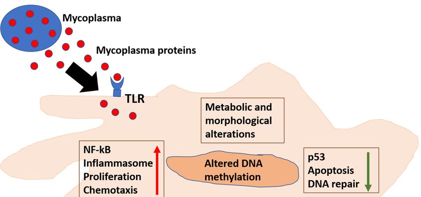
replication, inflammation is triggered [77] (Figure 1). The most important mechanism that triggers
inflammation is triggered [77] (Figure 1). The most important mechanism that triggers the immune
the immune response is the binding of Mycoplasma proteins to pattern-recognition receptors (PRRs)—
response is the binding of Mycoplasma proteins to pattern-recognition receptors (PRRs)—Toll-like
Toll-like receptors (TLRs) and NOD-like (nucleotide-binding and oligomerization domain) receptors
receptors (TLRs) and NOD-like (nucleotide-binding and oligomerization domain) receptors [32,34].
[32,34]. A series of cellular pathways are then engaged, and consequently a complex cascade of events
A series of cellular pathways are then engaged, and consequently a complex cascade of events
determines the specificity of the immune response. TLRs 1, 2, 4, and 6 were found to bind bacterial
determines the specificity of the immune response. TLRs 1, 2, 4, and 6 were found to bind bacterial
LPS [36,37]. However, Mollicutes lack typical bacterial PAMPs (e.g., lipoteichoic acid, flagellin, and
LPSsome
[36,37]. However, Mollicutes
lipopolysaccharides) andlack typical bacterial
consequently PAMPs
the exact (e.g., lipoteichoic
molecular mechanismsacid,of
flagellin, and some
Mycoplasmas’
lipopolysaccharides) and consequently
recognition by the cells of the immunethe exactismolecular
system mechanisms
the subjects of Mycoplasmas’
of several researches recognition
for its pathogenic
by implications.
the cells of the immune system is the subjects of several researches for its pathogenic implications.
Figure 1. Mycoplasmas
Figure 1. Mycoplasmas affect cellular pathways
affect cellular involved involved
pathways in inflammation and cellular transformation.
in inflammation and cellular
Mycoplasmas’
transformation. Mycoplasmas’ proteins interact with TLR or enter the cells, alter
proteins interact with TLR or enter the cells, where they can whereseveral pathways
they can alter
responsible for inflammation
several pathways responsibleand DNA repair. and
for inflammation In addition,
DNA repair.affecting methylation
In addition, affectingof cellular DNA
methylation of
results in alteration
cellular DNA resultsof cellular epigenetic
in alteration landscape.
of cellular TLR: landscape.
epigenetic Toll Like Receptor;
TLR: TollROS:
Like Reactive
Receptor;Oxygen
ROS:
Species.
ReactiveTGF: Transforming
Oxygen Species. TGF:Growth Factor;Growth
Transforming TNF: Tumor Necrosis
Factor; TNF: TumorFactor; and
Necrosis MCP-Monocyte
Factor; and MCP-
Monocyte Chemoattractant
Chemoattractant Protein. Protein.
To this regard, a protein able to bind TLRs is the macrophage-activating lipopeptide-2
(MALP-2) from Mycoplasma fermentans [38,39,78]. Upon binding, nuclear factor NF-kB [79] is
activated and induces the expression of pro-inflammatory mediators, such as TNF-α (tumor
necrosis factor-α), IL-6 (interleukin 6), MIP-1β (macrophage inflammatory protein-1β), GRO-αMicroorganisms 2020, 8, 1351 6 of 21
(growth-regulated oncogene-α), MCP-1 (monocyte chemoattractant protein-1), MIP-1α (macrophage
inflammatory protein-1α) [42], CXCL13 (chemokine CXCL13), CXL14 (chemokine CXL14),
RANTES (Regulated-on-Activation-Normal-T-cell-Expressed-and-Secreted chemokine) [43], and
MIP-2 (macrophage inflammatory protein-2) in monocytes [44]. Mycoplasma fermentans infection
of monocyte/macrophages increase also MMP-12 levels, a metalloproteinase which is both a
pro-inflammatory molecule and necessary for Monocyte Chemoattractant Protein-1 (MCP-1) cleavage
into its active form [80]. MCP-1 is involved in monocyte recruitment to the site of infection. All together,
these data indicate an evolutionarily conserved nature of the mycoplasmal ligands able to elicit the
same cellular signaling response. Of note, individual lipopeptides from Mycoplasmas can induce
inflammation, separated from the whole microorganism, pointing to a possible paracrine effect on
cells [81].
While the presence of high levels of Mycoplasmas and increased levels of inflammation can
easily explain their pathogenicity, in some cases the mechanisms underlying their negative effects
are not very clear. An example is chronic obstructive pulmonary disease (COPD), which in its two
pathological manifestations (chronic bronchitis and emphysema) is an increasing cause of morbidity
and mortality (130,000 death worldwide). Long-term exposure to irritants (mainly tobacco smoking
and air pollutants) triggers an inflammatory response in the lungs, resulting in narrowing of the small
airways, breakdown of lung tissue and progressive alveolar destruction (emphysema), and onset
of symptoms like dyspnea, cough, and sputum production [82]. Although respiratory symptoms
are the hallmarks of COPD, non-pulmonary manifestations occur frequently; thus, increasing risk of
significant cardiovascular, endocrine, and musculoskeletal comorbidities [83]. These non-pulmonary
manifestations are most likely mediated by immune-dysfunction initiated by inflammatory processes
that are initially triggered within the lungs and propagate systemically both causing and accentuating
comorbidities [84]. To this regard, increased levels of circulating inflammatory biomarkers observed in
COPD patients are potential mediators of these systemic effects [85]. In addition, COPD patients also
have significantly higher levels of circulating functional T-regulatory cells (Tregs), myeloid-derived
suppressor (MDSC) cells, and exhausted programmed Death (PD) 1 + cells, which contribute to
effector T-cell dysfunction and reduce their ability to fight infections [86,87]. The characterization of
lung microbiota lead to the discovery of a significant reduction in diversity, compared to microbiota
observed in healthy persons. In particular, in COPD patients the composition of microbiota seems
to be restricted to phyla which include potentially pathogenic microorganisms, such as Mycoplasma
pneumoniae [88–90], which is also associated with acute exacerbation [91,92].
2.2. Mycoplasmas Causing Diseases in Animals
Regarding their role as pathogenic agents for animals, we mention first Mycoplasma mycoides
subsp. mycoides Small Colony (SC), responsible for bovine pleuropneumonia (CBPP), which among
the several Mycoplasma species is arguably the most pathogenic. A massive inflammatory reaction
predominantly involving the lungs of the infected host is the most important pathological manifestation
of this Mycoplasma species, causing lung consolidation and leading to respiratory distress and death in
25–35% of the cases. In the remaining majority of infected animals, CBPP assumes a chronic form,
with recovery from the acute stage of the disease but where the animal host remains a potential carrier
and consequently a reservoir of Mycoplasma mycoides subsp. mycoides SC. The CD4 Th1-like T-cell
response to the pathogen was observed in animals recovered from disease over the entire duration of the
experiments lasting for over five months. This contrasted with the observation that symptomatic CBPP
progression correlated with PBMCs reduced capacity to produce interferon in animals that developed
an acute disease [93,94]. Morphological changes in mononuclear cells from bovine PBMCs were
observed in vitro upon infection with Mycoplasma mycoides subsp. mycoides SC. Such changes included
increased cell granularity and reduced cell size. The observation that heat inactivated Mycoplasma was
unable to induce the same changes, further highlights the requirement for viable Mycoplasma mycoides
subsp. mycoides SC and productive infection. These changes eventually lead to a cytopathic effectMicroorganisms 2020, 8, 1351 7 of 21
responsible for the apoptosis of the mononuclear cells. This effect was minimal when Mycoplasma
free culture supernatants were used, indicating that the responsible protein was probably released by
the infected cells upon infection [95]. Among the possible cytokines potentially responsible for the
cytopathic effect, it was demonstrated in a different study that Mycoplasma mycoides subsp. mycoides SC
strains can induce TNF-α production in bovine alveolar macrophages [96]. Other factors able to cause
cell death by damaging DNA are the reactive oxygen species (ROS), that are produced by Mycoplasma
mycoides infection through the metabolism of glycerol upon leukocytes activation. The proposed
mechanism involves: (i) Mycoplasma mycoides adhesion to the surface of the cells, (ii) activation of
TLRs and consequent promotion of their respiratory burst; and (iii) production and translocation of
increased ROS amounts within the phagocytic cell of the host; thus, causing an irreparable damage to
the cell membranes. The resulting inflammatory reaction could thus contribute to changes in lung
morphology and to function impairment [97].
Another Mycoplasma, Mycoplasma capricolum subsp. Capripneumoniae, is highly pathogenic when
localized in the caprine mammary gland. It causes acute mastitis, initially purulent. Massive fibrosis
ensues after a phase of infiltration of lymphonuclear cells, followed by fibroplasia in the interacinar
tissue [98]. Mycoplasma capricolum infection is also responsible for a disease in the goats (contagious
caprine pleuropneumonia—CCPP), where considerable inflammatory infiltrates are detected in the
injured lungs during CCPP development and lung damage is caused by increased IL-17 production
and consequent accumulation of neutrophil within the alveoli [99].
We also mention Mycoplasma agalactiae, responsible for the agalactia syndrome in sheep and goats, a
contagious disease that produces considerable economic losses worldwide. The primary mechanism(s)
whereby Mycoplasma agalactiae infection damages the host cells are not completely clear, and the most
credited hypothesis considers the host immune response as the major responsible for the excessive
inflammation and consequent tissue destruction. To this regard, in vitro infection of HeLa cells resulted
in some morphological changes, namely cell elongation, cytoplasm shrinkage, and membrane blebbing.
These changes, together with chromatin condensation and increased caspase-3 activation indicate an
apoptosis-like phenomenon leading to reduced cell viability and increased cell lysis [100]. Additionally,
it was observed an association with Mycoplasma agalactiae antigen and production of IL-10, IFN-γ, IL-4,
and TNF-α in an experimental in vitro model consisting of inflammatory cells of mammary tissues
from goats infected with Mycoplasma agalactiae, [101]. Finally, sheep infected with Mycoplasma agalactiae
showed prolonged depletion of peripheral CD3+ CD4+ and CD3+ CD8+ cells, possibly due to organ
infiltration. Real-time PCR assay allowed the detection of the infectious agent in different areas (ear,
nose, and milk) up to 50 days post infection [102].
Finally, MALP-2 from some strains of Mycoplasma gallisepticum induces the expression of TNF-α,
IL-6, and MIP-1β in chickens [43]. Interestingly, it was observed a differential role of TLR2-2 and TLR6
in Mycoplasma gallisepticum-infected DF-1 cells and chicken embryos [103].Microorganisms 2020, 8, 1351 8 of 21
Table 1. Association between several species of Mycoplasma, diseases, and proposed mechanism(s)
of inflammation.
Mycoplasma Types Diseases and Proposed Mechanism(s) of Inflammation
Human-Associated Mycoplasmas
Respiratory diseases [55], Urogenital diseases [104], Rheumatoid Arthritis [52],
Fibromyalgia [105,106], and Neurological diseases [107,108]. Mycoplasma proteins bind to
Mycoplasmas (general)
pattern-recognition receptors (PRRs)—Toll-like receptors (TLRs) and NOD-like
(nucleotide-binding and oligomerization domain) receptors [32,34,36,37].
Urogenital infections [47]. Adhesion to epithelial cells promotes acute inflammation via triggering
of innate immune sensors expressed on the cells’ surface. Activation of pro-inflammatory signals
ultimately results in recruitment of leucocytes to the infection site. The recombinant C-terminal
Mycoplasma genitalium
portion of the immunogenic protein MG309 (rMG309c) activates NF-κB via TLR2/6 in genital
epithelial cells (EC), which in turn secreted proinflammatory cytokines, including interleukin-6
(IL-6) and IL-8 [109,110].
Respiratory diseases [55]. Different adhesins and accessory adhesion proteins mediates the crucial
initial step of cytoadherence to respiratory tract epithelium, Subsequently, several mechanisms,
namely intracellular localization, direct cytotoxicity and toll-like receptors (TLRs)-mediated
activation of the inflammatory cascade cause tissue injury mediated by such cytokines. Infection is
associated with acute exacerbation of COPD [91,92], and COPD patients also have significantly
higher levels of circulating functional T-regulatory cells (Tregs), myeloid-derived suppressor
(MDSC) cells and exhausted programmed Death (PD) 1 + cells, which contribute to effector T-cell
Mycoplasma pneumoniae
dysfunction and reduce their ability to fight infections [86,87]. In infected mice is observed a
dysregulated Mycoplasma pneumoniae-derived immune response in lung [81,88–90]. Mycoplasma
pneumoniae also is responsible for Community-Acquired Respiratory Distress Syndrome toxin
(CARDS toxin), which activates adenosine diphosphate (ADP) ribosylation and inflammasome,
causing airway inflammation. [111]. Inflammatory mediators, namely interleukin IL-6, IL-8, IL-18,
interferon (INF)-g, tumor necrosis factor (TNF)-α, and transforming growth factor (TGF)-β 1 are
increased in serum of CNS [54].
Mycoplasma hominis Urogenital infections (pelvic inflammatory diseases and bacterial vaginosis) [112–117].
Urogenital infections [116], Autoimmune disorders: Immunoglobulin A nephropathy [118].
Mycoplasma penetrans Secreted P40 mediates (partly) cytotoxicity upon infection of Mycoplasma penetrans in vitro, by
inducing physiological modifications resembling apoptosis [119].
Septic arthritis [120,121], periodontal disease [122–124]. Cell membranes of Mycoplasma salivarium
Mycoplasma salivarium promote expression of IL-6 and IL-8 in human fibroblasts through stimulation of protein kinase C
(PKC) in Gin-1 cells, a human gingival fibroblast cell line [125].
Urogenital diseases [104], Rheumatoid Arthritis [52]. Mycoplasma fermentans increases the secretion
of macrophage-activating lipopeptide-2 (MALP-2) [38,39,78], TNF-α (tumor necrosis factor-α),
IL-6 (interleukin 6), MIP-1β (macrophage inflammatory protein-1β), GRO-α (growth-regulated
oncogene-α), MCP-1 (monocyte chemoattractant protein-1), MIP-1α (macrophage inflammatory
protein-1α) [39,42,79], CXCL13 (chemokine CXCL13), CXL14 (chemokine CXL14), RANTES
Mycoplasma fermentans
(Regulated-on-Activation-Normal-T-cell-Expressed-and-Secreted chemokine) [43], MCP-1
(monocyte chemoattractant protein-1), MIP-1α (macrophage inflammatory protein-1α) [42].
Mycoplasma fermentans infection of monocyte/macrophages increases also MMP-12 levels, a
metalloproteinase which is both a pro-inflammatory molecule and also necessary for the cleavage
of Monocyte Chemoattractant Protein-1 (MCP-1) into its active form [80]
Animal-Associated Mycoplasmas
In bovine hosts, it is observed: increased production of TNF-α in alveolar macrophages
Mycoplasma mycoides
(cattle) [96]; induction of morphological changes in mononuclear cells [95]; induction of ROS [97].
Contagious caprine pleuropneumonia (CCPP) is associated with increased IL-17 and neutrophil
Mycoplasma capricolum
accumulation, leading to lung injury [99]
Infection of HeLa cells lead to morphological changes including membrane blebbing, which
together with increased caspase-3 cleavage activity indicated an apoptosis-like phenomenon [100].
Mycoplasma agalactiae An in vitro model consisting of inflammatory cells of mammary tissues from goats infected with
Mycoplasma agalactiae demonstrated an association with Mycoplasma antigen(s) and production of
IL-10, IFN-γ, IL-4, and TNF-α [101]
MALP-2 from some strains of Mycoplasma gallisepticum induces the expression of TNF-α, IL-6, and
Mycoplasma gallisepticum MIP-1β in chickens [43]. Interestingly, it was observed a differential role of TLR2-2 and TLR6 in
Mycoplasma gallisepticum-infected DF-1 cells and chicken embryos [103].
3. Mycoplasmas and Cancer
Definitive establishment of the causal correlation between Helicobacter pylori and gastric cancer
provided the first demonstration that bacteria can cause cancer [126]. Since then, studies of the
human microbiome have elucidated an array of complex interactions between prokaryotes and
their hosts [127]. Recent examples of studies in human patients highlighted an association between
Fusobacterium nucleatum and colorectal cancer [128–134], and between Mycoplasmas and prostate andMicroorganisms 2020, 8, 1351 9 of 21
colorectal cancer, oral carcinoma associated with Fanconi anemia [123], as well as non-Hodgkin’s
lymphoma (NHL) in HIV-seropositive subjects [123,135–139]. These data strongly support them as
leading bacterial candidates with oncogenic properties (Figure 1).
Indeed, several data obtained by using mouse models with particularly mutated genes, or in vivo
experiments carried on with cancer-inducing agents, showed that tumor formation is reduced when
the mice colonies are grown and kept in a germ-free environment [140,141].
The precise pathogen–cancer relationships of a number of bacteria, including Mycoplasmas,
remain largely elusive. In particular, we note that some bacteria are able to establish persistent,
chronic infection by invading the host’s cell and remaining undetected by the immune system for
a long period of time. They produce proteins that interfere and alter the function of important
cellular pathways like cell cycle control, apoptosis, DNA repair. This, linked to the ability of these
pathogens to induce substances able to increase DNA damage may increase abnormal cell growth and
transformation [142,143].
A number of studies have established a firm link between chronic inflammation, tumor progression
and p53, which, undoubtedly, is the most important tumor suppressor protein in humans, given its
central role in preserving genome stability [144,145]. NF-κB reduces the activities of p53 and the
mutual regulation between antiapoptotic NF-κB and proapoptotic p53 is one of the major determinant
of a cell’s fate [146]. In fact, genetic or pharmacological inhibition of constitutively active NF-κB in
different tumor cell lines leads to the activation of p53 function and tumor cell death via p53-dependent
apoptosis [147]. Given that inflammation can reduce the activity of p53, it is possible that chronic
inflammation through the activation of NF-κB reduces the activity of p53; thus, promoting cellular
transformation [146].
Following DNA damage and other stress signals, low levels of cellular p53 protein increase,
causing growth arrest, DNA repair, or apoptosis. Interruption of cell cycle prevents replication of
damaged DNA, allowing p53 to activate the transcription of proteins involved in DNA repair. On the
other hand, when this pathway is compromised the cell activates the pathways leading to apoptosis,
which is the mechanism of choice to avoid proliferation of cells containing abnormal DNA [148].
For these reasons, the cellular concentration and activity of p53 must be tightly regulated, and the
major regulator of p53 is Mdm2, which functions by retaining p53 in the cytoplasm and activating
its degradation by the ubiquitin system [149–151]. Mdm2 is regulated by p53 through a feedback
mechanism, and by the genes involved in growth arrest, DNA repair, and apoptosis (such as p21,
Gadd45, BAX, and PUMA) [152,153].
It is well known that several strains of Mycoplasma suppress the transcriptional activity of p53,
resulting in reduced apoptosis of damaged cells and some Mycoplasmas (notably Mycoplasma fermentans,
Mycoplasma penetrans, and Mycoplasma hyorhinis) were reported to have oncogenic potential since they
demonstrated not just accumulation of abnormalities but also phenotypic changes of the cells [154–156]
(Table 2). Moreover, long-term Mycoplasma infections in cell cultures are associated with increased
frequency of chromosomal instability and malignant transformation such as the lost cell-to-cell contact,
the spindle morphology and the growth in multiple layers [154]. These changes were reversed when
earlier cultured cells (maintained for up to six passages in vitro) were treated with three cycles of
ciprofloxacin and returned to a normal growth pattern [154]. On the contrary, long-time cultured
cells (for more than 18 passages) were not able to acquire their previous morphology/growth pattern
when treated with the same antibiotic, demonstrating an irreversible change. These data indicate
that persistent infection with Mycoplasma induce cellular transformation through a series of cellular
events [154]. In addition, spontaneous transformation of mouse embryo fibroblasts and concomitant
overexpression of the H-ras and c-myc proto-oncogenes were observed upon long-term infection with
Mycoplasma fermentans or Mycoplasma penetrans [157].
Moreover, upon infection, several species of human Mycoplasmas would prevent apoptosis in
32D cells from undergoing in vitro in the absence of IL-3, indicating continuous growth even in the
absence of the important IL-3 growth signaling. To this regard, it was observed that infected 32DMicroorganisms 2020, 8, 1351 10 of 21
cells gradually underwent malignant transformation after a period of 4 to 5 weeks and no longer
needed the presence of either Mycoplasma fermentans, Mycoplasma penetrans, nor of IL-3 to grow. Not
surprisingly, these 32D cells were able to grow independently and were highly tumorigenic upon
injection into a nude mice model. Karyotyping analysis demonstrated chromosomal changes and
trisomy 19 associated with malignant transformation [155].
Another potential way that Mycoplasmas have to influence cancer formation, is by deregulating
expression of Bone morphogenetic protein 2 (BMP2), which is an essential growth factor and morphogen,
implicated in cancer promotion and growth [158,159]. In fact, it has been shown that infection by
Mycoplasma penetrans, Mycoplasma fermentans, and Mycoplasma hominis induces BMP2 RNA expression,
as well as secretion of mature BMP2 protein, in cells that usually do not express such protein, including
BEAS-2B cells (immortalized human bronchial epithelial cells), A549 cells (lung adenocarcinoma cells),
plus several other cell lines of different origins (mesenchymal, epithelial, and myeloid). This increase in
BMP2 expression in Mycoplasma-infected cells was mostly achieved by regulating RNA stability, rather
than influencing the transcriptional level. Additionally, it was demonstrated that BMP2 stimulated
proliferation of BEAS-2B cells transformed by chronic Mycoplasma infection, indicating the profound
effects of Mycoplasma infection on BMP2-regulated pathways, including the ones involved in cell
proliferation, differentiation, and apoptosis [136].
Mycoplasma hyorhinis expresses p37 protein on its surface, and this protein belongs to a high-affinity
transport system associated with cancers in animals and humans. Indeed, p37 induces rapid expression
of several genes involved in inflammation and cancer progression through TLR4 receptor triggering
in fibroblasts. As cancer associated fibroblasts favor growth, invasion, and metastasis by regulation
of tumor-related inflammation, p37 may influence cancer development by inducing expression of
pro-inflammatory genes [160]. To this regard, p37 increased migration in a transwell (Matrigel) assay
of human gastric carcinoma (AGS) cells by inducing the phosphorylation of epidermal growth factor
receptor (EGFR) and extracellular signal-regulated kinase and the activity of matrix metalloproteinase-2
(MMP-2) [161].These results indicate that p37 may be able to promote invasion by upregulating the
activity of MMP-2; thus, causing EGFR phosphorylation and increasing tumor metastasis upon
Mycoplasma hyorhinis infection. Additional type of cancers that seem to be influenced by p37 are PC-3
and DU145 (two prostate cell lines), since treatment with p37 increased invasivity and migratory ability,
as demonstrated by a Matrigel-based assay [161,162]. To this regard, it was observed a significant
nuclear enlargement, denoting active, anaplastic cells following incubation with recombinant p37.
Microarray analysis of p37-treated cells allowed to identify eight clusters of differentially expressed
genes broadly divided into three groups. The most represented categories of functional genes were
composed by signal transduction, cell cycle, and metabolic factors [163]. Treatment with p37 also
affected Ficoll-separated human peripheral blood mononuclear cells (PBMCs), increasing the expression
of tumor necrosis factor α (TNFα) gene transcription and the secretion of TNFα [160]. This also indicates
that p37 and its regulated molecules could be potentially targeted for anti-cancer intervention [161].
Expanding on these studies, it was also shown that Mycoplasma fermentans, is able to influence the
expression of hundreds of genes in cultured human cells; thus, affecting many pathways. This regulation
involved increased or reduced expression of many cytokines, stress-response genes, transport proteins,
receptors, ion channels, growth factors, oxidases, tumor suppressors, and oncogene during a two-stage
process; a reversible one, when the transformation process can be stopped by eradicating the Mycoplasma,
and an irreversible phase [138].
Further in vivo experiments demonstrated the oncogenic potential of Mycoplasma penetrans in
immunocompromised settings. Upon infection, mice immunosuppressed with cyclophosphamide
had lower expression of p53 and p21 and higher expression of H-ras in gastric mucosa, compared to
the uninfected animals. Moreover, NF-κB p65 subunit and TNF-α expression increased in infected
mice. On the other hand, Bax expression was lower while Bcl-2 expression was higher. These data
demonstrate that Mycoplasma infection reduces the levels of several oncogenes in the gastric mucosaMicroorganisms 2020, 8, 1351 11 of 21
of immunodeficient mice, and this could potentially facilitate the malignant transformation of these
cells [164].
Table 2. Association between several species of Mycoplasmas, cancer(s), and proposed mechanisms of
cellular transformation.
Mycoplasma Types Cancer(s) and Proposed Mechanisms of Cellular Transformation
Increased expression of BMP2 upon infection [136].
Mycoplasma fermentans and Mycoplasma penetrans infection induced malignant transformation of
32D cells (including autonomous growth in IL-3-conditions). After a few weeks, the presence of
Mycoplasmas was no longer needed for autonomous growth of the cells. Transformed 32D cells
Mycoplasma fermentans and
were able to form tumors when injected into nude mice. Karyotyping analysis showed
Mycoplasma penetrans
chromosomal abnormalities, including trisomy 19 associated with malignant transformation
[154–156]. Several mechanisms account for their potential cell-transforming effect: induction of
genetic instability, alterations in metabolism, changes in the expression of many genes, in
particular growth factors, tumor suppressors and oncogenes [164]
Infection promoted a malignant phenotype in benign human prostate cells (BPH-1), as assessed by
in vitro and in vivo assays showing anchorage-independent growth, greater percentage of
Mycoplasma genitalium
migrating cells with increased invasive capacity, generation of xenograft tumors in athymic mice
and accumulation of chromosomal aberrations and polysomy [137].
Infection promoted a malignant phenotype in benign human prostate cells (BPH-1), similar to
Mycoplasma genitalium [137]. Higher titers of antibodies against Mycoplasma hominis were observed
Mycoplasma hominis
in prostate cancer positive patients, together with higher average PSA levels [139]. Infection
promoted expression of BMP2, similar to Mycoplasma penetrans and Mycoplasma fermentans [136].
p37 seems to be the major determinant involved in events potentially leading to cell
transformation: (1) it induces the expression of genes implicated in inflammation and cancer
progression in fibroblasts, indicating that cancer associated fibroblasts may facilitate growth,
invasion and metastasis by regulating tumor associated inflammation [160]; (2) when added to
human gastric carcinoma cells (AGS) increased the migration in a transwell (Matrigel) assay, by
Mycoplasma hyorhinis promoting phosphorylation of epidermal growth factor receptor (EGFR) and extracellular
signal-regulated kinase and the activity of matrix metalloproteinase-2 (MMP-2) [161]; (3) it
induces significant nuclear enlargement, indicating the generation of active, anaplastic cells and
promoted the migratory capacity of both PC-3 and DU145 cells [162,163]; and (4) microarray
analysis of p37-treated cells identified eight gene expression clusters classified into three groups,
with cell cycle, signal transduction and metabolic factors among the most represented genes [163].
Infection in vivo is associated with lower expression of p53 and p21 and higher H-ras expression
in gastric mucosa. Moreover, expression of NF-κB p65 subunit increased together with TNF-α
expression are observed, and Bax expression was lower while Bcl-2 expression was higher. These
Mycoplasma penetrans
data indicate that persistent infection is associated with aberrant expression of multiple
proto-oncogenes in gastric mucosa of immunodeficient mice suggesting its potential influence on
malignant transformation. [164].
Mycoplasma salivarium Possible role in oral cancer [123,165].
Mycoplasma fermentans reduced activity and expression of Topo I [166].
Mycoplasma fermentans
Reduction of p53 activity [167,168], reduction of PARP-1 activity [168,169]
infection in vivo resulted in suppression of p53, activation of NF-kB and increased Ras mutagenic
Mycoplasma arginini
effects, similar to Mycoplasma penetrans [167].
Additionally, Mycoplasma infection reduced activation of p53 with a constitutive activation of
NF-κB in cells infected with Mycoplasma, further highlighting its effects of on these important regulatory
pathways [167]. This altered expression was consistent with many human tumors. Thus, infected
cells were able to evade apoptosis by inhibiting p53 [167,170,171]. Though the responsible Mycoplasma
protein was not identified, more recent works from our group point to a Mycoplasma chaperon protein,
DnaK, a chaperone protein belonging to the HSP70 family, as responsible for reduction of pathways
linked to DNA repair, cell cycle control and apoptosis [168]. In particular, following the isolation of
a strain of Mycoplasma fermentans able to induce lymphoma in a severe combined immuno-deficient
(SCID) mouse model [170–172], we characterized the molecular mechanisms in vitro. We showed that
this Mycoplasma DnaK, co-immunoprecipitates with USP10 (ubiquitin carboxyl-terminal hydrolase 10),
a key p53 regulator [173], and impairs p53-dependent anti-cancer activities [168].
We showed that the binding of DnaK to PARP1, which recognizes DNA breaks and participates in
DNA repair [174–177], reduces its activity and, following recognition of damaged DNA, PARylation of
certain proteins of very high MW is greatly reduced (> 150 KDa), while it seems it only marginally
affects proteins between 100–150 KDa [168,169]. We could abundantly find sequences of Mycoplasma
DnaK early in infected mice, while only a low amount of copy number was found in primary andMicroorganisms 2020, 8, 1351 12 of 21
secondary tumors, pointing to a “hit and run/hide” mechanism [168]. Given the fact that infections
with certain Mycoplasmas lead to ROS production [49,178], and ROS can cause direct damage to DNA,
our data provide a molecular link between a Mycoplasma protein, DnaK, and cellular transformation.
Further studies linking Mycoplasma to carcinogenesis are illustrated by its involvement in changes
in DNA methylation pattern. DNA methylation (that is the conversion of cytosine to 5-methylcytosine)
is an essential element in transcriptional regulation and is one of the major epigenetic mechanisms.
Many stresses or DNA damage can in fact interfere with the ability of DNA to be methylated at
CpG dinucleotides by DNA-methyltransferases (DNA-MTases) [179]. When specific Mycoplasma
MTases were expressed in human cell lines, their translocation to the nucleus has been observed.
The result was a change of the human genome methylation landscape because these bacterial enzymes
methylated cytosines within the respective CG and GATC sites in human genomic DNA, resulting in
the stimulation of pro-oncogenic pathways [180].
Additional reports have strongly suggested a role for Mycoplasma in cellular transformation and
the search for the link between Mycoplasma and cancer is currently actively being investigated. To this
regard, many studies demonstrated the effects of Mycoplasma on cell lines by showing that Mycoplasma
may facilitate tumorigenesis, for example in oral tissues [165], in human prostate cells [137,139] in
gastric carcinoma cells [181] and cervical cells both in vitro [182].
In vivo, several studies reported the isolation of Mycoplasma species in various neoplastic tissues
and body fluids, and in particular Mycoplasmas have been found in precancerous lesions as well as in
malignant tissues from patients with stomach, colon, ovarian and lung cancers, and hepatocellular
carcinoma [142,183,184], though no direct causal relationship with cellular transformation has been
demonstrated so far. Nonetheless, all the outlined studies and properties of Mycoplasmas strongly
suggest that these agents act as cancer-promoting factors.
4. Conclusions
Several different bacteria have been associated with human cancers. A widespread and concerted
scientific effort is ongoing to identify potentially responsible bacteria and characterize the molecular
mechanism(s). While Helicobacter pylori so far is the only one with clear data to support causality [126],
studies of other bacteria including Mycoplasmas [123,135,139] strongly support the idea that they
too have oncogenic properties. Experimental results have demonstrated the role of Mycoplasmas in
increasing inflammation and associated them to cancer initiation.
Although it seems plausible that accumulation of DNA-damage and inhibition of p53-activities
play a major role in driving transformation, molecular mechanisms whereby these bacteria dysregulate
cellular pathways and eventually result in cellular transformation are still largely unknown. By linking
inflammation, DNA damage and reduction of p53 activity, it may be possible to formulate a hypothesis
to better define the role of Mycoplasmas in causing cellular transformation and disease.
Author Contributions: F.B., S.C. and D.Z. wrote sections of the manuscript. All authors have read and agreed to
the published version of the manuscript.
Funding: This work was supported by IHV’s internal funding.
Conflicts of Interest: The authors declare no conflict of interest.
References
1. Razin, S.; Yogev, D.; Naot, Y. Molecular biology and pathogenicity of mycoplasmas. Microbiol. Mol. Biol. Rev.
1998, 62, 1094–1156. [CrossRef] [PubMed]
2. Razin, S. Peculiar properties of mycoplasmas: The smallest self-replicating prokaryotes. FEMS Microbiol. Lett.
1992, 100, 423–431. [CrossRef] [PubMed]
3. Kornspan, J.D.; Rottem, S. The phospholipid profile of mycoplasmas. J. Lipids 2012, 2012, 640762. [CrossRef]
[PubMed]Microorganisms 2020, 8, 1351 13 of 21
4. Dybvig, K.; Voelker, L.L. Molecular biology of mycoplasmas. Annu. Rev. Microbiol. 1996, 50, 25–57.
[CrossRef]
5. Paessler, M.; Levinson, A.; Patel, J.B.; Schuster, M.; Minda, M.; Nachamkin, I. Disseminated Mycoplasma
orale infection in a patient with common variable immunodeficiency syndrome. Diagn. Microbiol. Infect. Dis.
2002, 44, 201–204. [CrossRef]
6. Lo, S.C.; Hayes, M.M.; Wang, R.Y.H.; Pierce, P.F.; Kotani, H.; Shih, J.W.K. Newly discovered mycoplasma
isolated from patients infected with HIV. Lancet 1991, 338, 1415–1418. [CrossRef]
7. Lo, S.C.; Hayes, M.M.; Kotani, H.; Pierce, P.F.; Wear, D.J.; Newton, P.B., 3rd; Tully, J.G.; Shih, J.W. Adhesion
onto and invasion into mammalian cells by mycoplasma penetrans: A newly isolated mycoplasma from
patients with AIDS. Mod. Pathol. 1993, 6, 276–280.
8. Yavlovich, A.; Katzenell, A.; Tarshis, M.; Higazi, A.A.; Rottem, S. Mycoplasma fermentans binds to and
invades HeLa cells: Involvement of plasminogen and urokinase. Infect. Immun. 2004, 72, 5004–5011.
[CrossRef]
9. Yavlovich, A.; Tarshis, M.; Rottem, S. Internalization and intracellular survival of Mycoplasma pneumoniae
by non-phagocytic cells. FEMS Microbiol. Lett. 2004, 233, 241–246. [CrossRef]
10. Baseman, J.B.; Tully, J.G. Mycoplasmas: Sophisticated, reemerging, and burdened by their notoriety.
Emerg. Infect. Dis. 1997, 3, 21–32. [CrossRef]
11. Hentges, D.J. Anaerobes: General Characteristics. In Medical Microbiology, 4th ed.; Baron, S., Ed.; University
of Texas Medical Branch at Galveston: Galveston, TX, USA, 1996.
12. Yamaguchi, M.; Kikuchi, A.; Ohkusu, K.; Akashi, M.; Sasahara, J.; Takakuwa, K.; Tanaka, K. Abscess formation
due to Mycoplasma hominis infection after cesarean section. J. Obstet. Gynaecol. Res. 2009, 35, 593–596.
[CrossRef] [PubMed]
13. Mori, N.; Takigawa, A.; Kagawa, N.; Kenri, T.; Yoshida, S.; Shibayama, K.; Aoki, Y. Pelvic abscess due to
Mycoplasma hominis following caesarean section. JMM Case Rep. 2016, 3, e005059. [CrossRef] [PubMed]
14. Koshiba, H.; Koshiba, A.; Daimon, Y.; Noguchi, T.; Iwasaku, K.; Kitawaki, J. Hematoma and abscess formation
caused by Mycoplasma hominis following cesarean section. Int. J. Womens Health 2011, 3, 15–18. [CrossRef]
[PubMed]
15. Kennedy, K.J.; Prince, S.; Makeham, T. Mycoplasma hominis-Associated Parapharyngeal Abscess following
Acute Epstein-Barr Virus Infection in a Previously Immunocompetent Adult. J. Clin. Microbiol. 2009,
47, 3050–3052. [CrossRef]
16. Parsonson, F. Mycoplasma hominis infection following neurosurgical intervention in a patient with spinal
cord compression. JMM Case Rep. 2016, 3. [CrossRef]
17. Garner, C.; Hubbold, L.; Chakraborti, P. Mycoplasma detection in cell cultures: A comparison of four
methods. Br. J. Biomed. Sci. 2000, 57, 295–301. [PubMed]
18. Jaffe, J.D.; Berg, H.C.; Church, G.M. Proteogenomic mapping as a complementary method to perform genome
annotation. Proteomics 2004, 4, 59–77. [CrossRef]
19. Catrein, I.; Herrmann, R. The proteome of Mycoplasma pneumoniae, a supposedly “simple” cell. Proteomics
2011, 11, 3614–3632. [CrossRef]
20. Regula, J.T.; Boguth, G.; Gorg, A.; Hegermann, J.; Mayer, F.; Frank, R.; Herrmann, R. Defining the mycoplasma
‘cytoskeleton’: The protein composition of the Triton X-100 insoluble fraction of the bacterium Mycoplasma
pneumoniae determined by 2-D gel electrophoresis and mass spectrometry. Microbiology 2001, 147, 1045–1057.
[CrossRef]
21. Balasubramanian, S.; Schneider, T.; Gerstein, M.; Regan, L. Proteomics of Mycoplasma genitalium:
Identification and characterization of unannotated and atypical proteins in a small model genome.
Nucleic. Acids Res. 2000, 28, 3075–3082. [CrossRef]
22. Parraga-Nino, N.; Colome-Calls, N.; Canals, F.; Querol, E.; Ferrer-Navarro, M. A comprehensive proteome of
Mycoplasma genitalium. J. Proteome Res. 2012, 11, 3305–3316. [CrossRef] [PubMed]
23. Liu, Y.C.; Lin, I.H.; Chung, W.J.; Hu, W.S.; Ng, W.V.; Lu, C.Y.; Huang, T.Y.; Shu, H.W.; Hsiao, K.J.; Tsai, S.F.;
et al. Proteomics characterization of cytoplasmic and lipid-associated membrane proteins of human pathogen
Mycoplasma fermentans M64. PLoS ONE 2012, 7, e35304. [CrossRef] [PubMed]
24. Benedetti, F.; Krishnan, S.; Cocchi, F.; Tettelin, H.; Gallo, R.C.; Zella, D.; Curreli, S. Proteome analysis of
Mycoplasma fermentans cultured under aerobic and anaerobic conditions. Transl. Med. Commun. 2019, 4, 15.
[CrossRef]You can also read