The cryptic gonadotropin-releasing hormone neuronal system of human basal ganglia
←
→
Page content transcription
If your browser does not render page correctly, please read the page content below
RESEARCH ARTICLE

                                    The cryptic gonadotropin-releasing
                                    hormone neuronal system of human basal
                                    ganglia
                                    Katalin Skrapits1*, Miklós Sárvári1, Imre Farkas1, Balázs Göcz1, Szabolcs Takács1,
                                    Éva Rumpler1, Viktória Váczi1, Csaba Vastagh2, Gergely Rácz3, András Matolcsy3,
                                    Norbert Solymosi4, Szilárd Póliska5, Blanka Tóth6, Ferenc Erdélyi7, Gábor Szabó7,
                                    Michael D Culler8, Cecile Allet9, Ludovica Cotellessa9, Vincent Prévot9,
                                    Paolo Giacobini9, Erik Hrabovszky1*
                                    1
                                      Laboratory of Reproductive Neurobiology, Institute of Experimental Medicine,
                                    Budapest, Hungary; 2Laboratory of Endocrine Neurobiology, Institute of
                                    Experimental Medicine, Budapest, Hungary; 31st Department of Pathology and
                                    Experimental Cancer Research, Semmelweis University, Budapest, Hungary; 4Centre
                                    for Bioinformatics, University of Veterinary Medicine, Budapest, Hungary;
                                    5
                                      Department of Biochemistry and Molecular Biology, Faculty of Medicine, University
                                    of Debrecen, Debrecen, Hungary; 6Department of Inorganic and Analytical
                                    Chemistry, Budapest University of Technology and Economics, Budapest, Hungary;
                                    7
                                      Department of Gene Technology and Developmental Biology, Institute of
                                    Experimental Medicine, Budapest, Hungary; 8Amolyt Pharma, Newton, France;
                                    9
                                      Univ. Lille, Inserm, CHU Lille, Laboratory of Development and Plasticity of the
                                    Neuroendocrine Brain, Lille Neuroscience & Cognition, Lille, France

*For correspondence:
                                    Abstract Human reproduction is controlled by ~2000 hypothalamic gonadotropin-releasing
skrapits.katalin@koki.hu (KS);
                                    hormone (GnRH) neurons. Here, we report the discovery and characterization of additional
hrabovszky.erik@koki.hu (EH)        ~150,000–200,000 GnRH-synthesizing cells in the human basal ganglia and basal forebrain. Nearly
                                    all extrahypothalamic GnRH neurons expressed the cholinergic marker enzyme choline
Competing interests: The            acetyltransferase. Similarly, hypothalamic GnRH neurons were also cholinergic both in embryonic
authors declare that no
                                    and adult human brains. Whole-transcriptome analysis of cholinergic interneurons and medium
competing interests exist.
                                    spiny projection neurons laser-microdissected from the human putamen showed selective
Funding: See page 22                expression of GNRH1 and GNRHR1 autoreceptors in the cholinergic cell population and uncovered
Received: 19 February 2021          the detailed transcriptome profile and molecular connectome of these two cell types. Higher-order
Accepted: 14 June 2021              non-reproductive functions regulated by GnRH under physiological conditions in the human basal
Published: 15 June 2021             ganglia and basal forebrain require clarification. The role and changes of GnRH/GnRHR1 signaling
                                    in neurodegenerative disorders affecting cholinergic neurocircuitries, including Parkinson’s and
Reviewing editor: Margaret M
                                    Alzheimer’s diseases, need to be explored.
McCarthy, University of Maryland
School of Medicine, United
States
   Copyright Skrapits et al. This
article is distributed under the
                                    Introduction
                                    Mammalian reproduction is controlled by a few hundred/thousand preoptic/hypothalamic
terms of the Creative Commons
Attribution License, which          neurons, which release the decapeptide neurohormone gonadotropin-releasing hormone (GnRH)
permits unrestricted use and        into the hypophysial portal circulation. GnRH promotes fertility via increasing the synthesis and
redistribution provided that the    secretion of luteinizing hormone and follicle-stimulating hormone in the anterior pituitary (Herbi-
original author and source are      son, 2018). Unlike other neurons of the central nervous system, GnRH neurons are born in the olfac-
credited.                           tory placodes and migrate into the forebrain prenatally (Casoni et al., 2016; Schwanzel-Fukuda and

Skrapits et al. eLife 2021;10:e67714. DOI: https://doi.org/10.7554/eLife.67714                                                  1 of 26
Research article                                                                                     Cell Biology Neuroscience

                                    Pfaff, 1989; Wray et al., 1989). Recent developmental studies on embryos/fetuses determined the
                                    detailed spatio-temporal profile of this process in the human (Casoni et al., 2016). Approxi-
                                    mately 2000 neurons were observed along a ventral migratory path whereby GnRH neurons reach
                                    the hypothalamus to regulate reproduction after puberty. In addition, a previously unknown dorsal
                                    migratory route has been identified whereby ~8000 GnRH neurons migrated towards pallial and/or
                                    subpallial structures. The fate of these neurons at later stages of pre- and postnatal development
                                    has been unexplored so far.
                                       While GnRH neurons in adult laboratory rodents are mostly preoptic/hypothalamic and serve
                                    reproductive functions (Merchenthaler et al., 1980), a handful of anatomical studies on primates
                                    identified additional GNRH1 mRNA expression and/or GnRH immunoreactivity in extrahypothalamic
                                    regions unrelated to reproduction. These included several basal ganglia and the basal forebrain
                                    (Krajewski et al., 2003; Quanbeck et al., 1997; Rance et al., 1994; Terasawa et al., 2001). Initial
                                    enthusiasm to study these neurons further faded after suggestions that extrahypothalamic GnRH
                                    neurons in monkeys contain the GnRH degradation product GnRH1-5, instead of the bona fide
                                    GnRH decapeptide (Quanbeck et al., 1997; Terasawa et al., 2001).
                                       Here, we study human extrahypothalamic GnRH neurons in adult postmortem brains with immu-
                                    nohistochemistry (IHC), in situ hybridization (ISH), single-cell transcriptomics (RNA-seq), and high-
                                    performance liquid chromatography/tandem mass spectrometry (HPLC-MS/MS). We report and
                                    characterize a previously unexplored large GnRH neuron population with ~150,000–200,000 cell
                                    bodies scattered in different basal ganglia and the basal forebrain. Extrahypothalamic GnRH neu-
                                    rons, most of which are found in the putamen (Pu), contain bona fide GnRH decapeptide, as shown
                                    by HPLC-MS/MS. Deep transcriptome analysis reveals that these neurons express GnRH biosynthetic
                                    enzymes, GNRHR1 autoreceptors, and the molecular machinery of cholinergic and GABAergic co-
                                    transmission. Somewhat unexpectedly, hypothalamic GnRH neurons also exhibit cholinergic pheno-
                                    type in the embryonic and adult human brains. Altogether, these data indicate that GnRH is synthe-
                                    sized as a co-transmitter of many cholinergic neurons in the human basal ganglia and basal
                                    forebrain. At least in the Pu, GnRH appears to act on GnRHR1 autoreceptors to regulate higher-
                                    order non-reproductive functions associated with the cholinergic system.

                                    Results
                                    Human extrahypothalamic GnRH-immunoreactive neurons occur in the
                                    basal ganglia and the basal forebrain
                                    The primate central nervous system contains extrahypothalamic GnRH cell populations, which have
                                    unknown functions (Krajewski et al., 2003; Quanbeck et al., 1997; Rance et al., 1994;
                                    Terasawa et al., 2001). An earlier ISH study of adult human brains identified ~6000–7000 GNRH1
                                    mRNA-expressing neurons in the Pu and the nucleus basalis magnocellularis of Meynert (nbM),
                                    among other sites (Rance et al., 1994). Here, we used IHC to address the presence and map the dis-
                                    tribution of GnRH-immunoreactive (IR) neurons in extrahypothalamic sites of three adult human
                                    brains (#1–3). Every 24th 100-mm-thick coronal section between Bregma levels 22.5 and 33.1
                                    (Mai et al., 1997) was immunostained using a well-characterized guinea pig antiserum (#1018)
                                    against GnRH decapeptide (Hrabovszky et al., 2011; Figure 1A, B). This experiment revealed
                                    numerous extrahypothalamic GnRH-IR neurons in the Pu, moderate numbers in the nucleus accum-
                                    bens (nAcc) and the head of the nucleus caudatus (Cd), and lower numbers also in the nbM
                                    (Figure 1C). Labeled neurons were also scattered in the globus pallidus (GP), the ventral pallidum
                                    (VP), and the bed nucleus of the stria terminalis (BnST). The immunostained perikarya showed round
                                    or oval shape, with a mean diameter of 29 mm in the Pu (Figure 1B, C). Preabsorption of the working
                                    solution of this antiserum with 0.1 mg/ml GnRH decapeptide eliminated all labeling in control experi-
                                    ments using sections of three subjects (#17–19) (Figure 2A).

                                    Quantitative analysis detects ~ 150,000–200,000 extrahypothalamic
                                    GnRH neurons in the adult human brain most of which are located in
                                    the putamen
                                    GnRH neurons develop in the olfactory placodes and migrate to the brain prenatally (Schwanzel-
                                    Fukuda and Pfaff, 1989; Wray, 2001). Recent studies from Casoni and colleagues identified 10,000

Skrapits et al. eLife 2021;10:e67714. DOI: https://doi.org/10.7554/eLife.67714                                                    2 of 26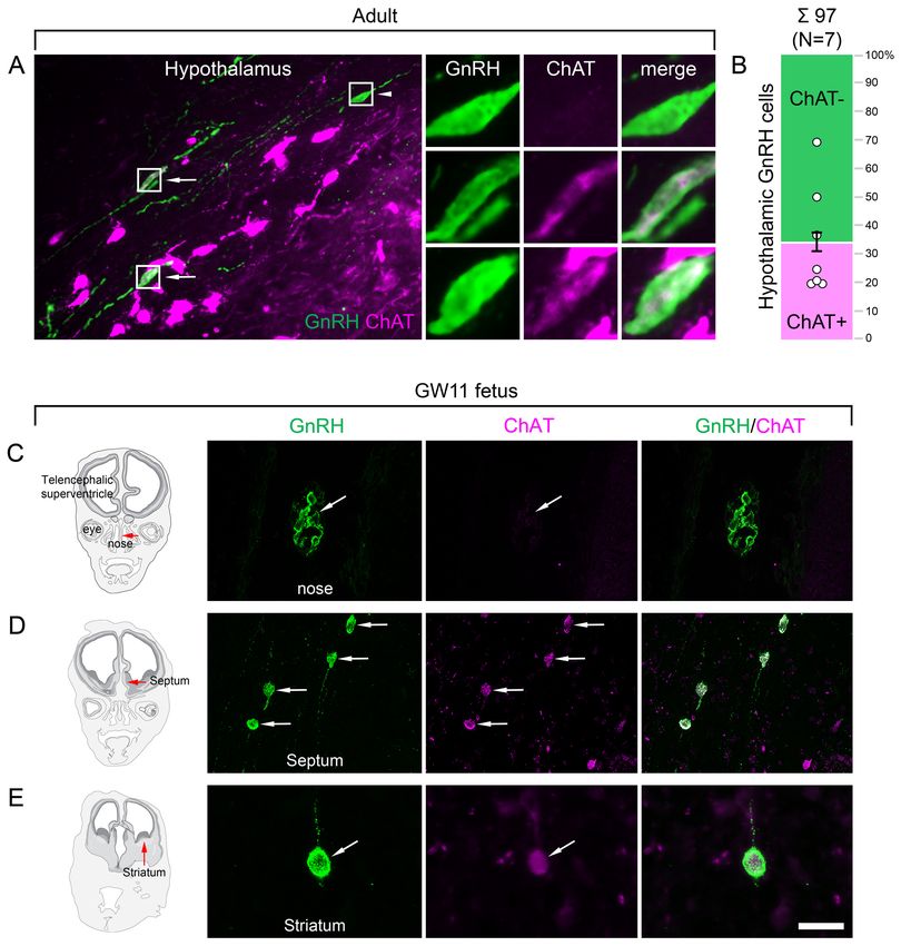
Research article                                                                                                      Cell Biology Neuroscience

Figure 1. Anatomical approaches unveil the distribution, number, fine structure, and cholinergic phenotype of extrahypothalamic gonadotropin-
releasing hormone (GnRH) neurons in the adult human brain. (A) Extrahypothalamic GnRH-immunoreactive (GnRH-IR) neurons were mapped with
immunohistochemistry (IHC) and quantified in the brain of three adult human individuals (#1–3). (B) Representative coronal section of a 31-year-old
female subject (#1) illustrates the caudate nucleus (Cd), putamen (Pu), claustrum (Cl), insular cortex (Ins), anterior limb of the internal capsule (aic),
Figure 1 continued on next page

Skrapits et al. eLife 2021;10:e67714. DOI: https://doi.org/10.7554/eLife.67714                                                                          3 of 26
Research article                                                                                                Cell Biology Neuroscience

Figure 1 continued
external capsule (ec), extreme capsule (ex), corpus callosum (cc), lateral medullary lamina (lml), lateral ventricle (LV), nucleus accumbens (nAcc),
and olfactory tubercle (Tu). High-power insets (1–3) reveal extrahypothalamic GnRH neurons many of which can be found in the Pu. (C) The majority
(82.2%) of the 163,168 ± 36,223 extrahypothalamic GnRH neurons occurred in the Pu, followed by the nAcc, Cd, nucleus basalis magnocellularis (nbM),
globus pallidus (GP), ventral pallidum (VP), and bed nucleus of the stria terminalis (BnST). (D) To visualize the fine structure of dendrites, the
immunofluorescent (IF) detection of GnRH was combined with cell membrane labeling using DiI delivered with a Gene Gun. (E) 3-D reconstruction of
the DiI-labeled (magenta) GnRH-IR (green) neurons revealed large multipolar cells, which exhibited only few dendritic spines. (F) Depth color coding,
where colors represent distance from the section surface, allowed better distinction between DiI-labeled processes of the GnRH neuron (upper inset;
GnRH+) from other DiI-labeled neuronal elements many of which belonged to medium spiny GABAergic projection neurons (lower inset; GnRH-). (G)
Double-IF experiments addressed the presence of the cholinergic marker enzyme choline acetyltransferase (ChAT) in GnRH neurons. Nearly all GnRH
neurons in the Pu contained ChAT signal. (H) The GnRH neuron population also overlapped with cholinergic projection neurons of the nbM. (I) With
few exceptions, GnRH neurons were ChAT-immunoreactive (green columns), whereas they represented relatively small subsets of cholinergic cells
(magenta columns) being the highest in the Pu (~35%). Scale bar (shown in H): 3.5 mm in low-power photomicrograph (B), 50 mm in (B, C, G and H)
(insets in G and H: 25 mm), 12.5 mm in (E) and (F) (insets: 3.7 mm).
The online version of this article includes the following figure supplement(s) for figure 1:
Figure supplement 1. Cell numbers determined with light microscopic analysis require compensation for overcounting using Abercrombie’s correction
factor.

                                    migrating GnRH neurons in human embryos/fetuses most of which (~8000) followed a previously
                                    unknown dorsal migratory route targeting subpallial and/or pallial structures, as opposed to
                                    the ~2000 neurons in the ventral route leading to the hypothalamus (Casoni et al., 2016). We
                                    addressed the possibility that extrahypothalamic GnRH-IR neurons of the adult brain originate from
                                    the 8000 neurons observed in the dorsal pathway. With this aim, we determined the total number of
                                    GnRH-IR neurons in the basal ganglia and the basal forebrain. Immunolabeled neurons were counted
                                    in every 24th section of a single hemisphere using light microscopy (Figure 1A, B). Cell counts were
                                    then multiplied by 24 and 2 (for the two hemispheres) and compensated for overcounting (Aber-
                                    crombie, 1946; Guillery, 2002; Figure 1—figure supplement 1A). The total number of extrahypo-
                                    thalamic GnRH neurons calculated this way in three subjects was 229,447 (31-year-old female; #1),
                                    155,357 (61-year-old male; #2), and 104,699 (62-year-old male; #3) (163,168 ± 36,223; mean ± SEM).
                                    These unexpectedly high GnRH cell numbers made it unlikely that human extrahypothalamic GnRH
                                    neurons develop from olfactory placodes and migrate into the brain along the dorsal migratory
                                    route (Casoni et al., 2016). The individual variations in total GnRH cell numbers of the three samples
                                    may be both biological and technical, which would be difficult to separate. 82.2 ± 1.1% of labeled
                                    cells were observed in the Pu, 5.5 ± 0.2% in the nAcc, 4.9 ± 0.7% in the Cd, 3.5 ± 1.1% in the nbM,
                                    1.8 ± 0.5% in the GP, 1.3 ± 0.1% in the VP, and 0.8 ± 0.2% in the BnST (Figure 1C).

                                    Extrahypothalamic GnRH neurons synthesize bona fide GnRH
                                    decapeptide derived from the GNRH1 transcript
                                    Results of previous studies with IHC on embryonic and fetal rhesus monkey brains questioned
                                    whether extrahypothalamic GnRH neurons synthesize bona fide GnRH decapeptide
                                    (Quanbeck et al., 1997; Terasawa et al., 2001). First, developing GnRH neurons in the septum,
                                    stria terminalis, amygdala, striatum, and internal capsule of the monkey brain were not detected by
                                    several GnRH antibodies (Quanbeck et al., 1997; Terasawa et al., 2001), including the widely used
                                    LR-1 rabbit GnRH polyclonal antiserum (Silverman et al., 1990). Second, these neurons exhibited
                                    immunoreactivity to EP24.15 (aka thimet oligopeptidase; THOP1), a metalloendopeptidase, which
                                    can cleave GnRH at the Tyr5-Gly6 position to generate GnRH1-5 (Terasawa et al., 2001). To investi-
                                    gate the possibility that GnRH neurons in the basal ganglia and the basal forebrain of the adult
                                    human brain use GnRH1-5, rather than GnRH decapeptide for signaling, we first tested a series of
                                    polyclonal antibodies against human GnRH-associated peptide (hGAP1) or GnRH decapeptide
                                    (Supplementary file 2) for their reactivity with GnRH neurons of the human Pu (N = 10; #5, 6, and
                                    12–19). All of the tested antibodies, including the LR-1 antiserum, recognized GnRH-IR neurons
                                    (Figure 2B), suggesting that these cells contain the bona fide GnRH. Neurons detected with differ-
                                    ent antibodies were identical as they were double-labeled (Figure 2C) in dual-immunofluorescence
                                    (IF) experiments (N = 2; #4 and 5) using two GnRH antibodies from different host species. Results of
                                    further control experiments with the combined use of IF and non-isotopic ISH (N = 5; #15–19)

Skrapits et al. eLife 2021;10:e67714. DOI: https://doi.org/10.7554/eLife.67714                                                                 4 of 26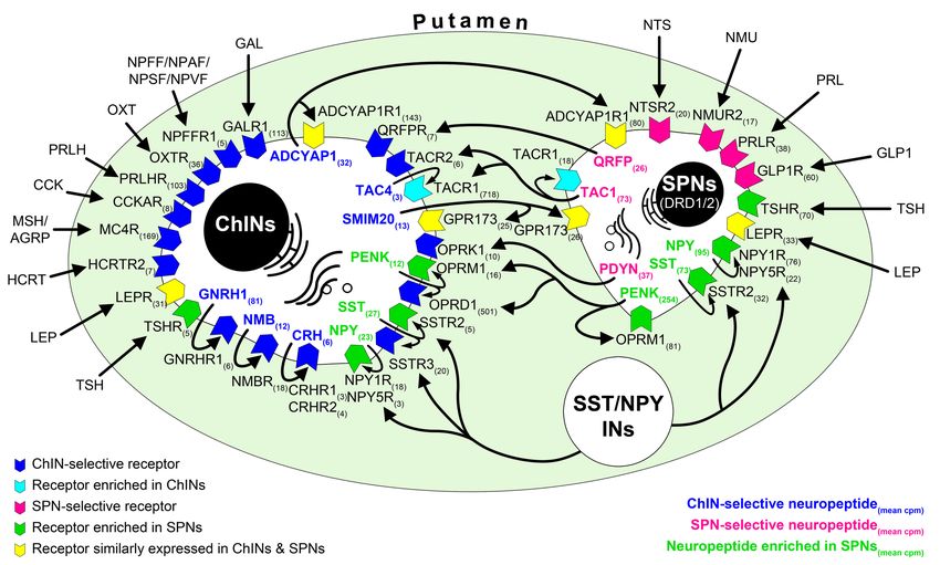
Research article                                                                                                    Cell Biology Neuroscience

Figure 2. Combined evidence from immunohistochemistry (IHC), in situ hybridization (ISH), and high-performance liquid chromatography-tandem mass
spectrometry (HPLC-MS/MS) indicates that extrahypothalamic gonadotropin-releasing hormone (GnRH) neurons synthesize bona fide GnRH
decapeptide derived from the GNRH1 transcript. (A) IHC labeling of the human putamen with the guinea pig polyclonal GnRH antiserum #1018 reveals
a large number of immunoreactive neurons in control sections (Ctrl) of a 64-year-old male subject (#19). All labeling is eliminated if the working solution
Figure 2 continued on next page

Skrapits et al. eLife 2021;10:e67714. DOI: https://doi.org/10.7554/eLife.67714                                                                      5 of 26
Research article                                                                                           Cell Biology Neuroscience

Figure 2 continued
of #1018 is preabsorbed overnight with 0.1 mg/ml GnRH. (B) A series of additional primary antisera against the human GnRH-associated peptide
(hGAP1) or GnRH also recognize immunoreactive neurons in the human putamen (#6). Such antibodies include the LR-1 rabbit primary antiserum, which
was reported previously not to label extrahypothalamic GnRH neurons in the developing monkey brain. (C) Positive control with the combined use of
two GnRH antibodies from different host species for dual-immunofluorescence (IF) experiments on sample #5 provides evidence that the antibodies
detect the same neuronal elements. (D) Non-isotopic ISH/IF dual-labeling studies reveal that GnRH-immunoreactive neurons express GNRH1 mRNA,
indicating that extrahypothalamic GnRH is a GNRH1 gene product (sample #15). (E) As illustrated in representative chromatograms, HPLC-MS/MS (
#21–28) detects bona fide GnRH decapeptide in tissue extracts from the mediobasal hypothalamus (MBH), putamen (Pu), and nucleus caudatus (Cd),
but not the claustrum (Cl). (F) The GnRH1-5 degradation product is present in the Pu and Cd and undetectable in the MBH and Cl. (G) Quantitative
analysis reveals the highest tissue concentrations of GnRH in the MBH, somewhat lower levels in the Pu and the Cd, and no detectable GnRH
decapeptide signal in the Cl. Note that tissue concentrations of GnRH in the Pu and the Cd are 3–4 times higher than those of GnRH1-5. Scale bar
(shown in D): 120 mm in (A, B), 50 mm in (C, D).

                                    showed that GnRH-IR neurons express GNRH1 mRNA (Figure 2D). Finally, to provide direct evi-
                                    dence for the biosynthesis of the GnRH decapeptide in these cells, tissue samples (#21–28) were
                                    microdissected from the mediobasal hypothalamus (MBH; N = 2), Pu (N = 5), Cd (N = 2), and Cl
                                    (N = 3). HPLC-MS/MS analysis of the tissue extracts established that the dominant peptide form in
                                    the Pu and Cd is the GnRH decapeptide. GnRH1-5 was also present, albeit at 3–4 times lower tissue
                                    concentrations (Figure 2E–G). Only GnRH decapeptide was detectable in the MBH (used as a posi-
                                    tive control) where hypophysiotropic GnRH neurons occur and neither peptide form was present in
                                    the Cl, in accordance with the absence of IHC labeling at this site (Figure 2E–G). Together with
                                    observations from the IHC and ISH experiments, HPLC-MS/MS results gave firm support to the
                                    notion that extrahypothalamic GnRH neurons mainly produce bona fide GnRH decapeptide derived
                                    from the GNRH1 gene.

                                    GnRH neurons of the putamen are large multipolar interneurons with
                                    smooth-surfaced dendrites
                                    The IHC method was unable to visualize the entire dendritic arbor of extrahypothalamic GnRH-IR
                                    cells (Figure 1B, C). This limitation could be due to the low amount and/or restricted subcellular dis-
                                    tribution of the peptide. To overcome this problem, we labeled the dendritic compartment of GnRH
                                    cells with the lipophilic dye DiI for further morphological analysis (Takács et al., 2018; Figure 1D).
                                    Following the immunofluorescent (IF) visualization of GnRH neurons in the Pu of a 72-year-old female
                                    (#20), DiI-coated tungsten particles were delivered into the sections using a Helios Gene Gun (Bio-
                                    Rad) (Figure 1D; Takács et al., 2018). Spreading of this lipophilic dye along the cytoplasmic mem-
                                    brane caused Golgi-like labeling of random-hit neurons, including 12 GnRH-IR cells (Figure 1E, F).
                                    Confocal microscopic analysis and 3-D reconstruction of the DiI signal revealed spider-like neurons
                                    with rich arborization of poorly spined dendrites. DiI-labeled GnRH neurons were clearly distinct
                                    from the main Pu cell type, the densely spined medium spiny GABAergic projection neurons (SPNs)
                                    (Figure 1E, F).

                                    Extrahypothalamic GnRH cells represent subpopulations of cholinergic
                                    neurons
                                    SPNs represent 80–98% of striatal neurons, the remainder being made up of cholinergic and differ-
                                    ent subclasses of GABAergic interneurons (Gonzales and Smith, 2015). DiI-labeled GnRH cells
                                    resembled cholinergic interneurons (ChINs) in size and dendritic morphology. Indeed, dual-IF experi-
                                    ments (N = 4; #3–5 and 19) established that GnRH neurons of the Pu contain the cholinergic marker
                                    enzyme choline acetyltransferase (ChAT) (Figure 1G). Similarly, GnRH neurons in the nbM
                                    (Figure 1H) and other extrahypothalamic sites also exhibited ChAT immunoreactivity. The extent of
                                    ChAT/GnRH colocalization was assessed quantitatively in five distinct regions of a 62-year-old male
                                    subject (#3). Confocal microscopic analysis of representative dual-labeled sections established that
                                    the vast majority of extrahypothalamic GnRH neurons are cholinergic (green bars in Figure 1I). In
                                    contrast, GnRH-IR neurons represented only 34.9% of all cholinergic neurons in the Pu, 6.3% in the
                                    nAcc, 1.8% in the head of the Cd, 3.6% in the nbM, and 28.4% in the GP (magenta bars in
                                    Figure 1I). GnRH-positive and GnRH-negative cholinergic neurons often intermingled, without gross
                                    morphological differences between the two phenotypes (Figure 1G, H).

Skrapits et al. eLife 2021;10:e67714. DOI: https://doi.org/10.7554/eLife.67714                                                            6 of 26
Research article Cell Biology Neuroscience
Hypothalamic GnRH neurons regulating reproduction also exhibit an
unexpected cholinergic phenotype
The ChAT phenotype emerged as a hallmark of extrahypothalamic GnRH neurons. To confirm this
notion by verifying the absence of ChAT in the hypothalamic GnRH neuron population, hypothalamic
tissue sections were processed for dual-IF detection of ChAT and GnRH, followed by confocal micro-
scopic analysis. Unexpectedly, 34.6 ± 7.1% of the hypothalamic GnRH neurons also exhibited ChAT
signal in seven adult human male and female subjects (Figure 3A, B; #3 and 6–11), a phenomenon
not observed in other species before.
Cholinergic phenotype of human GnRH neurons develops prenatally
To address when GnRH neurons gain the cholinergic phenotype, prenatal co-expression of ChAT
and GnRH was explored via dual-IF experiments in coronal sections of two fetal heads (#29 and 30)
at gestational week 11 (GW11). At this age, ~20% of GnRH neurons can still be found in the nasal
region, whereas the majority have already entered the brain to migrate toward hypothalamic and
extrahypothalamic target areas (Casoni et al., 2016). While GnRH-positive neurons within the nasal
compartment did not contain ChAT signal (Figure 3C), those in the septum (Figure 3D), the striatum
(Figure 3E) and elsewhere in the developing brain were already ChAT-IR. These data suggest that
migrating GnRH neurons become cholinergic after entering the brain and continue to express ChAT
immunoreactivity in hypothalamic as well as extrahypothalamic regions.
Neurons laser-capture microdissected from the postmortem putamen
provide sources for high-quality RNA suitable for RNA-seq
ChINs, at least one-third of which synthesize GnRH in the human Pu (Figure 1I), communicate with
SPNs locally (Ahmed et al., 2019). To localize the receptors mediating the effects of GnRH in the
adult human Pu, transcriptome profiling of cellular samples enriched in ChINs and SPNs was carried
out. Being the largest cell type, ChINs were readily recognizable in sections subjected to Nissl-stain-
ing under RNase-free conditions, whereas the medium-sized SPNs represent the most frequently
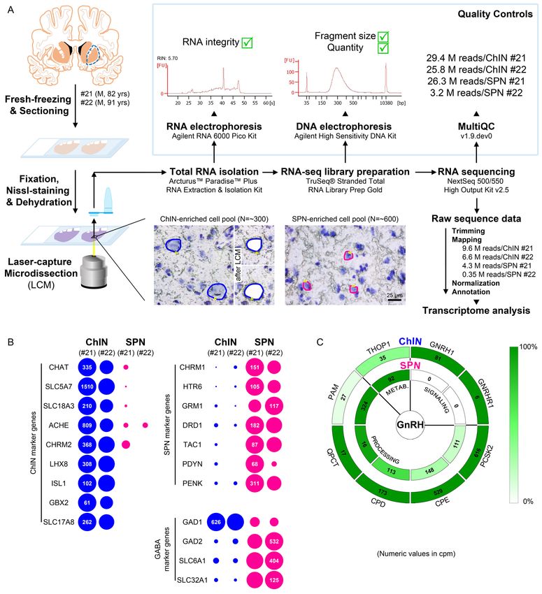
encountered neuronal phenotype in the Pu (Figure 4A). Laser-capture microdissection (LCM) was
used to collect neuronal pools enriched in ChINs and SPNs from cresyl violet-stained Pu sections of
two human subjects (#21 and 22). Each ChIN-enriched pool contained ~300 large neurons and each
SPN-enriched pool consisted of ~600 medium-sized neurons (Figure 4A). Total RNA was isolated
and RNA-seq libraries were prepared from the four cell pools and sequenced with the Illumina Next-
Seq 500/550 High Output (v2.5) kit. 29.4M and 25.8M reads were obtained from the two ChIN pools
and 26.3M and 3.2M reads from the two SPN pools. Approximately 9.6M and 6.6M reads from ChIN
and ~4.3M and 0.35M reads from SPN pools were mapped to the GRCh38.p13 human reference
genome; 13,664 and 12,637 identified transcripts occurred at cpm >5 in ChIN pools and 13,558 and
13,682 transcripts in SPN pools (Figure 4A and Supplementary file 3).
Size-based laser-capture microdissection allows adequate sampling of
striatal cholinergic interneurons and medium spiny projection neurons
Cholinergic markers, including CHAT, SLC5A7, SLC18A3, ACHE, and CHRM2, were highly enriched
in the ChIN pools from subjects #21 and 22. These transcripts were either absent or found at low lev-
els only in the two SPN pools (Figure 4B). Mouse ChINs arise from Nkx2.1+ progenitors. During
development, Nkx2.1 upregulates the expression of the LIM homeobox proteins LHX8, ISL1, and
GBX2, which, in turn, promote cell differentiation into ChINs (Allaway and Machold, 2017). These
LIM transcripts as well as type 3 vesicular glutamate transporter (SLC17A8) showed robust and exclu-
sive expression in ChINs (Figure 4B). The SPN pools, in turn, expressed much higher levels of known
SPN markers than ChINs, including various cholinergic (CHRM1), serotonergic (HTR6), glutamatergic
(GRM1), and dopaminergic (DRD1) receptor isoforms and several neuropeptides (TAC1, PDYN,
PENK) (Figure 4B). Differential distribution of the above transcripts verified that the size-based LCM
strategy efficiently separated ChINs from SPNs for transcriptome profiling. Relatively high levels of
expression of known GABAergic marker transcripts (GAD1, GAD2, SLC6A1, and SLC32A1) in ChINs,
in addition to SPNs (Figure 4B), revealed that ChINs use GABAergic co-transmission, as proposed
earlier for ChINs of the rodent CPU (Lozovaya et al., 2018).
Skrapits et al. eLife 2021;10:e67714. DOI: https://doi.org/10.7554/eLife.67714 7 of 26Research article Cell Biology Neuroscience Figure 3. Both hypothalamic and extrahypothalamic gonadotropin-releasing hormone (GnRH) neurons exhibit cholinergic phenotype gained during early fetal development. (A) The cholinergic phenotype is not a hallmark of human extrahypothalamic GnRH neurons because large subsets of GnRH neurons in the adult human hypothalamus (green immunofluorescent signal; subject #3) also exhibit choline acetyltransferase (ChAT; magenta) immunoreactivity. High-power insets show single- (arrowhead) and dual-labeled (arrows) GnRH neurons from framed regions. (B) Quantitative analysis of 97 GnRH neurons from seven subjects (#3; 6–11) reveals the ChAT phenotype in 34.6 ± 7.1% of hypothalamic GnRH neurons. (C–E) The cholinergic phenotype of GnRH neurons is gained during early fetal development. Left panels illustrate coronal views of the fetal head at GW11 (#29). Representative photomicrographs taken from sites indicated by the red arrows show results of dual-immunofluorescence experiments. (C) At this stage of development, a large subset of GnRH neurons (green immunofluorescent signal) migrate in the nasal region toward the brain and do not exhibit Figure 3 continued on next page Skrapits et al. eLife 2021;10:e67714. DOI: https://doi.org/10.7554/eLife.67714 8 of 26
Research article                                                                                                 Cell Biology Neuroscience

Figure 3 continued
ChAT signal. (D, E) In contrast, GnRH neurons migrating through the septal area (D, arrows) or located in the striatum (E, arrow) express ChAT
(magenta). Scale bar (shown in E): 50 mm in (A, C, D) (insets in A: 12.5 mm), 20 mm in (E).

                                     Cholinergic interneurons selectively express GNRH1 and GNRHR1 and
                                     contain GnRH biosynthetic enzymes
                                     GNRH1 was expressed exclusively in the two ChIN pools, in accordance with the morphological
                                     observations (Figure 4C). Processing of the proGnRH1 protein begins with endoproteolysis by pro-
                                     hormone convertases from which ChINs abundantly expressed the PCSK2 isoform. Enzymes catalyz-
                                     ing subsequent steps of GnRH biosynthesis, including carboxypeptidases (CPE, CPD),
                                     peptidylglycine a-amidating monooxygenase (PAM), and glutaminyl cyclase enzymes (QPCT), were
                                     also present in ChINs (Figure 4C). The promiscuous THOP1 enzyme accounts for the cleavage of
                                     multiple neuropeptides, including GnRH. This enzyme was expressed in both ChINs and SPNs, with
                                     a higher relative abundance in the latter. The most important finding of the RNA-seq study was that
                                     the seven-transmembrane receptor GNRHR1 was expressed selectively in ChINs. This observation
                                     indicated that GnRH in the human Pu acts on GnRHR1 autoreceptors and also made it unlikely that
                                     SPNs are affected directly by GnRH derived from ChINs. Altogether, transcriptome profiling of
                                     ChINs and SPNs provided molecular support to a concept that GnRH is synthesized by ChINs and
                                     acts locally via GnRHR1 autoreceptors.

                                     Transcriptome profiling provides novel insight into the molecular
                                     connectome of the human putamen
                                     Transcriptome profiling of ChINs and SPNs revealed a large set of genes that were expressed selec-
                                     tively or predominantly in one cell type only, in addition to many other genes expressed in both.
                                     Neurotransmitter and neurotransmitter receptor transcripts identified this way allowed us to propose
                                     signaling mechanisms that act in the bidirectional communication between ChINs and SPNs. Some
                                     receptors appear to serve as autoreceptors (e.g., GNRHR1, NMBR, CRHR1/2). Others may receive
                                     ligands from multiple neuronal sources within (e.g., QRFPR, NPY1R/5R, TACR1, SSTR2/3) or outside
                                     (e.g., OXTR, MC4R, GLP1R, PRLR) the striatum. Peptidergic mechanisms concluded from the tran-
                                     scriptome profiles are illustrated as a schematic model in Figure 5. A deeper insight into the molec-
                                     ular connectome of the human Pu can be obtained from the detailed receptor and neuropeptide
                                     expression profiles of ChINs and SPNs and from the full list of expressed genes in the two SPN and
                                     two ChIN cell pools (Supplementary file 3; BioProject accession number: PRJNA680536).

                                     Discussion
                                     Extrahypothalamic GnRH-IR neurons correspond to type III GnRH
                                     neurons detected earlier with in situ hybridization and synthesize the
                                     full-length GnRH decapeptide derived from the GNRH1 gene
                                     A pioneer ISH study by Rance and co-workers distinguished three types of GNRH1 mRNA-express-
                                     ing neurons in the human brain based on size, shape, and labeling intensity (Rance et al., 1994).
                                     The GnRH-IR neurons we detected in our study correspond to type III neurons characterized by
                                     round/oval shape, large nucleus and nucleolus, prominent Nissl substance, and GNRH1 mRNA levels
                                     intermediate between those of heavily labeled type I neurons in the MBH and lightly labeled type II
                                     neurons in the medial septum and the dorsal medial preoptic area (Rance et al., 1994). Our IHC
                                     studies also detected many type I hypothalamic GnRH neurons but found only few septal type II neu-
                                     rons, which latter had negligible contribution to the total GnRH cell numbers.
                                         Although type III GnRH neurons have not been reported in adult laboratory rodents, they occur
                                     in the striatum, amygdala, and nbM of non-human primates (Krajewski et al., 2003). These neurons
                                     were proposed to differ from the hypothalamic GnRH cell population in that they contain the
                                     GnRH1-5 degradation product of GnRH instead of the bona fide GnRH decapeptide
                                     (Quanbeck et al., 1997; Terasawa et al., 2001). Circumstantial IHC evidence to support this notion
                                     stemmed from IHC observations made on developing monkey embryos and fetal brains
                                     (Quanbeck et al., 1997; Terasawa et al., 2001). First, these neurons could not be immunolabeled

Skrapits et al. eLife 2021;10:e67714. DOI: https://doi.org/10.7554/eLife.67714                                                                   9 of 26
Research article Cell Biology Neuroscience Figure 4. Deep transcriptome profiling of cholinergic interneurons (ChINs) and spiny projection neurons (SPNs) provides new insight into extrahypothalamic gonadotropin-releasing hormone (GnRH) signaling mechanisms and the molecular connectome of the human putamen. (A) 20-mm- thick coronal sections were collected on PEN membrane slides from frozen putamen samples of two male human subjects (#21 and 22) and fixed with an ethanol/paraformaldehyde mixture. Neurons were visualized using Nissl-staining and isolated with laser-capture microdissection (LCM). 300 neurons included in each ChIN-enriched cell pool were recognized based on their large perikaryon size. The vast majority of the 600 medium-sized Figure 4 continued on next page Skrapits et al. eLife 2021;10:e67714. DOI: https://doi.org/10.7554/eLife.67714 10 of 26
Research article                                                                                                  Cell Biology Neuroscience

Figure 4 continued
microdissected and pooled neurons corresponded to SPNs, the major putamen cell type. Total RNA was isolated and RNA-seq library prepared from
both cell types and sequenced with the Illumina NextSeq 500/550 High Output (v2.5) kit. (B) Bioinformatic analysis verified high enrichment of known
cholinergic markers in the two ChIN pools and of SPN markers in the two SPN pools. Expression levels in dots reflect counts per million reads (cpm),
and in each case, dot areas reflect transcript abundances relative to the highest cpm (100%). (C) Key elements of proGnRH processing, GnRH signaling,
and GnRH metabolism are illustrated in two concentric circles. The GNRH1 and GNRHR1 transcripts are present in ChINs only (outer circle). ChINs
express all enzymes required for proGnRH processing. The promiscuous THOP1 enzyme, which may account for GnRH cleavage, occurs in both cell
types, at higher levels in SPNs than in ChINs. Color coding reflects relative transcript abundances, whereas numbers indicate cpms (mean cpms of
subjects #21 and 22).

                                     with the LR-1 rabbit polyclonal antiserum and a series of other antibodies against GnRH
                                     (Quanbeck et al., 1997). Second, they showed weaker labeling with GnRH4-10 and heavier labeling
                                     with GnRH1-5 antibodies than migrating GnRH neurons targeting the hypothalamus
                                     (Terasawa et al., 2001). Third, they showed immunoreactivity to the THOP1 enzyme, which can
                                     cleave GnRH at the Tyr5-Gly6 position, although notably, they did not differ from hypothalamic
                                     GnRH neurons in this latter respect (Terasawa et al., 2001).

Figure 5. RNA-seq studies reveal the neuropeptide and neuropeptide receptor expression profiles of cholinergic interneurons (ChINs) and spiny
projection neurons (SPNs) and provide insight into the molecular connectome of putamen cell types. Proposed signaling mechanisms are based on
neuropeptide and peptide receptor expression profiles of the two cell types. ChINs appear to use GNRHR1, CRHR1/2, and NMBR autoreceptor
signaling. SSTR2, NPY1R/5R, OPRM1, and TACR1 may serve, at least partly, as autoreceptors in SPNs. Proposed peptidergic communication between
the two cell types is also indicated by arrows. Other receptors receive ligands from different neuronal sources within (e.g., QRFPR, NPY1R/5R, TACR1,
SSTR2/3) or outside (e.g., OXTR, MC4R, GLP1R, PRLR) the putamen. Numbers in receptor symbols reflect transcript abundances expressed as mean
counts per million (cpms) from subjects #21 and 22. The figure illustrates receptors that were consistently observed in the given cell type of both human
samples. INs: interneurons.

Skrapits et al. eLife 2021;10:e67714. DOI: https://doi.org/10.7554/eLife.67714                                                                  11 of 26
Research article Cell Biology Neuroscience
In contrast with the above suggestion, our results indicate that the human Pu and Cd contain
mostly bona fide GnRH decapeptide because (1) human ChINs can be immunolabeled with the LR-1
antibodies and several other GAP1 and GnRH antibodies, unlike extrahypothalamic neurons of the
monkey. (2) They possess the full enzyme set of GnRH biosynthesis, as revealed by deep transcrip-
tome profiling. Finally, (3) human Pu extracts contain 3–4-times as much uncleaved GnRH decapep-
tide as GnRH1-5, as shown by results of HPLC-MS/MS studies. Different conclusion from the monkey
and human studies may reflect species differences and the use of different methodological
approaches.
We note that the human genome includes a fully functional GNRH2 gene (Stewart et al., 2009),
in addition to GNRH1. The GnRH signals we detected in the Pu are due to GNRH1, rather than
GNRH2 expression, because (1) extrahypothalamic GnRH-IR neurons also exhibit ISH signal for
GNRH1 mRNA, (2) they are IR to GAP1, which only has low homology with the corresponding GAP2
sequence, and (3) ChINs of the Pu express high levels of the GNRH1 transcript, according to RNA-
seq results.
Our data neither exclude nor support the possibility that the GnRH degradation product GnRH1-
5 plays a role in the regulation of the human striatal neurocircuitry. Indeed, we found that GnRH1-5
was also detectable with HPLC-MS/MS in the postmortem Pu and Cd, albeit at ~70% lower levels
than GnRH. Transcriptomic studies revealed that one of the putative GnRH1-5 receptors, GPR101
(Cho-Clark et al., 2014), was expressed at high levels in SPNs (mean cpm: 58.4) and at low levels in
ChINs (mean cpm: 6.4) (Supplementary file 3). This differential expression suggests that if GnRH1-5
is a physiological neurotransmitter of human ChINs, it may regulate SPNs via GPR101. We note that
THOP1 expression in ChINs is not a strong proof for the transmitter role of GnRH1-5. This endopep-
tidase has many substrates unrelated to GnRH degradation (Orlowski et al., 1983). Further, in RNA-
seq studies, THOP1 was expressed at higher levels in SPNs than in ChINs. Moreover, bona fide
hypothalamic GnRH neurons in the developing monkey brain also exhibited THOP1 immunoreactiv-
ity (Terasawa et al., 2001).
Overlap with cholinergic neurons and large cell numbers argue against
the placodal origin
Total GnRH-IR cell numbers we calculated for the basal forebrain and the basal ganglia of three
adult human brains (229,447, 155,357, and 104,699) exceeded all previous estimates of GnRH cell
numbers in any species. For technical reasons, our calculation could even underestimate the real
number of GnRH neurons. Perimortem and postmortem conditions could be suboptimal in some
samples to achieve maximal detection sensitivity. In addition, the use of 100-mm-thick sections for
IHC could also compromise the detection of low signal levels via reducing antibody penetration.
Rance and coworkers identified 5800 type III GnRH neurons in the human basal forebrain complex
rostral to the mammillary bodies, caudal to the optic chiasm, and ventral to the anterior commissure
(Rance et al., 1994). This low cell number may reflect that tissues with these anatomical guidelines
are devoid of the bulk of the Pu, which contained the majority (82%) of the extrahypothalamic GnRH
neurons in our study.
Our initial hypothesis was that extrahypothalamic GnRH neurons are derived from the ~8000
GnRH neurons reported recently along the dorsal migratory route during embryonic/fetal develop-
ment (Casoni et al., 2016). The placodal origin now seems to be very unlikely considering the much
larger GnRH cell numbers we observed in the adult brain.
Extrahypothalamic GnRH neurons of the human central nervous system also appear to be homol-
ogous to the ‘early type’ GnRH neurons reported in the developing monkey embryo and fetus
(Quanbeck et al., 1997). In this species, the ‘early’ and ‘late’ types of GnRH neurons were distin-
guished based on differences in their time of appearance, morphology, and immunoreactivity pat-
tern using GnRH antibodies against different GnRH epitopes (Quanbeck et al., 1997;
Terasawa et al., 2001). It was speculated that early GnRH neurons originated from the dorsal olfac-
tory placode before olfactory pit formation at E30, migrated into the brain along the olfactory nerve,
and settled in striatal and limbic structures of the fetal brain (Quanbeck et al., 1997). However, in a
subsequent study these authors noted that a 10- to 10,000-fold increase in the number of ‘early’
GnRH neurons in the basal forebrain within just a few days indicates that early GnRH neurons might
rather be derived from the ventricular wall of the telencephalic vesicle (Terasawa et al., 2001). The
possibility of non-placodal GnRH neuron development is compatible with the in vitro capability of
Skrapits et al. eLife 2021;10:e67714. DOI: https://doi.org/10.7554/eLife.67714 12 of 26Research article Cell Biology Neuroscience
hypothalamic and hippocampal progenitors to generate GnRH cells and all other neuroendocrine
cell types (Markakis et al., 2004).
It is worth noting that our RNA-seq studies provided transcriptomic information about a mixed
ChIN population of the Pu, whereas ChINs exhibit substantial diversity in their physiology, morphol-
ogy, and connectivity (Gonzales and Smith, 2015). Subclasses differ in their developmental origin
(medial ganglionic eminence, septal epithelium, or preoptic area) and transcription factor profiles
(Ahmed et al., 2019). It is unknown which ChIN subpopulation expresses GNRH1 and GNRHR1
because selective harvesting of well-preserved cellular RNA specifically from the GnRH-IR ChINs is
currently unresolved. Development of an ‘immuno-LCM/RNA-seq’ method would also allow us to
compare the gene expression profiles of hypothalamic and extrahypothalamic GnRH neurons.
Both hypothalamic and extrahypothalamic GnRH neurons use
cholinergic co-transmission
ChAT co-expression provided evidence that extrahypothalamic GnRH neurons correspond to subpo-
pulations of previously known cholinergic cells. These include ChINs of the Pu, which communicate
locally with SPNs as well as projection neurons of the nbM, which innervate distant limbic structures
(Ahmed et al., 2019). Although ChAT emerged as a hallmark of the extrahypothalamic GnRH sys-
tem, we found evidence that a relatively large subset of human hypothalamic GnRH neurons also
express the cholinergic marker enzyme. To our knowledge, this colocalization phenomenon has not
been reported in any other species before. Studies on GW11 human fetuses established that migra-
tory GnRH neurons in the nasal compartment are not cholinergic, whereas both hypothalamic and
extrahypothalamic GnRH neurons already express the ChAT signal at this age.
Rodent GnRH neurons are regulated by cholinergic afferents but not known to co-express cholin-
ergic markers (Turi et al., 2008). To address the possibility that the cholinergic phenotype of murine
GnRH neurons has only been overlooked in previous studies, we have recently tried to detect ChAT
immunoreactivity in GnRH neurons of the mouse preoptic area. The lack of success of this attempt
(Skrapits et al., unpublished) suggests that the cholinergic phenotype of GnRH neurons is a true spe-
cies difference between humans and rodents.
Laser microdissection of size-selected ChINs and SPNs is a highly efficient approach for transcrip-
tome profiling of the two cell types from the postmortem brain.
Deep transcriptome profiling of postmortem human neurons is technically challenging. Difficulties
include (1) compromised RNA quality, (2) lack of obvious marker signals to distinguish cell types,
and (3) low RNA yield from the LCM-isolated 300–600 neurons. Our strategy to isolate size-selected
ChINs and SPNs with LCM was justified by the RNA-seq results, which showed high enrichment of
known cell type-specific marker genes in the two cell pools and millions of reads in each. As one-
third of ChINs in the Pu also contain GnRH, deep transcriptome profiling of ChINs offered an insight
into the extrahypothalamic GnRH neuron transcriptome. Although it was beyond the focus of our
study, RNA-sequencing of ChINs and SPNs also unveiled the neurotransmitter and receptor profiles
of the two cell types and provided information about the putative molecular interactions taking
place in the Pu. The transcriptome databases allowed us to propose putative peptidergic mecha-
nisms and, thus, build the partial molecular connectome model of ChINs and SPNs.
GnRH acts outside the hypothalamus to regulate various reproductive
and non-reproductive functions
Clearly, the functions of GnRH are far from being restricted to the regulation of hypophysial gonado-
tropin secretion. Its receptor transcript, GNRHR1, is expressed in normal peripheral endocrine tis-
sues including the uterus, placenta, ovaries, testes, and prostate gland as well as in various tumor
cell types (Harrison et al., 2004). High levels of GNRHR1 mRNA and immunoreactivity were
reported in pyramidal neurons of the human hippocampus and cerebral cortex (Wilson et al., 2006).
GnRH analogues were anti-apoptotic in a rat model of ischemia/reperfusion (Chu et al., 2010). Fur-
ther, GnRH increased hippocampal estradiol levels and the spontaneous firing and GNRHR1 expres-
sion of pyramidal neurons and prevented memory deficits caused by amyloid b deposition
(Marbouti et al., 2020). While the source of GnRH acting on hippocampal neurons remains to be
explored, results of our studies indicate that GnRHR1 in ChINs of the basal ganglia can bind locally
synthesized GnRH neuropeptide. ChINs of the striatum contribute as interneurons to the regulation
Skrapits et al. eLife 2021;10:e67714. DOI: https://doi.org/10.7554/eLife.67714 13 of 26Research article Cell Biology Neuroscience
of cortico-striato-thalamocortical neural pathways. Functions associated with this circuitry include
motor control, learning, language, reward, cognitive function, and addiction (Fazl and Fleisher,
2018). The exact role of GnRH/GnRHR1 signaling in these functions requires clarification. We note
that various GABAergic interneurons of the Pu not studied here may also express GnRHR1 to serve
as additional target cells for GnRH signaling by ChINs. Cholinergic neurons of the nbM, which proj-
ect to the entire cortical mantle, olfactory tubercle, and amygdala, have been implicated in the con-
trol of attention, maintenance of arousal, and learning and memory formation (Koulousakis et al.,
2019). It remains to be determined if GnRH synthesized by these neurons binds to postsynaptic tar-
get cells or acts on autoreceptors, as we proposed for striatal ChINs.
Receptor profile of human cholinergic interneurons may offer new
therapeutic targets to treat neurodegenerative disorders
In the absence of animal models, it is currently difficult to estimate the role and importance of
GnRH/GnRHR1 signaling in the human basal ganglia and basal forebrain. Non-reproductive dysfunc-
tions have not been characterized in GnRH-deficient patients (Chan, 2011) or in the more common
cases of GnRHR1 deficiency (Chevrier et al., 2011; Seminara et al., 1998).
Future studies will need to clarify alterations of extrahypothalamic GnRH/GnRHR1 signaling in
neurodegenerative disorders affecting various cholinergic systems. Leading symptoms and cognitive
decline in Alzheimer’s disease are due to the loss of basal forebrain cholinergic neurons many of
which exhibited GnRH immunoreactivity in nbM. Parkinson’s disease (PD) is characterized by motor
symptoms such as abnormal involuntary movements, bradykinesia, rigidity, gait, and tremor. Non-
motor symptoms often include cognitive impairment, mood disorders, sleep alterations, dysautono-
mia, anosmia, and hallucinations (Perez-Lloret and Barrantes, 2016; Tubert and Murer, 2021).
Many of these malfunctions in PD can be explained with the loss of the nigrostriatal dopaminergic
input and ameliorated with levodopa. However, gait disorders and cognitive impairment/dementia
are most often unresponsive to dopamine precursor treatment. These data indicate the involvement
of other neurotransmitter systems. In particular, loss of striatal dopamine input causes a local hyper-
cholinergic state in the striatum with consequences reviewed recently (Tubert and Murer, 2021).
This hypercholinergic state explains the success of early PD therapies with Atropa belladonna deriva-
tives (Goetz, 2011). Although the low efficacy of anticholinergic drugs compared to levodopa and
unwanted side effects limit the use of general anticholinergic strategies (Katzenschlager et al.,
2003), selective inhibition of striatal ChINs has been proposed recently as a more promising strategy
to improve the transmitter balance in dopamine-deprived basal ganglia (Mallet et al., 2019;
Tubert and Murer, 2021). An important physiological mechanism to inhibit acetylcholine release
from ChINs is via M2-type (M2 and M4) muscarinic autoreceptors coupled to Gi proteins. Accord-
ingly, deletion of M2-type autoreceptors results in increased striatal acetylcholine release
(Bonsi et al., 2008). Autoinhibitory mechanism by muscarinic autoreceptors was found to be lost in
PD animal models (Ding et al., 2006). Indeed, our RNA-seq analysis confirmed that human ChINs
contain very high levels of CHRM2 autoreceptors (Figure 4B and Supplementary file 3). We note
that although the reproductive side effects would limit the use of GnRH analogues in clinical practice
(Almeida et al., 2004), the transcriptome profile of ChINs (Figure 5 and Supplementary file 3)
offers a few alternative receptorial mechanisms to counteract the hyperactivity of ChINs in PD.
Conclusions
This study reports the discovery and characterization of 150,000–200,000 GnRH-IR neurons, which
are located in the basal ganglia and basal forebrain of the adult human brain. These extrahypothala-
mic GnRH cells represent subsets of previously known cholinergic neurons that mainly synthesize
bona fide GnRH decapeptide. Unexpectedly, a large subpopulation of human hypothalamic GnRH
neurons share this cholinergic (ChAT) neurochemistry, which has not been detected in rodents.
RNA-seq experiments on ChINs and SPNs of the human Pu reveal that ChINs express GNRH1 and
GNRHR1, whereas their main target cells, the SPNs, do not, making it likely that GnRH acts via
autoreceptors. The role of GnRH/GnRHR1 signaling within extrahypothalamic neuronal circuitries of
the human brain requires clarification. RNA-seq studies, which revealed the transcriptome profiles of
ChINs and SPNs, also provide an insight into the molecular connectome of the human putamen.
Skrapits et al. eLife 2021;10:e67714. DOI: https://doi.org/10.7554/eLife.67714 14 of 26Research article Cell Biology Neuroscience
Materials and methods
Key resources table
Reagent type
(species) or Source or Additional
resource Designation reference Identifiers information
Biological sample Striatum, 1st Department See Supplementary file 1
(Homo sapiens) hypothalamus, of Pathology
putamen, n. and Experimental Cancer
caudatus, claustrum Research, Semmelweis
(adult) University,
Budapest, Hungary
Biological sample Head (fetus) Agence de la Biomédecine, See Supplementary file 1
(Homo sapiens) Saint-Denis la Plaine,
France, protocol n˚:
PFS16–002
Antibody Anti-GAP (rabbit Dr. M. D. Culler MC-2 IHC (1:5000)
polyclonal) Culler and
Negro-Vilar, 1986
Antibody Anti-GnRH (rabbit Dr. R. A. Benoit LR-1 IHC (1:10,000)
polyclonal) Silverman et al., 1990
Antibody Anti-GnRH (guinea Made in-house #1018 IHC, IF-TSA (1:30,000)
pig polyclonal) Hrabovszky IF (1:10,000)
et al., 2011
Antibody Anti-GnRH Made in-house #1014 IHC, IF-TSA (1:20,000)
(rat polyclonal) Skrapits et al., 2015
Antibody Anti-GnRH (sheep Made in-house #2000 IHC (1:1000)
polyclonal) Skrapits et al., 2015
Antibody Anti-ChAT (goat Merck Cat#AB144P; IF (1:150),
polyclonal) RRID:AB_2079751 IF-TSA (1:2000)
Antibody Anti-guinea pig IgG Jackson Cat#706-545-148; RRID:AB_2340472 IF (1:400)
(H + L) Alexa Fluor ImmunoResearch
488 (donkey
polyclonal)
Antibody Anti-goat IgG (H + L) Invitrogen Cat#A-11057; IF (1:400)
Alexa Fluor 568 RRID:AB_2534104
(donkey polyclonal)
Antibody Biotin-SP-AffiniPure anti- Jackson Cat#706-065-148; RRID:AB_2340451 IHC (1:500)
guinea pig IgG (H + L) ImmunoResearch
(donkey polyclonal)
Antibody Biotin-SP-AffiniPure Jackson Cat#711-065-152; RRID:AB_2340593 IHC (1:500)
anti-rabbit IgG (H + L) ImmunoResearch
(donkey polyclonal)
Antibody Biotin-SP-AffiniPure Jackson Cat#712-065-153; RRID:AB_2315779 IHC (1:500),
anti-rat IgG (H + L) ImmunoResearch IF-TSA (1:500)
(donkey polyclonal)
Antibody Biotin-SP-AffiniPure Jackson Cat#713-065-147; RRID:AB_2340716 IHC (1:500)
anti-sheep IgG (H + L) ImmunoResearch
(donkey polyclonal)
Antibody Biotin-SP-AffiniPure Jackson Cat#705-065-147; RRID:AB_2340397 IF-TSA (1:500)
anti-goat IgG (H + L) ImmunoResearch
(donkey polyclonal)
Antibody Peroxidase-AffiniPure Jackson Cat#706-035-148; RRID:AB_2340447 IF-TSA (1:250)
anti-guinea pig IgG ImmunoResearch
(H + L) (donkey
polyclonal)
Antibody Peroxidase- Roche 11207733910; ISH (1:100)
conjugated RRID:AB_514500
anti-digoxigenin, Fab
fragments (sheep
polyclonal)
Continued on next page
Skrapits et al. eLife 2021;10:e67714. DOI: https://doi.org/10.7554/eLife.67714 15 of 26Research article Cell Biology Neuroscience
Continued
Reagent type
(species) or Source or Additional
resource Designation reference Identifiers information
Other FITC-tyramide Synthesized IF-TSA (1:1000)
in-house
Hopman et al., 1998
Other Cy3-tyramide Synthesized IF-TSA, ISH (1:1000)
in-house
Hopman et al., 1998
Other ABC elite reagent Vector PK-6100; IHC, IF-TSA (1:1000)
Laboratories RRID:AB_2336819
Sequence- Antisense This paper ISH probe CATTCACAACACAGCA
based reagent proGnRH CTTTATTATGGAATATG
TGCAACTTGGTGTAAGG
ATTTCTGAAATTCATACC
ATTTACAGGTATTTAATG
GGTTATAAATTTTCAATG
TCAGAATTATACTTAAGT
CATGTTAGTAATGGTCAT
TCCTTCTGGCCCAATGG
ATTTAAATCTTCTTCTGC
CCAGTTTCCTCTTCAATC
AGACTTTCCAGAGCTCC
TTTCAGGTCTCGGAGGG
GAGAACGTGGCTGGTGC
GTGGTGCATTCGAAGCG
TTGGGTTTCTGCCAGTT
GACCAACCTCTTTGACT
ATCTCTTGGAAAGAATCA
ATCAAATTTTCGGCATCT
CTCTTTCCTCCAGGGCG
CAGTCCATAGGACCAGT
GCTGGCTGGAGCAGCCT
TCCACGCACCAAGTCAG
TAGAATAAGGCCAGCTA
GGAGTTTTTGAATTGGC
TTCATTCTGTTTAGAGG
CAGAGAGCCAAAAAGATCC
Peptide, GnRH decapeptide Merck Cat#L8008
recombinant
protein
Commercial Arcturus Paradise ThermoFisher Cat#KIT0312I
assay or kit Plus RNA Extraction
and Isolation Kit
Commercial RNA 6000 Pico Kit Agilent 5067-1513
assay or kit
Commercial TruSeq Stranded Illumina Cat#20020598
assay or kit Total RNA Library
Preparation Gold Kit
Commercial High Sensitivity Agilent 5067-4626
assay or kit DNA Kit
Commercial NextSeq 500/550 Illumina Cat#20024906
assay or kit High Output
(v2.5) Kit
Software, AxioVision Carl Zeiss RRID:SCR_002677
algorithm Imaging System 4.6
Software, Zen Black Carl Zeiss RRID:SCR_018163
algorithm v.14.0.12.201
Software, PALM MicroBeam Carl Zeiss RRID:SCR_020929
algorithm
Software, Agilent Bioanalyzer Agilent RRID:SCR_018043
algorithm 2100 Expert Technologies
Skrapits et al. eLife 2021;10:e67714. DOI: https://doi.org/10.7554/eLife.67714 16 of 26Research article Cell Biology Neuroscience
Human subjects
Adult human brain tissues from male and female individuals without known neurological disorders
were collected from autopsies (N = 28) at the 1st Department of Pathology and Experimental Can-
cer Research, Semmelweis University, Budapest, Hungary. Quantitative analyses were performed on
tissues from several subjects to compensate for unavoidable biological (sex, age, health status of
human subjects) and methodological (perimortem conditions and postmortem time) differences
among the samples. Ethic permissions were obtained from the Regional and Institutional Committee
of Science and Research Ethics of Semmelweis University (SE-TUKEB 251/2016), in accordance with
the Hungarian Law (1997 CLIV and 18/1998/XII.27. EÜM Decree/) and the World Medical Association
Declaration of Helsinki. The demographic data of donors and use of their tissue samples in the differ-
ent experiments are summarized in Supplementary file 1, whereas the most important technical
details of IHC studies are presented in Supplementary file 2. The dissected brain tissue blocks were
rinsed briefly with running tap water. Then, depending on use, they were either immersion-fixed
with buffered paraformaldehyde (PFA) as detailed below or snap-frozen on powdered dry ice.
Human fetuses
Fetal tissues (#29 and 30; Supplementary file 1) were made available in accordance with French
bylaws (Good Practice Concerning the Conservation, Transformation, and Transportation of Human
Tissue to Be Used Therapeutically, published on December 29, 1998). The studies on human fetal tis-
sue were approved by the French agency for biomedical research (Agence de la Biomédecine, Saint-
Denis la Plaine, France, protocol no.: PFS16-002). Non-pathological human fetuses were obtained at
GW11 from pregnancies terminated voluntarily after written informed consent of the parents
(Gynaecology Department, Jeanne de Flandre Hospital, Lille, France).
Mapping and quantitative analysis of extrahypothalamic GnRH neurons
in adult brains
Three brains (#1–3) were cut into ~15-mm-thick coronal slices. The tissue slabs were immersion-fixed
in several changes of buffered (0.1 M PBS; pH 7.4) 4% PFA for 21 days and then infiltrated with 20%
sucrose for 7 days (4˚C). The right hemispheres were isolated and processed to determine the distri-
bution and number of extrahypothalamic GnRH neurons in the nucleus caudatus (Cd), putamen (Pu),
globus pallidus (GP), nucleus accumbens (nAcc), bed nucleus of the stria terminalis (BnST), and
nucleus basalis of Meynert (nbM). Brain slices were embedded in Jung tissue freezing medium (Leica
Biosystems, Nussloch, Germany), snap-frozen on powdered dry ice. Then, 100-mm-thick coronal sec-
tions were collected with a Leica SM 2000R freezing microtome into tissue culture plates filled with
anti-freeze solution (30% ethylene glycol, 25% glycerol, 0.05 M phosphate buffer, pH 7.4) and stored
at 20˚C. Every 24th section between Bregma levels 22.5 and 33.1 (Mai et al., 1997) was immu-
nostained using a well-characterized guinea pig antiserum (#1018) against GnRH decapeptide
(Hrabovszky et al., 2011; Figure 1A). The sections were rinsed in PBS, pretreated with a mixture of
1% H2O2 and 0.5% Triton X-100 for 30 min, and subjected to antigen retrieval with 0.1 M citrate
buffer (pH 6.0) at 80˚C for 30 min. To maximize signal, immunohistochemical incubations were
extended: guinea pig anti-GnRH antibodies (#1018; 1:30,000) (Hrabovszky et al., 2011), 5 days;
biotinylated donkey anti-guinea pig IgG antibodies (Jackson ImmunoResearch Europe, Cambridge-
shire, UK; 1:500), 12 hr; ABC Elite reagent (Vector, Burlingame, CA; 1:1000), 4 hr. The signal was
visualized with nickel-diaminobenzidine (Ni-DAB) chromogen (10 mg diaminobenzidine, 30 mg
nickel-ammonium-sulfate, and 0.003% H2O2 in 20 ml Tris-HCl buffer solution [0.05 M; pH 8.0]).
Immunostained sections were mounted on 75 mm 50 mm microscope slides from 0.3% polyvinyl
alcohol, air-dried, dehydrated with 70, 95, and 100% ethanol (5 min each), cleared with xylenes (2
5 min), and coverslipped with DPX mounting medium (Merck, Darmstadt, Germany).
Anatomical sites to be analyzed separately were identified at each rostro-caudal level (Mai et al.,
1997) by macroscopic and microscopic analyses and their borders were marked on the coverslips.
Labeled cell bodies were counted in each region with light microscopy, and cell numbers were cor-
rected against overcounting (Figure 1—figure supplement 1A) using Abercrombie’s correction fac-
tor T/(T + h), where T is actual section thickness and h is the average diameter of GnRH neurons
along the Z axis (Guillery, 2002). Two Pu sections were used to determine T and h (#2, 3). These
sections were processed for the IF detection of GnRH neurons with guinea pig anti-GnRH antibodies
Skrapits et al. eLife 2021;10:e67714. DOI: https://doi.org/10.7554/eLife.67714 17 of 26You can also read