New Insights Into Monogenic Causes of Osteoporosis
←
→
Page content transcription
If your browser does not render page correctly, please read the page content below
REVIEW
                                                                                                                                                published: 25 February 2019
                                                                                                                                             doi: 10.3389/fendo.2019.00070

                                             New Insights Into Monogenic Causes
                                             of Osteoporosis
                                             Riikka E. Mäkitie 1,2 , Alice Costantini 3 , Anders Kämpe 3 , Jessica J. Alm 3 and
                                             Outi Mäkitie 1,2,3,4,5*
                                             1
                                              Folkhälsan Institute of Genetics and University of Helsinki, Helsinki, Finland, 2 Research Program for Clinical and Molecular
                                             Metabolism, Faculty of Medicine, University of Helsinki, Helsinki, Finland, 3 Department of Molecular Medicine and Surgery
                                             and Center for Molecular Medicine, Karolinska Institutet, Stockholm, Sweden, 4 Children’s Hospital, Pediatric Research
                                             Center, University of Helsinki and HUS Helsinki University Hospital, Helsinki, Finland, 5 Clinical Genetics, Karolinska University
                                             Hospital, Stockholm, Sweden

                                             Osteoporosis, characterized by deteriorated bone microarchitecture and low bone
                                             mineral density, is a chronic skeletal disease with high worldwide prevalence.
                                             Osteoporosis related to aging is the most common form and causes significant morbidity
                           Edited by:        and mortality. Rare, monogenic forms of osteoporosis have their onset usually in
                        David Karasik,       childhood or young adulthood and have specific phenotypic features and clinical
              Bar-Ilan University, Israel
                                             course depending on the underlying cause. The most common form is osteogenesis
                       Reviewed by:
                                             imperfecta linked to mutations in COL1A1 and COL1A2, the two genes encoding type
                  Graziana Colaianni,
    Università degli Studi di Bari, Italy    I collagen. However, in the past years, remarkable advancements in bone research
                Maria Felicia Faienza,       have expanded our understanding of the intricacies behind bone metabolism and
    Università degli Studi di Bari, Italy
                      Francesca Gori,        identified novel molecular mechanisms contributing to skeletal health and disease.
  Harvard School of Dental Medicine,         Especially high-throughput sequencing techniques have made family-based studies an
                        United States
                                             efficient way to identify single genes causative of rare monogenic forms of osteoporosis
                  *Correspondence:
                                             and these have yielded several novel genes that encode proteins partaking in type I
                           Outi Mäkitie
                outi.makitie@helsinki.fi     collagen modification or regulating bone cell function directly. New forms of monogenic
                                             osteoporosis, such as autosomal dominant osteoporosis caused by WNT1 mutations
                   Specialty section:
                                             or X-linked osteoporosis due to PLS3 mutations, have revealed previously unidentified
         This article was submitted to
                       Bone Research,        bone-regulating proteins and clarified specific roles of bone cells, expanded our
               a section of the journal      understanding of possible inheritance mechanisms and paces of disease progression,
           Frontiers in Endocrinology
                                             and highlighted the potential of monogenic bone diseases to extend beyond the skeletal
         Received: 26 October 2018
         Accepted: 24 January 2019
                                             tissue. The novel gene discoveries have introduced new challenges to the classification
        Published: 25 February 2019          and diagnosis of monogenic osteoporosis, but also provided promising new molecular
                             Citation:       targets for development of pharmacotherapies. In this article we give an overview of the
   Mäkitie RE, Costantini A, Kämpe A,
                                             recent discoveries in the area of monogenic forms of osteoporosis, describing the key
    Alm JJ and Mäkitie O (2019) New
   Insights Into Monogenic Causes of         cellular mechanisms leading to skeletal fragility, the major recent research findings and
                        Osteoporosis.        the essential challenges and avenues in future diagnostics and treatments.
              Front. Endocrinol. 10:70.
      doi: 10.3389/fendo.2019.00070          Keywords: early-onset osteoporosis, Wnt signaling, osteogenesis imperfecta, PLS3, bone metabolism

Frontiers in Endocrinology | www.frontiersin.org                                      1                                                February 2019 | Volume 10 | Article 70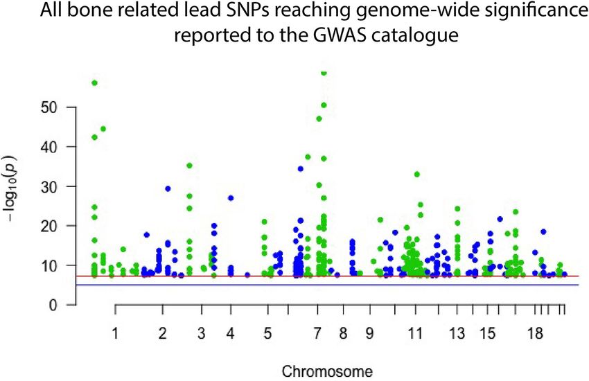
Mäkitie et al.                                                                                                   Insights to Monogenic Osteoporosis

INTRODUCTION                                                               DXA measurements (Z-scores) must be considered together with
                                                                           fracture history. A pathologic fracture history entails (i) ≥2
Bone Health                                                                clinically significant long bone fractures by age 10 years, (ii) ≥3
Bone is a rigid connective tissue composed mainly of organic               clinically significant long bone fractures by 19 years, or (iii) one
components (90% type I collagen, the rest other non-collagenous            or more vertebral compression fractures in the absence of high-
structural proteins and cells) and inorganic minerals (mostly              energy trauma, meaning a ≥20% loss in vertebral anterior, middle
calcium hydroxyapatite). These combined give bones their                   or posterior height. However, a vertebral compression fracture
sturdiness to withstand an individual’s weight and the elasticity to       alone suffices for the diagnosis of pediatric osteoporosis even in
enable movement and resist fractures. Bone comprises dense and             the presence of normal BMD (2, 9–12).
compact cortical bone and cancellous, loosely-webbed trabecular
bone, and serves as a reservoir for minerals, growth factors,
cytokines, and fat. Bone also functions as an endocrine organ by           GENETICS IN BONE HEALTH
secreting several systemic hormonal factors (1).                           Genetics in Bone Health
    Bone is all but a quiescent tissue—it undergoes active                 Genetics play a substantial role in determining an individual’s
renewing and remodeling throughout life. By coupled, successive            skeletal strength, bone microarchitectural properties and risk
processes of bone resorption and bone formation, together                  of osteoporosis. BMD is known to be a highly heritable trait
called “bone turnover,” old or damaged bone is eroded and                  and twin studies have shown genetic factors to determine up
replaced by new bone to maintain healthy and strong bone tissue.           to 80% of its variance (13, 14). Genetic factors influence bone
Throughout childhood and adolescent growth, the period of                  health in a polygenic manner and multiple gene variants, or
bone mass accrual, bone turnover is formation-favoring, until              single nucleotide polymorphisms (SNPs), in several different
the highest amount of bone mass, termed “peak bone mass,”                  genes each contribute to the overall risk for compromised
is attained by young adulthood. Thereafter, bone mass remains              bone health. Recent research, especially large-scale genome-wide
fairly constant until bone resorption begins to dominate by the            association studies in large cohorts, has elucidated the complexity
age of menopause and bone mass slowly declines.                            of genetic networks that are important for bone metabolism but
    Factors that impede skeletal growth in childhood or accelerate         also evidenced limitations in our current knowledge. On the
bone loss later in adulthood, such as long-term or chronic                 other hand, significant scientific advances have been made by
illnesses, glucocorticoid treatment and other medications,                 studying rare monogenic forms of osteoporosis in which one
hypogonadism and menopause, other endocrine disorders and                  mutation in a single gene with a major role in bone metabolism
cancers, impose a great risk for low bone mass and osteoporosis            dominates and is alone sufficient to cause osteoporosis. Technical
(1–3). In childhood, especially glucocorticoids play a major role          advancements in research methods, especially high through-
in secondary osteoporosis. Studies on patients receiving systemic          put sequencing techniques, have made family-based studies an
steroids for acute lymphoblastic leukemia (4), juvenile idiopathic         efficient way to identify new genes relevant to osteoporosis. Such
arthritis (5, 6), Duchenne muscular dystrophy (7) or asthma (8)            studies have enabled recognition of novel molecular mechanisms
all indicate increased peripheral and vertebral fracture rates.            and given leeway to understanding the intricacies behind bone
                                                                           metabolism (13, 14). In this article we only briefly summarize
Osteoporosis                                                               GWAS methodology and recent advancements while the main
Osteoporosis is a chronic skeletal disease with high prevalence            focus is on discoveries made from family-based research on
and mortality worldwide. It is characterized by low bone                   patients and families with monogenic forms of osteoporosis.
mass and bone mineral density (BMD), and by destructed
bone microarchitecture that often results from imbalanced                  Genome Wide Studies to Identify
bone formation and resorption or from abnormal matrix.                     Contributing Genetic Factors
Impaired bone quality leads to compromised bone strength                   Genome wide association studies (GWASs) have proven
and high propensity to low-energy fractures in long bones                  successful and robust in deciphering the genetic mechanisms
and vertebrae (9). Osteoporosis, with frequent fractures, pain             underlying complex diseases, including osteoporosis (14, 15).
and physical limitations, causes significant human suffering and           As mentioned, single nucleotide variants (SNVs) in several
burdens the health care system (9). BMD is considered to                   different genomic sites all contribute to bone quality and
define osteoporosis and risk of fractures. It is assessed using            strength and risk of osteoporosis but are often very common
dual-energy X-ray absorptiometry (DXA), where reduction of                 in the general population and, by themselves, have only a
more than 2.5 standard deviations from the normal mean for                 minor effect (16). The current GWAS catalog, released in
young adults (T-score) is diagnostic of osteoporosis. Of note,             September 2018, comprises 55 separate studies focusing on
osteopenia (T-score 1.0 to −2.5) together with a high probability          bone properties, fractures or osteoporosis. Together they report
of fractures, or a fragility fracture without another metabolic            425 different lead SNVs, in 118 different genomic regions,
bone disease and independent of BMD are also clinically                    that associate with some aspect of bone on a genome-wide
indicative of osteoporosis. Pediatric osteoporosis requires more           significant level (Figure 1). From these, altogether 144 different
than mere DXA-determined low BMD, as variation in growth                   genes are reported to be directly linked to, or plausible candidate
and pubertal maturation make interpretation of BMD values                  effectors, for the identified signals. Of note, this catalog is
challenging. Therefore, age-, gender-, and body size–adjusted              not entirely up to date due to the extensive curation required

Frontiers in Endocrinology | www.frontiersin.org                       2                                       February 2019 | Volume 10 | Article 70
Mäkitie et al. Insights to Monogenic Osteoporosis
FIGURE 1 | Manhattan plot displaying all lead SNPs independently associated with bone-related traits reported to the GWAS catalog as of September 2018. The
associated SNPs highlight genomic regions important to bone. However, they each have only a minor effect on an individual’s skeletal qualities and risk of
osteoporosis and hence have limited use in clinical practice.
before publication in the GWAS catalog. Recently, Kemp et al. authors were able to identify 60 genes likely to underlie the
undertook a colossal genome-wide search for genetic factors association signals. Of these, 13 genes (22%) had been implicated
correlating with BMD, estimated from quantitative ultrasound in monogenic skeletal disorders and 27 genes (45%) had a
of the heel (eBMD) (17). The GWAS is the largest to date, corresponding knockout mouse with a skeletal phenotype (14,
encompassing a total of 142,487 individuals from the UK 18). This demonstrates that even though the signals picked up
Biobank. The authors were able to identify 203 loci, of which by GWASs might indicate a weak effect from the measured
153 were novel, to be associated with eBMD. These together variation, it is likely that rare and more damaging genetic
explained about one third of the total variance in eBMD (17). variations in the same genomic locus might have a large effect.
Although highly successful, none of the previous GWA-studies The genomic areas implicated in these GWASs are therefore
with DXA-derived BMD have been as successful as the study likely to be of greater importance than the individual signal
by Kemp et al. divulges (24).
Despite these great advances, thus far, only 2,000 individuals, rare variants can be imputed and assessed. parameters when clinically evaluating a patient’s skeletal
These data have enabled low frequency variants with large health (27).
effect sizes for BMD in COL1A2 and LGR4 to be identified
(22, 23). Several genomic loci, identified through common Recent Advances in Genetic Research
genetic variation, have also been linked to genes known to As mentioned, several monogenic forms of osteoporosis have
underlie monogenic forms of skeletal pathology. In a large been described. Osteogenesis imperfecta (OI) is the best-
meta-analysis on BMD conducted by Estrada et al. (18), the known form of monogenic osteoporosis and comprises a
Frontiers in Endocrinology | www.frontiersin.org 3 February 2019 | Volume 10 | Article 70Mäkitie et al. Insights to Monogenic Osteoporosis
heterogeneous family of different heritable bone dysplasias patterns emphasize the importance of a molecular diagnosis in
with skeletal fragility (28). Parallel to new developments in these patients.
genetic methodology, new gene discoveries in variable forms of
monogenic osteoporosis have been made and, to date, the list PATHS TO MONOGENIC OSTEOPOROSIS
of genetic causes of OI and monogenic primary osteoporosis
comprises altogether 19 genes (Table 1). The novel genetic Defects in Bone Cell Function and Bone
findings have considerably enhanced our understanding of the Remodeling
complexities of bone metabolism and uncovered new molecular Normal osteoblast and osteoclast functions are key to sustaining
pathways that regulate bone metabolism and contribute to healthy bone tissue. Bone resorption by osteoclasts and
skeletal pathology. They span beyond the collagen-related formation by osteoblasts are tightly linked in successive
pathways to include signaling cascades regulating bone cell repetitive cycles at specific bone sites and the processes
function and the extracellular matrix, as described in detail are meticulously controlled by several locally produced and
below. The great variability in clinical features and inheritance circulating systemic factors (29). Communication between the
TABLE 1 | Different molecular mechanisms and genes underlying osteogenesis imperfecta.
Pathophysiological Gene Protein Inheritance Number of OMIM (Phenotype MIM number)
mechanism known
mutations
Defects in collagen type I COL1A1 Collagen alpha-1(I) chain AD >1,000* 166200; 166210; 259420; 166220
synthesis, structure, folding,
post-translational
modification, processing
and cross-linking
COL1A2 Collagen alpha-2(I) chain AD; AR◦ >600* 259420; 166210; 166220
CRTAP Cartilage-associated protein AR 32* 610682
PPIB Peptidyl-prolyl cis-trans isomerase B; AR 17* 259440
cyclophilin B
P3H1 Prolyl 3-hydroxylase 1 AR 69* 610915
FKBP10 Peptidyl-prolyl cis-trans isomerase AR 38* 610968
FKBP10
PLOD2 Procollagen-lysine,2-oxoglutarate AR 10* 609220
5-dioxygenase 2
SERPINH1 Serpin H1 AR 9* 613848
BMP1 Bone morphogenetic protein 1 AR 11* 614856
Defects in other proteins SPARC SPARC; osteonectin AR 2* 616507
leading to abnormal bone
mineralization
SERPINF1 Pigment epithelium-derived factor AR 38* 613982
(PEDF)
IFITM5 Interferon induced transmembrane AD 2* 610967
protein 5
PLS3 Plastin 3 XLD 17 300910
Defects in osteoblast TMEM38B Trimeric intracellular cation channel AR 6* 615066
differentiation and function type B
WNT1 Proto-oncogene Wnt-1 AR 35* 615220
SP7 Transcription factor Sp7; osterix AR 2* 613849
CREB3L1 Cyclic AMP-responsive AR 3* 616229
element-binding protein 3-like protein
1
MBTPS2 Membrane-bound transcription factor XLR 2 301014
site-2 protease
Unknown TENT5A (also Terminal nucleotidyltransferase 5A AR 3 617952
known as
FAM46A)
AD, autosomal dominant; AR, autosomal recessive; XLD, X-linked dominant; XLR, X-linked recessive.
◦ Seen only in a few consanguineous families.
* Information taken from the Osteogenesis imperfecta & Ehlers-Danlos syndrome variant databases.
Frontiers in Endocrinology | www.frontiersin.org 4 February 2019 | Volume 10 | Article 70Mäkitie et al. Insights to Monogenic Osteoporosis
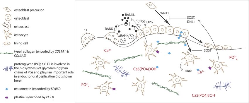
osteoclast and osteoblast is crucial for balanced bone turnover and blood calcium and phosphate concentrations (37).
and defects in either cell’s function can jeopardize bone health. Osteocytes express a range of proteins, such as dentin
Osteoblasts express the receptor activator of nuclear factor matric protein 1 (DMP1), phosphate-regulating neutral
kappa-B ligand (RANKL), which binds to its conjugate receptor endopeptidase on chromosome X (PHEX), and matrix
RANK on osteoclast cell surface (Figure 2) (30, 31). This extracellular phosphoglycoprotein (MEPE), that are crucial
activates osteoclastogenesis and osteoclastic bone resorption. for local matrix mineralization (38). Osteocytes are the primary
Osteoblasts also secrete osteoprotegerin (OPG) that serves as a source of sclerostin, RANKL, and fibroblast growth factor
decoy receptor for RANKL to inhibit RANKL-RANK–binding, 23 (FGF23), through which osteocytes exert their endocrine
therefore downplaying RANKL’s osteoclastogenesis-promoting functions in bone (Figure 2) (36, 38).
effect and, as its name implies, protecting bone from over- The WNT pathway has a key role in all aspects of bone
resorption (Figure 2) (30, 31). Recently, RANK was also noted health—from fetal skeletal development to childhood bone
to relay back by vesicular trafficking from mature osteoclasts mass accrual to adult bone homeostasis and microarchitectural
to osteoblasts to promote bone formation by reverse signaling sustenance (39). WNTs act locally by activating adjacent cells’
(32). The significance of the RANK-RANKL–communication WNT signaling in a paracrine manner: in developmental
is portrayed in several monogenic conditions with abnormal stages to partake in the cross-talk between osteoblasts and
bone mass resulting from defective RANK-RANKL-OPG–axis: hematopoietic stem cells (HSCs) in bone marrow and promote
osteoclast-poor osteopetrosis with excessive bone formation due bone cell development, differentiation and proliferation, and
to mutated RANKL, juvenile Paget’s disease with osteopenia and later in mature adult bone, to induce osteoblastic bone
progressive skeletal deformity from mutated OPG, and familial formation (39). WNTs can also act by autocrine means by
expansile osteolysis (FEO) with osteolytic lesions and increased regulating cells of the same osteoblast or osteoclast lineage
bone remodeling from mutated RANK (33–35). (40). The activated pathway is anabolic to bone, leading
Alongside osteoblasts and osteoclasts, osteocytes have to increased bone formation and decreased bone resorption.
emerged as key regulators of bone turnover, mineral homeostasis Three different WNT pathways are recognized: the canonical
and hematopoiesis (36). Osteocytes are terminally differentiated pathway (WNT/β-catenin pathway), the non-canonical planar
osteoblasts embedded throughout the mineralized matrix. cell polarity pathway, and the non-canonical WNT/Ca2+
They communicate with each other and other cells through an pathway. While the latter two, also known as the β-catenin-
extensive network of long cytoplasmic dendritic processes and independent pathways, participate in a range of development
are thought to orchestrate the interplay between osteoblasts process and in bone metabolism, the canonical WNT/β-catenin
and osteoclasts in bone modeling and remodeling by sensing pathway is considered the predominant pathway maintaining
mechanical loading and responding to endocrine factors, skeletal health (41).
FIGURE 2 | Schematic overview of bone cells and extracellular matrix components involved in regulating bone homeostasis. Receptor activator of nuclear factor
kappa-B ligand (RANKL) binds to its conjugate receptor RANK on osteoclast cell surface to stimulate osteoclast differentiation and activity. Osteoprotegerin (OPG)
inhibits RANK/RANKL-binding to inhibit bone resorption. WNT signaling pathway stimulates osteoblast function and bone formation. Sclerostin (SOST) and dickkopfs
(DKK1), produced by the osteocytes, are two WNT antagonists that promote osteoclasts differentiation. Osteonectin, produced by the osteoblasts, binds calcium,
hydroxyapatite and collagen type I and thus regulates bone mineralization. Plastin-3 (PLS3), expressed by the osteocytes, may also be involved in the mineralization of
the extracellular matrix but its role in osteoprogenitors and other bone cells is yet to be confirmed.
Frontiers in Endocrinology | www.frontiersin.org 5 February 2019 | Volume 10 | Article 70Mäkitie et al. Insights to Monogenic Osteoporosis Dysregulated WNT/β-catenin signaling leads to various example, WNT16 is considered an important ligand in skeletal disorders of both high and low bone mass. This bone WNT signaling and has been shown to mediate its was first recognized in 2001 when mutations in low-density bone-specific actions via both canonical and non-canonical lipoprotein 5 (LRP5), encoding a coreceptor for WNT ligands, WNT pathways (51). Although the specifics behind its were found to lead to low bone mass in the autosomal recessive mechanisms are unclear, GWASs show that polymorphisms osteoporosis pseudoglioma syndrome (OPPG, MIM 259770), of the WNT16 locus associate with cortical bone thickness, characterized by early-onset severe osteoporosis and blindness BMD, and osteoporotic fracture risk in large observational (42, 43). The LRP5 mutations inhibit normal WNT signaling studies and variations in WNT16 may also impact individual and lead to reduced osteoblast proliferation and function and peak bone mass (18, 52, 53). These findings are echoed in subsequently decreased bone formation (43). Since then, many in vivo studies as Wnt16 KO mice have reduced cortical other mutations in LRP5 have been shown to cause OPPG (44). thickness and bone strength leading to spontaneous peripheral In addition, functionally significant SNPs in LRP5 have been fractures (54). linked to adolescent bone mass accrual and peak bone mass In 2013, several groups identified WNT1 as a key ligand (45, 46), and genome-wide searches have found common LRP5 to the WNT pathway in bone; heterozygous WNT1 mutations polymorphisms that contribute to population-based variance in were reported to cause autosomal dominant osteoporosis, and BMD, confirming its significant role in osteoporosis risk also homozygous mutations, a more severe osteogenesis imperfecta in the general population (14, 18). The molecular mechanisms (55). Since then, various other mutations have been found by which these missense mutations in LRP5 decrease WNT worldwide, all reporting skeletal morbidity with frequent and signaling, however, remain largely unknown (46, 47). Conversely, childhood-onset peripheral and vertebral compression fractures inadequate WNT inhibition from mutations or deletions in the and successive changes in spinal stature (55–61). In our sclerostin-encoding SOST results in high bone mass phenotypes comprehensive clinical analyses of a large cohort of 25 WNT1 sclerosteosis (MIM 269500) and van Buchem disease (MIM mutation-positive subjects with the same heterozygous missense 239100), respectively (48, 49). In the absence of sufficient mutation p.C218G, the aberrant WNT1 signaling results in a sclerostin, WNT signaling is unrestrained, leading to continuous severe skeletal pathology (62). In addition to prevalent fractures, bone formation. long bone modeling is altered and BMD low in affected children, All in all, 19 different WNT proteins are known and while vertebral compression fractures are very common later together they initiate several intracellular signaling cascades in adulthood and result in severe kyphotic deformity and loss to regulate organogenesis, cell fate determination, primary of adult height soon after the age of 50 years (Figure 3). axis formation, and stem cell renewal (39). Several of the Bone biopsy histomorphometry demonstrated low-turnover WNT proteins are expressed in bone tissue and regulate osteoporosis with scarce and inactive bone cells and stagnant bone health at various phases during skeletal growth, bone turnover. Noted extra-skeletal traits included changes development, and e.g., osteoporosis pathogenesis (50). For in spinal cartilaginous structures, namely vertebral endplate FIGURE 3 | Spinal magnetic resonance images of four WNT1 p.C218G mutation-positive subjects. (A) Thoracic spine of a 17-years-old female showing multiple Schmorl nodes (arrow). (B) Thoracic spine of a 44-years-old female showing exaggerated thoracic kyphosis. (C) Thoracic spine of a 76-years-old male showing several compressed vertebrae, kyphotic stature, and Schmorl hernia (arrow). (D) Lumbar spine of a 74-years-old female showing several compressed vertebrae and enlarged intervertebral discs (arrows). Reprinted from Mäkitie et al. (63) with permission from Elsevier. Frontiers in Endocrinology | www.frontiersin.org 6 February 2019 | Volume 10 | Article 70
Mäkitie et al.                                                                                                  Insights to Monogenic Osteoporosis

deterioration and frequent Schmorl nodes, and increased                    the mineralization process, are normal despite altered bone
reticulin and early-phase–shifted granulopoiesis as signs of               formation (68). The detailed mechanisms of bone tissue
abnormal bone marrow function (63, 64).                                    mineralization are still debated, but extracellular mineral
    The latest finding of dysregulated WNT signaling in                    deposition through budding off of intracellular microvesicles
monogenic osteoporosis is SFRP4 mutations in Pyle’s disease                has emerged as one part of the process (80). This process
(65). Frizzled-related protein 4 (SFRP4) acts as an WNT                    requires dramatic changes in the cell membrane through a
inhibitor and biallelic, truncating mutations in its encoding              complex and well-orchestrated process involving the actin
gene SFRP4 result in aberrant regulation of WNT signaling,                 cytoskeleton. Thouverey et al. (81) and Piehl et al. (82)
osteoblasts and osteoclast function and bone remodeling                    have demonstrated congruently that plastin 3 is involved
(65). The patients’ clinical phenotype is predominated by                  in the formation of extracellular vesicles. It can thereby be
cortical-bone thinning and fragility and expanded metaphyseal              speculated that PLS3 mutations could have deleterious effects
trabecular bone, resulting in limb deformity and high propensity           on the mineralization process in bone through defective
to fracture. Correspondingly, Sfrp4-null mice present with                 microvesicle formation, although the details behind this too
increased trabecular bone, decreased cortical bone and failure in          remain undisclosed.
bone modeling (65).                                                           Lastly, a recent experimental animal study presented new
    Despite their important functions, known monogenic forms               findings suggesting involvement of osteoclast malfunction as
of bone diseases stemming from osteocyte defects are rare                  part of pathophysiology in PLS3 osteoporosis (83). In vivo
and often relate to defective mineral metabolism, especially               and in vitro studies using Pls3 knockout and overexpressing
hypophosphatemia due to disturbed FGF23 regulation. One of                 mice confirmed the osteoporotic phenotype in the former
the most recently identified monogenic forms of osteoporosis               and thickening cortical bone in the latter. In vitro studies of
is caused by mutations in the PLS3 gene (66–70), encoding the              osteoclasts derived from the animals demonstrated a regulatory
actin binding, actin bundling protein plastin 3. This X-linked             role of PLS3 in osteoclastogenesis. Additionally, a dysregulation
form of primary early-onset osteoporosis is characterized                  of osteoclast activity was found in cells from Pls3 knockouts,
by low BMD, frequent peripheral fractures and vertebral                    likely connected to impaired podosome organization due to
compression fractures, and subsequent severe thoracic kyphosis.            decreased actin regulation (83). These findings are yet to be
Due to its X-chromosomal inheritance, male patients are more               confirmed in humans.
severely affected, usually presenting with severe childhood-
onset osteoporosis. Clinical manifestations in females with                Defects in Bone Extracellular Matrix
heterozygous PLS3 mutations are variable ranging from                      In addition to bone cells, reduced bone strength and various
subclinical osteopenia to a more severe phenotype resembling               skeletal disorders can also stem from defects in the extracellular
that of males’ (68). The total number of diagnosed patients is still       matrix (ECM). The ECM is primarily composed of different
scarce and hence the comprehension of the clinical and genetic             collagenous proteins, non-collagenous proteins (in particular
spectrum, the disease progression and appropriate treatment                glycoproteins and proteoglycans), lipids, minerals and water
is limited.                                                                (84, 85). The most abundant protein is the type I collagen,
    While the role of PLS3 in bone fragility is yet unknown,               made of two alpha-1 and one alpha-2 chains intertwined in
one theory presumes PLS3 to alter osteocyte function through               a triple helical structure. Mutations in the encoding genes,
abnormal cytoskeletal microarchitecture. Plastins, in general,             COL1A1 and COL1A2, respectively, lead to qualitative or
are Ca-dependent actin binding and bundling proteins and as                quantitative defects in the protein and give rise to osteogenesis
such, are involved in cytoskeletal arrangements and partake in             imperfecta (OI), a skeletal dysplasia characterized by low BMD
regulating cellular morphology, motion, and adherence (71).                and enhanced bone fragility, and often extra-skeletal features,
Despite lack of systematic studies, plastin 3 (also called T-              such as blue sclerae, dentinogenesis imperfect, and hearing
plastin) is supposedly expressed in all solid tissues and through          loss (86, 87). Heterozygous glycine substitutions that affect
indicated functions in other tissues, such as spinal muscle,               the Gly-Xaa-Yaa pattern in the triple helix are the most
inner ear stereocilia, and periodontal ligaments, is suggested             common mutations and can cause mild to lethal OI (87).
to be involved in bone mechano-transduction (72–74). This is               However, multiexonic deletions or deletion of an entire allele
supported by the high expression of plastin 3 in chicken osteocyte         have been sporadically found (88–91). Interestingly, mutations
dendrites, especially during dendrite formation (Figure 2) (75–            that lead to a reduced amount of normal protein give rise
77). Although this is supported by clinical investigations from            to a milder phenotype than missense mutations affecting the
biochemical and bone biopsy findings indicating that osteocytes            primary structure of the triple helix (dominant negative effect)
appear affected in PLS3 mutation-positive subjects (78), the               (87). Furthermore, homozygous glycine substitutions in COL1A2
observation remains mostly theoretical.                                    have been identified in a handful of consanguineous families
    Another suggested role for PLS3 in bone is involvement in              (92–95). Surprisingly, the patients harboring biallelic COL1A2
mineralization. This is collectively supported by the patients’            mutations have a moderate to severe phenotype whereas the
low BMD and their bone biopsies’ histology. We have reported               mutation carriers are only mildly affected or free from any
accumulation of non-mineralized osteoid in trabecular bone in              obvious skeletal impairment. On the other hand, homozygous
patient biopsies (69, 70, 78, 79) and shown that biochemical               COL1A1 mutations are likely to be lethal since they have
markers of bone turnover, although not directly echoing                    never been reported in humans. Furthermore, some previous

Frontiers in Endocrinology | www.frontiersin.org                       7                                      February 2019 | Volume 10 | Article 70
Mäkitie et al. Insights to Monogenic Osteoporosis
reports have indicated that when the COL1A1 or COL1A2 TOOLS FOR DIAGNOSING MONOGENIC
mutation involves the C-propeptide cleavage site, the phenotypic OSTEOPOROSIS
manifestations may include high BMD and mild skeletal fragility
(96). A recent study on such cleavage site variants showed Uncovering the Genetics
that the mutations lead to a distinctive OI phenotype with As discussed, to make the diagnosis of osteoporosis in children
variable expression, mild to moderate disease severity, moderate two criteria need to be met; (1) low BMD or BMC (Z-score
fracture rate, high bone mass and increased bone mineral ≤ −2.0 SD) and (2) a clinically significant fracture history. A
density (97). vertebral fracture indicates severely compromised bone strength
Although COL1A1 or COL1A2 mutations are detected in and suffices alone for the diagnosis (12). The diagnosis of primary
∼85% of OI cases, to date, mutations in altogether 17 other osteoporosis in children can be made when potential causes
genes are also known to cause OI-like skeletal disorders (Table 1). of secondary osteoporosis, such as other underlying illnesses
Some of these genes play a role in the post-translational or medical treatments, have been excluded (2). Most forms of
modification of type I collagen while some are key regulators of childhood-onset primary osteoporosis are termed osteogenesis
osteoblast differentiation and function and/or lead to abnormal imperfecta, although the diagnosis is vague and merely appoints
bone mineralization (Table 1). One example of severe autosomal the disease to belong to a heterogeneous group of skeletal
recessive OI caused by a mineralization defect is linked to disorders with diverse clinical presentation (86, 87). As indicated
mutations in SPARC (98). The encoded protein Secreted Protein earlier, the genetic background of OI is heterogeneous and
Acidic and Cysteine Rich, better known as osteonectin, is a the phenotypic and genetic variability have complicated OI
glycoprotein that is mainly expressed by osteoblasts during bone classification. As of yet, there is no consensus indicating which
formation and binds calcium, hydroxyapatite and collagen type genotype-phenotype combinations should be classified under the
I and other proteins in the ECM (Figure 2). Null mutations in umbrella of OI and which should not. The current classification
SPARC lead to reduced accumulation of type I collagen in the of OI is based on phenotypic features, but the molecular cause is
ECM (99). Furthermore, the osteonectin-type I collagen complex often the key factor determining clinical prognosis, appropriate
is suggested to sequestrate calcium and phosphate in order to treatment approach and recurrence risk in the family, and
initiate bone mineralization (100). An impairment of two other should therefore be emphasized (28). A molecular diagnosis also
proteins expressed by the osteoblasts, the pigment epithelium- facilitates the refinement of future treatment and clinical care
derived factor (encoded by SERPINF1) and the interferon- protocols (87, 111). Although more than 85% of OI cases can still
induced transmembrane protein 5 (encoded by IFITM5), be traced to pathogenic variants in either of the two collagen type
respectively, can also compromise bone mineralization and I–coding genes COL1A1 or COL1A2 (112, 113), the several other
lead to OI (86, 87, 101, 102). Most recently, mutations in genes identified over the past 12 years in OI or monogenic forms
FAM46A, encoding the terminal nucleotidyltransferase 5A, have of primary osteoporosis need to be kept in mind (92, 114, 115).
been detected in four patients with OI. However, the molecular While most clinicians begin by screening COL1A1 and
function of this protein and the pathophysiological mechanism COL1A2 possibly in combination with MLPA, proceeding to
by which the mutations lead to OI are not yet known (103). a full OI gene panel using massive parallel sequencing is
Besides OI, there are several other skeletal syndromes that recommended (87). A sequencing-based gene panel will not only
feature osteoporosis and are caused by defects in the ECM. For capture sequence variants but also possible structural variations
example, mutations in XYLT2 lead to spondyloocular syndrome including larger deletions and duplications. Although the surge
characterized by childhood-onset osteoporosis, cataract, cardiac of new genetic findings has facilitated interpretation of sequence
defects and hearing impairment (104–106). The mutated variants, deep intronic splice variants or splice variants masked
protein xylosyltransferase 2 is involved in the biosynthesis as synonymous variants are still difficult to correctly annotate.
of glycosaminoglycan chains and plays an important role Transcriptome analysis using RNA sequencing together with
in endochondral ossification and chondrocyte differentiation DNA sequencing has proven successful in increasing the
and maturation. Proteoglycans are also important for other diagnostic yield and assessing functional impact of variants
tissues and organs, including brain, heart, and retina, which that are otherwise hard to interpret (116). This, however,
could explain why the clinical manifestations of spondyloocular requires that the disease in focus has a readily accessible proxy
syndrome are not only restricted to the skeleton (106). tissue, where the gene expression reflects the expression in
In addition to causing autosomal recessive OI, inadequate the affected tissue. Unfortunately, tissue accessibility is very
folding and post-translational modification of type I collagen can difficult in bone diseases and the method cost-restricted in
result in another skeletal syndrome characterized by congenital clinical settings.
contractures, named Bruck syndrome. Homozygous mutations Regarding structural variants, WGS has provided an
in FKBP10 and PLOD2 result in Bruck syndrome 1 and advantage in assessing structural variants compared to exome
2, respectively (107–110). FKBP10 encodes the immunophilin sequencing or other capture-based protocols. However, all short-
FKBP65, a molecular chaperon of type I collagen and PLOD2 read sequencing technologies have shortcomings in their ability
encodes the procollagen-lysine, 2-oxoglutarate 5-dioxygenase 2, to detect and identify structural variants, and, as concluded by
which catalyzes the hydroxylation of lysyl residues in type I Telenti et al. (117), after sequencing 10 000 human genomes
collagen. Mutations in both FKBP10 and PLOD2 can also cause the interpretation of structural variants on an individual level
autosomal recessive OI (Table 1). still remains challenging. Older methods to indirectly detect
Frontiers in Endocrinology | www.frontiersin.org 8 February 2019 | Volume 10 | Article 70Mäkitie et al. Insights to Monogenic Osteoporosis
structural variations, such as array-based comparative genomic easily confounded by other patient-related (e.g., body adiposity,
hybridization (array-CGH), are still applicable in specific inflammation, blood glucose level, time of sampling) and
cases and can help clinicians in their search for a molecular analytical factors. Furthermore, they often respond inadequately
diagnosis (91). to bisphosphonate treatment and correlate poorly with BMD
and bone histomorphometric parameters (55, 120–125). None of
Clinical Characterization the monogenic forms of osteoporosis have a specific biomarker
Owning to the wide spectrum of genetic causes, the clinical profile and these conventional markers are of little value in
presentation of different OI and primary osteoporosis forms differentiating between the various genetic forms of osteoporosis.
is unsurprisingly miscellaneous (87). The diseases vary in The limitations of the conventional bone markers have fueled
their primary skeletal traits, age-at-onset, natural progression, a field-wide search for new potential biomarkers. Zooming into
sensitivity to treatment, and presence and spectrum of extra- smaller cell-released particles, small microRNAs (miRNAs), as
skeletal characteristics. Although severely compromised bone one, have attained much attention and are proposed to hold
strength is usually a unifying finding, the DXA-derived promise in future diagnostic and treatment in skeletal disorders.
BMD, bone biopsy findings, prevalence and type of fractures, These small, non-coding fragments of RNA are highly conserved
and radiographic findings are inconsistent. The phenotypic and comprise, on estimate, 1% of our genome (126, 127). They
severity can vary from mild to severe and disease onset alter gene expression by RNA silencing and post-transcriptional
from childhood to early adulthood—at times provoked by regulation; each miRNA is predicted to regulate hundreds of
pregnancy-related calcium loss. Presentation may vary between different target genes, thus serving important functions in many
patients with different mutations and even between family tissues and biological processes (127, 128). While their exact
members with identical mutations (87). Classical OI-related function in gene regulation is still largely unknown, miRNAs
extra-skeletal findings include blue sclerae, increased joint are thought to mediate intercellular communications in various
laxity, dentinogenesis imperfecta and impaired hearing (28, metabolic processes and diseases and a unique imprint of
87, 118). Mutations in proteins affecting the collagen-related differentially expressed miRNAs is observed in e.g., certain
pathways all seem to exhibit similar traits; only the severity and cancers, metabolic diseases and viral infections. In bone, miRNAs
array of affected skeletal sites vary. Some typical presentations contribute to homeostasis and their dysfunctional expression
include popcorn epiphyseal plates in CRTAP, calcifications of relays to progression of skeletal disorders (129, 130). Their
interosseous membranes and hyperplastic callus formation in expressions change in result of low BMD, frequent fractures, or
IFITM5, and skull ossification defects in SEC24D-related OI menopausal osteoporosis (129, 130).
(Table 1) (86, 87, 118). The extra-skeletal manifestations of These findings have encouraged researchers to explore the
bone cell-related forms are still incompletely defined; with clinical potential of miRNAs in disease diagnostics and follow-
monoallelic WNT1 mutations patients have changes in spinal up. Several clinical studies have evaluated miRNA expression
cartilaginous structures (63) and mild abnormalities in bone in osteoporotic patients and distinguished specific miRNAs
marrow hematopoiesis and reticulin formation (119), while in correlating with the degree of osteoporosis (131). miR-133a was
biallelic mutations the phenotype is more severe and OI-like significantly elevated in postmenopausal Caucasian women with
but no bone marrow defects have been reported (55). However, low BMD (132), and miR-194-5p and miR-21-5p negatively
central nervous system manifestations have been reported in correlated with BMD in Chinese osteoporotic women (133,
some patients with homozygous WNT1 mutations (55, 61). 134). Seeliger et al. (135) also identified miR-21-5p, in addition
Patients with PLS3 mutations do not exhibit any apparent extra- to four other miRNAs (miR-23a-3p, miR-24-3p, miR-100-5p,
skeletal traits, though this is still scantily explored. and miR-125b-5p) to be differentially expressed in serum and
upregulated in bone tissue in patients with osteoporotic fractures.
Novel Biomarkers In vitro studies have observed miRNAs that interact with known
In addition to DXA and plain radiography, several factors key regulators of bone metabolism, such as miR-152-3p and
can be measured from systemic circulation and urine when miR-335-3p with Dickkopf-1 (136, 137), miR-30e-5p with Lrp6
diagnosing and monitoring patients’ disease state, progression (138), and the aforementioned miR-133 with Runx2 (139).
and treatment response. The conventional metabolic markers Furthermore, Anastasilakis et al. (140) reported that serum
reflect bone turnover and consist of enzymatic and proteinaceous levels of miRNAs changed in response to anti-osteoporotic
by-products; the most widely used resorption markers include treatment. While different studies pinpoint to varying miRNAs
mainly by-products of collagen breakdown, [urinary collagen depending on cohort size, demographic or other factors, a clear
type 1 cross-linked N-telopeptide (NTX), urinary/serum collagen congruency is echoed that a unique miRNA signature is observed
type 1 cross-linked C-telopeptide (CTX), and collagen fragments in osteoporosis.
from matrix-metalloproteases (ICTP)], and formation markers We have reported altered miRNA pattern in patients
procollagens from collagen synthesis [serum amino-terminal with WNT1 osteoporosis, with two upregulated and six
propeptide (PINP) and carboxyl-terminal propeptide (PICP)] or downregulated miRNAs, as compared with age and sex-matched
osteoblast-related proteins (serum osteocalcin (OC) and serum mutation-negative controls from the same family (119). While
bone isoenzyme of alkaline phosphatase (ALP) (120, 121). specific miRNA alterations may be recognized in certain
While these markers are commonly used and easily analyzed in monogenic forms of osteoporosis, the role of miRNAs in
automated routine laboratories, they do lack specificity and are complementing or substituting genetic testing remains to be
Frontiers in Endocrinology | www.frontiersin.org 9 February 2019 | Volume 10 | Article 70Mäkitie et al. Insights to Monogenic Osteoporosis
explored in future studies. Further, the utilization of miRNA Clinical care and follow-up are advised to be centered in special
assessments in clinical practice demands further methodological health care units with abilities to provide multidisciplinary care
development but based on present data, they hold great potential and expertise.
for future diagnosis and follow-up, including monogenic forms
of osteoporosis.
Novel Target-Drugs
Discoveries through rare, monogenic forms of skeletal disorders
OPTIONS FOR TREATMENT have provided new information on the biology of bone health
and revealed previously unidentified proteins that take part in
Conventional Osteoporosis Drugs and key regulatory pathways. Naturally, these proteins also present
Implications for Treatment as appealing target molecules for development of new treatment
Conventional osteoporosis drugs, namely bisphosphonates, have modalities. In early 2000s, inhibition of RANKL by a monoclonal
been the mainstay of pharmacological treatment in classical, antibody denosumab brought a novel approach for treatment of
type I collagen-related OI forms. These typically have high osteoporosis (147). The drug has been used to improve skeletal
bone turnover and thus the osteoclast-targeting and resorption- health in some forms of OI. Particularly patients with SERPINF1
decreasing bisphosphonates have proven effective in increasing mutations show a modest increase in BMD in response to
BMD, reducing fractures, and improving VCFs in patients (141– denosumab whereas treatment outcomes with bisphosphonates
143). Contrary to collagen I-related OI, bisphosphonates have are poor (148). Due to the coupled nature of osteoblast-
proven insufficient in improving BMD or fracture tendency in osteoclast–activity, blocking osteoclastogenesis through RANKL
several new forms of primary osteoporosis (55, 57, 60). These is also unfavorably accompanied by reduced osteoblast function.
OI forms often present with low-turnover osteoporosis and The previously mentioned discovery of RANKL reverse signaling
hence the benefits of anti-catabolic treatment are not optimal. could offer a novel solution to avoid this problem (32).
We have also shown that patients with prior bisphosphonate Also, inhibition of cathepsin K, an osteoclast-derived lysosomal
treatment have abnormal and apoptotic osteocytes, suggesting enzyme, seemed promising due to its coupled bone formation-
adverse effects of bisphosphonates in WNT1 osteoporosis (63). favoring action, but its development was later discontinued due
However, our longitudinal study on the effects of teriparatide- to increased risk of cardiovascular complications (149). As of
treatment in WNT1 osteoporosis indicated that exogenous recently, the effects of anti-TGF-β antibodies have been studied
PTH may be efficient in increasing bone formation and BMD in Crtap−/− and Col1a1frt/− mice with varying results; while
during a 24-months-long treatment in adults; however, there the Crtap−/− showed great improvements in bone mass and
may be simultaneous increase in bone marrow adiposity (79). biochemical qualities, Col1a1frt/− mice did not show significant
Thus far, the efficacy of anti-sclerostin antibodies have been changes in bone quality or strength (150).
experimented in mice only; subcutaneous administration of Along with the discovery of van Buchem disease and
Scl-Ab to the murine model of WNT1 OI Wnt1sw/sw mice sclerosteosis, two human models of sclerostin inhibition,
significantly improved fracture rate and increased bone mass that fueled the development of a new anabolic target drug named
seemed to result from increased osteoblast activity (144). romosozumab—a monoclonal anti-sclerostin antibody targeting
Besides WNT1-related skeletal pathologies, even less is known the WNT pathway (151, 152). Its efficacy has been evaluated
about the optimal treatments in other new forms of primary in several clinical trials with promising results; a placebo-
osteoporosis and OI, such as PLS3 and XYLT2 (105, 145). Efficacy controlled, multicenter, phase II study on 419 postmenopausal
of bisphosphonates in PLS3 osteoporosis has been evaluated in women with osteoporosis treated with subcutaneous injections
a handful of cases and indicate positive response (66, 67, 70). of romosozumab at 3-months intervals showed significant, and
Our above-mentioned clinical study on teriparatide also included superior to those attained by alendronate and teriparatide,
PLS3 mutation-positive subjects and they showed congruent, increase in areal BMD and a tilt in BTMs reflective of increased
although slightly lesser, improvement in bone parameters in bone formation (151), and another phase III study reported a
24-months follow-up, as compared with patients with WNT1 reduction in fracture risk in 7,180 postmenopausal osteoporotic
osteoporosis (79). Patients with XYLT2 mutations seem to women (153). Anti-DKK1 antibodies act similarly to oppose
benefit from pamidronate treatment with increase in BMD and WNT signaling and are potent as osteoanabolic agents. However,
improvement in vertebral morphology (104, 105). administration of anti-DKK1 is only mildly efficacious as the
Clinical care of OI patients, including both classical and newer WNT-neutralizing effect is compensated by upregulation of
forms of OI and monogenic osteoporosis, is often complex and sclerostin, although the opposite is not seen when given only
challenging. Means of treatment and pace of clinical follow-up anti-sclerostin antibodies. Thus, the benefits of anti-DKK1
are dependent on the patient’s age, clinical manifestations, and antibodies manifest only when given in conjunction with anti-
degree of impairment, and should be individually tailored and sclerostin (154).
regularly evaluated. Bisphosphonates are still the main treatment Another target of interest for new drug development is
option for pediatric patients and are often used to prevent greater Notum. It is a secreted enzyme that inhibits WNTs by removing
decrease in BMD and enable maximum yield in bone mineral the palmitoleic acid group that is essential for binding of
throughout childhood and adolescent bone mass accrual. The WNTs to Frizzled receptors, thereby inhibiting WNT signaling.
overall benefits of bisphosphonate treatment in most cases of Interestingly, experimental studies in rodents have shown
OI are non-negligible (146). Variable treatment protocols exist. that inhibiting Notum through either knockout, or by oral
Frontiers in Endocrinology | www.frontiersin.org 10 February 2019 | Volume 10 | Article 70Mäkitie et al. Insights to Monogenic Osteoporosis
administration of molecular inhibitors or neutralizing antibodies CONCLUSIONS
increase cortical bone formation and strength, but do not affect
trabecular bone mass (155, 156). Recent advances in genetic methodology have resulted in several
Possible undesired adverse and extra-skeletal effects of new new discoveries relating to the genetic architecture of bone
drugs are inevitable as many of the targeted proteins have tissue- homeostasis. Not only have the basic clinical and genetic pillars
wide expression and key roles in various biological processes. of classical OI been refined, but several new forms of monogenic
Side effects can be latent and subtle but also challenging and osteoporosis have also been identified that have pinpointed
life-threatening. Knowing the WNT pathway’s fundamental role novel molecular mechanisms contributing to skeletal health and
in embryonic development, tumorigenesis and pathogenesis of disease. The clinical presentation, inheritance mode, natural
other systemic or chronic diseases, romosozumab has been course and response to conventional osteoporosis drugs are
under careful scrutiny for its clinical safety. In mice receiving diverse, often variable and logically dependent on the affected
different doses, no malignancies were noted over a 98-weeks protein. Although uncovering the limitations in our current
follow up (157). However, along with the robust and positive diagnostic and treatment modalities, they have also provided
skeletal effects, use of romosozumab has been associated with new signaling pathways that hold promise in new targeted drug
cardiovascular and cerebrovascular events, and the drug is development. Future research will hopefully continue expanding
currently under FDA review (Amgen and UCB). the genetics and molecular mechanisms behind bone metabolism
and increasing our understanding of the specific skeletal and
MicroRNAs extra-skeletal characteristics of monogenic osteoporosis, while
Recently, researchers have acknowledged the opportunities in finding new avenues for improved diagnosis and treatment of
targeting miRNA pathways to develop new therapeutic means patients with severe bone diseases.
and genome editing approaches (128, 158). A few groups
have pursued clinical trials to evaluate efficacy of miRNAs in AUTHOR CONTRIBUTIONS
disease target treatment: an on-going clinical trial evaluates
the anticancer effect of miRNA lethal-7 in binding to Kirsten RM and OM initiated the manuscript. RM wrote the first
rat sarcoma viral oncogene homolog (KRAS) gene in patients draft. All authors contributed to the writing and approved the
suffering from stage III colon cancer, and miR-122 in hepatitis final manuscript.
C (159, 160). Bone-specific miRNAs have not been evaluated
clinically, but analyses have shown that for example in vitro FUNDING
miR-21 could promote osteogenesis in bone marrow stem cells,
and systemic administration of miR-214 induced BMD increase Our research is supported by research funding from Novo
and miR-92a enhance fracture healing in mice (161–163). In Nordisk Foundation, Academy of Finland, Vetenskapsrådet,
fracture healing, also angiogenesis is vital to the repair process Sigrid Jusélius Foundation, Folkhälsan Research Foundation,
and Li et al. (164) were able to demonstrate that implantation Foundation for Pediatric Research, Karolinska Institutet’s KID
of MSCs transfected with an angiogenesis-involved anti-miR- funding, Swedish Childhood Cancer Foundation, University of
26a showed good bone repair. Further, anti-miR-31-transfected Helsinki and Helsinki University Hospital through the Doctoral
MCSs efficiently repaired bone defects by increasing BMD and Programme in Clinical Research, Finnish Medical Foundation,
new bone volume (165). These findings and the efficacy, safety Biomedicum Helsinki Foundation, Finnish-Norwegian Medical
and possible side effects need to be confirmed and carefully Association, Helsinki Medical Association, Emil Aaltonen
evaluated in clinical settings in vivo. Foundation, and Jalmari and Rauha Ahokas Foundation.
REFERENCES idiopathic arthritis. J Rheumatol. (2012) 39:365–73. doi: 10.3899/jrheum.
110305
1. Florencio-Silva R, Sasso GR, Sasso-Cerri E, Simões MJ, Cerri PS. Biology of 6. LeBlanc CM, Ma J, Taljaard M, Roth J, Scuccimarri R, Miettunen P, et al.
bone tissue: structure, function, and factors that influence bone cells. Biomed Incident vertebral fractures and risk factors in the first three years following
Res Int. (2015) 2015:421746. doi: 10.1155/2015/421746 glucocorticoid initiation among pediatric patients with rheumatic disorders.
2. Mäkitie O. Causes, mechanisms and management of paediatric osteoporosis. J Bone Miner Res. (2015) 30:1667–75. doi: 10.1002/jbmr.2511
Nat Rev Rheumatol. (2013) 9:465–75. doi: 10.1038/nrrheum.2013.45 7. Joseph S, Wang C, Di Marco M, Horrocks I, Abu-Arafeh I, Baxter A, et al.
3. Baxter-Jones AD, Faulkner RA, Forwood MR, Mirwald RL, Bailey DA. Bone Fractures and bone health monitoring in boys with Duchenne muscular
mineral accrual from 8 to 30 years of age: an estimation of peak bone mass. J dystrophy managed within the Scottish Muscle Network. Neuromuscul
Bone Miner Res. (2011) 26:1729–39. doi: 10.1002/jbmr.412 Disord. (2018). doi: 10.1016/j.nmd.2018.09.005. [Epub ahead of print].
4. Ward LM, Ma J, Lang B, Ho J, Alos N, Matzinger MA, et al., Steroid- 8. Gray N, Howard A, Zhu J, Feldman LY, To T. Association between inhaled
Associated Osteoporosis in the Pediatric Population (STOPP) Consortium. corticosteroid use and bone fracture in children with asthma. JAMA Pediatr.
Bone morbidity and recovery in children with acute lymphoblastic leukemia: (2018) 172:57–64. doi: 10.1001/jamapediatrics.2017.3579
results of a six-year prospective cohort study. J Bone Miner Res. (2018) 9. Camacho PM, Petak SM, Binkley N, Clarke BL, Harris ST, Hurley DL,
33:1435–43. doi: 10.1002/jbmr.3447 et al. American Association of Clinical Endocrinologists and American
5. Markula-Patjas KP, Valta HL, Kerttula LI, Soini IH, Honkanen VE, College of Endocrinology clinical practice guidelines for the diagnosis and
Toiviainen-Salo SM, et al. Prevalence of vertebral compression fractures treatment of postmenopausal osteoporosis−2016. Endocr Pract. (2016) 22:1–
and associated factors in children and adolescents with severe juvenile 42. doi: 10.4158/EP161435.GL
Frontiers in Endocrinology | www.frontiersin.org 11 February 2019 | Volume 10 | Article 70You can also read