Vulvar Lichen Sclerosus from Pathophysiology to Therapeutic Approaches: Evidence and Prospects
←
→
Page content transcription
If your browser does not render page correctly, please read the page content below
biomedicines
Review
Vulvar Lichen Sclerosus from Pathophysiology to Therapeutic
Approaches: Evidence and Prospects
Monica Corazza, Natale Schettini, Pierantonia Zedde and Alessandro Borghi *

                                          Section of Dermatology and Infectious Diseases, Department of Medical Sciences, University of Ferrara,
                                          44121 Ferrara, Italy; czm@unife.it (M.C.); schntl@unife.it (N.S.); zddpnt@unife.it (P.Z.)
                                          * Correspondence: alessandro.borghi@unife.it; Tel.: +39-0532-239684

                                          Abstract: Vulvar lichen sclerosus (VLS) is a chronic, distressing, inflammatory disease with an
                                          enormous impact on quality of life. Treatment goals are relieving symptoms, reversing signs and
                                          preventing anatomical changes. Despite the availability of numerous therapeutic options, treatment
                                          outcome may not be entirely satisfactory and a definitive cure does not exist. This may be due to the
                                          fact that the exact VLS etiopathogenesis remains unknown. The objectives of this paper were to review
                                          the most up-to-date knowledge on VLS etiopathogenesis and to consider the available therapies
                                          through the lens of a plausible pathogenetic model. An electronic search on both VLS etiopathogenesis
                                          and its treatment was performed using the National Library of Medicine PubMed database. Based
                                          on current knowledge, it is conceivable that various, heterogeneous environmental factors acting on
                                          a genetic background trigger an autoimmune, Th-1 response, which leads to a chronic inflammatory
                                          state. This, in turn, can determine both tissue and micro-vascular injury and activation of signaling
                                          pathways involved in fibroblast and collagen metabolism. This pathogenetic sequence may explain
                                the effectiveness of anti-inflammatory treatments, mostly topical corticosteroids, in improving VLS

                                          clinical-pathological changes. Further deepening of the disease pathways will presumably allow key
Citation: Corazza, M.; Schettini, N.;
                                          mediators to become new therapeutic targets and optimize the available treatments.
Zedde, P.; Borghi, A. Vulvar Lichen
Sclerosus from Pathophysiology to
                                          Keywords: vulvar lichen sclerosus; etiopathogenesis; pathway; immunity; fibroblasts; collagen
Therapeutic Approaches: Evidence
                                          metabolism; therapy; corticosteroid
and Prospects. Biomedicines 2021, 9,
950. https://doi.org/10.3390/
biomedicines9080950

Academic Editor: Anna Campanati           1. Introduction
                                                Lichen sclerosus is a chronic inflammatory progressive skin disease. It mostly affects
Received: 14 July 2021                    the anogenital areas [1,2]. The disease occurs at all ages and in both sexes, but is more
Accepted: 30 July 2021                    common in females than in males. The exact prevalence of vulvar LS (VLS) is unknown
Published: 3 August 2021                  but is estimated to range between 0.1% and 3% in the pre-pubertal and postmenopausal
                                          periods, respectively [2,3]. Regardless of its exact incidence, VLS is among one of the most
Publisher’s Note: MDPI stays neutral      common referrals for vulvar distress and/or structural changes [4].
with regard to jurisdictional claims in         VLS primary lesions are flat, ivory or porcelain white spots, which may coalesce
published maps and institutional affil-   into pale, crinkly, thin patches and plaques. Erythema, ecchymosis and itching-related
iations.
                                          excoriations may occur and, occasionally, hyperkeratosis is prominent.
                                                The sites most characteristically involved by VLS are the inter-labial sulci, labia minora
                                          and labia majora, clitoris and clitoral hood. The entire vulva may be involved, with possible
                                          extension to perineum and perianus, giving rise to the characteristic ‘figure-of-eight’ shape.
Copyright: © 2021 by the authors.         The genitocrural folds, buttocks and thighs may be affected as well. The vagina is usually
Licensee MDPI, Basel, Switzerland.        spared. The main differential diagnoses are lichen planus, lichen simplex chronicus,
This article is an open access article    vitiligo, immunobullous disorders such as mucous membrane pemphigoid and vulvar
distributed under the terms and           intraepithelial neoplasia.
conditions of the Creative Commons
                                                Post-inflammatory progressive scarring may cause irreversible subversion of anogen-
Attribution (CC BY) license (https://
                                          ital architecture, including fusion or loss of the labia minora, narrowing of the vaginal
creativecommons.org/licenses/by/
                                          introitus and burying of the clitoris.
4.0/).

Biomedicines 2021, 9, 950. https://doi.org/10.3390/biomedicines9080950                                  https://www.mdpi.com/journal/biomedicines
Biomedicines 2021, 9, 950 2 of 24
There is also an increased risk of genital cancer. In fact, women affected with VLS
have a lifetime risk of developing a squamous cell carcinoma estimated to be 2–5%, while
up to 65% of vulvar carcinomas arise in a background of VLS [5–8].
The diagnosis of VLS is clinical, but a biopsy may be necessary when the clinical
picture is not diagnostic. Typical histological features consist of orthohyperkeratosis, loss
of rete ridges, epidermal atrophy, basal cell degeneration, dermal hyalinization with a
homogenized band of dense fibrosis at papillary dermis and a band-like lymphocytic
infiltrate [9]. In the early phases, VLS poses a diagnostic challenge since it presents as an in-
terface dermatitis without the classic papillary dermal homogenization [10]. Dermoscopic
examination may support a non-invasive diagnosis [11] as well as enhance differential di-
agnosis with respect to other inflammatory and neoplastic vulvar disorders [12]. A prompt
diagnosis seems to have favourable consequences on the course and prognosis of VLS,
since previous experiences have found that early intervention with topical corticosteroids
may revert skin changes and prevent both scarring and tumour evolution [13,14]. In spite
of this, delayed diagnosis in VLS is common [15].
Most patients with VLS complain of distressing symptoms which include itching,
burning, stinging and pain, the latter especially elicited by sexual intercourse [1,2,13,15–18].
Chronic inflammation, anatomic changes and presence of erosions and fissures are the
main causal factors of sexual pain and apareunia, which are frequently complained of by
patients [15,19,20].
Growing evidence has shown that VLS has a great impact on the well-being and quality
of life of affected subjects. Its annoying symptoms, chronic course, sexual dysfunction,
disfiguring anatomical changes, partial and temporary response to treatment and risk of
evolution towards cancer, are the main determinants of VLS-related burden [21–26].
In spite of these huge detrimental effects on health, a definitive cure for VLS does not
exist, since its exact etiopathogenesis remains unknown [1].
The present paper aims to analyse the most up-to-date knowledge on the etiopatho-
genesis and treatments of VLS through a full extensive review of the literature. The
etiopathogenetic framework of this disease may be the perspective from which to consider
the current therapeutic options and, ideally, address new ones.
All the studies evaluating either VLS etiopathogenesis or VLS treatment published in
the English literature were analysed in April 2021, with no date limitations. An electronic
search was performed using the National Library of Medicine PubMed database. Papers
not written in English were excluded. All studies identified as relevant, including research
articles, controlled studies, case series, guidelines, reviews and single case reports that
raised important issues were included in the present review.
2. Update on VLS Etiopathogenesis
To date, the etiology and pathogenesis of VLS are not well defined. The available
evidence indicates the role of two main pathomechanisms, which act on a susceptible
genetic background. Suspected triggering factors complete this pathogenetic scenario.
More in detail, immunological dysreactivity and chronic inflammation represent one
of the two aforesaid key pathomechanisms. On the other hand, promoting fibroblast
growth and activity—as well as abnormal collagen synthesis, which leads to a progressive
formation of hyalinised and sclerotic dermal tissue—is the other crucial event in VLS
pathogenesis [27,28].
It is conceivable that in VLS, as in other dermatological chronic inflammatory disor-
ders, immune dysreactivity and pathological changes are closely associated. However, the
link between these two events and their exact sequence, as well as the role of potential
triggers, have not yet been clarified. All the protagonists of this intricate scenario are
analysed below.Biomedicines 2021, 9, 950 3 of 24
2.1. Predisposing Background and Genetics
A family history of LS has been shown in several studies [29–33]. The rate of first-
degree relatives affected with LS ranges between 5.4% and 12% of the patients. Case reports
of LS in monozygotic and dizygotic twins support a genetic component as well [34,35].
A significant positive association with genes regulating HLA class II antigens further
strengthens the view that there is a genetic susceptibility to VLS [36]. In particular, HLA-
DQ7, -DQ8, -DQ9 and -DR12, mostly haplotype DRB1*12/DQB1*0301/04/09/010, are
shown to appear more frequently in LS patients than in controls. Associations between LS
and other both class I and class II antigens, namely HLA -B*08–B*18, -B*15, -B*57, -CW*03,
-CW*07, -CW*18, -DRB1*04, -DRB4*, -DRB1*07, -DRB1*12 have been documented [30,36–38].
On the other hand, some HLA antigens appear involved in protection from LS [36].
In addition to predisposing HLA haplotypes, both changes in the genomic sequence
and epigenetics, i.e., functionally relevant changes in gene expression or cellular phenotype,
have been described in VLS. A recent study profiled the genomes of VLS patients in com-
parison with an unaffected relative through the whole-exome sequencing technique [39]. It
found recurrent germ-line variants in genes encoding for proteins playing immunomod-
ulatory and/or tumour suppressor activities, such as CD177, CD200, ANKRD18A and
LATS2, which can be putative contributors to VLS pathogenesis. A number of further DNA
sequence and epigenetic alterations of some genes, mainly oncogenes, have been described
in VLS as well as in vulvar carcinomas associated with LS. These include, among others,
CDKN2a, TP53, KRAS, DAPK1 and RARβ [27,28,40–50]. It has been hypothesized that
aberrant immune response, and collagen synthesis as well, may have a genetic or epigenetic
background [28,51,52]. As in any multifactorial disease, in VLS a genetic or epigenetic
predisposition reflects the cumulative effect of variations at multiple loci sequence and/or
expression, which leads to susceptibility to disease. Susceptibility requires interaction with
exogenous factors to result in disease.
2.2. Immune Dysregulation and Inflammatory Response
Several lines of evidence suggest a pivotal role of autoimmunity in VLS pathogenesis.
A higher percentage of VLS patients, when compared with controls, have one or more
autoimmune-related disease or a family history of autoimmune disease or autoimmune an-
tibodies [53–60]. Thyroiditis, alopecia areata, vitiligo and pernicious anaemia are the most
common autoimmune diseases diagnosed in patients with VLS. Autoimmune bowel dis-
ease, localized scleroderma/morphea, rheumatoid arthritis, systemic lupus erythematosus
and multiple sclerosis are less frequent associations.
Immunological dysreactivity and autoimmune response appear relevant pathophys-
iological mechanisms. More in detail, an abnormal activation of a Th1 autoimmune re-
sponse has been found in affected tissue [61,62]. Th1-type pro-inflammatory cytokines and
immune-mediators, such as IL-1, IL-7, IL-15, IFN-γ, TNF-α, IL-2 receptor (CD25), caspase
1, ICAM-1 and its ligand CD11a, are recognized to be up-regulated in LS. This supports
the likelihood that a Th1 reactivity may promote the development of VLS. An increased
tissue expression of CXCR3 and CCR5, chemokine receptors known to characterize a Th1
response, is in line with this hypothesis [61,62].
On the other hand, a concurrent impairment in Treg activity may be assumed in VLS
tissues. Treg cells are recognized to have a critical role in the maintenance of immune
tolerance. Therefore, a reduced Treg function leads to both impaired immune tolerance
toward self-antigens and autoimmunity. An overexpression of microRNA-155 (miR-155)
in LS lesions compared to controls has been supposed to induce a suppression of Treg
cell activity [61,63]. The low expression of Foxp3, which is a marker and a function
regulator of Tregs, in the affected sites is a further possible determinant of the impaired
immunosuppressive function of this lymphocytic subtype in VLS [52]. A decrease of
circulating CD127, a specific phenotypic marker of CD4+ CD25+ Treg cells, in the blood
of VLS subjects, compared with healthy controls also suggests a systemic dysfunction ofBiomedicines 2021, 9, 950 4 of 24
Tregs. Data inconsistent with these are also available, highlighting the complexity of the
immune mechanisms and the need of further investigation [64].
In keeping with a likely reduced Treg cell activity, low IL-10 levels have also been
found in LS [28]. Taken together, these determinants and markers of abnormal immune
control create a microenvironment conducive to autoimmunity [28,61,63].
This immune imbalance perpetuates an up-regulation of pro-inflammatory cytokines
and, in turn, leads to a cell-mediated attack against self-antigens. In fact, in lesional tissue
a high number of activated T cells that express perforin, Tia1, and granzyme B have been
found [65,66]. Thus, a T cell-mediated cytotoxicity seems to play a major role in the
pathogenesis of LS and is sustained by the chronic release of pro-inflammatory cytokines,
mostly IFNγ [62]. The local expression of the interferon-inducible protein IP10/CXCL10
seems to act in the recruitment of CXCR3+ cells into the skin [61,66]
Chronic inflammation also induces the production of reactive oxygen species (ROS),
which are deemed responsible for contributing to tissue damage [67,68]. An increase of
lipid peroxidation products, particularly within the basal layers of the epidermis, and ox-
idative DNA and protein damage are direct consequences of the oxidative stress contextual
to LS [68]. Oxidative processes are putative contributors to autoimmunity, tumorigenesis,
tissue sclerosis and scarring. These latter lead to constriction and damage of dermal vascu-
lature which, in turn, further maintain and increase local oxidative stress. ROS, and chronic
tissue damage more in general, also seem to create new skin epitopes. These enhance
the development of autoantibodies, especially in subjects with an autoimmune diathesis.
Autoantibodies to extracellular matrix protein 1 (ECM1) and to the basement membrane
zone BP180 and BP230 have been found in LS patients [69–73]. ECM1 autoantibodies
potentially affect the functional binding of ECM1 to both collagen IV and other basement
proteins at the dermal-epidermis junction and, to a lesser extent, matrix metallopeptidase
9 (MMP-9), resulting in the focal basement membrane disruption observed in LS [74].
Moreover, ECM1 has a role in the structural organization of the dermis and controls ker-
atinocyte differentiation and angiogenesis. Loss-of-function mutation studies in ECM1
gene in lipoid proteinosis, an autosomal recessive genodermatosis that shows comparable
clinical and histopathological features to those of LS, implicates ECM1 as a strong putative
autoantigen in LS autoimmunity [75]. However, at the current state of knowledge, it seems
more plausible that the formation of autoantibodies and humoral autoimmunity are an
epiphenomenon, i.e., a consequence of the formation of novel, previously sequestered
tissue epitopes, rather than primary pathogenetic mechanisms [71]. This phenomenon,
called epitope spreading, consists in an immune response induced by previously hidden
antigens, which become exposed, and recognized by the immune system, after tissue
destruction via an autoimmune or inflammatory process.
2.3. Abnormal Collagen Metabolism
Promoting fibroblast growth and activity, as well as abnormal collagen synthesis,
is another crucial event in VLS pathogenesis and progressive formation of hyalinised
and sclerotic dermal tissue [28]. Several pathways seem to be involved in disrupting
fibroblast and collagen homeostasis. An miR-155 overexpression has been found to decrease
expression and activity of two tumour suppressor genes, namely FOXO3 and CDKN1B51.
Decrease in FOXO3 and CDKN1B expression seems to promote fibroblast proliferation.
Thus, miR-155 may contribute to the pathogenesis and development of VLS both inducing
autoimmunity and enhancing fibroblast proliferation.
TGF-β, which stimulates extracellular matrix deposition and is a known mediator
of fibrosis in scleroderma, was found overexpressed in a few studies [76–78], but strong
evidence is lacking on its involvement in VLS.
Growth differentiation factor-15 (GDF-15), which is a distant member of the TGF-β
superfamily, promotes fibroblast activation and has a crucial function in the pathogen-
esis of fibrotic diseases [79]. In accordance with this, the serum levels of GDF-15 are
increased in patients with systemic sclerosis, above all in those with diffuse cutaneousBiomedicines 2021, 9, 950 5 of 24
forms [80]. A significant increase in GDF-15 levels has recently been observed in VLS
fibroblast in comparison with healthy fibroblasts [81]. This finding further highlights the
critical role of fibroblasts in VLS physiopathology via multiple pathways, including GDF15
protein secretion
An involvement of matrix metalloproteinases has also been supposed: mostly MMP-
9 and MMP-2, and their inhibitors, i.e., tissue inhibitors of metalloproteinases (TIMPs),
whose interaction is essential for maintaining the balance between extracellular matrix
(ECM) synthesis and degradation [82]. In particular, an increase in the immunodistribution
of MMP-2 and -9 and TIMP-1 and -2 in VLS compared to normal vulvar skin has been
observed, suggesting their role in collagen remodelling found in this disease. Modulation
in decorin expression within VLS dermis may reflect the abnormal MMP activity as well as
contribute to pathological matricial remodelling [83].
Further potential actors of these histopathological changes are collagen V (COLV)
and galectin-7 which both appear increased in VLS [84,85]. Increased COLV expression,
and contextual ECM1 decrease, may contribute to both the disappearance of elastic fibres
in the upper homogenized dermis and the abnormal collagen metabolism. On the other
hand, the increased expression of galectin-7 in patients with VLS compared with healthy
tissue may influence the viability of fibroblasts. In fact, the synthesis of collagen I and
collagen III, which are the primary collagens generated by dermal fibroblasts, is stimulated
by galectin-7 in a dose dependent manner. Thus, the hyalinization mediated by fibroblasts
may be significantly enhanced by galectin-7.
2.4. Triggering Factors
Several agents have been investigated as possible causal or triggering agents of LS.
Convincing evidence is lacking but it cannot be excluded that exogenous agents can trigger
immune activation and inflammation, initially even in a non-specific way. Then, especially
in the case of prolonged action of the potential trigger and in predisposed subjects, the
inflammatory process may assume the typical phenotype underlying LS. Therefore, on the
basis of current knowledge, if an actual etiological role stricto sensu cannot be attributed
to any specific agent, the effect of exogenous stimuli on activation and perpetuation of an
immune response and inflammatory state appears plausible. Several heterogeneous factors
could induce autoimmune responses via different mechanisms. This may explain the
controversial and not univocal results arising from the research conducted in this regard.
Infectious agents, especially Borrelia burgdorferi [86,87], human papilloma virus
(HPV) [88], hepatitis C virus [89] and Epstein–Barr virus [90], have been researched in
either the skin or blood from LS patients with conflicting and in no case conclusive results.
The possible consequences of SARS-CoV-2 virus infection on the clinical course of VLS are
not known. In fact we have not had any known cases of VLS patients with intercurrent
infection with this virus, nor have there been any cases reported in the literature.
A dysbiosis has been observed in the skin and gut microbiotas of LS subjects [91]. In
particular, the bacterial genera Prevotella spp., Peptostrepococcus spp., Porphyromonas
spp. and Parvimonas spp have been found to be more abundant on several skin districts
of girls with LS compared to healthy individuals. In the gut samples, significantly higher
relative abundance of Dialister spp., Clostridiales spp., Paraprevotella spp., Escherichia
coli, Bifidobacterium adolescentis and Akkermansia muciniphila was observed in patients
than in controls. Alterations in both cutaneous and gut microbiotas may support inflam-
matory processes in LS, as in other inflammatory skin conditions, by modulating systemic
immunity [92–94].
Trauma and chronic irritation have been postulated to lead to LS development. The
Koebner phenomenon, also termed isomorphic response, which describes the occurrence
of clinically and histologically disease specific lesions on normal appearing skin after
trauma, is described in LS [2]. Mechanical factors like friction due to tight clothing, occlu-
sion, sexual intercourse, tissue damage during childbirth, genital jewellery and piercing,Biomedicines 2021, 9, 950 6 of 24
surgery, radiotherapy and scars, in general, are thought to trigger and maintain genital
LS [31,95–101].
Chronic irritation from exposure to urine as well as, in males, from the occlusion and
moisture determined by the prepuce, are further conditions permissive for the development
of genital LS [102].
In addition to exogenous determinants, also possible endogenous factors have been
proposed as conditioning the development of LS. For instance, a hormonal influence has
been supposed. In particular, a defect of androgen action, due to low circulating levels
or decreased specific receptors or, again, iatrogenic suppression, has been supposed to
enhance the development of the disease [103–105].
Overweight, obesity and blood hypertension were more frequent among a large cohort
of LS patients than among the general population, suggesting dysmetabolic conditions to
be possible triggers in predisposed individuals [29].
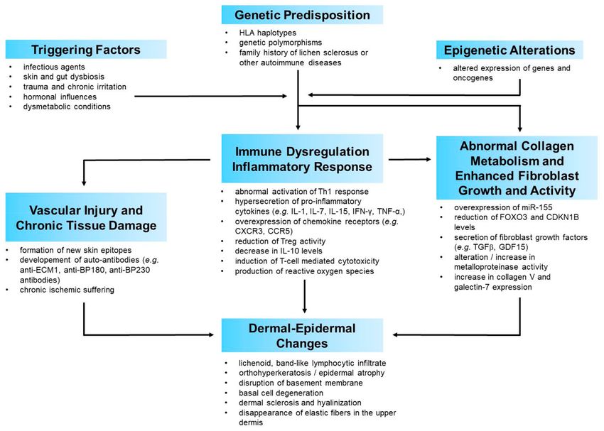
2.5. A Possible Pathogenetic Model
The current evidence on physiopathology of VLS points towards a complex, multifac-
torial process. Once the possible factors involved in the pathogenesis of VLS have been
focused, it is possible to try to define a credible scenario in which the interaction of the
aforementioned factors leads to the development of this disease. It is conceivable that
environmental factors acting on a genetic background trigger autoimmune processes, with
subsequent inflammation which, in turn, leads to tissue and microvascular injury as well
as to activation of signalling pathways involved in fibroblast and collagen metabolism.
Dysregulation of pro- and anti-fibrotic mechanisms determines the degree of dermal fibro-
sis and all the distinctive histopathological features of LS, which represent the culmination
of this process (Figure A1).
Exogenous triggers may act as generic damage-associated molecular patterns (DAMPs),
which ignite the immune response. This activates an inflammatory cascade with raised
levels of pro-inflammatory cytokines predominantly belonging to a Th1 type of inflam-
mation. A recent research quantified the cytokine/chemokine levels in independent
keratinocyte and fibroblast cultures derived from VLS samples and healthy controls [81].
The cytokines/chemokines which resulted most dysregulated in VLS keratinocytes and/or
fibroblasts were DKK1, a soluble antagonist of β-catenin-dependent Wnt signaling, growth
differentiation factor-15 (GDF-15), a member of the TGF-β superfamily, insulin-like growth
factor binding protein-2 (IGFBP-2) and chitinase-like protein Chi3L1. The most noteworthy
aspect of these findings is that all these mediators are implied in both inflammatory pro-
cesses and collagen/fibroblast metabolism. They share the common characteristic of being
a sort of “bridge” between inflammation and collagen metabolism. This not only confirms
the implication of both these pathways but also their close crosstalk in the pathophysiology
of VLS. The involvement of certain mediators which favour the interplay between inflam-
mation and fibroblast biology, or which are themselves junction points between these two
systems, could be crucial in switching the primary inflammatory process into the gradual
development of the histological and clinical changes characteristic of VLS. This seems to
be confirmed by the histopathological close approximation of immune cell infiltrates with
sclerosis in VLS tissue, further supporting the interplay between immune dysregulation
and increased extracellular matrix components.
In this perspective, from a chronological point of view, inflammation may be the
first pathogenetic event in VLS, followed by an alteration of the collagen metabolism,
which is the clinical-histological epiphenomenon of this process. In accordance with this
view, histologically early LS is typified by a lichenoid, lymphocyte-predominant band
of inflammation, basal vacuolization and focal cytotoxic damage to basilar keratinocytes.
Progressively over time, the inflammatory infiltrate is displaced by dermal oedema and
homogenization of papillary dermal collagen fibres [106,107].
This sequence would explain the efficacy of anti-inflammatory treatments, especially
topical corticosteroids or topical calcineurin inhibitors, in significantly improving dermalBiomedicines 2021, 9, 950 7 of 24
collagen changes [108]. In some ways, this resembles the histological and clinical improve-
ment obtained in other different immune-mediated inflammatory skin diseases following
the administration of molecules capable of blocking the immune mechanism. Psoriasis,
pemphigus vulgaris, lichen planus, hidradenitis suppurativa or drug-induced cutaneous
adverse reactions are just some examples of skin diseases in which clinical-histological
healing is obtained by antagonizing the immune and inflammatory cascades [109–113]. In
all these diseases, as well as in many other immune-mediated skin diseases of unknown
aetiology, nonspecific triggering factors activate the immune cascade. For instance, Koebner
phenomenon is a shared trigger for the development of several morphologically different
skin diseases such as psoriasis, lichen planus, vitiligo, LS and others [114]. In a similar
way, other nonspecific factors may trigger the occurrence or relapse, or worsening of het-
erogeneous skin conditions. These include infectious agents, drugs, emotional distress or
dysmetabolic syndrome. What differs from one disease to another is the triggered and
activated immune pathway. The immune pathway elicited in each disease appears to be
driven by a genetically determined background [115–122]. The different immune mediators
involved account, in turn, for the pathogenetic, histological and clinical differences among
these diseases.
Thus, in our view, the main pathomechanisms of VLS, despite the differences, may be
similar to those of other chronic inflammatory multifactorial diseases. Genetic or epigenetic
background determine the activation of a peculiar immune cascade, and the release of
the relative mediators, which is elicited by unspecific factors that probably simply act as
triggers. The type of activated immunity and inflammation mediators is a key determinant
of the histopathological changes and features of one disease or another. Anatomical sites
may contribute to a disease pathogenesis, also in the case of LS [123]. With specific reference
to VLS, with due distinctions, its physio-pathological process resembles that underlying
morphea. An inflammatory driven fibrosis is considered the main pathogenetic event of
morphea [124,125]. This could be a pathogenetic model shared by VLS. Consistent with
this, the histopathological features of the two disorders present similar aspects. Moreover,
genital LS has been found to be significantly more frequent in patients with morphea than
in controls, with a frequency of 38% in patients with morphea [126]. A recent cross-sectional
study found the frequent coexistence of extragenital LS and genital morphea [127]. These
epidemiological data, together with the aforesaid clinical and histological similarities, seem
to further support the strong link between the two clinical entities, which is probably
determined by pathogenetic similarities.
3. Treatment Options
A definitive cure for VLS does not exist, as its exact etiopathogenesis remains un-
known. The ideal treatment should aim at inducing relief of symptoms, reversing signs and
preventing further anatomical changes, including tumorigenesis. Due to its progressive
course and potential evolution towards cancer, all VLS cases should be treated, even when
asymptomatic. The chronic nature of VLS is one of its main troublesome features, because
recurrences are unavoidable when treatments are discontinued. Therefore, long-term
maintenance of sign and symptom remission, achieved with an effective initial therapy, is
an additional objective of treatment.
Considering the recognized main pathophysiological events of VLS, the following
two main potential therapeutic targets can be identified:
(1) auto-immunogenic mechanisms, and subsequent inflammation and oxidative stress.
(2) sclerotic tissue formation.
VLS treatment should act on one or both of these mechanisms.
Currently, there is no single option that can be recommended for the treatment of VLS.
Several strategies are available and the choice depends on various factors. These include
both disease-related aspects, mainly its length and severity, in terms of clinical features and
symptoms, and patient variables such as age, expectations and quality of life impairment.Biomedicines 2021, 9, 950 8 of 24
Treatment options vary as well, depending on the phase of therapy, namely either acute or
maintenance therapy.
The available treatment options are subdivided below on the basis of their main target,
namely immune response and inflammation or collagen metabolism (Tables A1 and A2).
3.1. Treatments Mainly Acting on Immune Dysreactivity and Inflammatory Response
3.1.1. Topical Corticosteroids
High potency topical corticosteroids represent the first-line treatment in the active
phase of VLS [1,128–130]. Topical corticosteroids’ effectiveness lies in their anti-inflammatory
effect via interaction with the intracellular glucocorticoid receptor and the inducement of
specific genes encoding signalling proteins with an immunosuppressive action. They exert
an antipruritic effect, as well. Consistent with this, there is strong evidence that potent and
ultra-potent corticosteroids have an excellent effect on both symptom control and reversal
of histopathological and clinical features.
Topical clobetasol propionate 0.05% ointment or cream is the gold standard treatment
for VLS, based on a long history of use [108,131–133] and wide clinical experience, as
well as on several randomized controlled trials (RCT) [134–140]. Mometasone furoate
0.1% is another molecule whose effectiveness in VLS is supported by quite strong evi-
dence [139,141–143]. Up to date, limited evidence on the effectiveness of alternative topical
corticosteroids is available. In the initial treatment phase, 12 weeks is the duration of a
corticosteroid treatment recommended for achieving a satisfying result with a favourable
safety profile and tolerability. Topical corticosteroids can be used daily (once or twice,
based on the molecule) for the entire 12-week duration. Tapering regimens have been
suggested in order to avoid tachyphylaxis and reduce the risk of dose-dependent side-
effects [129]. An RCT showed that in the initial VLS treatment, topical corticosteroids
can be administered equally well in a continuous or tapering regimen [144]. In fact, both
treatment regimens showed similar efficacy and tolerability. The ointment vehicle seems
to optimize the therapeutic action of potent corticosteroids [145]. Local adverse effects
potentially induced by the prolonged use of corticosteroids, such as atrophy, telangiectasia,
or infections, usually do not occur with a 12-week therapy. Intralesional steroid injection
is an alternative approach, to be reserved for cases resistant to topical treatment [146,147].
Setting up an effective long-term maintenance therapy, after an initial attack phase, is of
paramount importance in the adequate therapeutic management of a chronic disease, such
as VLS. With this specific goal, i.e., for preventing or, at least, delaying disease recurrences,
topical corticosteroids can be administered
(1) on an “as needed” basis (“reactive” scheme) [129,143]
(2) on a continuative prolonged regimen [13,14,148–150],
(3) on a low-dose, intermittent regimen (“proactive” scheme) [151,152].
3.1.2. Topical Calcineurin Inhibitors
Tacrolimus and pimecrolimus are calcineurin inhibitors used in topical preparations
in the treatment of atopic dermatitis. By virtue of their immunosuppressive activity, topical
calcineurin inhibitors (TCI) have been assessed in the treatment of many immune-mediated
skin disorders, including VLS. TCI have been shown to be very effective and safe in
treating VLS, in both adult and prepubertal patients [136,137,153–165]. Both tacrolimus
and pimecrolimus, applied twice a day for 8 to 24 weeks, resulted highly effective in
improving VLS signs and symptoms, with considerable complete response rates. A burning
sensation, usually mild and transient, at the point of application during the first weeks of
treatment, may occur. Patients should be informed of this so that they do not discontinue
therapy. Since comparative trials have shown that ultra-potent corticosteroid is superior to
TCI [136,137], tacrolimus and pimecrolimus are considered an effective alternative to strong
topical corticosteroids in the treatment of active VLS. With reference to the concerns of
potentiating squamous cell carcinoma occurrence with the use of TCI in patients with VLS,Biomedicines 2021, 9, 950 9 of 24
based on the available evidence, short-term application of TCI does not seem to increase
the risk of squamous cell carcinoma in these subjects.
3.1.3. Miscellaneous Topical Treatments
Calcipotriol (CPT) is a synthetic derivative of calcitriol, the active form of vitamin D,
which binds to the vitamin D receptor and exerts a gene regulatory effect. This leads to both
attenuation of the abnormal keratinocyte proliferation and differentiation and inhibition
of the inflammatory response induced by T cells, especially in psoriatic lesions [166].
CPT 0.005% ointment, applied once to twice a day for 16 weeks, induced a significant
improvement, mainly in symptoms, in 8 women affected with VLS [167].
Oxatomide 5% gel is a molecule with antihistamine and anti-inflammatory properties.
In a double-blind, cross-over, controlled trial that enrolled 22 VLS patients, oxatomide 5%
gel, applied twice daily, was compared with placebo. It induced a significant improvement
in itching compared with placebo but without relevant effects on clinical features [168].
Human fibroblast lysate cream (HFLC), also known as cutaneous lysate, is obtained
from cultured human foetal fibroblasts. It has been shown to contain anti-inflammatory
cytokines, such as interleukin 1 receptor antagonist (IL-1RA), IL-10, and IL-13. It also
contains wound-healing growth factors, like EGF, FGFs and VEGF. In a small double blind,
placebo-controlled trial including 30 VLS patients, HFLC applied for 12 weeks did not
provide significant improvement compared to placebo [169].
3.1.4. Systemic Treatments
Systemic agents exerting immunosuppressive or immune-modulating action, such as
cyclosporine and methotrexate, are not supported by adequate evidence in the treatment of
VLS. Thus, they may be considered an alternative therapeutic option exclusively in severe
and refractory forms of VLS.
A single open-label trial which enrolled five VLS patients assessed efficacy and safety
of oral cyclosporine, administered at 3 or 4 mg/kg/day for 12 weeks [170]. At the end
of the third month, subjective symptoms regressed significantly and the clinical findings
showed marked improvement. No severe side effects were observed.
There are no studies that have specifically assessed methotrexate in the treatment of
LS affecting the vulva. Considering a few trials carried out on extensive skin forms of
lichen sclerosus, in some cases also involving the genitals, methotrexate, 10 to 15 mg/week
for 6 months, showed improvement of clinical features [171,172]. In the study by Kreuter
et al., methotrexate was administered with pulsed intravenous high-dose corticosteroids,
for 3 consecutive days, monthly [172].
Promising perspectives, albeit supported by anecdotal experiences, derive from drugs
capable of blocking the immune cascade by acting on novel targets, such as janus kinase
(JAK) 1/2, which is inhibited by baricitinib [173].
3.2. Treatments Mainly Acting on Abnormal Fibroblast and Collagen Metabolism
3.2.1. Topical Retinoids
Topical retinoids are therapeutic options for the treatment of VLS due to their nor-
malizing action on both the keratinization process [174] and collagen metabolism [175].
Retinoids exert a mild anti-inflammatory effect as well [176]. Moreover, they have a role in
the prevention of some skin cancers [177].
Up to date, two non-comparative studies have found that tretinoin 0.025% cream,
applied either once a day, 5 days per week [178], or every other day [179] for prolonged
periods, i.e., 6 to 12 months, induced improvement in subjective and clinical parameters
and, when assessed, also in histological changes [178]. In another open, non-comparative
study, 20 VLS patients were treated with 0.5% cis-retinoic acid in ointment [180]. Complete
remission of clinical features was observed in 11 patients, usually after 1–2 months of daily
retinoid application. Other single case reports can be found in the literature [181]. In the
available literature, topical tretinoin produced an “irritant retinoid reaction”, mostly tran-Biomedicines 2021, 9, 950 10 of 24
sient mild erythema with a burning sensation, in about one third of patients [178,179]. In
an open label, nonrandomized, comparative study, tretinoin 0.05% cream, in short-contact
therapy, was combined with mometasone furoate 0.1% ointment applied for 5 consecutive
days a week, for 12 weeks. This treatment protocol was compared with the same corti-
costeroid, administered with the same regimen, simply combined with an emollient [182].
Surprisingly, no significant differences as regards the rate of response were found between
the two study groups [182,183]. On the other hand, occurrence of local side effects, mostly
burning, was higher in the patients treated with mometasone furoate plus tretinoin than
among those treated with mometasone plus cold cream. This study shows that adding a
topical retinoid, in short-contact therapy, to a potent corticosteroid does not enhance its
effectiveness in the treatment of VLS. A less favourable safety profile of this combination,
which may reduce patient adherence to the treatment, may explain the lack of therapeutic
benefits of combining complementary topical molecules.
Therefore, based on the available literature, topical retinoids may represent a third-line
choice, due to the lack of robust scientific evidence and their well-known local adverse effects.
3.2.2. Miscellaneous Topical Treatments
In the literature, avocado/soybean unsaponifiable (ASU), which is an extract prepared
from avocado and soybean oil, has been shown to exert an anti-inflammatory effect by re-
ducing several cytokines, prostaglandin E2, metalloproteinases and other pro-inflammatory
mediators [184,185]. Moreover, when administered percutaneously on the dorsal skin of
hairless rats for 15 days, it produced a modification of dermal connective tissue compo-
nents [186]. In fact it induced an increase of soluble collagen and a decrease of insoluble
collagen, as a result of the activation of connective tissue metabolism, mainly the inhibition
of lysyl oxidase activity. The ASU action on the skin collagen metabolism may explain
the beneficial effect on scleroderma observed by some authors [187–189]. An open-label,
non-comparative study assessed efficacy and tolerability of a topical product containing
avocado and soybean extracts and other lenitive and anti-oxidant principles administered
for 24 weeks, in association with a dietary supplement containing avocado and soybean
extracts vitamin E and para-aminobenzoic acid for the first 12 weeks, in the treatment
of active mild-to-moderate VLS. Among the 23 women included, mean symptom and
sign scores decreased significantly after treatment. The treatment was well tolerated [190].
Emollients and moisturizers do not have a direct action to improve the histopathological
features of VLS, but rather to make the skin softer while preserving its integrity. They
provide relief of symptoms and seem to delay flares [191,192]. This latter property makes
them particularly suitable for long-term maintenance therapy.
3.2.3. Physical Treatments
Phototherapy
The most convincing data on the potential benefits of phototherapy in VLS treatment
concern UV-A1 phototherapy. UV-A1 phototherapy has been shown to be effective in a
variety of sclerotic skin disorders, such as morphea, systemic sclerosis, eosinophilic fasciitis
and necrobiosis lipoidica [193], which share some histological and clinical similarities with
LS. UV-A1 enhances MMP activity, inhibits collagen synthesis and induces the fibroblasts’
release of cytokines which lead to an up-regulation of collagenase activity [193]. More-
over, it has strong melanogenic properties, which induce a re-pigmentation of LS pale
surfaces [194].
A randomized trial compared clobetasol propionate 0.05% ointment, applied once
daily for 12 weeks, with medium-dose UV-A1 (50 J/cm2 ) home-based phototherapy, per-
formed four times weekly for 12 weeks [195]. At the end of treatment, UV-A1 phototherapy
resulted in a significant clinical improvement, but was inferior to clobetasol propionate
with respect to practicability, relief of itching, and improvement in quality of life. In a
non-comparative study which included seven VLS patients, UV-A1 phototherapy was
given three to five times/week for variable periods, with an acceptable clinical responseBiomedicines 2021, 9, 950 11 of 24
in five patients [195]. Case series and anecdotal reports showed that 8-methoxypsoralen
(8-MOP)-containing cream plus UVA (PUVA) as well as narrow band UVB induced some
improvements in VLS-related symptoms and signs [196,197]. Some concerns remain about
the use of phototherapy in VLS treatment, especially UVB and PUVA therapy, due to its
well-known carcinogenic potential.
Photodynamic Therapy
The pathophysiologic rationale of photodynamic therapy (PDT) is based on the con-
version of ALA and methyl δ-ALA into the photosensitizing metabolic products called
protoporphyrins. By activating protoporphyrins with the appropriate wavelength, PDT
induces production of ROS, like superoxide, and singlet oxygen [198]. These reactions
ultimately result in apoptosis of lymphocytes and keratinocytes, as well as in alteration of
the expression of both cytokines and MMPs, which play a role in skin remodelling. Thus,
even if the exact mechanism of PDT in VLS treatment remains uncertain, it seems to mainly
act on dermal sclerosis.
To date, a single open-label RTD compared four sessions of ALA-PDT, administered
at 2-week intervals, with clobetasol propionate 0.05% ointment, administered once daily
for 8 weeks, in 43 VLS patients [140]. At the end of treatment, both groups showed im-
provement in terms of clinical signs and symptoms. ALA-PDT showed a longer remission
duration and a higher complete response rate than clobetasol propionate.
Further data are provided by some noncomparative studies including prospective
cohorts and case series [199–207]. The majority of the patients included in these studies
were treated with 5-ALA before the application of radiation (5-ALA PDT), whereas the
remaining patients with methyl-aminolevulinate (methyl-PDT). Red light (630–635 nm)
was the most commonly applied light, followed by green light (495–570 nm).
Even considering the differences in both treatment schemes and outcomes adopted in
the available studies, PDT was shown to induce symptom relief in most treated patients.
Improvement in objective features was less constant and appreciable. Relevant changes in
dermoscopic features have been documented as well [207].
No major adverse effects were reported during therapy and the post-treatment period,
with the exception of mild to moderate pain or burning during irradiation. Based on
a systematic review of the literature, a protocol for PDT in VLS has been recently pro-
posed [208]. It consists in 5% 5-ALA as a photosensitizer applied for 3 h under occlusion
before irradiation at the dose of 120 J/cm2 with red light (590–760 nm) and intensity of
204 mW/cm2 .
Laser
Lasers, both ablative and non-ablative, are expected to be a suitable option in VLS
treatment due to some collagen remodelling changes induced by these tools. In particular,
lasers lead to neovascularization, neocollagenesis, elastogenesis, and restoration of the
trabecular architecture of collagen, as well as reduction of epithelial degeneration and
atrophy [209].
A single RCT assessed efficacy and tolerability of non-ablative neodymium: yttrium
aluminium garnet (Nd:YAG) laser treatment of active VLS in comparison with a topical
corticosteroid [210]. More in detail, the patients in the laser group received three Nd:YAG
laser treatments every 14 days, whereas the control group received topical betamethasone
for 4 weeks with tapering dosage. Those treated with laser applied betamethasone one
week before the first laser treatment, and then for further 3 weeks with a decreasing dosage.
Patients in the laser group had significantly greater improvement in VLS symptoms, better
patient satisfaction, and greater reduction of sclerosis than those in the corticosteroid group,
at 1- and 3-month follow-up visits. Both the very short course of corticosteroid treatment
in the control group and the concomitant use of the same corticosteroid, albeit for a shorter
period, in the laser group as well should be taken into consideration when interpreting
these results.Biomedicines 2021, 9, 950 12 of 24
Ablative lasers, such as carbon dioxide lasers, have been used for recalcitrant and/or
severe VLS in non-comparative, case series, studies [211–216]. CO2 laser treatment, very
heterogeneous among the different studies for energy, treatment depth and regimens, led
to an almost constant improvement in signs and symptoms. Post-operative pain was a
common report.
The potential benefits of micro-ablative fractional radiofrequency (MFR) on epithelial
trophism and symptom improvement have been assessed in VLS patients [217]. After two
to three sessions of MFR, most participants reported an improvement in symptoms, which
persisted for about 11 months (range: 7–16 months) after the treatment. At histological
examination, type III collagen concentration significantly increased and was associated
with symptom improvement.
A recent systematic review supported the efficacy and tolerability of lasers, particularly
fractionated ablative lasers, as an adjunct to topical potent corticosteroids for symptomatic
relief in refractory VLS [218].
3.2.4. Injective Treatments
Adipose-Derived Stem Cells and Platelet-Rich Plasma
Adipose-derived stem cells (ADSCs) are deemed capable of inhibiting fibrosis and
regenerating damaged tissue by remodelling the extracellular matrix, especially in fibrotic
disorders [219,220]. Moreover, they exert anti-inflammatory and immune-modulatory
activity, due to the presence of pluripotent mesenchymal stem cells, endothelial progenitor
cells, T cells, B cells, mast cells and adipose-resident macrophages. Platelet-rich plasma
(PRP) contains platelets, which release growth factors supposed to reduce inflammation
and to promote mesenchymal cell proliferation, tissue repair and angiogenesis [221,222].
In keeping with their action on the immune system and extracellular synthesis, two altered
conditions in VLS, some observational studies and reports have assessed their efficacy
and safety for the treatment of this condition [223–228]. Overall, these small series, non-
comparative studies reported favourable short-term and patient-reported treatment out-
comes with both ADSCs and PRP. In particular, ADSCs and PRP may represent promising
therapeutic perspectives, especially for scarring, atrophy and the other sequelae of VLS
which are poorly responsive to topical therapies. Methodological biases, the small number
of patients included and the lack of standardized interventions, as well as of standardized
outcome measures, limit the strength of these results [227].
An RCT assessed the efficacy and safety of autologous PRP for the treatment of VLS
in 30 patients [229]. Enrolled patients were randomized to receive either placebo (saline
injections) (10 subjects) or two separate treatments of PRP 6 weeks apart (20 subjects). No
significant differences were found between the two study groups as regards the histological
and clinical outcome parameters considered in the study.
A recent study compared the effectiveness of local injection of purified adipose-
derived stromal vascular fraction (AD-SVF) compared with purified SVF plus PRP in two
randomized groups of LS patients [230]. Each patient received two surgical procedures, in
general anaesthesia, distanced by a 4-month period. The study assessed clinical features,
symptoms and quality of life before and after the treatment. The authors found that
both AD-SVF and AD-SVF plus PRP are safe and efficacious regenerative methods for LS,
especially in early disease stages. The addition of PRP did not improve treatment outcome
of SVF graft.
Heterologous Type I Collagen
Effectiveness and safety of intradermal injections of heterologous type I collagen
(HT1C) have been recently assessed in 3 VLS patients not responsive to ultra-potent
corticosteroid [231]. HT1C stimulates both fibroblast proliferation and the production of
extracellular matrix proteins [232]. Four injective treatments at 2-week intervals provided
substantial relief of symptoms and complete resolution of clinical features in all of the
treated subjects. VLS remission was maintained with further injections every 2 months.Biomedicines 2021, 9, 950 13 of 24
None of the patients complained of adverse events. The small number of cases and the lack
of a comparison render these results only preliminary, even though they are promising.
3.2.5. Systemic Treatments
In a multi-centre RCT, acitretin, a synthetic monoaromatic retinoid, administered
orally at daily dosage of 20 to 30 mg/day for 16 weeks, was compared with placebo [233].
At treatment completion, significant improvement in itching, clinical features, such as
atrophy, hyperkeratosis, erosions and lichenification, and the extent of VLS lesions were
observed in the acitretin-treatment group as compared with the placebo group. A few
noncomparative studies assessed the efficacy of etretinate, for many years now no longer
on the market [234,235].
Potassium para-aminobenzoate (PABA) is an intermediate in the bacterial synthesis of
folate (Vitamin Bx), which has been shown to have a favourable effect in several connective
tissue disorders and skin fibroses when taken orally. A double-blind, placebo-controlled
trial tested oral PABA (3 g capsules four times daily for 8 weeks) in 25 patients (22 women)
suffering from genital and extra-genital LS [236]. After treatment, improvements recorded
among the patients treated with oral PABA were not significantly different from those of
the placebo group.
4. Concluding Remarks and Research Agenda
The ultimate objective for VLS patients undergoing specific treatments is to be no longer
impaired by the disease in such aspects of their daily lives as distressing symptoms and
sexual dysfunction. Achieving objective normality of skin colour and texture is a further key
endpoint of treatment. Moreover, treatment should ideally protect against the progression
to more severe forms of the disease and development of vulvar malignant neoplasm.
Considering both the available evidence and their action against the main pathophysi-
ological mechanism underlying VLS, that is immune dysreactivity and inflammation, high
potency topical corticosteroids represent the first-line treatment in the active phase of the
disease. Topical calcineurin inhibitors, which inhibit the immune cascade as well, are an
effective and safe alternative. The other available therapeutic options, including medi-
cal, physical and, to a lesser extent, systemic treatments, which mainly affect VLS tissue
changes, should currently be considered third-line, at least until they are supported by
stronger evidence. Further RCT, longer follow up, and better standardization of protocols
and outcomes are required to shed light on the real potential of these treatments.
A maintenance treatment is necessary and should always be planned in order to
stabilize the improvement obtained with the initial treatment and prevent flares [130].
However, the available treatment strategies have been shown to moderately satisfy
women affected with VLS [237]. In agreement with this, recent studies highlighted the
fact that complete healing of VLS occurs in a minority of patients treated with standard
pharmacological treatments, despite the achievement of major improvements in symptoms
and signs [238,239]. This means that, at treatment conclusion, a substantial rate of patients
may still have residual disease. Thus, persistent negative effects on their well-being and
quality of life may be supposed.
This may be due to the fact that, despite the advances in the knowledge of the
pathogenetic mechanisms of VLS, many aspects and mediators remain unclear. Further
investigation is needed to better define the following issues:
(1) The exact sequence of events underlying VLS pathogenesis;
(2) The key mediators involved in VLS immune response and those which, more than
others, trigger an abnormal fibroblast and collagen metabolism; in other words, the
agents that convert inflammation into fibrosis;
(3) To what extent keratinocytes and fibroblasts actively participate in VLS pathogenesis
and how they interact; andBiomedicines 2021, 9, 950 14 of 24
(4) How a genetic background predisposes certain individuals to an abnormal release
of pro-inflammatory and pro-fibrotic mediators, in response to still not fully under-
stood triggers.
Advancement in this knowledge will presumably lead to making key mediators
and events become therapeutic targets. This is the requirement for the development of
extremely selective drugs, such as biologics, capable of modulating or suppressing specific
pathogenetic sequences. These highly selective molecules have changed the natural course
of many skin diseases that were not optimally managed until recently, such as psoriasis or
atopic dermatitis. It is conceivable, and desirable, that the increasingly detailed definition
of the mediators of the VLS pathogenetic process may allow us to have more effective
drugs for patients suffering from this disabling disease.
Author Contributions: Conceptualization, M.C. and A.B.; methodology, A.B., N.S., P.Z.; writing—
original draft preparation, A.B.; writing—review and editing, A.B., M.C., N.S., P.Z.; supervision,
M.C., A.B. All authors have read and agreed to the published version of the manuscript.
Funding: This research received no external funding.
Institutional Review Board Statement: Not applicable.
Informed Consent Statement: Not applicable.
Conflicts of Interest: The authors declare no conflict of interest.
Appendix A
Table A1. Treatments mainly acting on immune dysreactivity and inflammatory response.
Treatment Posology Notes
Topical treatments
- first line treatment in the active phase
- anti-inflammatory and immunosuppressive activity
Topical Corticosteroids - effectiveness on both symptoms and objective features
- Clobetasol Propionate 0.05% Ointment - tachyphylaxis and dose-dependent side effects may be avoided by tapering
or Cream Once or twice a day for 12 weeks regimens
- Mometasone Furoate 0.1% Ointment or - ointment formulation seems to be more effective in comparison with cream
Cream - intralesional corticosteroid injection in recalcitrant forms
- long-term maintenance treatment (reactive, continuative or proactive
regimens)
- second-line choice with lower effectiveness than ultra-potent corticosteroid
Topical Calcineurin Inhibitors
- immunosuppressive activity
- Tacrolimus 0.1% Ointment Twice a day for 8 to 24 weeks
- effectiveness on both symptoms and objective features
- Pimecrolimus 1% Cream
- possible transient burning sensation during the first weeks of treatment
- inhibition of inflammatory response
- attenuation of abnormal keratinocyte proliferation and differentiation
Calcipotriol 0.005% Ointment Once to twice a day for 16 weeks
- effectiveness on symptoms
- alternative to standard treatment (weak evidence)
- antihistamine and anti-inflammatory properties
Oxatomide 5% gel Twice a day for periods of 14-days - effectiveness on both symptoms and objective features
- alternative to standard treatment (weak evidence)
- presence of anti-inflammatory cytokines and wound-healing grow factors
Human Fibroblast Lysate Cream Twice daily for 12 weeks
- no more effective than placebo
Systemic treatments
- immunosuppressive effect
- regression of symptoms and improvement of clinical features in resistant
Oral Cyclosporine 3–4 mg/kg/day for 12 weeks
case
- weak evidence
- immunosuppressive effect
- regression of symptoms and improvement of clinical features in resistant
Oral or Subcutaneous Metothrexate 10 to 15 mg/week
case
- weak evidence
- inhibition of JAK 1/2
Baricitinib
- anecdotal reportsYou can also read