Ferroptosis Mechanisms Involved in Neurodegenerative Diseases - MDPI
←
→
Page content transcription
If your browser does not render page correctly, please read the page content below
International Journal of
            Molecular Sciences

Review
Ferroptosis Mechanisms Involved in
Neurodegenerative Diseases
Cadiele Oliana Reichert 1 , Fábio Alessandro de Freitas 1 , Juliana Sampaio-Silva 1 ,
Leonardo Rokita-Rosa 1 , Priscila de Lima Barros 1 , Debora Levy 1 and
Sérgio Paulo Bydlowski 1,2, *
 1    Lipids, Oxidation, and Cell Biology Group, Laboratory of Immunology (LIM19), Heart Institute (InCor),
      Hospital das Clínicas HCFMUSP, Faculdade de Medicina, Universidade de São Paulo, São Paulo 05403-900,
      Brazil; kadielli@hotmail.com (C.O.R.); fabio.alessandro@usp.br (F.A.d.F.); jukisbio@gmail.com (J.S.-S.);
      rokita@usp.br (L.R.-R.); pri_limabarros@hotmail.com (P.d.L.B.); d.levy@hc.fm.usp.br (D.L.)
 2    Instituto Nacional de Ciencia e Tecnologia em Medicina Regenerativa (INCT-Regenera), CNPq,
      Rio de Janeiro 21941-902, Brazil
 *    Correspondence: spbydlow@usp.br

 Received: 6 October 2020; Accepted: 28 October 2020; Published: 20 November 2020

 Abstract: Ferroptosis is a type of cell death that was described less than a decade ago. It is caused
 by the excess of free intracellular iron that leads to lipid (hydro) peroxidation. Iron is essential
 as a redox metal in several physiological functions. The brain is one of the organs known to be
 affected by iron homeostatic balance disruption. Since the 1960s, increased concentration of iron in
 the central nervous system has been associated with oxidative stress, oxidation of proteins and lipids,
 and cell death. Here, we review the main mechanisms involved in the process of ferroptosis such
 as lipid peroxidation, glutathione peroxidase 4 enzyme activity, and iron metabolism. Moreover,
 the association of ferroptosis with the pathophysiology of some neurodegenerative diseases, namely
 Alzheimer’s, Parkinson’s, and Huntington’s diseases, has also been addressed.

 Keywords: ferroptosis; cell death; iron metabolism; neurodegenerative diseases; glutathione
 peroxidase 4; GSH; system xc− ; Alzheimer’s disease; Parkinson’s disease; Huntington’s disease

1. Introduction
      The current classification system of cell death has been updated by the Nomenclature Committee
on Cell Death (NCCD), according to their guidelines for the definition and interpretation of all aspects
of cell death [1,2]. Accidental cell death (ACD) is an instantaneous and catastrophic demise of cells
exposed to severe insults of physical or mechanical forces. On the other hand, regulated cell death
(RCD) is a dedicated molecular machinery [3]. RCD can occur in two ways: firstly, as programmed
cell death that can occur in the absence of any exogenous environmental perturbation. Secondly,
RCD can originate from disturbances of the intracellular or extracellular microenvironment that cannot
be restored to cellular homeostasis [4–6].
      In 2012, Brent R. Stockwell described a unique form of cell death that results from the overwhelming
iron-dependent accumulation of lethal amounts of lipid-based reactive oxygen species and named
it ferroptosis [7]. Ferroptosis is morphologically and biochemically distinct from other RCDs.
It occurs without the chromatin condensation and nuclear reduction seen in apoptosis, cellular
and organellar swelling of necrosis, and without the common features of autophagy. Morphologically,
only mitochondrial shrinkage distinguishes it from other forms of death [8,9]. Ferroptotic cell death
is associated with the iron-dependent mechanism and formation of extremely reactive free radicals,

Int. J. Mol. Sci. 2020, 21, 8765; doi:10.3390/ijms21228765                          www.mdpi.com/journal/ijms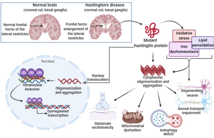
Int. J. Mol. Sci. 2020, 21, 8765 2 of 27
along with severe peroxidation of membrane phospholipids (PLs) rich in polyunsaturated fatty acids
(PUFAs),
Int. J.mainly of arachidonic
Mol. Sci. 2020, 21, x FOR PEER orREVIEW
adrenic acids from phosphatidyl ethanolamine (PE) molecules [10–12].
2 of 27
The complex balance between reactive oxygen species (ROS) and the antioxidant system maintains
polyunsaturated
cell homeostasis fatty acidsdangerous
by removing (PUFAs), mainly
stimuliof and
arachidonic or adrenic
controlling acidsstress
oxidative from byphosphatidyl
several factors,
ethanolamine (PE) molecules [10–12].
and is also present in the central nervous system (CNS) [13,14]. Among these factors, system xc− ,
The complex balance between reactive oxygen species (ROS) and the antioxidant system
an amino acid cell
maintains antiporter,
homeostasis maintains
by removingthe dangerous
synthesisstimuli
of glutathione (GSH)
and controlling and oxidative
oxidative protection.
stress by several
Inhibition of system xc − causes a rapid drop of intracellular glutathione levels and cell death caused
factors, and is also present in the central nervous system (CNS) [13,14]. Among these factors, system
by thexcaccumulation
-, an amino acidof lipid-derived
antiporter, maintainsROS. Lipid and
the synthesis protein oxidation
of glutathione (GSH) andlead to inflammation
oxidative protection. and
changes in DNA,
Inhibition and are
of system xc− the
causestrigger
a rapidfor premature
drop aging,
of intracellular loss of function
glutathione levels andand
cell death of neurons.
death caused
by the accumulation
The increase in oxidative of lipid-derived
stress generated ROS.
by Lipid and protein
free radicals oxidation
associated leaduncontrolled
with to inflammation and
intracellular
changes in DNA, and are the trigger for premature aging, loss of
iron metabolism has been associated with the pathophysiology of neurodegenerative diseasesfunction and death of neurons. The[13–15].
increase in oxidative stress generated by free radicals associated with uncontrolled intracellular iron
Here we address the main pathways of ferroptosis and its role in the pathophysiology of Alzheimer’s
metabolism has been associated with the pathophysiology of neurodegenerative diseases [13–15].
disease, Parkinson’s disease, and Huntington’s disease, the main neurodegenerative diseases in which
Here we address the main pathways of ferroptosis and its role in the pathophysiology of Alzheimer’s
ferroptosis
disease, hasParkinson’s
been shown to be and
disease, involved.
Huntington’s disease, the main neurodegenerative diseases in
which ferroptosis has been shown to be involved.
2. Ferroptosis
2. Ferroptosis
Ferroptosis is a particular form of cell death that is induced by lipid hydroperoxides derived from
Ferroptosis
the oxidation is a particular
of reactive form of cell
species generated bydeath that isCell
free iron. induced
deathbyinlipid hydroperoxides
ferroptosis involvesderived
three main
from
factors: the oxidation
increased of reactive species
free intracellular iron,generated
depletionbyoffree
theiron. Cellglutathione/GPx4/system
redox death in ferroptosis involvesxc
three
− and the
main factors: increased free intracellular iron, depletion of the redox glutathione/GPx4/system xc- and
oxidation of membrane PUFAs [16–18] (Figure 1).
the oxidation of membrane PUFAs [16–18] (Figure 1).
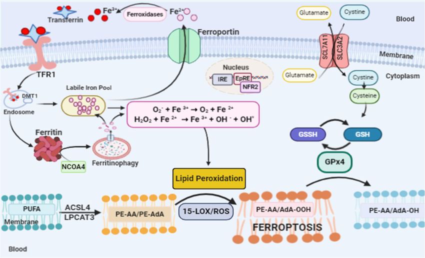
FigureFigure 1. Ferroptosis pathway. Ferroptosis can be initiated through transferrin endocytosis linked to
1. Ferroptosis pathway. Ferroptosis can be initiated through transferrin endocytosis linked
transferrin receptor 1 (TFR1). After endocytosis, ferric iron is released from the Transferrin–TRF1
to transferrin receptor 1 (TFR1). After endocytosis, ferric iron is released from the Transferrin–TRF1
complex and is reduced to ferrous iron (Fe2+). Fe2+ can be stored in ferritin or remain in the cytoplasm
complex and is reduced to ferrous iron (Fe2+ ). Fe2+ can be stored2+in ferritin or remain in the cytoplasm
as a labile iron Pool (LIP). The LIP is composed mainly of Fe , which through Fenton reaction
as a labile iron Pool (LIP). 2+ , which through Fenton reaction generates
generates species suchThe
as: LIP is composed
the hydroxyl mainly
radical of Fewith
that reacts membrane lipids, providing the lipid
speciesperoxidation
such as: theofhydroxyl
arachidonicradical that reacts
acid (AA) withacid
or adrenic membrane lipids,
(AdA). Lipid providingcan
peroxidation thealso
lipidoccur
peroxidation
via
of arachidonic acid (AA)itor
enzyme. However, is adrenic
necessary acid
for (AdA).
the free Lipid peroxidation
polyunsaturated fattycan also
acids occur via
(PUFAs) enzyme.
to be esterifiedHowever,
as
membranefor
it is necessary PUFA by the
the free enzymes ACSL4
polyunsaturated andacids
fatty LPCAT3, forming
(PUFAs) arachidonic
to be esterifiedorasadrenic
membrane acidsPUFA
by theesterified
enzymesinACSL4phosphatidyl ethanolamine
and LPCAT3, (PE-AA/PE-AdA).
forming arachidonic or Dioxigenation
adrenic acids by esterified
15-LOX generates PE-
in phosphatidyl
AA/AdA-OOH, which reacts with other membrane lipids, forming
ethanolamine (PE-AA/PE-AdA). Dioxigenation by 15-LOX generates PE-AA/AdA-OOH, which reactspores in the lipid bilayer,
destabilizing it and then rupturing the membrane. Ferroptosis is inhibited by GPx4, which converts
with other membrane lipids, forming pores in the lipid bilayer, destabilizing it and then rupturing the
PE-AA/AdA-OOH to alcohol and water. This reaction occurs through the use of glutathione (GSH)
membrane. Ferroptosis is inhibited by GPx4, which converts PE-AA/AdA-OOH to- alcohol and water.
as a substrate. GSH synthesis occurs via the entry of cystine into the cell by system xc .
This reaction occurs through the use of glutathione (GSH) as a substrate. GSH synthesis occurs via the
entry of cystine into the cell by system xc− .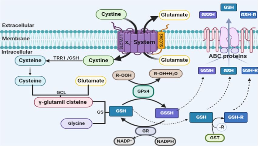
Int. J. Mol. Sci. 2020, 21, 8765                                                                       3 of 27

2.1. Lipid Peroxidation and Ferroptosis
      Lipid peroxidation is the trigger for the activation of ferroptosis [13,18]. Lipid peroxides (PL-OOH),
mainly lipid hydroperoxides (L-OOH), have the ability to cause damage to the lipid bilayer of the plasma
membrane due to the accelerated oxidation of the membrane lipids which leads to ferroptosis. The increase
in the concentration of lipid peroxides can alter the structure and function of nucleic acids and proteins,
as well as the Michael acceptors and aldehydes. In fact, it can generate additional toxicity due to its
degradation products [19–22]. The cellular lipids include thousands of lipid species that vary in quantity,
intra- and extracellular distribution, functions and cell type [8]. Thus, the higher the concentration of free
polyunsaturated fatty acids (PUFAs) in the cell, the greater the damage caused by lipid hydroperoxidation
and the extent of ferroptosis, which can vary among diseases and organs/tissues [4,8,17].
      PUFAs are good substrates for autoxidation because the C–H bonds of the methylene groups flanked by
C-C double bonds are among the weakest C–H bonds known [19–22]. The structure of the PUFA molecule
contains bis-allyl hydrogen atoms that can be abstracted. Then, there is a rearrangement of the resonance
radical structure, with subsequent addition of molecular oxygen, giving rise to the peroxyl radical and the
formation of the primary molecular product, lipid hydroperoxide (L-OOH). Soon after, the cleavage of the
L-OOH molecule occurs, giving rise to highly electrophilic secondary oxidation products, including epoxy,
oxo- or aldehyde groups, which are highly reactive and toxic to membranes and cells [8,23].
      First, PUFAs are esterified with membrane phospholipids, such as phosphatidyl ethanolamine
(PE). The esterification reaction is catalyzed by acyl-CoA synthetase long-chain family member 4 (ACSL4),
which binds coenzyme A to long-chain PUFAs, which can then be used for esterification of lysophospholipids
by lysophosphatidylcholine acyltransferase 3 (LPCAT3); the substrates can undergo peroxidation resulting
in the formation of arachidonoyl (AA) and adrenoyl (AdA) acids, which can lead to ferroptosis. Suppression
of the ACSL4 enzyme inhibits ferroptosis by depleting the substrates for lipid peroxidation [24,25].
      The PUFA oxidation process that leads to ferroptosis can occur enzymatically or non-enzymatically [26].
The non-enzymatic oxidation process occurs through ROS and hydroxyl radical, from the Fenton reaction.
This process is both non-selective and non-specific. Thus, oxidation rates are proportional to the number of
readily abstractable bis-allyl hydrogens in the PUFA molecule, resulting in the accumulation of a highly
diversified pattern of oxidation products with the predominance of oxygenated PUFA-PLs with 6, 5, 4, 3 and
2 double bonds [8,19]. Enzymatic oxidation of PUFAs occurs through lipoxygenases (LOXs) [27]. LOXs are
dioxigenases containing iron in their catalytic region that promote the dioxigenation of polyunsaturated
fatty acids containing at least two isolated cis-double bonds. In humans, there are different isoforms of LOX
(5-LOX, 12S-LOX, 12R-LOX, 15-LOX-1, 15-LOX-2 and eLOX3) [19,27].
      Membrane ester lipids are cleaved by cytosolic phospholipase A2 in different fatty acids:
arachidonic acid (AA), eicosapentaenoic acid (EPA) and docosahexaenoic acid (DHA). Oxygenation
by cyclooxygenases (COXs) generates prostanglandins-G (PGG2, PGG3 and PGG4, respectively).
However, oxygenation by LOX generates doubly and triply oxygenated (15-hydroperoxy)-diacylated
PE species [28–31]. Oxidation induced by 15-LOX is selective and specific, occurring preferably in
arachidonic acid-phosphatidylethanolamine (AA-PE) or adrenoyl acid (AdA)-PE. The product of
this oxidation is 15-hydroperoxy-arachidonic acid-phosphatidylethanolamines (15-HOO-AA-PEs) or
15-hydroperoxy-adrenoyl acid-phosphatidylethanolamines (15-HOO-AdA-PEs) (Figure 1) [28–31].
The catalytic activity 15-LOX is dependent on the pro-ferroptotic PEBP1 protein [32].
      Stoyanovsky et al. [33] showed that the ferroptosis process includes two stages: (i) selective and
specific enzymatic production of 15-HOO-AA-PE by 15-LOX; (ii) oxidative cleavage of these initial
HOO derivatives to proximate electrophiles capable of interacting with protein targets to cause the
formation of pores in plasma membranes, or to rupture them. The two types of oxidatively truncated
products can be formed from 15-HOO-AA-PE with the carbonyl function either on the shortened
AA-residue esterified into PE, or on the leaving aldehyde.
      In addition, tocopherols and tocotrienols suppress LOX and protect against ferroptosis [24]. On the
other hand, ferrostatins inhibit ferroptosis by efficiently scavenging free radicals in lipid bilayers [34].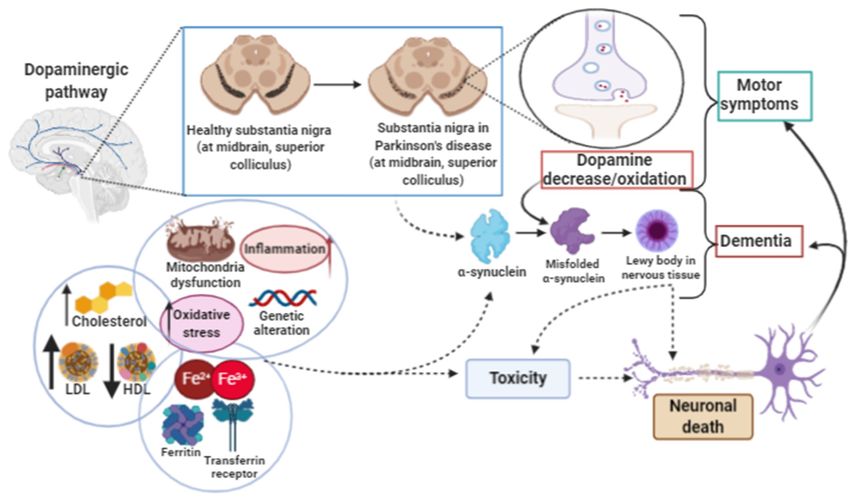
Int. J. Mol. Sci. 2020, 21, 8765                                                                      4 of 27

     Recently, Zou et al. [35] have shown that cytochrome P450 oxidoreductase (POR) is a key mediator
in the induction of ferroptosis in cells that exhibit intrinsic and induced susceptibility to ferroptosis
by enabling membrane polyunsaturated phospholipid peroxidation. POR depletion suppressed
arachidonic acid-induced sensitivity to ML210/RSL3 in a dose-dependent manner. In addition to
suppressing PUFA-induced ferroptosis susceptibility, POR depletion by constitutive or inducible
knockout also compromised the intrinsic ferroptosis sensitivity in ccRCC cells 786-O and 769-P.

2.2. Glutathione Peroxidase 4 and Ferroptosis
     Cells have several escape mechanisms against cell death [36,37]. In the ferroptotic process,
one of the most important and most studied so far is the enzyme glutathione peroxidase 4 (GPx4),
(also called Phospholipid Hydroperoxide Glutathione Peroxidase (PHGPx)) [38,39]. In the human
organism, there are several isozymes of glutathione peroxidase, which vary in cell location and substrate
specificity [40]. The GPx4 enzyme is a selenoprotein, with approximately 20–21 kDa, composed of
197 amino acids, and encoded by the GPx4 gene in chromosome 19 localization [41]. GPx4 has in its
active site the amino acid selenocysteine, which is necessary for protection against ferroptosis [42].
The catalytic site of selenocysteine involves three different redox states: selenol, selenenic acid and
seleninic acid. These different forms of the redox state allow the regulation of the catalytic efficiency of
the peroxide reduction, which is dependent on the cellular redox state [43]. The enzymatic activity
of GPx4 is vital to cells, since the enzyme can reduce H2 O2 and is the only enzyme that can reduce
phospholipid hydroperoxides [44].
     In addition, by structural similarity, GPx4 can reduce both peroxidized fatty acids and esterified
cholesterol hydroperoxides, as well as thymine hydroperoxide, a product of free radical attack on
DNA. The reduction reaction can occur in membranes, in the cytoplasm and/or in lipoproteins [45,46].
In the antiferroptotic process, the GPx4 enzyme directly reduces toxic lipid peroxides (PL-OOH) to
non-toxic lipid alcohols (PL-OH) using reduced glutathione (GSH) as a substrate [47–49]. The synthesis
of GSH through the cystine/glutamate antiporter system xc− is a limiting step for the function of
detoxification of lipid peroxides by GPx4 [50]. The rate-limiting compound of GSH synthesis is the
non-essential amino acid cysteine. Cysteine can be imported into cells directly or in its oxidized form,
cystine, through system xc− . Within the cell, cystine is reduced to cysteine by biosynthesis of GSH [51].
Figure 2 shows the complete GSH biosynthesis pathway.
     GPx4 inhibitors, including ML210, ML162 and (1S), (3R)-RSL3 (RSL3), are used as specific
ferroptosis inducers [52–55]. Moreover, the overexpression or silencing of the gene coding for
14-3-3 proteins controls the inactivation of GPx4 by RSL3 [56]. In addition, liproxstatin-1 is able to
suppress ferroptosis in cells, inhibits mitochondrial lipid peroxidation, and restores the expression of
GSH, GPX4 and ferroptosis suppressor protein 1 [57,58]. A variety of ferroptosis inducers can inhibit
cystine absorption by inhibiting system xc− , such as: erastin, sulfasalazine and sorafenib, resulting
in reduced GPx4 activity in different cells lines. Thus, there is no synthesis of GSH and the activity
of GPx4 decreases. As a consequence, there is a reduction in the cell antioxidant capacity and hence
increased L-ROS, ultimately leading to ferroptosis [59–67].
     GSH biosynthesis is regulated by the ubiquitously expressed transcription factor nuclear factor
erythroid-2 related factor 2 (Nrf2). In baseline conditions, Nrf2-dependent transcription is suppressed
due to proteasomal degradation in the cytosol by Keap1 (kelch-like ECH-associated protein 1). However,
due to the exposure to a variety of different stimuli, including oxidative stress, the ubiquitination
and degradation of Nrf2 are blocked, leading to the stabilization and nuclear accumulation of Nrf2,
where it induces dependent electrophilic response element (EpRE) gene expression to restore cellular
redox homeostasis [72]. Nrf2 regulates several steps of GSH biosynthesis transcription enzymes,
such as catalytic and regulatory subunits of glutamate cysteine ligase (GCL), GSH synthase, GPx2,
GSH S-transferases (GSTs) and GSH reductase (GR) as well as the light-chain subunit of the xc- [72–75].
Nrf2 is also associated with the regulation of antioxidant enzymes, NADPH: quinine oxidoreductase-1
(NQO-1 and NQO-2) and nicotinamide adenine dinucleotide phosphate oxidase 2 (NOX2). In addition,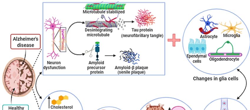
Int. J. Mol. Sci. 2020, 21, 8765 5 of 27
Nrf2 can regulate iron metabolism enzymes [76,77] and proteins associated with multiple drug
resistance (ABCG2, MRP3, MRP4, glutathione S-transferase P (GSTP)) [72,78].
Int. J. Mol. Sci. 2020, 21, x FOR PEER REVIEW 5 of 27
Figure
Figure 2.2. Glutathione
Glutathione (GSH) (GSH) biosynthesis
biosynthesis pathway.pathway. GSH GSH is known as
is known one of
as one of the
the small-molecule
small-molecule
water-soluble antioxidants, the most important of somatic cells.
water-soluble antioxidants, the most important of somatic cells. GSH is a linear tripeptide GSH is a linear tripeptide formed
formed by
three amino acids: glutamic acid, cysteine and glycine. The thiol
by three amino acids: glutamic acid, cysteine and glycine. The thiol group present in the amino group present in the amino acid
cysteine is considered
acid cysteine is considered the the active
activesitesite
responsible
responsible forforthetheantioxidant
antioxidantbiochemical
biochemical propertiesproperties of of
glutathione.
glutathione. In In biological
biologicalsystems,
systems,glutathione
glutathione cancanbebe found
found in reduced
in reduced formform (GSH) (GSH)or inor in oxidized
oxidized form
form (GSSG).
(GSSG). The oxidized
The oxidized form isform is a heterodimerization
a heterodimerization of the reduced
of the reduced form. The form. The GSH/GSSG
GSH/GSSG ratio to
ratio is used is
used to estimate the redox state of biological systems [51]. The
estimate the redox state of biological systems [51]. The rate-limiting compound of GSH synthesis is rate-limiting compound of GSH
synthesis is the non-essential
the non-essential amino acid cysteine.amino acid cysteine.
Cysteine canCysteine
be imported can be imported
into into cells
cells directly or indirectly or in
its oxidized
its oxidized
form, cystine,form,
through cystine, through the cystine/glutamate
the cystine/glutamate antiporter system −
antiporter
xc . In system
humans,xcon - . In humans, on
chromosome 4,
chromosome
the SLC7A11 gene 4, the(solute
SLC7A11 carriergene (solute
family carrier
7A11) family
encodes the7A11)
SLCA11 encodes the SLCA11
antiporter, which is antiporter, which
part of a system
is part system
called of a systemxc− . called system xc
The structure of-.this
Theprotein
structure of this proteinand
is heterodimeric is heterodimeric
includes two and chains:includes two
a specific
chains: a specific light chain, xCT (SLCA11), and a heavy chain, 4F2hc
light chain, xCT (SLCA11), and a heavy chain, 4F2hc (SLC3A2), which are linked by a disulfide bridge. (SLC3A2), which are linked by
aThe
disulfide
xCT chain bridge. The
has 12 xCT chain hasdomains
transmembrane 12 transmembrane
consisting of domains
501 amino consisting
acids, with of 501theamino
N andacids, with
C terminal
the N and
regions C terminal
located regions located
intracellularly; it is not intracellularly;
glycosylated and it has
is not glycosylated
a molecular mass and has a molecular
of approximately 55mass
kDa.
of
Theapproximately
heavy chain, 4F2hc, 55 kDa.is a typeTheII heavy
glycoproteinchain,with 4F2hc, is atransmembrane
a single type II glycoprotein domain, an with a single
intracellular
transmembrane
NH 2 terminal and domain, an intracellular
a molecular NH 2 terminal 85
weight of approximately and a molecular
kDa. The 4F2hcweight chain is ofaapproximately
subunit common 85
to amino
kDa. The acid
4F2hc transport
chain issystems,
a subunit while
commonthe xCT chain isacid
to amino unique to cystine/glutamate
transport systems, whileexchange. the xCT chain System is
xc− transports
unique amino acids, independently
to cystine/glutamate exchange. System of sodium and dependent
xc- transports amino on acids,chloride, which areof
independently specific
sodium to
import
and cystine and
dependent on export
chloride, glutamate
which are at the same time
specific through
to import the plasma
cystine and exportmembrane.glutamate Both at amino acids
the same
are transported
time through theinplasmaanionicmembrane.
form. The ratio Both of counter
amino acidstransport betweenin
are transported cystine
anionic and form.glutamate
The ratio is 1:1.
of
Currently, it is known that system xc − is involved in (a) cystine uptake to maintain the extracellular
counter transport between cystine and glutamate is 1:1. Currently, it is known that system xc is -
balance ofincysteine/redox
involved (a) cystine uptake cystine, to(b) cysteine/cystine
maintain uptake for
the extracellular GSH synthesis
balance and (c) non-vesicular
of cysteine/redox cystine, (b)
glutamate export
cysteine/cystine [68]. Within
uptake for GSHthe cell, cystine
synthesis and (c) is reduced
non-vesicularto cysteine.
glutamate Thisexport
reduction[68]. reaction
Within the cancell,
be
performed
cystine by intracellular
is reduced to cysteine. GSHThis or by the enzyme
reduction thioredoxin
reaction can be reductase
performed1 by (TRR1) [69]. TheGSH
intracellular beginning
or by
of GSH
the enzyme synthesis is the reductase
thioredoxin formation1 of the γ-glutamylcysteine
(TRR1) [69]. The beginningmolecule, of GSH synthesiswhich isiscatalyzed
the formation by the of
enzyme
the glutamate cysteine
γ-glutamylcysteine molecule,ligasewhich
(GCL). GCL catalyzes
is catalyzed by thethe binding
enzyme of glutamate
glutamate cysteine and cysteine
ligase (GCL). in
the presence
GCL catalyzesofthe adenosine
binding of triphosphate
glutamate and (ATP). Then,
cysteine in the enzyme GSH
the presence synthasetriphosphate
of adenosine (GS) catalyzes the
(ATP).
formation
Then, of GSH through
the enzyme GSH synthasethe link (GS)
between the formationand
γ-glutamylcysteine
catalyzes glycine
of GSH [69]. GSH
through the linkreduces radicals
between γ-
(R•) non-enzymatically and organic hydroperoxides catalyzed
glutamylcysteine and glycine [69]. GSH reduces radicals (R•) non-enzymatically and organic by GSH peroxidase (GPx) and is thus
hydroperoxides catalyzed by GSH peroxidase (GPx) and is thus converted to GSH disulfide (GSSG).
GSSG is recycled to GSH by GSH reductase (GR), a reaction that uses reduced nicotinamide adenine
dinucleotide phosphate (NADPH) as a cofactor [69]. GSH S-transferase (GST) forms GSH (GS-R)
adducts from organic molecules (R) and GSH, which together with GSH and GSSG are exported from
the cell by ABC transporters, mainly ABC-1 and ABC-G2 [70,71]. Extracellular GSH is metabolized by
the γ-glutamyl transferase (GGT) ectoenzyme, which transfers the γ-glutamyl residue to different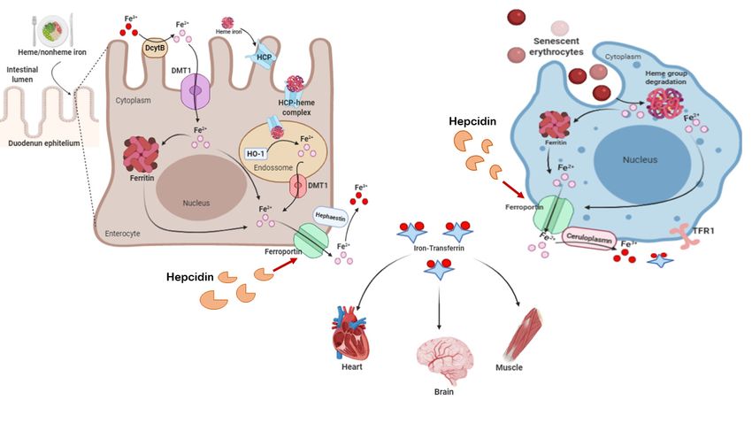
Int. J. Mol. Sci. 2020, 21, 8765                                                                             6 of 27

      converted to GSH disulfide (GSSG). GSSG is recycled to GSH by GSH reductase (GR), a reaction that uses
      reduced nicotinamide adenine dinucleotide phosphate (NADPH) as a cofactor [69]. GSH S-transferase
      (GST) forms GSH (GS-R) adducts from organic molecules (R) and GSH, which together with GSH and
      GSSG are exported from the cell by ABC transporters, mainly ABC-1 and ABC-G2 [70,71]. Extracellular
      GSH is metabolized by the γ-glutamyl transferase (GGT) ectoenzyme, which transfers the γ-glutamyl
      residue to different acceptor amino acids, leading to the formation of a dipeptide containing γ-glutamyl
      and the cysteine glycine dipeptide, which is cleaved by extracellular dipeptides to generate cysteine
      and glycine that can be taken up by cells, starting the glutathione biosynthesis cycle [69].

      Recently, Doll et al. [79] described an in vitro model, a parallel pathway that included
FSP1-CoQ10-NADPH, which cooperates with GPx4 and the glutathione system to suppress lipid
peroxidation of phospholipids. Ferroptosis suppressor protein 1 (FSP1) provides protection against
ferroptosis induced by the deletion of the GPx4 enzyme via RSL3. This effect is mediated by coenzyme
Q10 (CoQ10). The reduced form of the enzyme, ubiquinol, captures lipid peroxyl radicals that mediate
lipid peroxidation, while FSP1 catalyzes the regeneration of CoQ10 using NADPH as a cofactor. Moreover,
the authors described that the antiferroptotic function of FSP1 is independent of cellular glutathione
concentration, GPx4 activity, ACSL4 expression and oxidizable fatty acid content [79]. Coenzyme Q10
is an endogenous lipophilic antioxidant produced in the mevalonate pathway, as well as a part of the
mitochondrial respiratory chain, and from the metabolism of fatty acid and pyrimidine [80,81]. Indeed,
the homologous proteins MDM2 and MDMX, negative regulators of the tumor suppressor p53, promote
ferroptosis by regulating lipid peroxidation by altering PPARα activity. MDM2–MDMX complex inhibition
increased the levels of both FSP1 proteins and coenzyme Q10 [82].

2.3. Iron and Ferroptosis
     In the human body, iron metabolism is regulated by means of a perfectly adjusted balance between
plasma proteins. They are associated with the transport, absorption and recycling of iron, in order
to avoid the accumulation of iron, which is highly harmful and reactive in tissues. Figure 3 shows
several aspects of human iron metabolism. Biochemically, iron is capable of accepting and donating
electrons, interconverting between the ferric (Fe3+ ) and ferrous (Fe2+ ) forms, which are both found in
the human body. The Fe3+ /Fe2+ redox potential participates in a large number of protein complexes,
especially those that involve oxygen reduction for adenosine triphosphate (ATP) synthesis and the
reduction of DNA precursors. Iron is also a necessary component in the formation of molecules that
bind and transport oxygen (hemoglobin and myoglobin) and for the activities of cytochrome enzymes,
as well as in many enzymes that perform the redox process, functioning as electron carriers [83–85].
     Physiologically, iron that will be distributed to tissues needs to bind to transferrin (apotransferrin),
giving rise to holotransferrin [87]. The distribution of iron to the tissues occurs through the endocytosis
of holotransferrin, mediated by binding to transferrin receptor type 1 (TFR1) and type 2 (TFR2) [88].
After endocytosis, the holotransferrin–TFR1 complex is mobilized to the endosomes. In the acid
environment of the endosome, Fe3+ is released from TF and converted to Fe2+ by oxidation-reduction,
by six-transmembrane epithelial antigen of the prostate 3 (STEAP3) and then exported into cytosol by
divalent metal transporter 1 (DMT-1). Iron can be stored in ferritin/hemosiderin or remain labile [89].
     The labile iron pool (LIP), composed mostly of Fe2+ , is a pool of chelable iron and active redox
present in the cell; it can be present in mitochondria, lysosomes, cytosol and in the nucleus [90].
The concentration of LIP is essentially regulated by the absorption, use, distribution and export of iron
in the cell and in the body. Labile iron has high chemical reactivity and exhibits high cytotoxic potential.
Cytotoxicity is associated with the fact that labile iron catalyzes the formation of hydroxyl radicals
(OH·) derived from hydrogen peroxide (H2 O2 ) through the Fenton reaction (Figure 1). Moreover, H2 O2
has a lower capacity to react with molecules, while OH· generated from the iron-dependent Fenton
reaction has high reactivity with biological molecules, such as proteins and DNA, and generates lipid
(hydro) peroxidation in ferroptosis [12–17].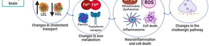
Int. J. Mol. Sci. 2020, 21, 8765                                                                                                              7 of 27

        Indeed, glutathione has a high affinity with Fe2+ and the major component of LIP in cytosol is
presented as the glutathione-Fe2+ conjugates [90]. This is important because the decrease in intracellular
glutathione increases the concentration of Fe2+ , facilitating the Fenton reaction [90–93]. The storage of
labile iron in ferritin serves to prevent its high reactivity, avoiding the generation of reactive species [94].
The ferritin structure is formed by 24 subunit-composed chains both light (L) and heavy (H) with a
spherical “shell” shape, which accommodates about 4500 iron atoms. H-ferritin contains a ferroxidase,
which oxidizes Fe2+ to Fe3+ , to store iron inside the nucleus. When necessary, iron stocks are mobilized
 Int. J.exported
and      Mol. Sci. 2020, 21, x FOR PEER
                     by ferroportin     REVIEW
                                      (FPN). This process is downregulated by hepcidin [83–85].           7 of 27

      Figure 3. Human Iron metabolism. Iron concentration in the body is maintained through diet and the
      recycling
        Figure 3.ofHumansenescentIronerythrocytes.
                                       metabolism.The     Irondaily  diet provides
                                                               concentration              approximately
                                                                                     in the                 1–2 mg through
                                                                                              body is maintained       of iron. Enterocytes,
                                                                                                                                  diet and the
      present
        recycling in the
                       of duodenum           and in the proximal
                            senescent erythrocytes.            The daily portiondietofprovides
                                                                                         the jejunum,     can absorb both
                                                                                                      approximately       1–2 ferrous
                                                                                                                                 mg of ironiron.
      (heme     iron)   and    ferric iron    (non-heme).      However,       it  is necessary     to reduce
        Enterocytes, present in the duodenum and in the proximal portion of the jejunum, can absorb both       ferric  iron  to ferrous   iron,
      by   apicaliron
        ferrous     ferric   reductase
                          (heme    iron) enzymes,
                                            and ferricsuchiron as enzyme duodenal
                                                                (non-heme).        However,   cytochrome    b (Dcytb),
                                                                                                 it is necessary           for absorption
                                                                                                                    to reduce    ferric irontoto
      occur.
        ferrous Then,
                   iron,iron    is transported
                           by apical                 by the enzymes,
                                        ferric reductase      divalent metalsuch as  type   transporter-1
                                                                                        enzyme      duodenal (DMT-1)     and stored
                                                                                                                cytochrome              inside
                                                                                                                                b (Dcytb),   for
      the   cell [86].toFerrous
        absorption                   iron from
                            occur. Then,       ironthe    diet is internalized
                                                     is transported                   by the heme-1
                                                                         by the divalent                 carrier
                                                                                                metal type         protein (HCP)
                                                                                                              transporter-1           in cells,
                                                                                                                                (DMT-1)     and
      where
        storeditinside
                    is stored    as hemosiderin
                            the cell   [86]. Ferrousand/or        ferritin,
                                                           iron from           to prevent
                                                                         the diet               the Fenton
                                                                                      is internalized         reaction.
                                                                                                          by the   heme-1Physiologically,
                                                                                                                              carrier protein
      iron
        (HCP) in cells, where it is stored as hemosiderin and/or ferritin, to prevent the when
             stores   are   mobilized      from    intracellular   to the    extracellular      by  ferroportin   (FPN)     Fenton thereaction.
                                                                                                                                        serum
      iron   is low.   Iron   released    in  its ferrous   state is oxidized       to ferric  iron  and  binds
        Physiologically, iron stores are mobilized from intracellular to the extracellular by ferroportin (FPN)  to  serum    apotransferrin
      to  be transported
        when     the serum iron  through
                                       is low.theIron
                                                    body,   givingin
                                                         released    rise
                                                                        its to   holotransferrin.
                                                                             ferrous                    This oxidation
                                                                                         state is oxidized                  reaction
                                                                                                               to ferric iron           occursto
                                                                                                                                 and binds
      through      the   action   of oxidase      enzymes:     hephestine       present     in  enterocytes,
        serum apotransferrin to be transported through the body, giving rise to holotransferrin. This          ceruloplasmin       present   in
      hepatocytes        macrophages.          The   distribution    of  iron    to  the  tissues   occurs
        oxidation reaction occurs through the action of oxidase enzymes: hephestine present in enterocytes, through     the  endocytosis     of
      holotransferrin,
        ceruloplasmin present  mediated  in by    the binding
                                              hepatocytes         to transferrin
                                                               macrophages.          Thereceptors    type 1of(TFR1)
                                                                                           distribution        iron toandthetype    2 (TFR2).
                                                                                                                               tissues   occurs
      Ferroportin      mediates     the  efflux   of  iron  within  cells,  maintaining        systemic
        through the endocytosis of holotransferrin, mediated by the binding to transferrin receptors     iron  homeostasis.      This  process
                                                                                                                                         type 1
      is(TFR1)
          negatively      regulated     by   hepcidin,     which   promotes        ferroportin    endocytosis
                  and type 2 (TFR2). Ferroportin mediates the efflux of iron within cells, maintaining systemic   and   then  proteolysis    in
      lysosomes by induced ubiquitination [83]. The recycling of iron by macrophages occurs through the
        iron homeostasis. This process is negatively regulated by hepcidin, which promotes ferroportin
      phagocytosis of senescent erythrocytes and hemoglobin and the heme group of intravascular hemolysis.
        endocytosis and then proteolysis in lysosomes by induced ubiquitination [83]. The recycling of iron
      Once internalized in the macrophage, the heme group releases ferrous iron through the activity of the
        by macrophages occurs through the phagocytosis of senescent erythrocytes and hemoglobin and the
      enzyme heme oxygenase, which can be exported to the extracellular medium by ferroportin or stored
        heme group of intravascular hemolysis. Once internalized in the macrophage, the heme group
      as ferritin [83,84].
        releases ferrous iron through the activity of the enzyme heme oxygenase, which can be exported to
        the extracellular medium by ferroportin or stored as ferritin [83,84].
      The intracellular iron content is regulated by the iron regulatory proteins (IRP1 and IRP2) and
the iron-responsive element (IRE) [95,96]. IRPs can bind to RNA stem-loops containing an IRE in the
       Physiologically, iron that will be distributed to tissues needs to bind to transferrin
untranslated region (UTR), affecting the translation of target Mrna: 30 UTR of H-ferritin mRNA and in
 (apotransferrin), giving rise to holotransferrin [87]. The distribution of iron to the tissues occurs
the 50 UTR of TFR1 mRNA. In response to cellular iron demand IRE/IRP interaction promotes TFR1
 through the endocytosis of holotransferrin, mediated by binding to transferrin receptor type 1 (TFR1)
 and type 2 (TFR2) [88]. After endocytosis, the holotransferrin–TFR1 complex is mobilized to the
 endosomes. In the acid environment of the endosome, Fe3+ is released from TF and converted to Fe2+
 by oxidation-reduction, by six-transmembrane epithelial antigen of the prostate 3 (STEAP3) and then
 exported into cytosol by divalent metal transporter 1 (DMT-1). Iron can be stored in
 ferritin/hemosiderin or remain labile [89].
Int. J. Mol. Sci. 2020, 21, 8765 8 of 27
mRNA stability and inhibits H-ferritin translation, thus modulating cellular iron uptake and storage.
Overexpression of both TF and TFR1 sensitizes cells to ferroptosis by enhancing iron uptake; on the
other hand, silencing TFR1 can inhibit erastin-induced ferroptosis [16,97–99]. In addition, anti-TfR1
antibodies identify tumor ferroptotic cells from different tissues [100].
Autophagy in fibroblasts leads to erastin-induced ferroptosis through the degradation of ferritin
and induction of TfR1 expression [101]. On the other hand, cellular senescence has been associated
with intracellular iron accumulation and impaired ferritinophagy [102]. Ferritinophagy induces
ferroptosis by increasing the nuclear receptor coactivator 4 (NCOA4), followed by an increase in ferritin
degradation in the phagolysosome and release of Fe2+ (labile iron pool) in the cytoplasm. In fact,
some authors consider ferroptosis to be a type of autophagy [103–107].
Another molecule involved in ferroptosis is the sigma-1 receptor (S1R), which protects
hepatocellular carcinoma cells against ferroptosis. S1R regulates ROS accumulation via Nrf2.
Knockdown of S1R blocks the expression of GPx4 and HO-1. Moreover, knockdown of S1R significantly
increases Fe2+ levels and MDA production in HCC cells treated with erastin and sorafenib, as well as
the upregulation of H-ferritin chain and TRF1 [108]. In addition, it has been shown that heat shock
protein β-1 (HSPβ1) is a negative regulator of ferroptotic cancer cell death, and erastin stimulates heat
shock factor 1 (HSF1)-dependent HSPβ1. Knockdown of HSF1 and HSPβ1 enhances erastin-induced
ferroptosis, whereas heat shock pretreatment and overexpression of HSPβ1 inhibits erastin-induced
ferroptosis by protein kinase C. Moreover, the increase in cellular iron in HSPβ1 knockdown cells
has been associated with increased expression of TFR1 and a mild decrease in the expression of the
H-ferritin chain [109]. HSPA5, an endoplasmic reticulum (ER)-sessile chaperone, was shown to bind
and stabilize GPx4, thus indirectly counteracting lipid peroxidation in ferroptosis [97].
3. Ferroptosis in Neurodegenerative Diseases
Iron is vital to the physiology of all human tissues. However, under certain conditions it can
be harmful, especially for the brain. Although the cellular metabolism of the CNS requires iron as
a redox metal for energy generation, mainly the production of ATP, nervous tissue is vulnerable to
oxidative damage generated by excess iron and decreased antioxidant systems [110–113]. The explicit
identification of ferroptosis in vivo is hampered by the lack of specific biomarkers, due to several
factors that may be associated with the ferroptotic process. However, there is considerable evidence
that implicates ferroptosis in the pathophysiology of neurodegeneration. Ferroptosis involves the
accumulation of brain iron, glutathione depletion and lipid peroxidation simultaneously, which triggers
a cascade of events including activation of inflammation, neurotransmitter oxidation, neuronal
communication failure, myelin sheath degeneration, astrocyte dysregulation, dementia and cell death.
Iron or free iron overload can initiate lipid peroxidation in neurons, astrocytes, oligodendrocytes,
microglia and Schwann cells. In addition, the low activity of GPx4 and the glutathione system have
been shown to be associated with ferroptosis in motor neurodegeneration [114–117].
Recently, it has been proposed that the modulation of ferroptosis may be beneficial for
neurodegenerative diseases and that inhibition of ferroptosis by GPx4 could provide protective
mechanisms against neurodegeneration [118]. First, it was demonstrated that the non-oxidative
form of dopamine is a strong inhibitor of ferroptotic cell death. Dopamine reduced erastin-induced
ferrous iron accumulation, glutathione depletion, and malondialdehyde production. Moreover,
dopamine increased the stability of GPx4 [119]. The GPx4 enzyme is essential for the survival of
parvalbumin-positive interneurons and prevention of seizures, as well as protection against ferroptosis
in animal models [42,120]. Next, in a study with PC12 cell line (a model system for neurobiological
and neurochemical studies), cell death was induced by tert-butylhydroperoxide (t-BHP), a widespread
inducer of oxidative stress; it was observed that t-BHP increased the generation of lipid ROS, decreased
the expression of GPx4 and the ratio of GSH/GSSG. All these effects could be reversed by the ferroptosis
inhibitor, ferrostatin-1 and deferoxamine, iron chelator. In addition, JNK1/2 and ERK1/2 were activated
upstream from the ferroptosis and mitochondrial dysfunction [121].Int. J. Mol. Sci. 2020, 21, 8765 9 of 27
In addition, in C57BL/6 J male mice treated with arsenite for 6 months, it was observed that
arsenite induced ferroptotic cell death in neurons by the accumulation of reactive oxygen species
and lipid peroxidation products, disruption of Fe2+ homeostasis, depletion of glutathione and
adenosine triphosphate, inhibition of system xc− , activation of mitogen-activated protein kinases and
mitochondrial voltage-dependent anion channel pathways, and upregulation of endoplasmic reticulum
stress [122]. This is an important issue because arsenite (inorganic arsenic) has been associated
with neural loss and Alzheimer’s and Parkinson’s diseases as well as amyotrophic lateral sclerosis
(ALS) [123–130]. An in vitro study showed that exposure to paraquat and maneb induced ferroptosis
in dopaminergic SHSY5Y cells, associated with the activation of NADPH oxidase. The activation of
NADPH oxidase contributed to the dopaminergic neurodegeneration associated with lipid peroxidation
and neuroinflammation [131].
In a multiple sclerosis model and in an experimental autoimmune encephalomyelitis (EAE) animal
model [124], it has been observed that mRNA expression of the cytoplasmic, mitochondrial and nuclear
GPx4 enzyme decreased in multiple sclerosis gray matter and in the spinal cord of EAE. Neuronal
GPx4 was lower in EAE spinal cords than in controls. Moreover, γ-glutamylcysteine ligase and
cysteine/glutamate antiporter were diminished in EAE, which is associated with high accumulation
of lipid peroxidation products and the reduction in the proportion of the docosahexaenoic acid in
non-myelin lipids. These results, together with the presence of abnormal neuronal mitochondrial
morphology, which includes an irregular matrix, ruptured external membrane and reduced/absent
ridges, are consistent with the occurrence of ferroptotic damage in inflammatory demyelinating
disorders [132].
In fact, iron can bind to IRPs, leading to the dissociation of IRPs from the IRE and altered translation
of the target transcripts. Recently, an IRE was found in the 50 -UTR of the amyloid precursor protein
(APP) and α-synuclein transcripts (α-Syn). The levels of α-Syn, APP and amyloid β (Aβ) peptide,
as well as protein aggregation, can be negatively regulated by IRPs, but are regulated positively in the
presence of iron accumulation. Therefore, it has been suggested that the inhibition of the IRE-modulated
expression of APP and α-Syn or iron chelation in patient brains has therapeutic significance for human
neurodegenerative diseases [133].
3.1. Ferroptosis in Alzheimer’s Disease
Alzheimer’s disease (AD) is considered a neurodegenerative disease associated with multiple
brain complications. It was initially described by the German Alois Alzheimer in 1907 [134,135].
AD is characterized by progressive disorder in the cortical and hippocampal neuronal areas which
leads to both loss of neuronal function and cell death, and is the most common type of dementia
(Figure 4). The hallmark of AD is the histopathological presence of an extracellular β-amyloid (Aβ)
deposition in senile plaques (SPs) and intracellular neurofibrillary tangles (NFTs) formed from the
hyperphosphorylation of the tau protein. Neurocognitive decline is associated with synapse decrease
and neurotransmitter oxidation. These changes are due to the increase in oxidative stress, mainly an
increase in ROS and intra- and extracellular hydrogen peroxides [136–138]. Moreover, genetic changes
include alterations in amyloid precursor protein (APP), apolipoprotein E (APOE), presenilin 1 (PSEN1)
and presenilin 2 (PSEN 2) genes [139].deposition in senile plaques (SPs) and intracellular neurofibrillary tangles (NFTs) formed from the
hyperphosphorylation of the tau protein. Neurocognitive decline is associated with synapse decrease
and neurotransmitter oxidation. These changes are due to the increase in oxidative stress, mainly an
increase in ROS and intra- and extracellular hydrogen peroxides [136–138]. Moreover, genetic
changes
Int. include
J. Mol. Sci. alterations
2020, 21, 8765 in amyloid precursor protein (APP), apolipoprotein E (APOE), presenilin
10 of 27
1 (PSEN1) and presenilin 2 (PSEN 2) genes [139].
Figure4.4.Alzheimer’s
Figure Alzheimer’s disease. The development
disease. The developmentand andprogression
progressionofofAlzheimer’s
Alzheimer’s disease
disease (AD)
(AD) lead
lead to to
atrophy,loss
atrophy, lossand
anddysfunction
dysfunctionof of both
both neurons
neurons andand glial
glial cells.
cells.AD
ADbegins
beginsininthe
thedorsal
dorsalraphe
raphe nucleus
nucleus
withsubsequent
with subsequentprogression
progressionto to the
the cortex, which
which is isthe
thecenter
centerofofinformation
informationprocessing
processing andandmemory
memory
storage.The
storage. Thefactors
factorsthat
that promote
promote the development
developmentof ofADADarearestill
stillunknown.
unknown.However,
However, it seems
it seems that
that
theintracellular
the intracellularaccumulation
accumulation in neuronsneurons of of the
thephosphorylated
phosphorylatedTau Tauprotein
protein(neurofibrillary
(neurofibrillary tangle)
tangle)
andthe
and theformation
formationof ofamyloid-B
amyloid-B plaqueplaque (senile
(senile plaque)
plaque)in inthe
theextracellular
extracellularenvironment
environment andandbrain
brain
tissue both lead to neuron loss and dysfunction. In addition, the formation of
tissue both lead to neuron loss and dysfunction. In addition, the formation of neurofibrillary tangleneurofibrillary tangle
andsenile
and senileplaque
plaquealters
alters the
the functions
functions of of glial
glial cells,
cells, such
suchas asoligodendrocytes
oligodendrocytes(responsible
(responsible forfor
thethe
myelination of neurons), microglia cells (phagocytic cells) and astrocytes (responsible
myelination of neurons), microglia cells (phagocytic cells) and astrocytes (responsible for the absorption for the
absorption
and exchangeand exchangebetween
of nutrients of nutrients
neurons between
and blood neurons andDysregulation
vessels). blood vessels). Dysregulation
of cholesterol of
transport
cholesterol
and transportinand
iron metabolism theiron metabolism
central nervous in the central
system nervous
contributes to system contributes
poor prognosis to poor prognosis
of Alzheimer’s disease.
of these
All Alzheimer’s
associateddisease. Alllead
factors these to associated
an increasefactors lead to an increase
in neuroinflammation andinoxidative
neuroinflammation and
stress associated
oxidative stress associated with mitochondrial dysfunction, compromising
with mitochondrial dysfunction, compromising the production of ATP, altering the concentration of the production of ATP,
altering the concentration of neurotransmitters in the synaptic cleft, finally promoting cell death.
neurotransmitters in the synaptic cleft, finally promoting cell death.
Evidence of
Evidence of the
the association
associationbetween
betweenthe theaccumulation
accumulation of iron in the
of iron in cerebral cortexcortex
the cerebral and theand
development of Alzheimer’s disease emerged in the early 1960s [140,141].
the development of Alzheimer’s disease emerged in the early 1960s [140,141]. Since Since then, several studies
then,
have demonstrated
several studies havethedemonstrated
direct association
the between free iron, oxidative
direct association between stress,
freelipid
iron,peroxidation
oxidative and
stress,
cell death of neurons, usually associated with apoptosis and/or necrosis, due to increased
lipid peroxidation and cell death of neurons, usually associated with apoptosis and/or necrosis,
neuroinflammation. The iron dyshomeostasis is associated with ROS production and
due to increased neuroinflammation. The iron dyshomeostasis is associated with ROS production and
neurodegeneration in AD [138]. Furthermore, aging and changes in iron metabolism are associated
neurodegeneration in AD [138]. Furthermore, aging and changes in iron metabolism are associated
with the development of Aβ plaques and NFTs. Svobodová et al. [142] demonstrated in an APP/PS1
with the development of Aβ plaques and NFTs. Svobodová et al. [142] demonstrated in an APP/PS1
transgenic mice model that free iron and ferritin accumulation follows amyloid plaque formation in
transgenic mice model that free iron and ferritin accumulation follows amyloid plaque formation in
the cerebral cortex area. In fact, iron deposition has been involved in the misfolding process of the Aβ
plaques and NFTs [143].
Iron is related to the development of Tau protein and, consequently, NFTs. In fact, iron is present
through the induction and regulation of tau phosphorylation [143,144]. The association of NFT with
neurodegenerative dysfunctions is termed tauopathy [145–147]. The oxidation process slows down or
excludes the regular action of the Aβ and tau protein [148]. In animal models of tauopathies, increased
iron associated with aging and neurodegeneration has been observed [149]. Indeed, animals withInt. J. Mol. Sci. 2020, 21, 8765 11 of 27
tauopathies treated with the iron chelator deferiprone showed a trend toward improved cognitive
function associated with the decrease in brain iron levels and sarkosyl-insoluble tau [150].
APP is a type 1 transmembrane protein and its function in heathy individuals appears to be associated
with the development of synaptic activity [145]. Proteolytic cleavage of the β-amyloid precursor protein
(APP) to form the β-amyloid peptide (Aβ) is related to the pathogenesis of AD because APP mutations that
influence this process induce familial AD or decrease the risk of AD [145]. The amyloid cascade hypothesis
states that the agglomeration and production of Aβ plaques in the brain would occur, resulting in cell
death. Presenilins 1 (PSEN1) and presenilins 2 (PSEN2) precisely cleave the APP and other proteins as they
are part of the catalytic protease compounds [151]. Acetylcholinesterase participates in the aggregation
of Aβ plaques [152]. Moreover, the Aβ plaques in the presence of free iron participate efficiently in the
generation of ROS resulting in increased lipid peroxidation, protein oxidation and DNA damage [153].
Deferiprone derivatives act as acetylcholinesterase inhibitors and in iron chelation [154].
The proteolytic cleavage in APP occurs by enzymatic complexes involving α-secretase or
β-secretase and γ-secretase. The proteolytic cleavage in APP by β-secretase produces a neurotoxic
40 to 42 amino acid amyloid [155]. Tsatsanis et al. [156] showed that APP promotes neuronal iron efflux
by stabilizing the cell-surface presentation of ferroportin, and that β-cleveage of APP depletes surface
ferroportin, leading to intracellular iron retention independently on the generation of Aβ. Furthermore,
these findings indicate how β-secretase’s processing of APP might indirectly promote ferroptosis.
Iron overload alters the neuronal sAPPα distribution and directly inhibits β-secretase activity [157].
Cortical iron has been strongly associated with the rate of cognitive decline [158]. Iron in the brain
increases lipid peroxidation, oxidative stress, and neuroinflammation due to the depletion of neuronal
antioxidant systems—mainly the glutathione system [143]. In addition, increased hepcidin expression
in APP/PS1 mice astrocytes improves cognitive decline and partially decreases the formation of Aβ
plaques in the cortex and hippocampus. Indeed, decreased iron levels in neurons led to a reduction
in oxidative stress (induced by iron accumulation), decrease in neuroinflammation and decreased
neuronal cell death in the cortex and the hippocampus. [159]. As mentioned before, the hepcidin
peptide binds ferroportin, which is followed by cell internalization and further degradation [160].
In order to investigate whether neurons of the cerebral cortex and hippocampus severely affected
in patients with AD may be vulnerable to ferroptosis, Hambright et al. [161] have shown in GPx4BIKO
mice (a mice model with a conditional deletion in neurons of the forebrain of GPx4) that tamoxifen led
to the deletion of GPx4 mainly in neurons of the forebrain. GPx4BIKO mice exhibited significant deficits
in spatial learning and memory function, as well as hippocampal neurodegeneration. These results
were associated with ferroptosis markers, such as increased lipid peroxidation, ERK activation and
neuroinflammation. In addition, GPx4BIKO mice fed a vitamin E-deficient diet had an accelerated
rate of hippocampal neurodegeneration and behavioral dysfunction. On the other hand, treatment
with Liproxstatin-1, a ferroptosis inhibitor, improved neurodegeneration in these mice. Moreover,
in an in vitro model, iron increased nerve cell death in conditions where GSH levels were reduced,
by decreasing the activity of glutamate cysteine ligase [162].
The HT22 cell line has high concentrations of extracellular glutamate, which inhibit the glutamate-cystine
antiport, leading to the depletion of intracellular GSH and resulting in excessive ROS production. In a
study with these cells, Hirata et al. [163] found that an oxindole compound, GIF-0726-r, prevented cell death
induced by oxidative stress, including oxytosis induced by glutamate and ferroptosis induced by erastin.
Moreover, an excess of extracellular glutamate associated with high levels of extracellular iron cause the
overactivation of glutamate receptors. As a consequence, there was an increase in iron uptake in neurons
and astrocytes, increasing the production of membrane peroxides. Glutamate-induced neuronal death can
be mitigated by iron chelating compounds or free radical scavenging molecules. Ferroptosis is induced by
reactive oxygen species in the excitotoxicity of glutamate [110,164,165]. In addition, the sterubin compound
maintained GSH levels in HT22 cell lines treated with erastin and RSL3, suggesting protection against
ferroptosis [166]. 7-O-esters of taxifolin 1 and 2 were described as having neuroprotective action against
ferroptosis induced by RSL3 in HT22 cells [167].Int. J. Mol. Sci. 2020, 21, 8765 12 of 27
Chalcones 14a–c were shown to inhibit β-amyloid aggregation, and in addition, protect neural
cells against toxicity induced by Aβ aggregation and from erastin and RSL3-induced ferroptosis in
human neuroblastoma SH-SY5Y cells [160]. The authors suggested that the inhibition of toxicity
induced by Aβ plaques’ aggregation and of ferroptosis occurs due to the presence of hydroxyl groups
in the chalcone derivatives. Chalcone 14a-c can react with lipid peroxyl radicals by transferring the
hydrogen (H) atom, thus inhibiting lipid peroxidation [168].
After treatment with high dietary iron (HDI), WT (wild type) mice and the APP/PS1 double
Tg mouse model of ADon (HDI) showed upregulation of divalent metal transporter 1 (DMT1) and
ferroportin expression, and downregulation of TFR1 expression, with fewer NeuN-positive neurons in
both animal models. Moreover, the iron-induced neuron loss may involve increased ROS production
and oxidative mitochondria dysfunction, decreased DNA repair, and exacerbated apoptosis and
autophagy [169]. Using X-ray spectromicroscopy and electron microscopy it was found that the
coaggregation of Aβ and ferritin resulted in the conversion of the ferritin inert ferric core into more
reactive low oxidation states [170].
Ates et al. [171] showed in an animal model that inhibition of fatty acid synthase (FASN) by CMS121
decreased lipid peroxidation. CMS121 treatment reduced the levels of 15LOX2 in the hippocampus
compared to those of untreated WT mice. Relative levels of endocannabinoids, fatty acids, and PUFAs
were significantly higher in untreated AD mice as compared to CMS121-treated AD mice, suggesting
that other enzymes may be involved in the process of ferroptosis in Alzheimer’s disease.
It is important to highlight the heterogeneity of Alzheimer’s disease and the involvement of
multiple metabolic pathways which contribute to the poor prognosis of this disease (Figure 4). In fact,
multiple patterns of cell death are involved in the neurodegeneration process, such as apoptosis, necrosis,
and autophagy associated with disturbed BBB (brain blood barrier) permeability. In vitro experiments are
the main evidence of ferroptosis in human neurodegenerative processes. The identification of ferroptosis
in in vivo models of Alzheimer’s disease is difficult since specific markers for ferroptotic cells, such as
specific antibodies, are not available. In addition, other metal ions, such as copper, can also regulate
ferroptosis and lipid peroxidation [172,173]. Taking all under consideration, we still do not know whether
ferroptosis is the cause or consequence of neurodegeneration processes such as Alzheimer’s disease.
3.2. Ferroptosis in Parkinson’s Disease
Parkinson’s disease (PD) is one of the most common and best-known diseases of the nervous system,
affecting roughly 0.1–0.2% of the general population and 1% of the population above 60 years [174]. PD is
characterized as a slowly progressing neurodegenerative ailment with motor and non-motor clinical
manifestations, due to an intense decrease in dopamine production [175]. Classic hallmarks in PD are
still related to the motor manifestation such as bradykinesia, resting tremor and rigidity [176]. However,
non-motor symptoms associated with PD have recently gained more attention due to their relevance and
impact on the patient’s quality of life. Non-motor symptoms of PD include anosmia, constipation, pain,
anxiety, depression, psychosis and cognitive disorders that can progress to dementia [177–179].
The pathophysiological characteristics of PD include the slow and progressive degeneration
of dopaminergic neurons in the pars compacta of the substantia nigra (SNpc), which is associated
with a systematic and progressive accumulation of iron, leading to striatum dopamine depletion,
disappearance of neuromelanin and the appearance of intracellular Lewy bodies having aggregated
α-synuclein as the main component [180,181]. During PD progression there is an increase in oxidative
stress, lipid peroxidation, and mitochondrial dysfunction associated with the depletion of antioxidant
enzymes in the glutathione systems. All of these associated factors lead to neuronal death and the
functional disability of the organism (Figure 5). Currently, the pharmacological treatment of PD aims
to increase dopamine levels in the synaptic cleft. Levodopa is the drug of choice, being associated with
dopamine agonists, dopamine metabolism inhibitors and decarboxylase inhibitors. Treatment is stable
for a period of 5–6 years. Then, however, the disease progresses with marked neurodegeneration and
development of dementia [182–184].You can also read