Lung Cancer (Non-Small Cell)
←
→
Page content transcription
If your browser does not render page correctly, please read the page content below

Lung Cancer (Non-Small Cell)
What is cancer?
The body is made up of trillions of living cells. Normal body cells grow, divide into new
cells, and die in an orderly fashion. During the early years of a person’s life, normal cells
divide faster to allow the person to grow. After the person becomes an adult, most cells
divide only to replace worn-out or dying cells or to repair injuries.
Cancer begins when cells in a part of the body start to grow out of control. There are
many kinds of cancer, but they all start because of out-of-control growth of abnormal
cells.
Cancer cell growth is different from normal cell growth. Instead of dying, cancer cells
continue to grow and form new, abnormal cells. Cancer cells can also invade (grow into)
other tissues, something that normal cells cannot do. Growing out of control and invading
other tissues is what makes a cell a cancer cell.
Cells become cancer cells because of damage to DNA. DNA is in every cell and directs
all its actions. In a normal cell, when DNA gets damaged the cell either repairs the
damage or the cell dies. In cancer cells, the damaged DNA is not repaired, but the cell
doesn’t die like it should. Instead, this cell goes on making new cells that the body does
not need. These new cells will all have the same damaged DNA as the first cell does.
People can inherit damaged DNA, but most DNA damage is caused by mistakes that
happen while the normal cell is reproducing or by something in our environment.
Sometimes the cause of the DNA damage is something obvious, like cigarette smoking.
But often no clear cause is found.
In most cases the cancer cells form a tumor. Some cancers, like leukemia, rarely form
tumors. Instead, these cancer cells involve the blood and blood-forming organs and
circulate through other tissues where they grow.
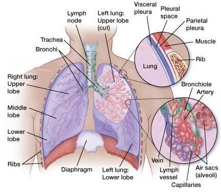
Cancer cells often travel to other parts of the body, where they begin to grow and form
new tumors that replace normal tissue. This process is called metastasis. It happens when
the cancer cells get into the bloodstream or lymph vessels of our body.
No matter where a cancer may spread, it is always named (and treated) based on the place
where it started. For example, breast cancer that has spread to the liver is still breast
cancer, not liver cancer. Likewise, prostate cancer that has spread to the bone is still
prostate cancer, not bone cancer.
Different types of cancer can behave very differently. For example, lung cancer and
breast cancer are very different diseases. They grow at different rates and respond to
different treatments. That is why people with cancer need treatment that is aimed at their
particular kind of cancer.
Not all tumors are cancerous. Tumors that aren’t cancer are called benign. Benign tumors
can cause problems – they can grow very large and press on healthy organs and tissues.
But they cannot grow into (invade) other tissues. Because they can’t invade, they also
can’t spread to other parts of the body (metastasize). These tumors are almost never life
threatening.

What is non-small cell lung cancer?
Note: This document is specifically for the non-small cell type of lung cancer. The
treatment for the 2 main types of lung cancer (small cell and non-small cell) is very
different, so much of the information for one type will not apply to the other type. If you
are not sure which type of lung cancer you have, ask your doctor so you can be sure the
information you receive is correct.
Lung cancer is a cancer that starts in the lungs. To understand lung cancer, it helps to
know about the normal structure and function of the lungs.

The lungs
Your lungs are 2 sponge-like organs found in your chest. Your right lung is divided into 3
sections, called lobes. Your left lung has 2 lobes. The left lung is smaller because the
heart takes up more room on that side of the body.
When you breathe in, air enters through your mouth or nose and goes into your lungs
through the trachea (windpipe). The trachea divides into tubes called the bronchi
(singular, bronchus), which enter the lungs and divide into smaller bronchi. These divide
to form smaller branches called bronchioles. At the end of the bronchioles are tiny air
sacs known as alveoli.
Many tiny blood vessels run through the alveoli. They absorb oxygen from the inhaled air
into your bloodstream and pass carbon dioxide from the body into the alveoli. This is

expelled from the body when you exhale. Taking in oxygen and getting rid of carbon
dioxide are your lungs’ main functions.

A thin lining layer called the pleura surrounds the lungs. The pleura protects your lungs
and helps them slide back and forth against the chest wall as they expand and contract
during breathing.
Below the lungs, a thin, dome-shaped muscle called the diaphragm separates the chest
from the abdomen. When you breathe, the diaphragm moves up and down, forcing air in
and out of the lungs.

Start and spread of lung cancer
Lung cancers can start in the cells lining the bronchi and parts of the lung such as the
bronchioles or alveoli.
Lung cancers are thought to start as areas of pre-cancerous changes in the lung. The first
changes in the genes (DNA) inside the lung cells may cause the cells to grow faster.
These cells may look a bit abnormal if seen under a microscope, but at this point they do
not form a mass or tumor. They cannot be seen on an x-ray and they do not cause
symptoms.
Over time, the abnormal cells may acquire other gene changes, which cause them to
progress to true cancer. As a cancer develops, the cancer cells may make chemicals that
cause new blood vessels to form nearby. These blood vessels nourish the cancer cells,
which can continue to grow and form a tumor large enough to be seen on imaging tests
such as x-rays.
At some point, cells from the cancer may break away from the original tumor and spread
(metastasize) to other parts of the body. Lung cancer is often a life-threatening disease
because it tends to spread in this way even before it can be detected on an imaging test
such as a chest x-ray.

The lymph (lymphatic) system
The lymph system is one of the ways in which lung cancers can spread. This system has
several parts:
 • Lymph nodes are small, bean-shaped collections of immune system cells (cells that
   fight infections) that are connected by lymphatic vessels.
 • Lymphatic vessels are like small veins, except that they carry a clear fluid called
   lymph (instead of blood) away from the lungs.
 • Lymph contains excess fluid and waste products from body tissues, as well as immune
   system cells.
Lung cancer cells can enter lymphatic vessels and begin to grow in lymph nodes around
the bronchi and in the mediastinum (the area between the 2 lungs). Once lung cancer cells
have reached the lymph nodes, they are more likely to have spread to other organs of the
body as well. The stage (extent) of the cancer and decisions about treatment are based in
part on whether or not the cancer has spread to the nearby lymph nodes in the
mediastinum. These topics are discussed later in the section “How is non-small cell lung
cancer staged?”

Types of lung cancer
There are 2 major types of lung cancer:
 • Small cell lung cancer (SCLC)
 • Non-small cell lung cancer (NSCLC)
(If a lung cancer has some cells with characteristics of SCLC and other cells with
characteristics of NSCLC it is called a combined small cell/non-small cell cancer. This is
uncommon.)
These 2 types of lung cancer are treated very differently. This document focuses on
non-small cell lung cancer. Small cell lung cancer is discussed in a separate document,
Lung Cancer (Small Cell).

Non-small cell lung cancer
About 85% to 90% of lung cancers are non-small cell lung cancer (NSCLC). There are 3
main subtypes of NSCLC. The cells in these subtypes differ in size, shape, and chemical
make-up when looked at under a microscope. But they are grouped together because the
approach to treatment and prognosis (outlook) are often very similar.
Squamous cell (epidermoid) carcinoma: About 25% to 30% of all lung cancers are
squamous cell carcinomas. These cancers start in early versions of squamous cells, which
are flat cells that line the inside of the airways in the lungs. They are often linked to a
history of smoking and tend to be found in the middle of the lungs, near a bronchus.
Adenocarcinoma: About 40% of lung cancers are adenocarcinomas. These cancers start
in early versions of the cells that would normally secrete substances such as mucus. This
type of lung cancer occurs mainly in current or former smokers, but it is also the most
common type of lung cancer seen in non-smokers. It is more common in women than in
men, and it is more likely to occur in younger people than other types of lung cancer.
Adenocarcinoma is usually found in outer parts of the lung. It tends to grow slower than
other types of lung cancer, and is more likely to be found before it has spread outside of
the lung.
People with a type of adenocarcinoma called adenocarcinoma in situ (previously called
bronchioloalveolar carcinoma) tend to have a better outlook (prognosis) than those with
other types of lung cancer.
Large cell (undifferentiated) carcinoma: This type of cancer accounts for about 10% to
15% of lung cancers. It can appear in any part of the lung. It tends to grow and spread
quickly, which can make it harder to treat. A subtype of large cell carcinoma, known as
large cell neuroendocrine carcinoma, is a fast-growing cancer that is very similar to
small cell lung cancer (see below).
Other subtypes: There are also a few other subtypes of non-small cell lung cancer, such
as adenosquamous carcinoma and sarcomatoid carcinoma. These are much less common.
Small cell lung cancer
About 10% to 15% of all lung cancers are small cell lung cancer (SCLC), named for the
size of the cancer cells when seen under a microscope. Other names for SCLC are oat
cell cancer, oat cell carcinoma, and small cell undifferentiated carcinoma. It is very rare
for someone who has never smoked to have small cell lung cancer.
SCLC often starts in the bronchi near the center of the chest, and it tends to spread widely
through the body fairly early in the course of the disease. This cancer is discussed in the
document called Lung Cancer (Small Cell).

Other types of lung cancer
Along with the 2 main types of lung cancer, other tumors can occur in the lungs.
Lung carcinoid tumors: Carcinoid tumors of the lung account for fewer than 5% of lung
tumors. Most are slow-growing tumors that are called typical carcinoid tumors. They are
generally cured by surgery. Some typical carcinoid tumors can spread, but they usually
have a better prognosis than small cell or non-small cell lung cancer. Less common are
atypical carcinoid tumors. The outlook for these tumors is somewhere in between typical
carcinoids and small cell lung cancer. For more information about typical and atypical
carcinoid tumors, see the separate document Lung Carcinoid Tumor.
Other lung tumors: Other types of lung cancer such as adenoid cystic carcinomas,
lymphomas, and sarcomas, as well as benign lung tumors such as hamartomas are rare.
These are treated differently from the more common lung cancers and are not discussed
in this document.
Cancers that spread to the lungs: Cancers that start in other organs (such as the breast,
pancreas, kidney, or skin) can sometimes spread (metastasize) to the lungs, but these are
not lung cancers. For example, cancer that starts in the breast and spreads to the lungs is
still breast cancer, not lung cancer. Treatment for metastatic cancer to the lungs is based
on where it started (the primary cancer site). For information on these primary cancers,
see our separate documents on each.

What are the key statistics about lung
cancer?
Most lung cancer statistics include both small cell and non-small cell lung cancers.
Lung cancer (both small cell and non-small cell) is the second most common cancer in
both men and women (not counting skin cancer). In men, prostate cancer is more
common, while in women breast cancer is more common. Lung cancer accounts for
about 13% of all new cancers.
The American Cancer Society’s estimates for lung cancer in the United States for 2015
are:
  • About 221,200 new cases of lung cancer (115,610 in men and 105,590 in women)
  • An estimated 158,040 deaths from lung cancer (86,380 in men and 71,660 among
    women).
Lung cancer accounts for about 27% of all cancer deaths and is by far the leading cause
of cancer death among both men and women. Each year, more people die of lung cancer
than of colon, breast, and prostate cancers combined.
Lung cancer mainly occurs in older people. About 2 out of 3 people diagnosed with lung
cancer are 65 or older; fewer than 2% of all cases are found in people younger than 45.
The average age at the time of diagnosis is about 70.
Overall, the chance that a man will develop lung cancer in his lifetime is about 1 in 13;
for a woman, the risk is about 1 in 16. These numbers include both smokers and non-
smokers. For smokers the risk is much higher, while for non-smokers the risk is lower.
Black men are about 20% more likely to develop lung cancer than white men. The rate is
about 10% lower in black women than in white women. Both black and white women
have lower rates than men, but the gap is closing. The lung cancer rate has been dropping
among men over the past 2 decades and has just recently begun to drop in women.
Statistics on survival in people with lung cancer vary depending on the stage (extent) of
the cancer when it is diagnosed. Survival statistics based on the stage of the cancer are
discussed in the section “Non-small cell lung cancer survival rates by stage.”
Despite the very serious prognosis (outlook) of lung cancer, some people with earlier
stage cancers are cured. More than 430,000 people alive today have been diagnosed with
lung cancer at some point.

What are the risk factors for non-small cell
lung cancer?
A risk factor is anything that affects a person’s chance of getting a disease such as cancer.
Different cancers have different risk factors. Some risk factors, like smoking, can be
changed. Others, like a person’s age or family history, can’t be changed.
But risk factors don’t tell us everything. Having a risk factor, or even several risk factors,
does not mean that you will get the disease. And some people who get the disease may
not have had any known risk factors. Even if a person with lung cancer has a risk factor,
it is often very hard to know how much that risk factor may have contributed to the
cancer.
Several risk factors can make you more likely to develop lung cancer. Tobacco smoke Smoking is by far the leading risk factor for lung cancer. In the early 20th century, lung cancer was much less common than some other types of cancer. But this changed once manufactured cigarettes became readily available and more people began smoking. At least 80% of lung cancer deaths are thought to result from smoking. The risk for lung cancer among smokers is many times higher than among non-smokers. The longer you smoke and the more packs a day you smoke, the greater your risk. Cigar smoking and pipe smoking are almost as likely to cause lung cancer as cigarette smoking. Smoking low-tar or “light” cigarettes increases lung cancer risk as much as regular cigarettes. There is concern that menthol cigarettes may increase the risk even more since the menthol allows smokers to inhale more deeply. Secondhand smoke: If you don’t smoke, breathing in the smoke of others (called secondhand smoke or environmental tobacco smoke) can increase your risk of developing lung cancer by almost 30%. Workers who have been exposed to tobacco smoke in the workplace are also more likely to get lung cancer. Secondhand smoke is thought to cause more than 7,000 deaths from lung cancer each year. Some evidence suggests that certain people are more susceptible to the cancer-causing effect of tobacco smoke than others. If you or someone you care about needs help in quitting, see our document called Guide to Quitting Smoking or call the American Cancer Society at 1-800-227-2345 Radon Radon is a naturally occurring radioactive gas that results from the breakdown of uranium in soil and rocks. It cannot be seen, tasted, or smelled. According to the US Environmental Protection Agency (EPA), radon is the second leading cause of lung cancer in this country, and is the leading cause among non-smokers. Outdoors, there is so little radon that it is not likely to be dangerous. But indoors, radon can be more concentrated. When it is breathed in, it enters the lungs, exposing them to small amounts of radiation. This may increase a person’s risk of lung cancer. The lung cancer risk from radon is much lower than that from tobacco smoke. However, the risk from radon is much higher in people who smoke than in those who don’t. Radon levels in the soil vary across the country, but they can be high almost anywhere. Homes in some parts of the United States built on soil with natural uranium deposits can have high indoor radon levels (especially in basements). Studies from these areas have
found that the risk of lung cancer is higher in those who have lived for many years in a
radon-contaminated house.
If you are concerned about radon exposure, you can use a radon detection kit to test the
levels in your home. State and local offices of the EPA can also give you the names of
reliable companies that can test your home (or other buildings) for radon and help you fix
the problem, if needed. For more information, see our document called Radon.

Asbestos
Workplace exposure to asbestos fibers is an important risk factor for lung cancer. Studies
have found that people who work with asbestos (in some mines, mills, textile plants,
places where insulation is used, shipyards, etc.) are several times more likely to die of
lung cancer. In workers exposed to asbestos who also smoke, the lung cancer risk is
much greater than even adding the risks from these exposures separately. It’s not clear to
what extent low-level or short-term exposure to asbestos might raise lung cancer risk.
Both smokers and non-smokers exposed to asbestos also have a greater risk of
developing mesothelioma, a type of cancer that starts in the pleura (the lining surrounding
the lungs). Because it is not usually considered a type of lung cancer, mesothelioma is
discussed in our document called Malignant Mesothelioma.
In recent years, government regulations have greatly reduced the use of asbestos in
commercial and industrial products. It is still present in many homes and other older
buildings, but it is not usually considered harmful as long as it is not released into the air
by deterioration, demolition, or renovation. For more information, see our document
called Asbestos.

Other cancer-causing agents in the workplace
Other carcinogens (cancer-causing agents) found in some workplaces that can increase
lung cancer risk include:
  • Radioactive ores such as uranium
  • Inhaled chemicals or minerals such as arsenic, beryllium, cadmium, silica, vinyl
    chloride, nickel compounds, chromium compounds, coal products, mustard gas, and
    chloromethyl ethers
  • Diesel exhaust
The government and industry have taken steps in recent years to help protect workers
from many of these exposures. But the dangers are still present, so if you work around
these agents, you should be careful to limit your exposure whenever possible.
Air pollution In cities, air pollution (especially near heavily trafficked roads) appears to raise the risk of lung cancer slightly. This risk is far less than the risk caused by smoking, but some researchers estimate that worldwide about 5% of all deaths from lung cancer may be due to outdoor air pollution. Radiation therapy to the lungs People who have had radiation therapy to the chest for other cancers are at higher risk for lung cancer, particularly if they smoke; for example, people who have been treated for Hodgkin disease or women who get radiation after a mastectomy for breast cancer. Women who receive radiation therapy to the breast after a lumpectomy do not appear to have a higher than expected risk of lung cancer. Arsenic in drinking water Studies of people in parts of Southeast Asia and South America with high levels of arsenic in their drinking water have found a higher risk of lung cancer. In most of these studies, the levels of arsenic in the water were many times higher than those typically seen in the United States, even in areas where arsenic levels are above normal. For most Americans who are on public water systems, drinking water is not a major source of arsenic. Personal or family history of lung cancer If you have had lung cancer, you have a higher risk of developing another lung cancer. Brothers, sisters, and children of those who have had lung cancer may have a slightly higher risk of lung cancer themselves, especially if the relative was diagnosed at a younger age. It is not clear how much of this risk might be due to genetics and how much might be from shared household exposures (such as tobacco smoke or radon). Researchers have found that genetics does seem to play a role in some families with a strong history of lung cancer. For example, people who inherit certain DNA changes in a particular chromosome (chromosome 6) are more likely to develop lung cancer, even if they don’t smoke or only smoke a little. At this time these DNA changes cannot be routinely tested for. Research is ongoing in this area. Certain dietary supplements Studies looking at the possible role of vitamin supplements in reducing lung cancer risk have not been promising so far. In fact, 2 large studies found that smokers who took beta
carotene supplements actually had an increased risk of lung cancer. The results of these
studies suggest that smokers should avoid taking beta carotene supplements.

Factors with uncertain or unproven effects on lung cancer
risk
Marijuana smoking
There are some reasons to think that marijuana smoking might increase lung cancer risk.
Marijuana smoke contains tar and many of same cancer-causing substances that are in
tobacco smoke. (Tar is the sticky, solid material that remains after burning, and is thought
to contain most of the harmful substances in smoke.) Marijuana cigarettes (joints) are
typically smoked all the way to the end, where tar content is the highest. Marijuana is
also inhaled very deeply and the smoke is held in the lungs for a long time, which gives
any cancer causing substances more opportunity to deposit in the lungs. And because
marijuana is often an illegal substance, it may not be possible to control what other
substances it might contain.
But those who use marijuana tend to smoke fewer marijuana cigarettes in a day or week
than the amount of tobacco consumed by cigarette smokers. For example, a light smoker
may smoke half of a pack (10 cigarettes) a day, but 10 marijuana cigarettes in a day
would be very heavy use of marijuana. In one study, most people who smoked marijuana
did so 2 to 3 times per month. The lesser amount smoked would make it harder to see an
impact on lung cancer risk.
It has been hard to study whether there is a link between marijuana and lung cancer
because marijuana was illegal in many countries for so long, and it is not easy to gather
information about the use of illegal drugs. Also, in the studies that looked at past
marijuana use in people who had lung cancer, most of the marijuana smokers also
smoked cigarettes. This can make it hard to know how much of the risk is from tobacco
and how much might be from marijuana. More research is needed to know the cancer
risks from smoking marijuana.

Talc and talcum powder
Talc is a mineral that in its natural form may contain asbestos. Some studies have
suggested that talc miners and millers might have a higher risk of lung cancer and other
respiratory diseases because of their exposure to industrial grade talc. But other studies
have not found an increase in lung cancer rate.
Talcum powder is made from talc. By law since 1973, all home-use talcum products
(baby, body, and facial powders) in the United States have been asbestos-free. The use of
cosmetic talcum powder has not been found to increase the risk of lung cancer.
Do we know what causes non-small cell lung
cancer?
We don’t know what causes each case of lung cancer. But we do know many of the risk
factors for these cancers (see “What are the risk factors for non-small cell lung cancer?”)
and how some of them cause cells to become cancerous.

Smoking
Tobacco smoking is by far the leading cause of lung cancer. At least 80% of lung cancer
deaths are caused by smoking, and many others are caused by exposure to secondhand
smoke.
Smoking is clearly the strongest risk factor for lung cancer, but it often interacts with
other factors. Smokers exposed to other known risk factors such as radon and asbestos
are at even higher risk. Not everyone who smokes gets lung cancer, so other factors like
genetics likely play a role as well (see below).

Lung cancer in non-smokers
Not all people who get lung cancer are smokers. Many people with lung cancer are
former smokers, but many others never smoked at all.
Lung cancer in non-smokers can be caused by exposure to radon, secondhand smoke, air
pollution, or other factors. Workplace exposures to asbestos, diesel exhaust, or certain
other chemicals can also cause lung cancers in some people who do not smoke.
A small portion of lung cancers occur in people with no known risk factors for the
disease. Some of these might just be random events that don’t have an outside cause, but
others might be due to factors that we don’t yet know about.
Lung cancers in non-smokers are often different in some ways from those that occur in
smokers. They tend to occur at younger ages. Lung cancers in non-smokers often have
certain gene changes that are different from those in tumors from smokers. In some cases,
these changes can be used to guide treatment.

Gene changes that may lead to lung cancer
Scientists now know how some of the risk factors for lung cancer can cause certain
changes in the DNA of lung cells. These changes can lead to abnormal cell growth and,
sometimes, cancer. DNA is the chemical in each of our cells that makes up our genes –
the instructions for how our cells function. We usually look like our parents because they
are the source of our DNA. But DNA affects more than how we look. It also can
influence our risk for developing certain diseases, including some kinds of cancer.
Some genes contain instructions for controlling when cells grow, divide to make new
cells, and die. Genes that help cells grow, divide, or stay alive are called oncogenes.
Genes that slow down cell division or cause cells to die at the right time are called tumor
suppressor genes. Cancers can be caused by DNA changes that turn on oncogenes or turn
off tumor suppressor genes.

Inherited gene changes
Some people inherit DNA mutations (changes) from their parents that greatly increase
their risk for developing certain cancers. But inherited mutations alone are not thought to
cause very many lung cancers.
Still, genes do seem to play a role in some families with a history of lung cancer. For
example, some people seem to inherit a reduced ability to break down or get rid of certain
types of cancer-causing chemicals in the body, such as those found in tobacco smoke.
This could put them at higher risk for lung cancer.
Other people may inherit faulty DNA repair mechanisms that make it more likely they
will end up with DNA changes. Every time a cell divides into 2 new cells, it must make a
new copy of its DNA. This process is not perfect, and copying errors sometimes occur.
Cells normally have repair enzymes that proofread the DNA to help prevent this. People
with repair enzymes that don’t work as well might be especially vulnerable to cancer-
causing chemicals and radiation.
Researchers are developing tests that may help identify such people, but these tests are
not yet used routinely. For now, doctors recommend that all people avoid tobacco smoke
and other exposures that might increase their cancer risk.

Acquired gene changes
Gene changes related to lung cancer are usually acquired during life rather than inherited.
Acquired mutations in lung cells often result from exposure to factors in the environment,
such as cancer-causing chemicals in tobacco smoke. But some gene changes may just be
random events that sometimes happen inside a cell, without having an outside cause.
Acquired changes in certain genes, such as the TP53 or p16 tumor suppressor genes and
the K-RAS or ALK oncogenes, are thought to be important in the development of non-
small cell lung cancer. Changes in these and other genes may also make some lung
cancers more likely to grow and spread than others. Not all lung cancers share the same
gene changes, so there are undoubtedly changes in other genes that have not yet been
found.
Can non-small cell lung cancer be
prevented?
Not all lung cancers can be prevented, but there are some ways you can reduce your risk
of getting lung cancer.
The best way to reduce your risk of lung cancer is not to smoke and to avoid breathing in
other people’s smoke.
If you stop smoking before a cancer develops, your damaged lung tissue gradually starts
to repair itself. No matter what your age or how long you’ve smoked, quitting may lower
your risk of lung cancer and help you live longer. People who stop smoking before age
50 cut their risk of dying in the next 15 years in half compared with those who continue
to smoke. If you would like help quitting smoking, see our document Guide to Quitting
Smoking or call the American Cancer Society at 1-800-227-2345.
Radon is an important cause of lung cancer. You can reduce your exposure to radon by
having your home tested and treated, if needed. For more information, see the document
Radon.
Avoiding exposure to known cancer-causing chemicals, in the workplace and elsewhere,
may also be helpful (see “What are the risk factors for non-small cell lung cancer?”).
When people work where these exposures are common, they should be kept to a
minimum.
A healthy diet with lots of fruits and vegetables may also help reduce your risk of lung
cancer. Some evidence suggests that a diet high in fruits and vegetables may help protect
against lung cancer in both smokers and non-smokers. But any positive effect of fruits
and vegetables on lung cancer risk would be much less than the increased risk from
smoking.
Attempts to reduce the risk of lung cancer in current or former smokers by giving them
high doses of vitamins or vitamin-like drugs have not been successful so far. In fact,
some studies have found that beta-carotene, a nutrient related to vitamin A, appears to
increase the rate of lung cancer in these people.
Some people who get lung cancer do not have any clear risk factors. Although we know
how to prevent most lung cancers, at this time we don’t know how to prevent all of them.

Can non-small cell lung cancer be found
early?
Usually symptoms of lung cancer do not appear until the disease is already in an
advanced, non-curable stage. Even when symptoms of lung cancer do appear, many
people may mistake them for other problems, such as an infection or long-term effects
from smoking. This may delay the diagnosis.
Some lung cancers are diagnosed early because they are found by accident as a result of
tests for other medical conditions. For example, lung cancer may be found by imaging
tests (such as a chest x-ray or chest CT scan), bronchoscopy (viewing the inside of lung
airways through a flexible lighted tube), or sputum exam (microscopic examination of
cells in coughed up phlegm) done for other reasons in patients with heart disease,
pneumonia, or other lung conditions. A small portion of these patients do very well and
may be cured of lung cancer.
Screening is the use of tests or exams to detect a disease in people without symptoms of
that disease. Doctors have looked for many years for a test to find lung cancer early and
help people live longer, but only in recent years has a study shown that a lung cancer
screening test can help lower the risk of dying from this disease.

The National Lung Screening Trial
The National Lung Screening Trial (NLST) was a large clinical trial that looked at using
a type of CT scan known as low-dose CT (sometimes called low-dose spiral or helical
CT) to screen for lung cancer. CT scans of the chest provide more detailed pictures than
chest x-rays and are better at finding small abnormalities in the lungs (discussed in more
detail in the next section). Low-dose CT (LDCT) of the chest uses lower amounts of
radiation than a standard chest CT and does not require the use of intravenous (IV)
contrast dye.
The NLST compared LDCT of the chest to chest x-rays in people at high risk of lung
cancer to see if these scans could help lower the risk of dying from lung cancer. The
study included more than 50,000 people aged 55 to 74 who were current or former
smokers and were in fairly good health. To be on the study, they had to have at least a 30
pack-year history of smoking. A pack-year is the number of cigarette packs smoked each
day multiplied by the number of years a person has smoked. Someone who smoked a
pack of cigarettes per day for 30 years has a 30 pack-year smoking history, as does
someone who smoked 2 packs a day for 10 years and then a pack a day for another 10
years. Former smokers could enter the study if they had quit within the past 15 years. The
study did not include people if they had a prior history of lung cancer or lung cancer
symptoms, if they had part of a lung removed, if they needed to be on oxygen at home to
help them breathe, or if they had other serious medical problems.
People in the study got either 3 LDCT scans or 3 chest x-rays, each a year apart, to look
for abnormal areas in the lungs that might be cancer. After several years, the study found
that people who got LDCT had a 16% lower chance of dying from lung cancer than those
who got chest x-rays. They were also 7% less likely to die overall (from any cause) than
those who got chest x-rays.
Screening with LDCT was also shown to have some downsides that need to be
considered. One drawback of this test is that it also finds a lot of abnormalities that have
to be checked out with more tests, but turn out not to be cancer. (About 1 out of 4 people
in the NLST had such a finding.) This may lead to additional tests such as other CT scans
or more invasive tests such as needle biopsies or even surgery to remove a portion of lung
in some people. These tests can sometimes lead to complications (like a collapsed lung)
or rarely, death, even in people who do not have cancer (or who have very early stage
cancer).
LDCTs also expose people to a small amount of radiation with each test. It is less than
the dose from a standard CT, but it is more than the dose from a chest x-ray. Some people
who are screened may end up needing further CT scans, which means more radiation
exposure. When done in tens of thousands of people, this radiation may cause a few
people to develop breast, lung, or thyroid cancers later on.
The NLST was a large study, but it left some questions that still need to be answered. For
example, it’s not clear if screening with LDCT scans would have the same effect on
people different than those allowed in the study, such as those who smoke less (or not at
all), or people younger than age 55 or older than 74. Also, in the NLST, patients got a
total of 3 scans over 2 years. It’s not yet clear what the effect would be if people were
screened for longer than 2 years.
These factors, and others, need to be taken into account by people and their doctors who
are considering whether or not screening with LDCT scans is right for them.

American Cancer Society’s guidelines for lung cancer
screening
The American Cancer Society has thoroughly reviewed the subject of lung cancer
screening and issued guidelines that are aimed at doctors and other health care providers:
Patients should be asked about their smoking history. Patients who meet ALL of the
following criteria may be candidates for lung cancer screening:
  • 55 to 74 years old
  • In fairly good health (discussed further down)
  • Have at least a 30 pack-year smoking history (this was discussed above)
  • Are either still smoking or have quit smoking within the last 15 years
These criteria were based on what was used in the NLST.
Doctors should talk to these patients about the benefits, limitations, and potential harms
of lung cancer screening. Screening should only be done at facilities that have the right
type of CT scan and that have a great deal of experience in LDCT scans for lung cancer
screening. The facility should also have a team of specialists that can provide the
appropriate care and follow-up of patients with abnormal results on the scans.

For patients
If you fit all of the criteria for lung cancer screening listed above, you and your doctor (or
other health care provider) should talk about starting screening. He or she will talk to you
about what you can expect from screening, including possible benefits and harms, as well
as the limitations of screening.
The main benefit is a lower chance of dying of lung cancer, which accounts for many
deaths in current and former smokers. Still, it is important to be aware that, like with any
type of screening, not everyone who gets screened will benefit. Screening with LDCT
will not find all lung cancers, and not all of the cancers that are found will be found early.
Even if a cancer is found by screening, you may still die from lung cancer. Also, LDCT
often finds things that turn out not to be cancer, but have to be checked out with more
tests to know what they are. This can mean more CT scans, or even invasive tests such as
a lung biopsy, in which a piece of lung tissue is removed with a needle or in surgery.
These tests have risks of their own (see above).
At this time, government and private insurance programs are not likely to provide
coverage for a LDCT done for lung cancer screening.
Screening should only be done at facilities that have the right type of CT scanner and that
have experience in LDCT scans for lung cancer screening. The facility should also have a
team of specialists that can provide the appropriate care and follow-up of patients with
abnormal results on the scans. You might not have the right kind of facility nearby, so
you may need to travel some distance to be screened.
If you and your doctor decide that you should be screened, you should get a LDCT every
year until you reach the age of 74, as long as you remain in good health.
If you are a current smoker, you should receive counseling about stopping. You should be
told about your risk of lung cancer and referred to a smoking cessation program.
Screening is not a good alternative to stopping smoking. For help quitting smoking, see
our document Guide to Quitting Smoking or call the American Cancer Society at 1-800-
227-2345.

What does “in fairly good health” mean?
Screening is meant to find cancer in people who do not have symptoms of the disease.
People who already have symptoms that might be caused by lung cancer may need tests
such as CT scans to find the underlying cause, which in some cases may be cancer. But
this kind of testing is for diagnosis and is not the same as screening. Some of the possible
symptoms of lung cancer that kept people out of the NLST were coughing up blood and
weight loss without trying.
To get the most potential benefit from screening, patients need to be in good health. For
example, they need to be able to have surgery and other treatments to try to cure lung
cancer if it is found. Patients who require home oxygen therapy most likely could not
withstand having part of a lung removed, and so are not candidates for screening. Patients
with other serious medical problems that would shorten their lives or keep them from
having surgery may also not be able to benefit enough from screening for it to be worth
the risks, and so should also not be screened.
Metal implants in the chest (like pacemakers) or back (like rods in the spine) can interfere
with x-rays and lead to poor quality CT images of the lungs. People with these types of
implants were also kept out of the NLST, and so should not be screened with CT scans
for lung cancer according to the ACS guidelines.
People who have been treated for lung cancer often have follow-up tests, including CT
scans to see if the cancer has come back or spread. This is called surveillance and is not
the same as screening. (People with a prior history of lung cancer were not eligible for
the NLST.)

Signs and symptoms of non-small cell lung
cancer
Most lung cancers do not cause any symptoms until they have spread too far to be cured,
but symptoms do occur in some people with early lung cancer. If you go to your doctor
when you first notice symptoms, your cancer might be diagnosed at an earlier stage,
when treatment is more likely to be effective. The most common symptoms of lung
cancer are:
  • A cough that does not go away or gets worse
  • Chest pain that is often worse with deep breathing, coughing, or laughing
  • Hoarseness
  • Weight loss and loss of appetite
  • Coughing up blood or rust-colored sputum (spit or phlegm)
  • Shortness of breath
  • Feeling tired or weak
  • Infections such as bronchitis and pneumonia that don’t go away or keep coming back
• New onset of wheezing
When lung cancer spreads to distant organs, it may cause:
 • Bone pain (like pain in the back or hips)
 • Neurologic changes (such as headache, weakness or numbness of an arm or leg,
   dizziness, balance problems, or seizures), from cancer spread to the brain or spinal
   cord
 • Yellowing of the skin and eyes (jaundice), from cancer spread to the liver
 • Lumps near the surface of the body, due to cancer spreading to the skin or to lymph
   nodes (collections of immune system cells), such as those in the neck or above the
   collarbone
Most of the symptoms listed above are more likely to be caused by conditions other than
lung cancer. Still, if you have any of these problems, it’s important to see your doctor
right away so the cause can be found and treated, if needed.
Some lung cancers can cause a group of very specific symptoms. These are often
described as syndromes.

Horner syndrome
Cancers of the top part of the lungs (sometimes called Pancoast tumors) may damage a
nerve that passes from the upper chest into your neck. This can cause severe shoulder
pain. Sometimes these tumors can affect certain nerves to the eye and part of the face,
causing a group of symptoms called Horner syndrome:
 • Drooping or weakness of one eyelid
 • Having a smaller pupil (dark part in the center of the eye) in the same eye
 • Reduced or absent sweating on the same side of the face
Conditions other than lung cancer can also cause Horner syndrome.

Superior vena cava syndrome
The superior vena cava (SVC) is a large vein that carries blood from the head and arms
back to the heart. It passes next to the upper part of the right lung and the lymph nodes
inside the chest. Tumors in this area may push on the SVC, which can cause the blood to
back up in the veins. This can cause swelling in the face, neck, arms, and upper chest
(sometimes with a bluish-red skin color). It can also cause headaches, dizziness, and a
change in consciousness if it affects the brain. While SVC syndrome can develop
gradually over time, in some cases it can become life-threatening, and needs to be treated
right away.

Paraneoplastic syndromes
Some lung cancers can make hormone-like substances that enter the bloodstream and
cause problems with distant tissues and organs, even though the cancer has not spread to
those tissues or organs. These problems are called paraneoplastic syndromes. Sometimes
these syndromes may be the first symptoms of lung cancer. Because the symptoms affect
organs besides the lungs, patients and their doctors may suspect at first that a disease
other than lung cancer is causing them.
Some of the more common paraneoplastic syndromes that can be caused by non-small
cell lung cancer include:
  • High blood calcium levels (hypercalcemia), which can cause frequent urination,
    thirst, constipation, nausea, vomiting, belly pain, weakness, fatigue, dizziness,
    confusion, and other nervous system problems
  • Excess growth of certain bones, especially those in the finger tips, which is often
    painful
  • Blood clots
  • Excess breast growth in men (gynecomastia)
Again, many of the symptoms listed above are more likely to be caused by conditions
other than lung cancer. Still, if you have any of these problems, it’s important to see your
doctor right away so the cause can be found and treated, if needed.

How is non-small cell lung cancer
diagnosed?
Lung cancers can be found by screening, but most lung cancers are found because they
are causing problems. If you are having signs or symptoms of lung cancer, you should
see your doctor, who will examine you and order some tests. The actual diagnosis of lung
cancer is made by looking at a sample of lung cells under a microscope.

Medical history and physical exam
If you have any signs or symptoms that suggest you might have lung cancer, your doctor
will want to take a medical history to check for risk factors and learn more about your
symptoms. Your doctor will also examine you to look for signs of lung cancer and other
health problems.
If the results of the history and physical exam suggest you might have lung cancer, more involved tests will be done. These could include imaging tests and/or getting biopsies of lung tissue. Imaging tests Imaging tests use x-rays, magnetic fields, sound waves, or radioactive substances to create pictures of the inside of your body. Imaging tests may be done for a number of reasons both before and after a diagnosis of lung cancer, including: • To help find a suspicious area that might be cancerous • To learn how far cancer may have spread • To help determine if treatment has been effective • To look for possible signs of cancer coming back after treatment Chest x-ray This is often the first test your doctor will do to look for any masses or spots on the lungs. Plain x-rays of your chest can be done at imaging centers, hospitals, and even in some doctors’ offices. If the x-ray is normal, you probably don’t have lung cancer (although some lung cancers may not show up on an x-ray). If something suspicious is seen, your doctor may order more tests. Computed tomography (CT) scan A CT (or CAT) scan is more likely to show lung tumors than routine chest x-rays. A CT scan can also provide precise information about the size, shape, and position of any lung tumors and can help find enlarged lymph nodes that might contain cancer that has spread from the lung. This test can also be used to look for masses in the adrenal glands, liver, brain, and other internal organs that might be due to the spread of lung cancer. The CT scan uses x-rays to produce detailed cross-sectional images of your body. Instead of taking one picture, like a regular x-ray, a CT scanner takes many pictures as it rotates around you while you lie on a table. A computer then combines these pictures into images of slices of the part of your body being studied. Unlike a regular x-ray, a CT scan creates detailed images of the soft tissues in the body. Before the CT scan, you may be asked to drink a contrast solution or you may get an injection of a contrast solution through an IV (intravenous) line. This helps better outline structures in your body. The contrast may cause some flushing (a feeling of warmth, especially in the face). Some people are allergic and get hives. Rarely, more serious reactions like trouble breathing or low blood pressure can occur. Be sure to tell the doctor
if you have any allergies or if you ever had a reaction to any contrast material used for x-
rays.
A CT scanner has been described as a large donut, with a narrow table that slides in and
out of the middle opening. You will need to lie still on the table while the scan is being
done. CT scans take longer than regular x-rays, and you might feel a bit confined by the
ring while the pictures are being taken.
CT-guided needle biopsy: If a suspected area of cancer lies deep within the body, a CT
scan can be used to guide a biopsy needle precisely into the suspected area. For this
procedure, you stay on the CT scanning table, while the doctor advances a biopsy needle
through the skin and toward the mass. CT scans are repeated until the doctor can see that
the needle is within the mass. A biopsy sample is then removed and looked at under a
microscope.

Magnetic resonance imaging (MRI) scan
MRI scans are most often used to look for possible spread of lung cancer to the brain or
spinal cord.
Like CT scans, MRI scans provide detailed images of soft tissues in the body. But MRI
scans use radio waves and strong magnets instead of x-rays. The energy from the radio
waves is absorbed and then released in a pattern formed by the type of body tissue and by
certain diseases. A computer translates the pattern into a very detailed image of parts of
the body. A contrast material called gadolinium is often injected into a vein before the
scan to better see details.
MRI scans take longer than CT scans (often up to an hour), and are a little more
uncomfortable. You have to lie inside a narrow tube, which is confining and can upset
people with a fear of enclosed spaces. Special “open” MRI machines can sometimes help
with this if needed, but the images may not be as sharp in some cases. MRI machines
make buzzing and clicking noises, so some centers provide earplugs to help block this
out.

Positron emission tomography (PET) scan
A PET scan can be a very important test if you appear to have early stage lung cancer.
Your doctor can use this test to help see if the cancer has spread to nearby lymph nodes
or other areas, which can help determine if surgery may be an option for you. This test
can also be helpful in getting a better idea whether an abnormal area on a chest x-ray or
CT scan might be cancer.
PET scans are also useful if your doctor thinks the cancer may have spread but doesn’t
know where. PET can reveal spread of cancer to the liver, bones, adrenal glands, or some
other organs. It is not as useful for looking at the brain, since all brain cells use a lot of
glucose.
For this test, a form of radioactive sugar (known as fluorodeoxyglucose or FDG) is
injected into the blood. (The amount of radioactivity used is very low and will pass out of
the body over the next day or so.) Because cancer cells in the body are growing rapidly,
they absorb more of the radioactive sugar. After about an hour, you will be moved onto a
table in the PET scanner. You lie on the table for about 30 minutes while a special
camera creates a picture of areas of radioactivity in the body. The picture is not finely
detailed like a CT or MRI scan, but it provides helpful information about your whole
body.
Often a PET scan is combined with a CT scan using a special machine that can do both at
the same time (PET/CT scan). This lets the doctor compare areas of higher radioactivity
on the PET with the more detailed appearance of that area on the CT. This is the type of
PET scan most often used in patients with lung cancer.

Bone scan
A bone scan can help show if a cancer has spread to the bones.
For this test, a small amount of low-level radioactive material is injected into a vein
(intravenously, or IV). The substance settles in areas of bone changes throughout the
entire skeleton over the course of a couple of hours. You then lie on a table for about 30
minutes while a special camera detects the radioactivity and creates a picture of your
skeleton.
Areas of active bone changes attract the radioactivity and show up as “hot spots.” These
areas may suggest metastatic cancer, but arthritis or other bone diseases can also cause
the same pattern. To distinguish among these conditions, your cancer care team may use
other imaging tests such as plain x-rays or MRI scans to get a better look at the areas that
light up, or they may even take biopsy samples of the bone.
Bone scans aren’t needed very often because PET scans, which are often done in patients
with non-small cell lung cancer, can usually show if cancer has spread to the bones. Bone
scans are done mainly when there is reason to think the cancer may have spread to the
bones (because of symptoms such as bone pain) and other test results aren’t clear.

Tests to diagnose lung cancer
Symptoms and the results of certain tests may strongly suggest that a person has lung
cancer, but the actual diagnosis of non-small cell lung cancer is made by looking at lung
cells under a microscope.
The cells can be taken from lung secretions (sputum or phlegm), found in fluid removed
from the area around the lung (thoracentesis), or removed from a suspicious area using a
needle or surgery (known as a biopsy). The choice of which test(s) to use depends on the
situation.

Sputum cytology
A sample of sputum (mucus you cough up from the lungs) is looked at under a
microscope to see if it contains cancer cells. The best way to do this is to get early
morning samples from you 3 days in a row. This test is more likely to help find cancers
that start in the major airways of the lung, such as most squamous cell lung cancers. It
may not be as helpful for finding other types of non-small cell lung cancer.

Thoracentesis
If there is a buildup of fluid around the lungs (pleural effusion), doctors can perform
thoracentesis to find out if it was caused by cancer spreading to the lining of the lungs
(pleura). The buildup might also be caused by other conditions, such as heart failure or an
infection.
For this procedure, the skin is numbed and a hollow needle is inserted between the ribs to
drain the fluid. (In a similar test called pericardiocentesis, fluid is removed from within
the sac around the heart.) The fluid is checked under a microscope for cancer cells.
Chemical tests of the fluid are also sometimes useful in telling a malignant (cancerous)
pleural effusion from a benign (non-cancerous) one.
If a malignant pleural effusion has been diagnosed, thoracentesis may be repeated to
remove more fluid. Fluid buildup can keep the lungs from filling with air, so
thoracentesis can help the patient breathe better.

Needle biopsy
Doctors can often use a hollow needle to get a small sample from a suspicious area
(mass). In a fine needle aspiration (FNA) biopsy, the doctor uses a syringe with a very
thin, hollow needle (thinner than the ones used for blood tests) to withdraw (aspirate)
cells and small fragments of tissue. In a core biopsy, a larger needle is used to remove
one or more small cylinders (cores) of tissue. Core biopsies provide a larger sample than
FNA biopsies, so they are often preferred.
An advantage of needle biopsies is that they don’t require a surgical incision. The
drawback is that they remove only a small amount of tissue. In some cases (particularly
with FNA biopsies), the amount of tissue removed might not be enough to both make a
diagnosis and to classify DNA changes in the cancer cells that can help doctors choose
anticancer drugs.
If the suspected tumor is in the outer portion of the lungs, either kind of biopsy needle
can be inserted through the skin on the chest wall. This is called a transthoracic needle
biopsy. The area where the needle is to be inserted may be numbed with local anesthesia
first. The doctor then guides the needle into the area while looking at the lungs with
either fluoroscopy (which is like an x-ray, but creates a moving image on a screen rather
than a single picture on film) or CT scans. Unlike fluoroscopy, CT doesn’t give a
constant picture, so if CT is used, the needle is inserted toward the mass (tumor), a CT
image is taken, and the direction of the needle is guided based on the image. This is
repeated a few times until the needle is within the mass.
A possible complication of this procedure is that air may leak out of the lung at the
biopsy site and into the space between the lung and the chest wall. This can cause part of
the lung to collapse and may cause trouble breathing. This complication, called a
pneumothorax, often gets better without any treatment. If not, it is treated by putting a
small tube into the chest space and sucking out the air over a day or two, after which it
usually heals on its own.
An FNA biopsy may also be done to check for cancer in the lymph nodes between the
lungs:
 • Transtracheal FNA or transbronchial FNA is done by passing the needle through the
   wall of the trachea (windpipe) or bronchi (the large airways leading into the lungs)
   during bronchoscopy or endobronchial ultrasound (described below).
 • In some cases an FNA biopsy is done during endoscopic esophageal ultrasound
   (described below) by passing the needle through the wall of the esophagus.

Bronchoscopy
Bronchoscopy can help the doctor find some tumors or blockages in the larger airways of
the lungs which can often be biopsied during the procedure.
For this exam, a lighted, flexible fiber-optic tube (called a bronchoscope) is passed
through the mouth or nose and down into the windpipe and bronchi. The mouth and
throat are sprayed first with a numbing medicine. You may also be given medicine
through an intravenous (IV) line to make you feel relaxed.
Small instruments can be passed down the bronchoscope to take biopsies (samples of
tissue). The doctor can also sample cells from the lining of the airways with a small brush
(bronchial brushing) or by rinsing the airways with sterile saltwater (bronchial washing).
These tissue and cell samples are then looked at under a microscope.
Tests to find lung cancer spread in the chest
Endobronchial ultrasound
Ultrasound is a type of imaging test that uses sound waves to create pictures of the inside
of your body. For this test, a small, microphone-like instrument called a transducer gives
off sound waves and picks up the echoes as they bounce off body tissues. The echoes are
converted by a computer into a black and white image on a computer screen.
For endobronchial ultrasound, a bronchoscope is fitted with an ultrasound transducer at
its tip and is passed down into the windpipe. This is done with numbing medicine (local
anesthesia) and light sedation.
The transducer can be pointed in different directions to look at lymph nodes and other
structures in the mediastinum (the area between the lungs). If suspicious areas such as
enlarged lymph nodes are seen on the ultrasound, a hollow needle can be passed through
the bronchoscope and guided into these areas to obtain a biopsy. The samples are then
sent to a lab to be looked at under a microscope.

Endoscopic esophageal ultrasound
This test is like endobronchial ultrasound, except the doctor passes an endoscope (a
lighted, flexible scope) down the throat and into the esophagus (the tube connecting the
throat to the stomach). This is done with numbing medicine (local anesthesia) and light
sedation.
The esophagus lies just behind the windpipe and is close to some lymph nodes inside the
chest to which lung cancer may spread. As with endobronchial ultrasound, the transducer
can be pointed in different directions to look at lymph nodes and other structures inside
the chest that might contain lung cancer. If enlarged lymph nodes are seen on the
ultrasound, a hollow needle can be passed through the endoscope to get biopsy samples
of them. The samples are then sent to a lab to be looked at under a microscope.

Mediastinoscopy and mediastinotomy
These procedures may be done to look more directly at and get samples from the
structures in the mediastinum (the area between the lungs). They are done in an operating
room while you are under general anesthesia (in a deep sleep). The main difference
between the two is in the location and size of the incision.
Mediastinoscopy: A small cut is made in the front of the neck and a thin, hollow, lighted
tube is inserted behind the sternum (breast bone) and in front of the windpipe to look at
the area. Instruments can be passed through this tube to take tissue samples from the
lymph nodes along the windpipe and the major bronchial tube areas. Looking at the
samples under a microscope can show whether cancer cells are present.
You can also read