The functional genomic circuitry of human glioblastoma stem cells - bioRxiv
←
→
Page content transcription
If your browser does not render page correctly, please read the page content below
bioRxiv preprint first posted online Jun. 28, 2018; doi: http://dx.doi.org/10.1101/358432. The copyright holder for this preprint
 (which was not peer-reviewed) is the author/funder, who has granted bioRxiv a license to display the preprint in perpetuity.
                              It is made available under a CC-BY-NC-ND 4.0 International license.

   The functional genomic circuitry of human glioblastoma stem cells
   Graham MacLeod1,#, Danielle A. Bozek2,#, Nishani Rajakulendran1, Vernon Monteiro1, Moloud
   Ahmadi1, Zachary Steinhart1, Michelle M. Kushida3, Helen Yu3, Fiona J. Coutinho3, Ian Restall2,
   Xiaoguang Hao2, Traver Hart4, H. Artee Luchman2, Samuel Weiss2, Peter B. Dirks3,5,6,*, Stephane
   Angers1,7,*,@

   1
       Leslie Dan Faculty of Pharmacy, University of Toronto, Ontario M5S 3M2, Canada.
   2
   Hotchkiss Brain Institute, Department of Cell Biology and Anatomy, Cumming School of Medicine,
   University of Calgary, Calgary, Alberta, T2N 4N1, Canada.
   3
    Developmental and Stem Cell Biology Program and Arthur and Sonia Labatt Brain Tumour Research
   Centre, The Hospital for Sick Children, Toronto, Ontario, M5G 0A4, Canada.
   4
    Department of Bioinformatics and Computational Biology, The University of Texas MD Anderson Cancer
   Center, Houston, Texas, 77030, USA.
   5
   Department of Molecular Genetics, Department of Laboratory Medicine and Pathobiology, Division of
   Neurosurgery, Department of Surgery, University of Toronto, Ontario, Canada.
   6
       Division of Neurosurgery, The Hospital for Sick Children, Toronto, Ontario, M5G 0A4, Canada.
   7
       Department of Biochemistry, Faculty of Medicine, University of Toronto, Ontario, M5G 0A4, Canada.

   # Denotes first co-authors
   * denotes co-corresponding authors
   @ denotes lead contact

   Correspondence: peter.dirks@sickkids.ca, stephane.angers@utoronto.ca

                                                                                                                                    1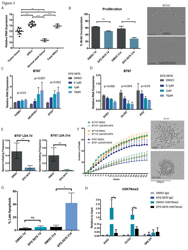
bioRxiv preprint first posted online Jun. 28, 2018; doi: http://dx.doi.org/10.1101/358432. The copyright holder for this preprint
 (which was not peer-reviewed) is the author/funder, who has granted bioRxiv a license to display the preprint in perpetuity.
                              It is made available under a CC-BY-NC-ND 4.0 International license.

   Summary

   Successful glioblastoma (GBM) therapies have remained elusive due to limitations in understanding

   mechanisms of growth and survival of the tumorigenic population. Using CRISPR-Cas9 approaches in

   patient-derived GBM stem cells to interrogate function of the coding genome, we identify diverse

   actionable pathways responsible for growth that reveal the gene-essential circuitry of GBM stemness. In

   particular, we describe the Sox developmental transcription factor family; H3K79 methylation by DOT1L;

   and ufmylation stress responsiveness programs as essential for GBM stemness. Additionally, we find

   mechanisms of temozolomide resistance and sensitivity that could lead to combination strategies with

   this standard of care treatment. By reaching beyond static genome analysis of bulk tumors, with a genome

   wide functional approach, we dive deep into a broad range of biological processes to provide new

   understanding of GBM growth and treatment resistance.

   Keywords (up to 10): Glioblastoma, Glioma Stem Cells, CRISPR-Cas9, essentiality

                                                                                                                                    2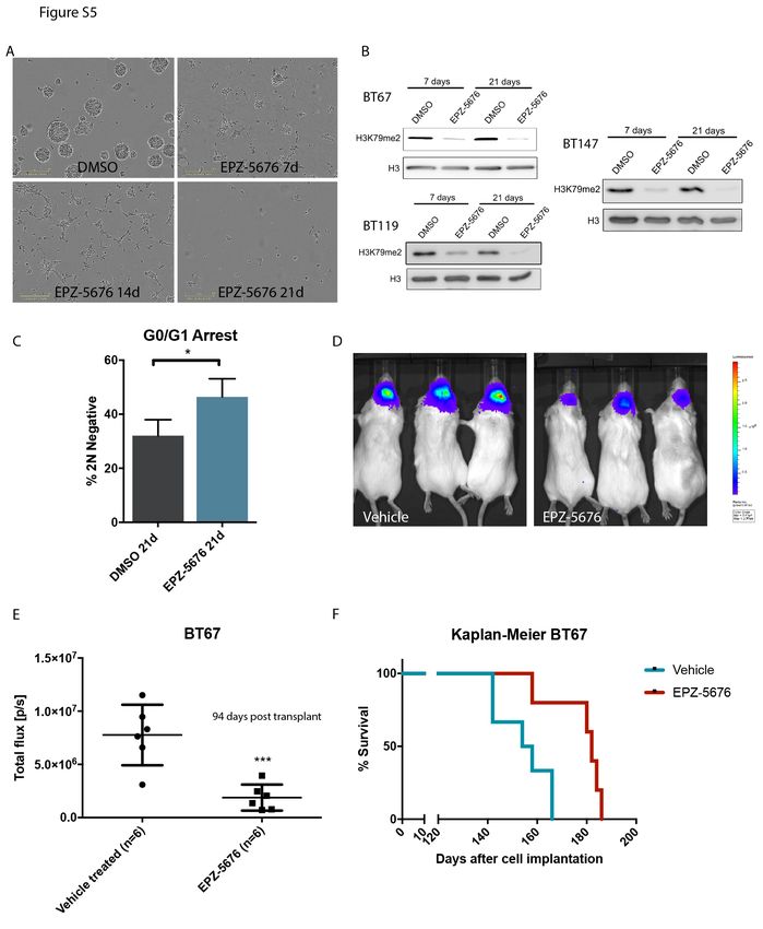
bioRxiv preprint first posted online Jun. 28, 2018; doi: http://dx.doi.org/10.1101/358432. The copyright holder for this preprint
 (which was not peer-reviewed) is the author/funder, who has granted bioRxiv a license to display the preprint in perpetuity.
                              It is made available under a CC-BY-NC-ND 4.0 International license.

   Significance

   Glioblastoma (GBM) remains an incurable disease despite an increasingly thorough depth of knowledge

   of the genomic and epigenomic alterations of bulk tumors. Evidence from multiple approaches support

   that GBM reflects an aberrant developmental hierarchy, with GBM stem cells (GSCs), fueling tumor

   growth and invasion. The properties of this tumor subpopulation may also in part explain treatment

   resistance and disease recurrence. Unfortunately, we still have a limited knowledge of the molecular

   circuitry of these cells and progress has been slow as we have not been able, until recently, to interrogate

   function at the genome-wide scale. Here, using parallel genome-wide CRISPR-Cas9 screens, we identify

   the essential genes for GSC growth. Further, by screening in the presence of low and high dose

   temozolomide, we identify mechanisms of drug resistance and sensitivity. These functional screens in

   patient derived cells reveal new aspects of GBM biology and identify a diversity of actionable targets such

   as genes governing stem cell traits, epigenome regulation and the response to stress stimuli.

                                                                                                                                    3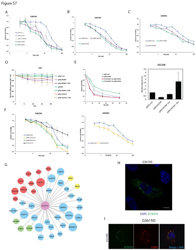
bioRxiv preprint first posted online Jun. 28, 2018; doi: http://dx.doi.org/10.1101/358432. The copyright holder for this preprint
 (which was not peer-reviewed) is the author/funder, who has granted bioRxiv a license to display the preprint in perpetuity.
                              It is made available under a CC-BY-NC-ND 4.0 International license.

   Introduction

             Glioblastoma (GBM) is the most common type of primary malignant brain tumor in adults (Wen

   and Kesari, 2008). Although there are many reasons underlying the treatment-refractory nature of these

   tumors, inter- and intra-patient tumor heterogeneity and a complete lack of durable responses to therapy

   are critical contributors to the dismal overall prognosis (Sturm et al., 2014). Temozolomide (TMZ)

   chemotherapy represents the last major widespread applied advance in GBM therapy (Stupp et al., 2005),

   although its efficacy is remarkably limited, and it endows tumors with an additional mutational burden

   that may ultimately drive disease progression (Cancer Genome Atlas Research Network, 2008; Hunter et

   al., 2006). While large-scale genomic analyses of human GBM tumor samples has revealed a complex

   genomic architecture (Brennan et al., 2013; Parsons et al., 2008; Uhm, 2009), this has been difficult to

   link to GBM growth and functional cell properties, particularly due to the tumors’ proliferative and cellular

   heterogeneity. Indeed, a more comprehensive understanding of molecular determinants of growth and

   drug responsiveness across this heterogenous cancer is needed, both to identify new therapeutic

   opportunities and to also provide new strategies that could be partnered with TMZ that is now

   entrenched in upfront GBM therapy.

             GBM growth is thought to be driven by a small subpopulation of GBM stem cells (GSCs) that are

   capable of self-renewal and generation of progeny that are more limited in their tumorigenic capacity

   and associated with expression of markers of more differentiated lineages. This concept is supported by

   recent single cell RNA sequencing approaches (Patel et al., 2014; Venteicher et al., 2017) and our recent

   fate mapping study of fresh human GBM cells following orthotopic transplantation in mice, which also

   highlights the presence of a chemotherapy-resistant tumorigenic subpopulation (Lan et al., 2017).

   Although tumorigenic properties of GBM stem cells are best interrogated in vivo, GSC culture systems

   have enabled expansion of these patient derived cells from many GBM samples for functional

   experiments on a scale that is difficult to achieve in vivo (Pollard et al., 2009). Importantly, these GSC

                                                                                                                                    4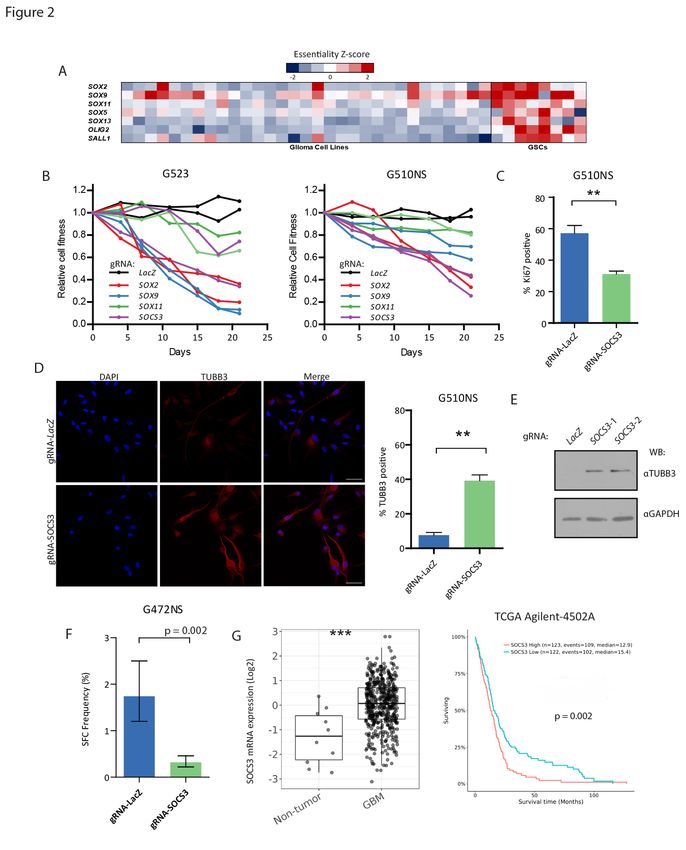
bioRxiv preprint first posted online Jun. 28, 2018; doi: http://dx.doi.org/10.1101/358432. The copyright holder for this preprint
 (which was not peer-reviewed) is the author/funder, who has granted bioRxiv a license to display the preprint in perpetuity.
                              It is made available under a CC-BY-NC-ND 4.0 International license.

   cultures reliably preserve patient specific phenotypic and genotypic characteristics, including at a single

   clonal level, retain in vivo tumorigenic capacity and recapitulate the hierarchical growth behavior

   observed in vivo (Lee et al., 2006; Pollard et al., 2015; Meyer et al., 2015; Lan et al., 2017).

             The emergence of the CRISPR-Cas9 technology has enabled genome-wide forward genetic

   screens, including “cell fitness screens”, which allow for the systematic identification of “core” and

   “context-specific” essential genes governing cell proliferation across all cell types or in a given cellular

   genetic background (Aguirre et al., 2016; Shalem et al., 2013; Wang et al., 2013, 2015). Using this

   approach, we and others reported that approximately 2000 genes are required for the optimal growth of

   all the cancer cell lines examined, including 400 genes that are unique to each cell type examined (Hart

   et al., 2015; Wang et al., 2015; Blomen et al., 2015). Context-specific essential genes represent unique

   genetic vulnerabilities that underlie the concept of synthetic lethality and may, in some cases, be

   therapeutically exploited. For example, using genome-wide CRISPR-Cas9 essentiality screens, we recently

   identified the Wnt receptor FZD5 as specifically required for the growth of RNF43 mutant PDAC cells and

   showed that FZD5 blocking antibodies robustly inhibit the proliferation of these cells in vitro and tumor

   growth in orthotopic mouse models (Steinhart et al., 2017).

             Given the complex patient to patient heterogeneity observed in GBM, additional gene essentiality

   screens are needed to capture the genetic dependencies common across all patients and essential genes

   unique to individual GBM genotypes. Here, we performed parallel genome-wide CRISPR screens in eight

   low passage patient-derived GSC cultures that harbor a range of typical GBM genomic abnormalities

   matched with their primary tumors (Lan et al., 2017; Pollard et al., 2009) to define the molecular circuitry

   governing GSC growth and survival. We concurrently performed chemogenomic screens to identify genes

   modulating TMZ responsiveness revealing mechanisms of therapeutic resistance and strategies for

   combinatorial therapy. Our results provide the largest series of forward-genetic screens for a solid tumor

   in patient-derived tumor initiating cells and provide a functional blueprint for GBM tumorigenicity, at the

                                                                                                                                    5
bioRxiv preprint first posted online Jun. 28, 2018; doi: http://dx.doi.org/10.1101/358432. The copyright holder for this preprint
 (which was not peer-reviewed) is the author/funder, who has granted bioRxiv a license to display the preprint in perpetuity.
                              It is made available under a CC-BY-NC-ND 4.0 International license.

   GSC level. These findings identify new GBM vulnerabilities and nominate a diverse repertoire of strategies

   for additional therapeutic development.

   Results

   Genome-wide CRISPR-Cas9 Screens Identify Genetic Vulnerabilities of GBM Stem Cells.

             To identify essential fitness genes in GSCs, we performed parallel genome-wide CRISPR-Cas9

   screens in 8 well-characterized (Figure S1A) low passage patient-derived GSC cultures engineered to

   express the Cas9 nuclease (GSC-Cas9) (Figure 1A). Cells were infected with lentivirus carrying the 90K

   gRNA TKOv1 library (Hart et al., 2015). After selection for infected cells, and T0 sampling, cells were grown

   for 14 doublings and genomic DNA collected at each passage. We also performed chemogenomic screens

   (Figure 1A) with lethal or sub-lethal doses (LD90 and LD20 respectively) of TMZ, to identify genes

   underlying TMZ sensitivity and resistance. The BAGEL algorithm was used to derive a Bayes Factor (BF)

   for each tested gene by comparing changes in gRNA abundance throughout the course of the screen to

   experimentally determined gold-standard reference essential and non-essential genes (Hart and Moffat,

   2016; Hart et al., 2017). The Bayes Factor is a measure of the confidence that knockout of a specific gene

   causes a decrease in cell fitness (high BF indicates increased confidence that the knockout of the gene

   results in a decrease in fitness). In this manner, we identified an average of 1,722 essential genes in each

   GSC culture at a 5% false discovery rate (FDR) (Table S1). Despite patient to patient genetic differences

   between these samples, a total of 1,157 genes were hits in at least 5 of the 8 patient-derived GBM cultures

   screened (Figure S1B). These findings suggest that GSCs require a core set of genes required for

   proliferation and survival, reminiscent of shared functional properties that we identified in our previous

   in vivo fate mapping study (Lan et al., 2017).

                                                                                                                                    6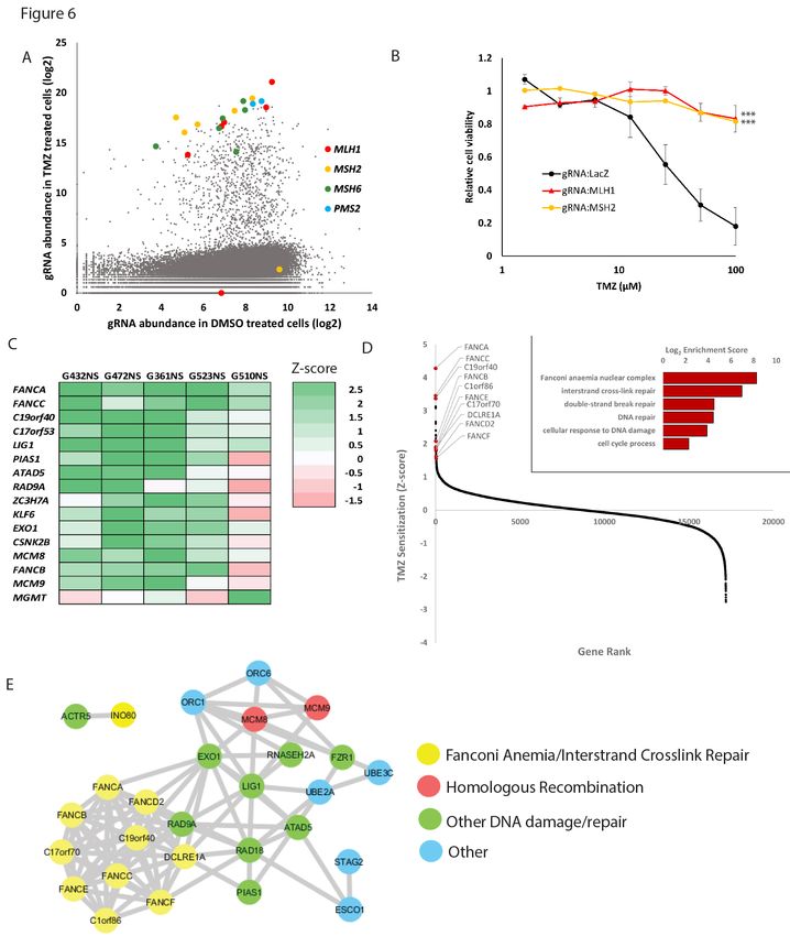
bioRxiv preprint first posted online Jun. 28, 2018; doi: http://dx.doi.org/10.1101/358432. The copyright holder for this preprint
 (which was not peer-reviewed) is the author/funder, who has granted bioRxiv a license to display the preprint in perpetuity.
                              It is made available under a CC-BY-NC-ND 4.0 International license.

             To identify fitness genes that are unique to the stem cell fraction of GBM, we compared the core

   set of fitness genes derived from our 6 highest quality GSC screens (Figure S1C) to 8 previously reported

   essentiality screens performed in cancer and epithelial cell lines from diverse tissues of origin (“non-

   GBM”) (Hart et al., 2015). As expected, GSC screens formed a distinct cluster as defined by the essentiality

   profile from non-CNS cancer cells, emphasizing the shared context-specific mechanisms governing growth

   and survival of this cancer type (Figure 1B; Figure S1D). We calculated a Z-score for the difference in

   average BF-score between GBM and non-GBM screens and derived a ranked list of GSC specific essential

   genes (Figure 1C; Table S2). Generation of an enrichment map for GBM-specific essential genes using

   Gene Ontology (GO) Biological Processes and the Reactome knowledge base terms revealed genetic

   vulnerabilities within a diverse set of networks, including GO terms related to Chromatin Organization

   and DNA Repair, Cell Fate and Gliogenesis, Cholesterol Biosynthesis, DNA Replication and Cell Cycle, and

   protein Ufmylation (Figure 1D). To validate genome-wide screening results, we selected 20 conserved

   GBM-specific essential genes of particular interest on the basis of high Z-score and spanning the key GO

   processes identified above. We validated all of these hits by testing individual gene knockouts in three

   GSCs using competitive cell proliferation assays in which we co-cultured GSC-Cas9 cells expressing GFP +

   a gRNA targeting a gene of interest with cells expressing mCherry + a non-targeting gRNA. The

   GFP/mCherry ratio was tracked over a period of three weeks (Figure 1E; Figure S2A-F). In most cases,

   cells expressing the gRNA targeting genes identified in the screen were outcompeted by cells expressing

   a non-targeting gRNA. We therefore identified a core set of GSC essential genes that represent genetic

   vulnerabilities crosscutting the majority of GBM patients.

   Careful examination of the relationship between gene essentiality profiles and the genetic background of

   the tested GSCs also identified synthetic lethal interactions unique to patient-specific genetic contexts.

   For example, 3 of the 8 lines harbored EGFR activating mutations or amplification and displayed EGFR

   essentiality, whereas EGFR disruption in EGFR wild-type lines had no effect on growth (Figure S3A). In

                                                                                                                                    7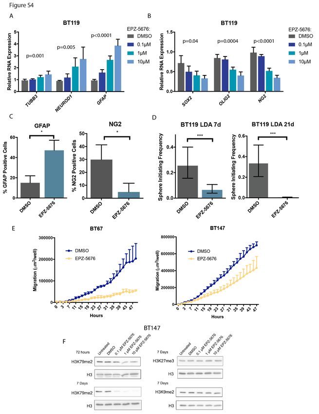
bioRxiv preprint first posted online Jun. 28, 2018; doi: http://dx.doi.org/10.1101/358432. The copyright holder for this preprint
 (which was not peer-reviewed) is the author/funder, who has granted bioRxiv a license to display the preprint in perpetuity.
                              It is made available under a CC-BY-NC-ND 4.0 International license.

   addition, given the universal disruption of the Rb pathway in GBM we observed a dependency on the

   CDK4/6 cyclin-dependent kinase family (Wiedemeyer et al., 2010). We saw a specific reliance on CDK4 or

   CDK6 depending on whether the cells had CDK4 amplification or CDNK2A/B disruption (Figure S3B). We

   validated these findings using single gene knockouts of CDK4 and CDK6 in 5 GSC cultures, including one

   not included in the screen panel. Consistent with our screen results, the proliferation of GSCs harboring

   CDK4 amplification was exclusively dependent on CDK4, while GSCs with CDKN2A/B deletion were

   exclusively dependent upon CDK6 (Figure S3C). These findings illustrate the potential for genome-wide

   CRISPR-Cas9 functional genomics screens to identify both core and genotype-specific therapeutic

   vulnerabilities in the GBM patient population. In turn, this provides exciting insights into novel therapeutic

   strategies for this devastating disease.

   Genetic Programs Governing Stemness are Essential in GSC Cultures

             One striking GSC essential GO network contained genes important for cell fate and gliogenesis.

   Importantly, we identified genes associated with stem cells such as members of the Sox family of

   transcription factors, OLIG2 and SALL1 (Figure 2A). These findings are consistent with previous studies

   demonstrating a role for these genes in normal neural precursor compartments and their capacity to

   reprogram more differentiated GBM cells into a stem cell state and sustain tumor propagation in vivo

   (Suvà et al., 2014).

             In particular, SOX2 and SOX9 were found to be essential in 5 and 6 cultures respectively,

   supporting their role as core regulators of stemness in GSCs. The GSC screens also identified SOX13,

   SOX11 and SOX5 as differentially essential across GSC cultures suggesting patient specific stem cell

   transcription factor programs that cooperatively sustain GSC self-renewal in patient-specific contexts.

   Individual knockout of SOX2 and SOX9 in cell competition assays confirmed strong negative effects on cell

   proliferation whereas SOX11 disruption conferred a milder and more variable anti-proliferation

                                                                                                                                    8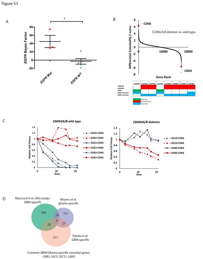
bioRxiv preprint first posted online Jun. 28, 2018; doi: http://dx.doi.org/10.1101/358432. The copyright holder for this preprint
 (which was not peer-reviewed) is the author/funder, who has granted bioRxiv a license to display the preprint in perpetuity.
                              It is made available under a CC-BY-NC-ND 4.0 International license.

   phenotype (Figure 2B; Figure S2C-E). We conclude that stem cell gene networks fuel the growth of

   patient-derived GSCs. Strikingly, comparison of our screens with recently reported CRISPR-Cas9 screens,

   performed in 31 serum-based glioma cell lines (Meyers et al., 2017) and two GSCs (Toledo et al., 2015)

   showed a different repertoire of essential genes (Figure S3D). Notably, the cell fate and gliogenesis genes

   were unique to our screens, perhaps reflecting the different growth conditions and duration of analysis

   of dropouts (Figure 2A). Indeed, while GSCs cultured in growth factor supplemented stem cell media have

   been demonstrated to be a robust model of GBM and are highly enriched for tumor initiating cells in vivo,

   serum cultured glioma cell lines are less capable of tumor formation and bear little histological

   resemblance to patient disease (Lee et al., 2006; Pollard et al., 2009).

             Taking advantage of this unique feature, we next asked whether additional, less well recognized

   stem cell genes could also be identified. Among the top scoring GBM-specific essential genes was

   Suppressor of Cytokine Signaling 3 (SOCS3), which has been previously shown to maintain stemness in

   neural stem cells (Cao et al., 2006). Intriguingly, SOCS3 was reported to have opposing effects in glioma

   and function as either a tumor suppressor or oncogene depending on context (Jiang et al., 2017; Martini

   et al., 2008; Zhou et al., 2007). To gain a better understanding of the functional mechanisms underlying

   SOCS3 in GSCs, we first knocked out its expression with 2 individual gRNAs to validate its essentiality in

   multiple GSC cultures. We found that SOCS3 knockout led to a reduction in GSC proliferation as measured

   by competitive cell proliferation assays (Figure 2B; Figure S2B-E) and Ki67 staining (Figure 2C), increased

   expression of the neuronal marker TUBB3 (Figure 2D and 2E) and decreased sphere forming ability in

   limiting dilution assays (Figure 2F). Analysis of TCGA datasets revealed an upregulation of SOCS3 levels in

   GBM when compared with non-tumor tissue and an association between high SOCS3 expression and

   worse prognosis (Figure 2G). Together these results imply a role of SOCS3 in GSC stemness and underlie

   its importance during GBM progression.

                                                                                                                                    9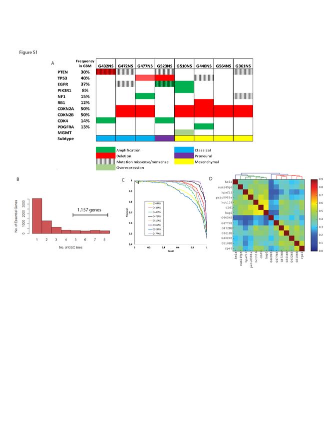
bioRxiv preprint first posted online Jun. 28, 2018; doi: http://dx.doi.org/10.1101/358432. The copyright holder for this preprint
 (which was not peer-reviewed) is the author/funder, who has granted bioRxiv a license to display the preprint in perpetuity.
                              It is made available under a CC-BY-NC-ND 4.0 International license.

   The Histone Methyltransferase DOT1L Regulates Stemness and Proliferation in GSCs

             Our previous GBM fate mapping work pointed to epigenetic compounds as disruptors of GSC

   growth (Gallo et al., 2015; Lan et al., 2017). In line with these findings, our screens identified epigenetic

   dependencies that are unique to GSCs compared to other non-GBM cancer types, suggesting an

   opportunity to develop CNS specific epigenetic modifiers for GBM treatment. We observed the epigenetic

   modulators DOT1L, EZH2 and SUV420H1 were among our top scoring hits as GSC-specific essential genes.

   The H3K79 methyltransferase DOT1L is highly expressed in GSCs compared to bulk tumor (Figure 3A) and

   is an essential gene in 7 of 8 GSC cultures, supporting a near pan-GSC essential function. We next

   validated DOT1L essentiality in GSCs using CRISPR-Cas9 gene editing (Figure 1E; Figure S2C,D and F) and

   found that the clinically relevant small molecule inhibitor of DOT1L, EPZ-5676, inhibited GSC growth

   (Figure 3B). The effects of EPZ-5676 were prominent but only after longer periods, consistent with

   inhibition of an epigenetic process (Figure 3B,G; Figure S5A, B). Importantly, in line with regulation of a

   stem cell program, morphologic changes consistent with differentiation were observed in a sphere culture

   model of GSCs (Figure S5A) with increased expression of neuronal and glial markers and decreased

   expression of stem cell markers (Figure 3C and D; Figure S4A,B and C) after 7 days of treatment. These

   changes translated into marked reduction in self-renewal as measured by assays of sphere forming

   capacity and, in addition, reduction of cellular migration/invasion (Figure 3E and F; Figure S4D and E).

   Critically, assessment of histone marks following treatment of GSCs with EPZ-5676 demonstrated on-

   target DOT1L inhibition as H3K79me2 levels were specifically decreased without affecting other histone

   marks (Figure S4F), and this was further diminished over time (Figure S5B). Consistent with this

   observation, we found that longer treatment periods of 21 days led to decreased BrdU incorporation,

   G0/G1 cell cycle arrest, as well as increased apoptosis (Figure 3B and G; Figure S5C). Importantly, EPZ-

   5676 treatment led to a significant loss of the H3K79me2 mark at the key neural stem cell regulators and

                                                                                                                                    10
bioRxiv preprint first posted online Jun. 28, 2018; doi: http://dx.doi.org/10.1101/358432. The copyright holder for this preprint
(which was not peer-reviewed) is the author/funder, who has granted bioRxiv a license to display the preprint in perpetuity.
It is made available under a CC-BY-NC-ND 4.0 International license.
GBM-specific essential genes SOX2 and OLIG2 (Figure 3H). Therefore, GSC stemness properties are
attenuated through inhibition of H3K79me2.
To investigate the potential of targeting DOT1L for the treatment of GBM, we tested the effect of
EPZ-5676 treatment on GSCs in an immunodeficient mouse model. As our data support that EPZ-5676
poorly crosses the blood brain barrier (Waters et al., 2016) in control animals (Figure 4A), we pretreated
GSC cultures in vitro followed by orthotopic transplantation. Animals that received tumors pre-treated
with EPZ-5676 had reduced tumor growth and longer survival (Figure 4B, C and D; Figure S5D,E and F).
Importantly, dosing of mice with EPZ-5676 led to tumor growth inhibition following subcutaneous
establishment of tumors. In these in vivo-treated tumors, we saw erasure of the H3K79me2 mark and
increased expression of genes associated with differentiation (Figure 4E, F). Given these data, further pre-
clinical studies are warranted to investigate DOT1L histone methylation in GBM, particularly if more CNS
penetrant compounds can be developed. The efficacy across multiple patient-derived cell populations
supports broad targeting of processes such as chromatin regulation (Bulstrode et al., 2017; Jin et al., 2017;
Lan et al., 2017), which could be a strategy to treat the diversity of GBM genotypes, and overcome GBM
interpatient heterogeneity.
Regulators of Cell Stress Response Pathways are Essential in GSCs
Several highly ranked GBM-specific essential genes were also enriched with functions within
stress response pathways. For example, c-JUN was the top ranked GBM-specific essential gene across our
panel of GSC cultures, and the upstream kinase MAP2K7 was also identified (Figure 5A). The JNK-JUN
pathway functions to protect cells from stress-induced apoptosis (Dhanasekaran and Reddy, 2008) and
has recently been reported to be activated in GBM (Matsuda et al., 2012; Yoon et al., 2012). Individual
CRISPR-Cas9 mediated knockout of either c-JUN or MAP2K7 significantly reduced cell fitness in multiple
11bioRxiv preprint first posted online Jun. 28, 2018; doi: http://dx.doi.org/10.1101/358432. The copyright holder for this preprint
(which was not peer-reviewed) is the author/funder, who has granted bioRxiv a license to display the preprint in perpetuity.
It is made available under a CC-BY-NC-ND 4.0 International license.
GSC cultures (Figure 1A; Figure S2C-E), validating the screen results. Additionally, treatment of GSC
cultures with the pan-JNK inhibitors SP600125 and bentamapimod caused a robust reduction in cell
viability (Figure 5B).
One recently uncovered stress-related pathway overrepresented amongst GBM-specific
essential genes was the protein ufmylation system (Figure 1D), a ubiquitin-like post-translational
modification linked to ER stress during development and stem cell homeostasis (DeJesus et al., 2016; Wei
and Xu, 2016; Zhang et al., 2012). All known ufmylation pathway members were striking hits across the
screened GSCs (Figure 5C), suggesting that this pathway may be highly critical for the maintenance of the
stem cell compartment in GBM. To further test this dependency on the ufmylation system, we first
knocked out the four highest ranking genes in this pathway and subjected these cells to competitive cell
growth assays with control cells. Single gene knockout of UBA5 (E1), UFC1 (E2), UFL1 (E3) and UFSP2
(UFM1 protease) all caused a significant reduction of cell fitness (Figure 5D) confirming that disruption of
any of the key catalytic steps in this pathway represents a potential therapeutic vulnerability. Next, we
treated a panel of 14 patient-derived GSCs with DKM 2-93 (Roberts et al., 2017), a small molecule inhibitor
of UBA5. We observed a robust inhibition of proliferation (Figure 5E) confirming the essentiality of this
pathway across all tested GSC cultures. The Endoplasmic Reticulum-Associated Degradation (ERAD)
pathway (Kim et al., 2015) members SEL1L and HRD1 (SYVN1) were also identified as GBM specific
essential genes (Figure 5A). We conclude that GSCs are under high proteotoxic stress and as a result rely
on activation of different stress signaling pathways for survival. These results also hint at an opportunity
to target the ufymylation proteostasis gene network as a potential therapeutic strategy for GBM.
Chemogenomic screens reveal mechanisms of Temozolomide sensitivity in GSCs
12bioRxiv preprint first posted online Jun. 28, 2018; doi: http://dx.doi.org/10.1101/358432. The copyright holder for this preprint
(which was not peer-reviewed) is the author/funder, who has granted bioRxiv a license to display the preprint in perpetuity.
It is made available under a CC-BY-NC-ND 4.0 International license.
The GBM chemotherapy agent temozolomide has poor efficacy and concerns exist with its
mutagenesis propensity due to DNA alkylation. However, it is used ubiquitously given that a small subset
of patients displays longer term survival. For patients who derive no benefit from TMZ treatment, it is
necessary to nominate potential treatment combinations that may expand clinical efficacy for this current
chemotherapy standard of care. To achieve this goal, further studies are needed to understand the
genetic mechanisms of intrinsic TMZ resistance. We therefore performed additional CRISPR-Cas9 screens
in the context of low dose and high dose TMZ (Figure 1A). Positive selection screens were performed in
two patient-derived GBM cultures (G361NS, G432NS) treated with a lethal dose of TMZ (100 µM) to
define genes required for TMZ sensitivity. This dose of TMZ resulted in over 90% cell death over 10 days
of treatment, with resistant clones emerging thereafter. Sequencing of integrated gRNAs in resistant cells
after 3 weeks of TMZ treatment revealed dramatic enrichment for gRNAs targeting only four genes, the
core members of the mismatch repair (MMR) pathway MLH1, MSH2, MSH6 and PMS2 (Figure 6A, Figure
S6A). The MMR pathway initiates futile cycling of attempted DNA repair in response to TMZ lesions and
is required for its cytotoxicity (Wang and Edelmann, 2006). Since MSH6 mutations are commonly found
in recurrent GBM (Hunter et al., 2006; Lan et al., 2017), the screen hence faithfully models tumor
evolution in vitro. Validating these results, introduction of MLH1 and MSH2 mutations in GSCs conferred
acquired TMZ resistance (Figure 6B). Given that the four core MMR genes were the sole hits in two
separate genome-wide screens, we conclude that this pathway is the primary mediator of TMZ sensitivity,
and that further clinical efforts can be directly focused on MMR integrity to improve treatment
responsiveness.
To identify mechanisms of intrinsic resistance of the GSC population to TMZ, we performed
negative selection screens in the presence of a sublethal dose of TMZ (LD20) in 5 patient-derived GSCs
(Figure 1A). Z-scores reflecting the difference in gene essentiality in the presence or absence of TMZ
revealed that 4/5 cultures displayed a core subset of genes amongst the top TMZ sensitizers (Figure 6C;
13bioRxiv preprint first posted online Jun. 28, 2018; doi: http://dx.doi.org/10.1101/358432. The copyright holder for this preprint
(which was not peer-reviewed) is the author/funder, who has granted bioRxiv a license to display the preprint in perpetuity.
It is made available under a CC-BY-NC-ND 4.0 International license.
Table S3). GO term enrichment (Figure 6D and Table S4) and STRING analysis (Szklarczyk et al., 2017)
revealed gene and protein networks involved in multiple DNA repair pathways. Notably, GO terms for the
Fanconi Anemia/interstrand crosslink repair and homologous recombination pathways were the most
significantly enriched (Figure 6D and 6E). Supporting these results, individual knockout of the Fanconi
Anemia pathway genes FANCA and C19orf40 confirmed sensitization of GSCs to TMZ (Figure S7A).
Conversely, analysis of the TMZ-resistant G510NS revealed the TMZ adduct repairing enzyme MGMT as
the top hit restoring TMZ sensitivity (Figure 6C), consistent with RNA-seq results showing this line to be
the only MGMT expressing line in our screen panel. The identification of a core set of genes involved in
intrinsic TMZ resistance across multiple, heterogeneous GSCs, many of which with potentially druggable
enzymatic functions, suggests rational strategies for the development of combinatorial therapies. This
further provides novel strategies for targeting mechanisms of intrinsic resistance to hypersensitize tumor
cells to standard of care chemotherapy.
Along these lines, MCM8 and MCM9 which together form a dimeric helicase complex involved in
homologous recombination, downstream of the Fanconi Anemia/interstrand crosslink repair pathways as
well as the DNA mismatch repair pathway (Nishimura et al., 2012; Traver et al., 2015), were top hits in
multiple TMZ chemogenomic screens. We performed functional experiments to determine the effects of
these genes on TMZ responsiveness. Knockout of MCM8 or MCM9 had no significant effect on cell fitness
in untreated cells but strongly sensitized GSC lines to TMZ and had little effect in human neural stem cells
(Figure 7A and 7B; Figure S7A-D). Treatment of GSCs with low dose TMZ resulted in double strand breaks
that were rapidly repaired in cells expressing a non-targeting gRNA, while cells expressing gRNAs targeting
MCM8 and MCM9 displayed persistent γH2AX foci, a hallmark of DNA repair defects (Figure 7C and 7D).
We further validated the role of MCM8 in contributing to TMZ resistance using a Dox-inducible MCM8
construct, which rescued the TMZ hypersensitivity displayed by MCM8 knockout GSCs (Figure S7E). It has
been previously shown that the cancer stem cell population demonstrates a highly efficient DNA damage
14bioRxiv preprint first posted online Jun. 28, 2018; doi: http://dx.doi.org/10.1101/358432. The copyright holder for this preprint
(which was not peer-reviewed) is the author/funder, who has granted bioRxiv a license to display the preprint in perpetuity.
It is made available under a CC-BY-NC-ND 4.0 International license.
response system which contributes to disease recurrence (Bao et al., 2006). Given their enzymatic
function and previous success in development of small molecule DNA helicases inhibitors, we nominate
the MCM8/9 complex as a target to be addressed in the context of TMZ. This finding reinforces the power
of chemogenomic screens to identify mechanisms of intrinsic drug resistance that could be
therapeutically leveraged for more efficient treatments.
A number of completely uncharacterized genes were also identified to mediate intrinsic TMZ
resistance in GSCs. C17orf53 and ZC3H7A were each identified as hits in at least 3 GSC cultures (Figure
6C). Knockout or overexpression of the zinc finger gene ZC3H7A led to increased sensitivity and resistance
to TMZ respectively (Figure 7B; Figure S7F). To determine the function of ZC3H7A, we performed BioID
affinity purification-mass spectrometry to identify ZC3H7A-associated proteins (Table S5). Proximal
interactors of ZC3H7A were enriched for biological processes relating to regulation of translation, mRNA
processing, cytoplasmic stress granule and p53-mediated DNA damage signaling (Figure 7E; Figure S7G,
Table S6). Expression of Venus-tagged full-length ZC3H7A cDNA in both Hela and GSCs showed localization
in cytoplasmic granules (Figure 7F, Figure S7H). Since known stress granule (UBAP2L, G3BP1) and RNA
Processing body (CNOT complex) proteins were found to interact with ZC3H7A in our BioID experiments,
we tested whether ZC3H7A colocalized with markers of either structure. We found that ZC3H7A localized
to cytoplasmic granules that co-localized with the stress granule marker G3BP1 upon sodium arsenite
treatment (Figure 7F, Figure S7H and I), but did not significantly co-localize with the P-body marker
DCP1A. ZC3H7A is therefore a novel stress-responsive regulator of chemoresistance in GBM.
15bioRxiv preprint first posted online Jun. 28, 2018; doi: http://dx.doi.org/10.1101/358432. The copyright holder for this preprint
(which was not peer-reviewed) is the author/funder, who has granted bioRxiv a license to display the preprint in perpetuity.
It is made available under a CC-BY-NC-ND 4.0 International license.
Discussion
There has been disappointingly slow progress in identifying effective therapies for GBM, in part
due to a lack of understanding of the genes essential for functional properties of these tumors and of
GSCs, the most primitive cell types shown to lie at the root of GBM growth and resistance (Chen et al.,
2012; Lan et al., 2017). The complex cellular ecosystem of GBM tumors certainly suggests that we still
have much to learn about cellular mechanisms of tumor growth, particularly in vivo, but we need
accessible human cell models to interrogate functional properties of enriched populations of tumorigenic
cells. Fortunately, patient-derived GSC cultures represent a relatively simple system to generate a cell
model of GBM on a very efficient patient to patient basis. These cells, as close as currently attainable,
reflect the properties of primary GBM tumors (Lan et al., 2017; Lee et al., 2006; Meyer et al., 2015; Pollard
et al., 2009) including following orthotopic xenograftment (Ben-David et al., 2017; Lan et al., 2017).
Critically, these GSC cultures are scalable for functional interrogation at the genome-wide level.
In this study, parallel genome-wide CRISPR-Cas9 screens in multiple patient-derived GSCs,
identified core growth programs for this heterogeneous disease and uncovered mechanisms underlying
TMZ resistance and sensitization. The identification of common GSC essential genes, despite inherent
intratumoral heterogeneity and irrespective of patient tumor genotype, provides insight into the
underlying biology of GBM and identifies new avenues for drug therapy. In particular, it suggests genes
and pathways that are important for GSC properties that were not previously identified following genomic
characterization. Moreover, our findings point to a convergence of the large diversity of GBM molecular
changes on a potentially smaller number of essential pathways, likely based on aberrant stemness
function. Comparison of our GSC screens to our previously performed essentiality screens in cancer and
epithelial cell lines from diverse tissues of origin also identifies genetic vulnerabilities unique to GSCs,
suggesting specific targeting strategies. These findings provide evidence of the GBM-specific mechanisms
used to maintain cancer stem cell characteristics and promote GSC survival.
16bioRxiv preprint first posted online Jun. 28, 2018; doi: http://dx.doi.org/10.1101/358432. The copyright holder for this preprint
(which was not peer-reviewed) is the author/funder, who has granted bioRxiv a license to display the preprint in perpetuity.
It is made available under a CC-BY-NC-ND 4.0 International license.
Equally important is the combination of functional approaches with a detailed understanding of
the GBM mutational landscape to begin linking genotypes with phenotypes. By performing a limited
number of genome-wide CRISPR essentiality screens in GSCs, characterized by exome-sequencing and
RNA-seq, we were able to identify genotype-specific essential genes. For example, within the frequently
altered Retinoblastoma pathway, we identified genotype-specific dependencies on either CDK4 or CDK6.
GSCs with amplification of CDK4 were predictably reliant on CDK4 for survival. However, we also found
that GSCs with CDKN2A/B deletion had an unexpected dependency on CDK6 alone, despite sharing
common regulation and function with CDK4. These findings suggest genotype-specific mechanisms of RB
pathway misregulation in GBM that would not have been apparent from genomic characterization of a
tumor sample and could be differentially targeted during treatment. Our findings thus warrant further
investigation to identify the mechanistic differences and non-redundant roles between CDK4 and CDK6
in GSCs to explain these genotype-specific dependencies. While our ability to uncover additional patient-
specific vulnerabilities is currently limited by the relatively small number of genome-wide screens
performed in patient-derived GSCs sharing the same genomic alterations, additional efforts will provide
the required power. Given the heterogeneity of GBM, it is very unlikely that a single therapeutic strategy
will be universally successful, and thus we foresee that patient stratification based on genotype-specific
vulnerabilities will be an area of important focus for future studies, particularly if they are superimposed
on common vulnerable pathways.
A comparison of our essentiality screens performed on freshly isolated GSCs from patients to
screens conducted in serum-cultured glioma cell lines (Meyers et al., 2017), revealed striking differences
in essentiality profiles, with the GSC screens described herein uniquely identifying genes involved in cell
fate determination and gliogenesis. These findings highlight the strength of our GBM cell model which
captures the stem cell characteristics of the disease. Consistent with this notion, we identified a
substantial number of master neurodevelopmental transcription factor genes that have been linked to
17bioRxiv preprint first posted online Jun. 28, 2018; doi: http://dx.doi.org/10.1101/358432. The copyright holder for this preprint
(which was not peer-reviewed) is the author/funder, who has granted bioRxiv a license to display the preprint in perpetuity.
It is made available under a CC-BY-NC-ND 4.0 International license.
GBM stemness, including an interesting core gene network that was described as being capable of
reprogramming differentiated GBM cells into a stem cell state (Suvà et al., 2014). SOX2 and SOX9, in
particular, were identified as essential genes across patient GSC cultures. SOX2 has been described to be
a core GBM reprogramming factor and SOX9 has been shown to have an important role in maintaining
neural stem cells, regulating astrogliogenesis (Caiazzo et al., 2015; Scott et al., 2010) and sphere formation
in serum cultured glioma cell lines (Wang et al., 2018). Based on these studies and our screen results,
SOX2 and SOX9 likely have foundational roles in driving GBM progression and maintaining stem cell
properties in GSCs. Furthermore, we also identified genetic dependencies on additional SOX transcription
factors that were variable across GSC cultures. Further studies are needed to help elucidate the
mechanisms underlying how these neural stem cell genes are co-opted in GBM and how they work
together to maintain the tumor initiating stem cell fraction. Although these transcription factors are
notoriously difficult to target using small molecule approaches, these studies reinforce a need to continue
to derive strategies that target the core GBM developmental/stemness state. Importantly, we also
uncovered additional genes that may contribute to GBM stemness function, such as SOCS3. In contrast
to a previous report of SOCS3’s predominant tumor suppressive function via negative regulation of
JAK/STAT signaling (Croker et al., 2008), our data support a context-specific role as a positive regulator of
GSC function, consistent with other reports showing SOCS3 enhances GBM tumor cell survival and
treatment resistance (Zhou et al., 2007).
Recent advances in the field of epigenetics demonstrate the importance of epigenetic regulation
in determining cell fate (Hsieh and Zhao, 2016; Podobinska et al., 2017). We identified several epigenetic
modulators as unique essential genes in GSCs. We studied the histone methyltransferase DOT1L in depth
due to its conserved essentiality. Recently, DOT1L was identified as an in vivo-specific genetic dependency
in GBM using a focused RNAi screen (Miller et al., 2017). Intriguingly, shRNA-mediated knockdown of
DOT1L was deemed dispensable for the growth of GSCs in vitro, while we found that CRISPR-mediated
18bioRxiv preprint first posted online Jun. 28, 2018; doi: http://dx.doi.org/10.1101/358432. The copyright holder for this preprint
(which was not peer-reviewed) is the author/funder, who has granted bioRxiv a license to display the preprint in perpetuity.
It is made available under a CC-BY-NC-ND 4.0 International license.
knockout of DOT1L robustly inhibits the growth of all GSC cultures. The reason for this discrepancy is
unclear but may point to residual DOT1L activity due to incomplete knockdown obtained with shRNA or
to the delay in observable effect upon DOT1L loss of function. Nevertheless, together with the previous
report, the evidence strongly points to the importance of epigenetic control in regulation of functional
stem cell properties. We extend the previous findings by providing evidence that two GSC essential genes,
SOX2 and OLIG2, are epigenetically regulated by DOT1L histone methylation, suggesting epigenetic
mediated regulation, in part is involved in targeting core stemness programs. Development of brain-
permeable DOT1L inhibitors or drug delivery modalities to enable inhibitors such as EPZ-5676 to access
the tumors will be worthwhile for pre-clinical testing. Overall, the data support the continued design of
therapeutics that specifically exploit epigenetic vulnerabilities in order to access GBM stemness function.
One of the more exciting findings from our screens was the identification of multiple stress
signaling pathways as essential for the growth of GSCs. The top GSC-specific hit in this study, c-JUN, plays
an important role in the protection of cells from stress-induced apoptosis. Supporting these results, the
kinase upstream to the JNK-JUN signaling axis, MAP2K7 was also identified as essential. Together with
previous studies (Matsuda et al., 2012; Yoon et al., 2012), our findings support further investigation of
the effectiveness of therapeutic strategies targeting JNK-JUN signaling for GBM. Along the same lines, the
identification of several components of the protein ufmylation pathway as essential genes, further
highlights the importance of stress response mechanisms for the survival and/or growth of GSCs. Indeed,
given that this ubiquitin-like post-translational modification is linked to modulation of the ER stress
response (Zhang et al., 2012), these results suggest that GSCs are under high proteotoxic stress and
addicted to ER stress signaling pathways, rendering them vulnerable to perturbation of proteostasis gene
networks (Deshaies, 2014). This finding is in line with new studies describing unique molecular
mechanisms employed by stem cells or their malignant counterparts to maintain their unique functional
properties (Dolma et al., 2016; van Galen et al., 2014; Tang and Rando, 2014). Our findings therefore
19bioRxiv preprint first posted online Jun. 28, 2018; doi: http://dx.doi.org/10.1101/358432. The copyright holder for this preprint
(which was not peer-reviewed) is the author/funder, who has granted bioRxiv a license to display the preprint in perpetuity.
It is made available under a CC-BY-NC-ND 4.0 International license.
provide a molecular basis for triggering proteotoxic crisis (Urra et al., 2017), either by inhibiting stress
response pathways or by increasing ER stress over homeostatic levels, such as by inhibition of proteasome
activity (Raizer et al., 2016), as a consideration for GBM treatment.
To complement our efforts to uncover new genetic vulnerabilities in GBM, we also performed
chemogenomic screens to better understand the genes and pathways that govern the variable
responsiveness of GSCs to TMZ. We also aimed to identify targets for combination therapy to increase
the efficacy of chemotherapy for GBM patients by blocking mechanisms of intrinsic TMZ resistance.
Positive selection screens in two GSC cultures identified just four genes, all core members of the MMR
pathway, whose loss confers resistance to high dose TMZ, faithfully modeling acquired TMZ resistance
observed in GBM recurrence (Wang et al., 2016). More importantly, from a therapeutic perspective, our
negative selection screens with sub-lethal TMZ doses revealed numerous genes underlying intrinsic TMZ
resistance whose loss of function mutations increase sensitivity to TMZ. Most of these genes converge
on the Fanconi Anemia/interstrand crosslink repair and homologous recombination pathways.
Supporting these results, several studies have linked the Fanconi Anemia pathway to TMZ resistance
(Chen et al., 2007; Kondo et al., 2011) Our screens also identify multiple genes involved in Base Excision
Repair (BER) and Nucleotide Excision Repair (NER), such as APEX1 and MPG, consistent with TMZ-induced
DNA adducts also being BER substrates. These hits were however not conserved across all GSC cultures
indicating variable activity or engagement of resistance pathways dependent on patients. Nevertheless,
many of the conserved hits have potentially druggable enzymatic functions including, for example, MCM8
and MCM9 which we validated to result in TMZ hypersensitivity when knocked out. In addition to genes
with known functions in DNA damage repair, we also identified several uncharacterized factors, such as
the zinc finger domain containing protein ZC3H7A, which we describe to play a role in GSC resistance to
TMZ.
20bioRxiv preprint first posted online Jun. 28, 2018; doi: http://dx.doi.org/10.1101/358432. The copyright holder for this preprint
(which was not peer-reviewed) is the author/funder, who has granted bioRxiv a license to display the preprint in perpetuity.
It is made available under a CC-BY-NC-ND 4.0 International license.
In summary, our genome-wide CRISPR screens in patient-derived GSC cultures have identified a
diversity of genetic vulnerabilities in GBM tumorigenic cells. We identify a substantial number of
pathways that previously were not linked to GBM biology, but now can be nominated for further
preclinical exploration. As highlighted here, we suggest that advances in GBM therapies will need to
combine in depth genomic characterization with functional approaches to unveil the genes and pathways
functionally connected to properties of the key malignant population. Despite the concern for
tremendous interpatient heterogeneity and the implications of nihilism that this view brings to future
therapeutic development, the finding of common essential programs for GSC growth and survival lends
hope that we will develop therapies that can be used on larger groups of GBM patients, with patient
specific vulnerabilities providing complementary personalized strategies.
Acknowledgments:
Research was supported by SU2C Canada Cancer Stem Cell Dream Team Research Funding (SU2C-AACR-
DT-19-15) provided by the Government of Canada through Genome Canada and the Canadian Institutes
of Health Research, with supplementary support from the Ontario Institute for Cancer Research through
funding provided by the Government of Ontario. Stand Up To Cancer Canada is a program of the
Entertainment Industry Foundation Canada. Research funding is administered by the American
Association for Cancer Research International – Canada, the scientific partner of SU2C Canada.
S.A. was also supported by the Canadian Institute of Health Research and the Terry Fox Research Institute.
P.B.D. is also supported by the Terry Fox Research Institute, the Canadian Cancer Society, the Hospital for
Sick Children Foundation, Jessica’s Footprint Foundation, the Hopeful Minds Foundation, the Bresler
family, and B.R.A.I.N. Child. P.B.D. holds a Garron Family Chair in Childhood Cancer Research at The
Hospital for Sick Children.
21bioRxiv preprint first posted online Jun. 28, 2018; doi: http://dx.doi.org/10.1101/358432. The copyright holder for this preprint
(which was not peer-reviewed) is the author/funder, who has granted bioRxiv a license to display the preprint in perpetuity.
It is made available under a CC-BY-NC-ND 4.0 International license.
S.W. and H.A.L. are also supported by the Canadian Institutes of Health Research.
T.H. was supported by MD Anderson Cancer Center Support Grant P30 CA016672 (the Bioinformatics
Shared Resource) and the Cancer Prevention Research Institute of Texas (CPRIT) grant RR160032.
We thank Dr. Mark Bernstein (University Health Network, Toronto), and Dr. Sunit Das and Dr. Michael D
Cusimano (St. Michael's Hospital, Toronto) for the generous provision of tumor tissue. In addition, we
thank Dr. Alice Yijun Wang for help in data acquisition and analysis, Dr. Michael Johnston for feedback on
the manuscript, Rozina Hassam and Orsolya Cseh (University of Calgary) for technical support and Dr.
Ahmed Aman (Ontario Institute for Cancer Research) for the mass spectrometry analysis for
pharmacokinetic studies.
Author Contributions:
S.A, P.B.D. and G.M. conceptualized the study. D.A.B., I.R., X.H., H.A.L. and S.W. conceptualized the
validation of DOT1L. G.M. and N.R. performed the genome-wide CRISPR-Cas9 screens. T.H., G.M., Z.S. and
S.A. analyzed screen data. G.M., D.A.B., V.M., N.R., M.M.K., M.A., and H.Y. performed in vitro
experiments. F.C. contributed to the drug response experiments in GSCs. D.A.B., X.H. and H.A.L.
performed in vivo experiments. P.B.D., H.A.L. and S.W. provided the GBM culture. S.A., P.B.D., H.A.L. and
S.W. supervised the study and acquired funding for research. S.A. P.B.D. and G.M. wrote the manuscript
with input from all authors.
22bioRxiv preprint first posted online Jun. 28, 2018; doi: http://dx.doi.org/10.1101/358432. The copyright holder for this preprint
(which was not peer-reviewed) is the author/funder, who has granted bioRxiv a license to display the preprint in perpetuity.
It is made available under a CC-BY-NC-ND 4.0 International license.
Declarations of Competing Interests:
The authors declare no competing financial interests.
23bioRxiv preprint first posted online Jun. 28, 2018; doi: http://dx.doi.org/10.1101/358432. The copyright holder for this preprint
(which was not peer-reviewed) is the author/funder, who has granted bioRxiv a license to display the preprint in perpetuity.
It is made available under a CC-BY-NC-ND 4.0 International license.
References
Aguirre, A.J., Meyers, R.M., Weir, B.A., Vazquez, F., Zhang, C.-Z., Ben-David, U., Cook, A., Ha, G.,
Harrington, W.F., Doshi, M.B., et al. (2016). Genomic Copy Number Dictates a Gene-Independent Cell
Response to CRISPR/Cas9 Targeting. Cancer Discov. 6, 914–929.
Babicki, S., Arndt, D., Marcu, A., Liang, Y., Grant, J.R., Maciejewski, A., and Wishart, D.S. (2016).
Heatmapper: web-enabled heat mapping for all. Nucleic Acids Res. 44, W147–W153.
Bao, S., Wu, Q., McLendon, R.E., Hao, Y., Shi, Q., Hjelmeland, A.B., Dewhirst, M.W., Bigner, D.D., and
Rich, J.N. (2006). Glioma stem cells promote radioresistance by preferential activation of the DNA
damage response. Nature 444, 756–760.
Ben-David, U., Ha, G., Tseng, Y.-Y., Greenwald, N.F., Oh, C., Shih, J., McFarland, J.M., Wong, B., Boehm,
J.S., Beroukhim, R., et al. (2017). Patient-derived xenografts undergo mouse-specific tumor evolution.
Nat. Genet. 49, 1567–1575.
Bowman, R.L., Wang, Q., Carro, A., Verhaak, R.G.W., and Squatrito, M. (2017). GlioVis data portal for
visualization and analysis of brain tumor expression datasets. Neuro. Oncol. 19, 139–141.
Brennan, C.W., Verhaak, R.G.W., McKenna, A., Campos, B., Noushmehr, H., Salama, S.R., Zheng, S.,
Chakravarty, D., Sanborn, J.Z., Berman, S.H., et al. (2013). The somatic genomic landscape of
glioblastoma. Cell 155, 462–477.
Brinkman, E.K., Chen, T., Amendola, M., and van Steensel, B. (2014). Easy quantitative assessment of
genome editing by sequence trace decomposition. Nucleic Acids Res. 42, e168.
Bulstrode, H., Johnstone, E., Marques-Torrejon, M.A., Ferguson, K.M., Bressan, R.B., Blin, C., Grant, V.,
Gogolok, S., Gangoso, E., Gagrica, S., et al. (2017). Elevated FOXG1 and SOX2 in glioblastoma enforces
neural stem cell identity through transcriptional control of cell cycle and epigenetic regulators. Genes
Dev. 31, 757–773.
Caiazzo, M., Giannelli, S., Valente, P., Lignani, G., Carissimo, A., Sessa, A., Colasante, G., Bartolomeo, R.,
Massimino, L., Ferroni, S., et al. (2015). Direct conversion of fibroblasts into functional astrocytes by
defined transcription factors. Stem Cell Reports 4, 25–36.
Campeau, E., Ruhl, V.E., Rodier, F., Smith, C.L., Rahmberg, B.L., Fuss, J.O., Campisi, J., Yaswen, P.,
Cooper, P.K., and Kaufman, P.D. (2009). A versatile viral system for expression and depletion of proteins
in mammalian cells. PLoS One 4, e6529.
Cancer Genome Atlas Research Network (2008). Comprehensive genomic characterization defines
human glioblastoma genes and core pathways. Nature 455, 1061–1068.
Cao, F., Hata, R., Zhu, P., Ma, Y.-J., Tanaka, J., Hanakawa, Y., Hashimoto, K., Niinobe, M., Yoshikawa, K.,
and Sakanaka, M. (2006). Overexpression of SOCS3 inhibits astrogliogenesis and promotes maintenance
of neural stem cells. J. Neurochem. 98, 459–470.
Chen, C.C., Taniguchi, T., and D’Andrea, A. (2007). The Fanconi anemia (FA) pathway confers glioma
24bioRxiv preprint first posted online Jun. 28, 2018; doi: http://dx.doi.org/10.1101/358432. The copyright holder for this preprint
(which was not peer-reviewed) is the author/funder, who has granted bioRxiv a license to display the preprint in perpetuity.
It is made available under a CC-BY-NC-ND 4.0 International license.
resistance to DNA alkylating agents. J. Mol. Med. 85, 497–509.
Chen, J., Li, Y., Yu, T.-S., McKay, R.M., Burns, D.K., Kernie, S.G., and Parada, L.F. (2012). A restricted cell
population propagates glioblastoma growth after chemotherapy. Nature 488, 522–526.
Choi, H., Larsen, B., Lin, Z.-Y., Breitkreutz, A., Mellacheruvu, D., Fermin, D., Qin, Z.S., Tyers, M., Gingras,
A.-C., and Nesvizhskii, A.I. (2010). SAINT: probabilistic scoring of affinity purification–mass spectrometry
data. Nat. Methods 8, 70–73.
Coyaud, E., Mis, M., Laurent, E.M.N., Dunham, W.H., Couzens, A.L., Robitaille, M., Gingras, A.-C., Angers,
S., and Raught, B. (2015). BioID-based Identification of Skp Cullin F-box (SCF)β-TrCP1/2 E3 Ligase
Substrates. Mol. Cell. Proteomics 14, 1781–1795.
Croker, B.A., Kiu, H., and Nicholson, S.E. (2008). SOCS regulation of the JAK/STAT signalling pathway.
Semin. Cell Dev. Biol. 19, 414–422.
DeJesus, R., Moretti, F., McAllister, G., Wang, Z., Bergman, P., Liu, S., Frias, E., Alford, J., Reece-Hoyes,
J.S., Lindeman, A., et al. (2016). Functional CRISPR screening identifies the ufmylation pathway as a
regulator of SQSTM1/p62. Elife 5.
Deshaies, R.J. (2014). Proteotoxic crisis, the ubiquitin-proteasome system, and cancer therapy. BMC
Biol. 12, 94.
Dhanasekaran, D.N., and Reddy, E.P. (2008). JNK signaling in apoptosis. Oncogene 27, 6245–6251.
Dolma, S., Selvadurai, H.J., Lan, X., Lee, L., Kushida, M., Voisin, V., Whetstone, H., So, M., Aviv, T., Park,
N., et al. (2016). Inhibition of Dopamine Receptor D4 Impedes Autophagic Flux, Proliferation, and
Survival of Glioblastoma Stem Cells. Cancer Cell 29, 859–873.
Eden, E., Navon, R., Steinfeld, I., Lipson, D., and Yakhini, Z. (2009). GOrilla: a tool for discovery and
visualization of enriched GO terms in ranked gene lists. BMC Bioinformatics 10, 48.
van Galen, P., Kreso, A., Mbong, N., Kent, D.G., Fitzmaurice, T., Chambers, J.E., Xie, S., Laurenti, E.,
Hermans, K., Eppert, K., et al. (2014). The unfolded protein response governs integrity of the
haematopoietic stem-cell pool during stress. Nature 510, 268–272.
Gallo, M., Coutinho, F.J., Vanner, R.J., Gayden, T., Mack, S.C., Murison, A., Remke, M., Li, R., Takayama,
N., Desai, K., et al. (2015). MLL5 Orchestrates a Cancer Self-Renewal State by Repressing the Histone
Variant H3.3 and Globally Reorganizing Chromatin. Cancer Cell 28, 715–729.
Hart, T., and Moffat, J. (2016). BAGEL: a computational framework for identifying essential genes from
pooled library screens. BMC Bioinformatics 17, 164.
Hart, T., Chandrashekhar, M., Aregger, M., Steinhart, Z., Brown, K.R., MacLeod, G., Mis, M.,
Zimmermann, M., Fradet-Turcotte, A., Sun, S., et al. (2015). High-Resolution CRISPR Screens Reveal
Fitness Genes and Genotype-Specific Cancer Liabilities. Cell 163, 1515–1526.
Hart, T., Tong, A.H.Y., Chan, K., Van Leeuwen, J., Seetharaman, A., Aregger, M., Chandrashekhar, M.,
Hustedt, N., Seth, S., Noonan, A., et al. (2017). Evaluation and Design of Genome-Wide CRISPR/SpCas9
Knockout Screens. G3 7, 2719–2727.
25You can also read