REVIEW Structure and Function of the Egg Cortex from Oogenesis through Fertilization
←
→
Page content transcription
If your browser does not render page correctly, please read the page content below
Developmental Biology 241, 1–23 (2002)
doi:10.1006/dbio.2001.0474, available online at http://www.idealibrary.com on

REVIEW
Structure and Function of the Egg Cortex
from Oogenesis through Fertilization

Christian Sardet, 1 François Prodon, Rémi Dumollard, Patrick Chang,
and Janet Chênevert
BioMarCell, UMR 7009 Biologie du Developpement, CNRS/UNIV P et M Curie,
Observatoire, Station Zoologique, Villefranche sur Mer 06230, France

A. WHAT IS THE CORTEX?
    The Egg Cortex
    The Scope of This Review

B. STORING INFORMATION AND BUILDING THE CORTEX DURING OOGENESIS
     Localizing Maternal Determinants in the Drosophila Cortex
     Structuring and Positioning Information in the Xenopus Cortex
     Cortical Polarity and Storage of Information in Other Oocytes
     The Cortex during Vitellogenesis

C. MATURATION PREPARES THE CORTEX FOR FERTILIZATION
    Restructuring Cortical Endoplasmic Reticulum and Organelles during Maturation
    Restructuring the Cytoskeleton during Maturation
    Relocalization of Cortical mRNAs and Domains during Maturation
    The Cortex as the Site of Attachment and Fusion of Sperm and Egg

D. FERTILIZATION TRANSFORMS THE EGG CORTEX
     Sperm-Induced Calcium Waves and Membranes
     Waves of Exocytosis and Endocytosis at Fertilization
     Fertilization-Induced Microfilament Reorganizations
     The Fertilized Egg Cortex

E. WHAT THE CORTEX IS
    The Cortex as a Repository for Spatiotemporal Information
    The Questions That Remain © 2001 Elsevier Science

   Key Words: egg; oocyte; fertilization; maturation; oogenesis; cortex; cytoskeleton; polarity; mRNA localization.

A. WHAT IS THE CORTEX?                                                          periphery of the cell. The cortex functions as a boundary
                                                                                zone that gives the cell its shape, a region of selective
  Today, as in the early days of experimental cell biology,                     exchanges and transductions, an active partner for the
the word cortex is used rather imprecisely to designate the                     cytosolic cytoskeleton and organelles, and a preferred site
                                                                                for the localization of RNA and protein determinants. It has
  1
    To whom correspondence should be addressed. Fax: 33-4-93-                   properties of a visco-elastic gel and functions as a unit
76-37-92. E-mail: Sardet@obs-vlfr.fr; Web site: www.obs-vlfr.fr/                during many cell behaviors. There is no consensus defini-
biomarcell.html.                                                                tion of the limits of the cell cortex. It generally refers to a

0012-1606/01 $35.00
© 2001 Elsevier Science
All rights reserved.                                                                                                                         1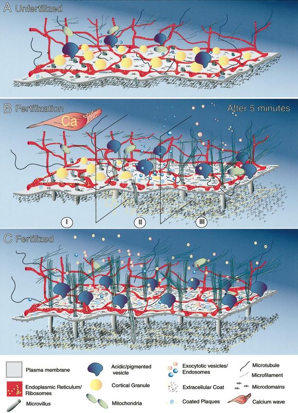
2                                                   Sardet et al.

    © 2001 Elsevier Science. All rights reserved.
What Is the Egg Cortex? 3
layer immediately beneath the cell surface, but depending vitro following addition of ATP or calcium (Sakai, 1960;
on the researcher’s point of view this layer may include Kane and Stephens, 1969; Guerrier, 1972; Mabuchi and
only the plasma membrane (PM) and subsurface coating of Sakai, 1972; Vacquier, 1975; Schatten and Mazia, 1976;
actin microfilaments (MFs) or continue into the deeper Begg and Rebhun, 1979). It is now possible to isolate the egg
cytoplasm. Therefore the thickness of the cell cortex may cortex from echinoderms, molluscs, ascidians, mouse, the
be anywhere between 0.1 and 20 m, and may vary with the amphibian Xenopus laevis, and the annelid Tubifex hattai.
cell type, its functional state, the biological process under Since the multiple connections with the deeper cytoplasm
study, and the resolution of the observations. are severed during the procedure, isolated in vitro cortices
only retain part of the interconnected networks of or-
ganelles and cytoskeletal elements which adhere to the PM
The Egg Cortex in in vivo cortices (Figs. 1D, 1E, 1G, 1H, and 1K). This
The egg cortex is a large assemblage of the plasma situation resembles that of isolated nuclei, which can be
membrane, extracellular coat, and subsurface skeleton to obtained only at the expense of severing the endoplasmic
which a characteristic set of cytoskeletal elements, or- reticulum (ER) and cytoskeletal elements that connect it
ganelles and macromolecules adhere. It behaves as a func- with the rest of the cell. Despite this limitation, isolated
tional unit during the reorganizations that precede and cortices, like isolated nuclei, retain functional properties.
follow fertilization. The egg cortex is also the repository for They have been shown to pump and release calcium (Ter-
spatiotemporal developmental information since it con- asaki and Sardet, 1991), to retain localized mRNAs (Elinson
tains determinants of axis and tissue formation. The exis- et al., 1993; Alarcon and Elinson, 2001), and to sustain
tence of a dynamic egg cortex was recognized in the first organelle and cytoskeleton movements as well as furrow
half of the 20th century when histological observations and progression (Shimizu, 1985; Walker et al., 1994).
experimental manipulations with rods, micropipettes, mag- Our present knowledge is relatively limited when one
netic particles, and using centrifugation showed that cer- considers published work devoted exclusively to the struc-
tain properties of the cortex (morphology, stiffness, thick- ture, function, or polarity of the egg cortex. At the same
ness) changed during maturation, fertilization and the first time, the amount of information concerning the cortex is
cell cycles (reviewed in Harvey, 1956; Chambers and Cham- enormous if one takes into account papers in which the egg
bers, 1961; Raven, 1970; Hiramoto, 1970; Sardet and Chang, cortex is discussed with respect to PM permeability, cal-
1987). Then in the 1970s several groups achieved prepara- cium release, exocytosis, endocytosis, the dynamics and
tions of “isolated cortices”, “cortical lawns”, “cell surface positioning of microtubules (MTs) and MFs, or the localiza-
complexes,” or “cortical hulls” (Figs. 1B and 1C) that tion of mRNAs, to name just a few topics. Reviews treating
retained the ability to contract and undergo exocytosis in the egg or oocyte cortex proper are few and far apart. Those
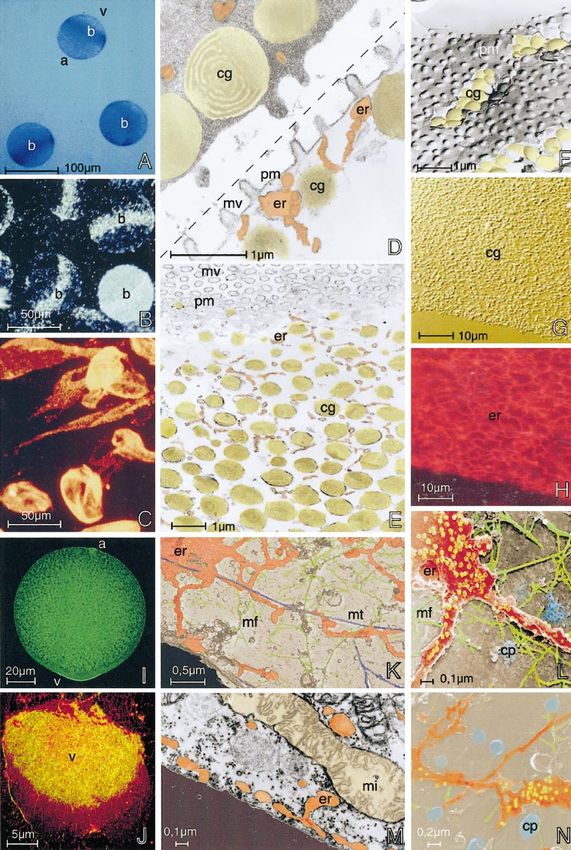
FIG. 1. Views of the egg cortex. Additional material can be found on our Web site: http://www.obs-vlfr.fr/biomarcell.html. Eggs and
isolated cortices of sea urchin (A–H), ascidian (I–M), and amphibian (N). The ER is colorized orange/red in D, E, K, L, M, and N; ribosomes
on the ER are colorized yellow in K, L, and N; the CGs are colorized ocre in D, E, and F; the MFs are colorized green in K, L, and N; the
MTs are colorized dark blue in K; the cytoplasmic face of the PM is light brown in K, L, and N; and coated plaques and vesicles are colorized
light blue in L and N. (A) Living eggs of the sea urchin Paracentrotus lividus display a band of pigmented vesicles (b), one of the earliest
described cortical markers of animal (a)–vegetal (v) polarity (bright field optics, blue filter). (B) Isolated cortices attached to a polylysine
coated surface. The band of pigmented vesicles (b) is retained in cortices. The cortex in the lower right corner has also retained cortical
granules (dark field optics, blue filter). (C) Isolated cortices (cortical hulls) suspended in homogenization milieu (dark field optics). (D) An
egg (top left) and an isolated cortex (bottom right) side by side. In the isolated cortex, the cER network (er) and cortical granules (cg) remain
attached to the plasma membrane (pm) which is covered by microvilli (mv) (electron micrograph (EM) thin section). (E) Oblique tangential
section through an isolated cortex showing the cER network (er) surrounding cortical granules (cg). Microvilli (mv) and plasma membrane
(pm) are on top (EM, thin section). (F) External face of the egg plasma membrane (pm) studded with short micropapillae. A lawn of cortical
granules is situated beneath (EM, freeze fracture replica). (G and H) The same isolated egg cortex. The cortical granules (cg) visualized in
G are surrounded by cER (er) labeled with the lipophilic dye DiI(C16)3 in H (DIC optics in G and fluorescence microscopy in H). (I) A
fertilized egg of the ascidian Phallusia mammillata. The animal (a)–vegetal (v) polarity is evident by the concentration of actin
microfilaments (labeled with fluorescent Phalloidin) which forms near the vegetal pole (v) 5 min after fertilization (confocal microscopy).
(J) Cortex isolated from the vegetal (v) region of an egg 5 min after fertilization (such as the egg in I). The cER network labeled with a
lipophilic dye (red) and MF network labeled with fluorescent phalloidin (green) are concentrated in the vegetal (v) pole (confocal
microscopy). (K) Fragment of isolated cortex from an unfertilized egg showing the networks of cER (er), microfilaments (mf), and
microtubules (mt) (EM, fast freeze/deep etch replica). (L) Detail of an isolated cortex like that shown in K. The cER (er) is studded with
ribosomes and particles. Microfilaments (mf) and coated plaques (cp) are visible as well as the cytoplasmic face of the plasma membrane
(EM, fast freeze/deep etch replica). (M) Vegetal cortical region of an egg showing the cER (er) and one mitochondrion (mi) (EM, thin section).
(N) Fragment of a Xenopus oocyte cortex isolated during vitellogenesis. A large number of coated plaques and vesicles (cp) are present on
the underside of the plasma membrane (EM, fast freeze/deep etch replica).
© 2001 Elsevier Science. All rights reserved.4 Sardet et al.
of Elinson (1980), Vacquier (1981), Longo (1985), and Sardet cytoplasmic reorganizations and polarity when necessary to
and Chang (1987) provide an access to the earlier literature understand causes and consequences of cortical events. It is
on cortical changes in starfish, sea urchins, amphibians, our hope that this review constitutes a useful access to
mammals and a half dozen other organisms. More recent sources and concepts for the many biologists who like to
publications by Sardet et al., (1992), Larabell (1993), use the word cortex freely while describing their favorite
Shimizu (1995), and Chang et al., (1999b) concern cortices process, cytoskeletal element, organelle or molecule.
from oocytes and eggs of the ascidian Phallusia mammil-
lata, the annelid Tubifex and the amphibian Xenopus.
Attention is also given to cortical changes in a few general B. STORING INFORMATION AND
reviews dealing with cytoplasmic reorganizations in BUILDING THE CORTEX DURING
leeches, annelids, the nematode Caenorhabditis elegans, OOGENESIS
ascidians and Xenopus (Elinson and Houliston,1990; Sardet
et al.; 1994; Shimizu, 1995; Fernandez et al., 1998; Gard, The oocyte cortex evolves and enlarges with the rest of
1999). We are also in a somewhat paradoxical situation with the cell as the small germ cell precursors pass through
regard to the contributions of different species to the cortex successive stages of migration, proliferation and then dif-
field. A large part of what we know regarding the structure ferentiation and growth within the gonads, a process that
and the transformations of the cortex is the result of studies can take days, months, or even years (Matova and Cooley,
using eggs of genetically intractable echinoderms, mol- 2001). The largest part of the oocyte’s content generally
luscs, and annelids, while very little is known about the comes from surrounding follicle cells and/or neighboring
structure or evolution of the cortex in Caenorhabditis nurse cells via endocytosis and/or by bulk cytoplasmic
elegans and Drosophila melanogaster, the two species in transfer through intercellular bridges.
which rapid progress is being made in defining cortical Embryonic polarity in Xenopus and Drosophila and
determinants of axis and tissue formation. An intermediate muscle differentiation in ascidians are dependent on the
situation prevails in eggs of Xenopus and ascidians, where localization of mRNA determinants in the oocyte cortex
identified cortical determinants can be studied both in vivo (Van Eeden and St Johnston, 1999; Houston and King,
and in the isolated cortex. 2000b; Mowry and Cote, 1999; Nishida and Sawada, 2001).
A cortical scaffold of cytoskeletal elements and organelles
The Scope of This Review beneath the PM must be assembled in preparation for the
localization of determinant mRNAs during oogenesis,
We will first examine the structural and functional prop- maturation, and fertilization, and for the expression of
erties of the egg cortex during oogenesis as information is these determinants during development. The early stages of
stored in the form of localized determinants, as it endocy- oogenesis are best understood in Drosophila and Xenopus
toses and recycles surface components during vitellogen- while the later steps of maturation and cortex reorganiza-
esis, and as it matures after hormone stimulation and egg tion in preparation for fertilization have been best described
laying. We then detail how the cortex is profoundly reorga- in Xenopus, echinoderms, and mammals.
nized during fertilization or artificial activation and briefly
introduce the establishment and amplification of cortical
polarity, a subject which will be covered in detail in a
Localizing Maternal Determinants in the
second review. What emerges from the synthesis of a large
Drosophila Cortex
and diverse body of literature is that the basic cortical The key event in the establishment of embryonic axes in
scaffold, comprised of the PM, a 3-dimensional matrix of Drosophila is the localization of maternal mRNA determi-
submembranous skeleton based on MFs, and an attached nants and proteins to the cortex during oogenesis. These
network of cortical ER, appears conserved in many oocytes cortical macromolecules essential for axis formation and
and eggs of different species (see Figs. 1 and 2). The presence germ plasm formation are a set of mRNA determinants
or absence in the cortex of many other components (MTs, (bicoid, nanos, oskar) and an increasing number of proteins
intermediate filaments (IFs), organelles, granules, particles) (Vasa, Tudor, Staufen, Egl, Exu, Bic-D, Lis-1, Par-1, . . .)
depends on what type of oocyte, egg, zygote, or embryo is which are synthesized in the 15 nurse cells, translocated
considered, and if the cortex is isolated, what isolation into the oocyte, and anchored in its anterior or posterior
procedures and what milieu are used (reviewed in Vacquier, cortex or poles (these terms are used interchangeably in the
1981; Schroeder, 1981; Sardet and Chang, 1987; Chang et field to designate the 1–20 micron thick region beneath the
al., 1999b). oocyte surface). As a result of these precocious cortical
We have emphasized recent references and reviews, only localizations of determinants, and in contrast to most other
mentioning the older literature where it remains essential species, the future antero-posterior and dorso-ventral po-
or represents an interesting historical source. We have also larities of the Drosophila embryo are established in the
attempted to point out areas where research on somatic gonad well before maturation and fertilization of the egg
cells contributes concepts and models that could be applied (reviewed in Van Eeden and St Johnston, 1999; Lasko, 1999;
to the egg cortex. In addition, we introduce notions of Reichmann and Ephrussi, 2001). The localization of deter-
© 2001 Elsevier Science. All rights reserved.What Is the Egg Cortex? 5
minant mRNAs and proteins produced in the nurse cells dependent repositioning of the GV from the posterior to
requires first the formation of large particles containing anterior cortex which is the essential step in the establish-
messages and proteins and their passage from the nurse ment of oocyte polarity (Cooley and Theurkauf, 1994; Van
cells into the oocyte, and then the translocation of these Eeden and St Johnston, 1999; Reichmann and Ephrussi,
particles in the oocyte to reach their cortical destination. 2001). The relocation of the bulk of the MT network from
These events depend on several phases of reorganizations the posterior to the antero-dorsal cortex is triggered by
during early, mid, and late oogenesis, involving principally extracellular signals from neighboring posterior follicle
the MT network, the cortex and the GV of the oocyte cells in response to the transcription, synthesis and secre-
(Theurkauf, 1997; Megraw and Kaufman, 2000; Cha et al., tion of the growth factor-like protein Gurken in the poste-
2001). rior region of the oocyte where the GV is first localized
During early Drosophila oogenesis (previtellogenic stages (Gonzalez-Reyes and St Johnston, 1998; Micklem et al.,
1– 6), the microtubular network is established by transfer- 1997). After relocalization of the GV, the MTOC and
ing centrosomal foci from the nurse cells into the oocyte via radiating MTs are nested in a crater of the GV in the
a distinctive intracellular structure (the fusome) which antero-dorsal corner of the oocyte and many MT foci are
interconnects all 16 cells of the cyst. The fusome, an located in the anterior and lateral cortex (Paré and Suter,
asymmetric structure made of proteins generally associated 2000; Cha et al., 2001). This reorganization of the MT
with the cortex (spectrins, annexins, ankyrins, adducins, network is again dependent on the cortical protein Par-1
Par-1), apparently plays a major role in determining which which localizes first transiently in the anterior cortex and
of the 16 cells in the cyst will be the oocyte (DeCuevas and then more stably in the posterior cortex by stage 9 (Toman-
Spradling, 1998). By using the fusome as a polarized guide, cak et al., 2000; Shulman et al., 2000). The understanding of
all the centrosomal foci (harboring the minus ends of MTs) the interaction between the MTs and the cortex is far from
and the centrioles from the 15 other cyst cells are trans- complete. The MT reorganization could result from a
ported through cytoplasmic bridges called ring canals disassembly of the MTs at the posterior pole and their
towards the differentiating oocyte (Theurkauf, 1997; selective growth from foci located in the anterior cortex, or
Gonzalez-Reyes and St Johnston, 1998; Grieder et al., 2000; possibly from a rotation of the centrosomal aster and
Navarro et al., 2001). Most centrioles and microtubule attached GV with respect to the cortex. The ER and the
organizing centers (MTOCs) are later found between the secretory pathway, which is known to carry important
posterior cortex and the large oocyte nucleus (the germinal constituents for MT-cortex interactions in yeast, may be
vesicle: GV). Although some ␥–tubulin foci still remain in involved (Roth et al., 1995; Heil-Chapdelaine et al., 1999).
the anterior cortex (Mahowald and Strassheim, 1970), the The changes in the position of the GV and the reorgani-
posterior centrosomal cluster sandwiched between the cor- zation of the MT network in the anterior cortex during
tex and the GV radiates the bulk of the oocyte’s MTs. This midoogenesis have two consequences. First, a second round
posterior centrosomal cluster accumulates important pro- of gurken transcription, localized synthesis, secretion and
teins such as Bicaudal-D (Bic-D), a protein essential for interaction with neighboring follicle cells takes place, fix-
oocyte specification and polarization of the oocyte (Paré and ing the antero-dorsal axis of the embryo. Second, the
Suter, 2000). The positioning of the complex containing presence of numerous MTs in the anterior cortex mediates
Bic-D and other proteins such as Egalitarian (Egl) apparently the arrival of particles containing the anterior determinant
requires the previous localization in the oocyte cortex of bicoid mRNA which is produced in the nurse cells. Al-
DLis-1, a regulator of cytoplasmic dynein implicated in though it is generally believed that an asymmetric MT
nuclear migration in Drosophila and also in fungi and network accounts for the anterior localization of bicoid,
mammals (Swan et al., 1999). How the translocation and recent observations show that the MT network lacks suffi-
posterior positioning of the MT network is accomplished is cient antero-posterior polarity to explain the vectorial
not entirely clear but it has been recently discovered that translocation of the large irregular particles containing
the Drosophila homolog of the Par-1 protein is essential for bicoid to the anterior cortex (Theurkauf and Hazelrigg,
this process and for specifying which of the 16 cells of the 1998; Cha et al., 2001). The specificity of anterior bicoid
cyst will become the oocyte (Cox et al., 2001; Huynh et al., localization probably lies in the specialized properties of the
2001). Par-1 protein is a serine/threonine kinase whose MT network in the anterior cortex to which a translocating
polarized cortical distribution is necessary for axis specifi- machinery attaches. Some of the translocating machinery is
cation of the Caenorhabditis zygote and for polarity of in the process of being uncovered, since bicoid mRNAs
mammalian epithelial cells (Böhm et al., 1997; Kemphues, apparently attach to MTs coursing along the anterior cortex
2000). Drosophila oocyte Par-1 is involved like its mamma- via the linker protein Swallow and the minus end-directed
lian homologues (called MARKs) in the establishment and motor Dynein I (Schnorrer et al., 2000). The nature of the
stabilization of the MT network (Drewes et al., 1997; large bicoid and Exuperantia (Exu)-containing particles re-
Navarro et al., 2001). mains mysterious; they could be the so-called “sponge
The next major reorganization of the MT network with bodies,” which are microdomains identified by electron
respect to the cortex occurs during midoogenesis (stages microscopy containing ER and Vasa-containing nuage ma-
7– 8, when vitellogenesis starts). It is linked to the MT- terial (Wilsch-Brauninger et al., 1997). By stage 8 –9, bicoid
© 2001 Elsevier Science. All rights reserved.6 Sardet et al.
has been translocated and anchored by a process which Structuring and Positioning of Information in the
requires Bic-D and DLis, (Swan et al., 1999; Vallee et al., Xenopus Cortex
2001). These molecules are themselves localized and an-
chored in the cortex during earlier stages of oogenesis by Oocyte growth, cortical transformations and acquisition
unknown mechanisms apparently independent of MTs (Liu of cortical RNAs have been reviewed extensively in Xeno-
et al., 1999; Paré and Suter, 2000). pus (Etkin, 1997; Mowry and Cote, 1999; Gard, 1999; King
A somewhat similar scenario directs the posterior corti- et al., 1999; Chang et al., 1999b; Houston and King, 2000b).
cal localization of oskar mRNA, a determinant important As in Drosophila, a cyst of 16 pear-shaped cells give rise to
for the formation of polar granules (Drosophila germ germ cell precursors, but in Xenopus all cells in the cluster
plasm), and for the subsequent localization and expression become oocytes. Germ cell precursors (stage 0 oocytes) are
of the posterior determinant nanos (Ephrussi and Lehman, polarized with acetylated MTs, mitochondria and golgi
1992). After transient anterior localization, around stage situated at one pole towards the center of the cluster, but it
8 –9 oskar mRNA migrates to the posterior pole in close is not known if that asymmetry bears a relationship to the
association with the cortex. Oskar is thought to travel in future animal-vegetal axis of the oocyte because this polar-
large RNP particles containing the protein Staufen and ity is lost by stage I (reviewed in Gard, 1995, 1999).
plus-end-directed MT motors of the Kinesin I family Early stage I oocytes have no identifiable centrosomes or
obvious polarity in the structure of their MF, MT or
(Micklem et al., 1997; Brendza et al., 2000; Reichmann and
intermediate filament (IF) networks. The IFs are principally
Ephrussi, 2001). It is not known what the exact composi-
type I and type II cytokeratins (keratin filaments: KFs)
tion of these RNP particles is, or if they travel freely along
which appear at mid stage I (Gard and Klymkowsky, 1998).
MTs or in association with a “sponge body”-like structure
Cortical polarity first arises at stage II with the arrival at the
containing ER. A role for internal membranes in Oskar
cortex of the “mitochondrial cloud” with its associated
localization has also been suggested (Ruden et al., 2000;
MTs and KFs. The site at which it attaches to the cortex
Jankovics et al., 2001). A possible involvement of ER in
defines the vegetal pole. The mitochondrial cloud appears
mRNA localization in Drosophila is further implied by the
to develop from one of several fibrous aggregates of mito-
fact that Staufen, a double stranded RNA binding protein
chondria, ER, and electron dense nuage material found
necessary for oskar mRNA and protein localization, is
around the GV in early stage I oocytes (Heasman et al.,
strongly associated with rough ER in fibroblasts and nerve
1984; Kloc et al., 1996; Wylie, 1999; Houston and King,
cells. In these cells, Staufen is also involved in the translo-
2000b). By stage II, cortical MTs with their minus ends in
cation of particulate mRNAs (Wickham et al., 1999; Ro- the cortex are present, along with cortical ER (cER), cortical
egiers and Jan, 2000). granules (CGs), and numerous coated pits which populate
In late oogenesis (stages 10b-14), large quantities of nurse the underside of the PM (Chang et al., 1999b). Vitellogen-
cell cytoplasm are transferred to the oocyte through ring esis begins at this stage, with yolk uptake occurring uni-
canals by cytoplasmic streaming motions (Cooley and formly over the surface (Danilchik and Gerhart, 1987).
Theurkauf, 1994). The process apparently involves non- During stages III and IV, dark pigment granules concentrate
muscle Myosin II and the establishment of sarcomere-like in the animal hemisphere cortex, and the vegetal accumu-
actin MF bundles in the nurse cells (Riparbelli and Callaini, lation of large yolk platelets displaces the GV animally. At
1995; Wheatly et al., 1995). The cytoplasm mixing move- stage III the MT and KF networks are organized radially and
ments are thought to depend in part on acto-myosin and to symetrically throughout the oocyte. The MTs emanate
participate in the anchoring of many cytoplasmic compo- from ␥-tubulin foci in the cortex with many plus ends
nents to the polarized cortical scaffold. This bulk cytoplas- pointing inwards towards the GV (Gard, 1994, 1995). MT
mic transport completes the recruitment of macromol- and KF networks become polarized along the AV axis at
ecules initiated in the cortex at earlier stages of oogenesis. stage IV. Cortical KFs bundle into thicker arrays detected
Such is the case for nanos which accumulates in the principally in the vegetal hemisphere, whereas radial arrays
posterior pole at the end of oogenesis (Mahajan-Miklos and of MTs are more obvious in the animal hemisphere where
Cooley, 1994; Lasko, 1999). yolk platelets are smaller. ␥-Tubulin foci accumulate in the
How mRNA determinants are anchored to the cortex vegetal cortex during this period (reviewed in Gard, 1999).
after being targeted to the proper location is still a matter of By stage VI the oocyte, still surrounded by follicle cells to
debate. MFs and proteins that bind RNA or are involved in which it is connected by numerous gap junctions, has
the control of polyadenylation, such as Staufen or Orb, have grown and accumulated yolk platelets through a cortical
been implicated (Chang et al., 1999a). A precise role for a endocytic process (see vitellogenesis below). It has acquired
specific organelle or cytoskeletal component is difficult to a submembranous layer of CGs and a cortical network of
pinpoint because the close interactions of the cortical ER tubes and cisternae continuous with stacks of subcorti-
constituents make it likely that perturbing one partner, cal annulate lamellae (Campanella and Andreucetti, 1977;
whether MT, MF, or ER, will destabilize the others (Lantz Campanella et al., 1984; Terasaki et al., 2001). The organi-
et al., 1999; Sider et al.,1999; Waterman-Storer and Salmon, zation and localization of KFs has been modified to form
1999). anastomosing networks 3– 4 m thick in the vegetal cortex
© 2001 Elsevier Science. All rights reserved.What Is the Egg Cortex? 7
and around the GV. A finer and deeper array of KFs can be cortex (Deshler et al., 1997, 1998; reviewed in Mowry and
discerned in the animal hemisphere 10 to 15 m beneath Cote, 1999). Other mRNAs such as fatvg seem to use both
the PM (Gard et al., 1995a; Pfeiffer and Gard, 1999). The early and late pathways to localize to the vegetal cortex
MTs are organized radially with minus ends predominantly (Kloc et al., 1998; Chan et al., 1999).
close to the vegetal cortex where ␥-tubulin foci are present Although limited in their interpretation because of the
(reviewed in Gard, 1999). In the animal hemisphere MTs are interdependancy of cytoskeletal and organelle networks,
more abundant and more acetylated, suggesting that they experiments using drugs which depolymerize MTs and MFs
are less dynamic (Gard, 1991). The network of MFs in the and injection of antibodies which interfere with KFs at first
animal cortex is thicker and more contractile than that in suggested that MFs and KFs as well as noncoding RNAs
the vegetal cortex, however there is no detailed description (Xlsirts) are involved in anchoring the 2 populations of
of the organization or evolution of MFs during oogenesis. mRNAs in the vegetal cortex (Klymkowsky et al., 1991;
The use of inhibitors suggests that the cortical actin layer is Kloc and Etkin, 1994). Studies based on the isolation of
necessary for the anchoring of the MT and IF networks, and cortices from stage VI oocytes show that at least three
that ␥-tubulin foci in the cortex are enmeshed in the mRNAs localized through the early and late pathways
cortical MF network (Gard et al., 1997). (Xcat2, Xwnt11, and Vg1) are retained in the cortices
Many specific mRNAs associated with germ cell deter- (Elinson et al., 1993; Alarcon and Elinson, 2001). More
mination such as Xcat2, Xdazl (related to Drosophila nanos recent selective treatment of eggs or isolated cortices with
and boule, respectively), and DEADSouth (an RNA helicase drugs, antibodies, or detergents which perturb or extract the
related to the eukaryotic initiation factor eIF4A), are found major components of the isolated cortex (the MF, KF, and
in the germinal granules (derived from nuage material) of ER networks) indicate that Vg 1 and Xwnt11 interact with
the mitochondrial cloud (Mosquera et al., 1993; Houston et the cortical KF network and possibly the cER, while Xcat2
al., 1998; MacArthur et al., 1999, 2000). When the mito- is apparently not tethered to any of these major constitu-
chondrial cloud arrives at the vegetal cortex, it fragments ents (Alarcon and Elinson, 2001).
into islands of germ plasm measuring about 5 um in Most of the cortically anchored mRNAs have not been
diameter. These microdomains disperse across the vegetal assigned clear functions for the moment except for the
cortex and form characteristic patches rich in mitochondria endoderm determinant VegT and Xdazl which is involved
and germ plasm that contain a number of mRNAs (Xlsirts, in primordial germ cell differentiation (Zhang et al., 1996,
Xwnt11, Xcat2, Xdazl, DEADSouth, Xpat, and Xotx1), 1998; Houston and King, 2000a,b). Experiments using UV
individual mRNAs taking up different positions in the treatment (which acts only within a few microns below the
patches (Ku and Melton, 1993; Kloc et al., 1993; Kloc et al., surface), oocyte inversion, and transfer of cortical material
1998; Hudson and Woodland, 1998; King et al., 1999; from full-grown but not matured oocytes suggest that
Mowry and Cote, 1999; Houston and King 2000a,b; Pannese mRNAs localized close to the surface are important for axis
et al., 2000). This early transport pathway of RNA localiza- establishment and germ cell determination (Holowacz and
tion (the so-called METRO pathway, Kloc and Etkin, 1995) Elinson, 1993, 1995; Kageura, 1997; Marikawa et al., 1997;
is likely due to the bulk translocation of germ plasm discussed and reviewed in Chang et al., 1999b)
material towards the vegetal cortex. Its reliance on the MT
and MF interdependent networks is still unclear. The
ER-rich particles transported by the METRO pathway may
Cortical Polarity and Storage of Information in
be analagous to the large mRNA particles or the nuage- and
Other Oocytes
ER-rich “sponge bodies” described in Drosophila oocytes Compared with Drosophila and Xenopus our knowledge
which are translocated in a MT-dependent fashion (Cha et of cortical polarity and mRNA localization in other organ-
al., 2001; Wilsh-Brauninger et al., 1997; Saffman and Lasko, isms is very poor. There are indications that mRNAs are
1999). localized in the animal and vegetal poles of zebrafish
There is also a late pathway of RNA localization in oocytes (Howley and Ho, 2000; Suzuki et al., 2000). In this
Xenopus (stage II-IV oocytes), during which a second set of organism the arrival of some of these RNAs precedes
mRNAs arrives at the cortex via another route which may morphological manifestations of polarity, so whether their
require the early METRO pathway (Kloc and Etkin 1998; localization causes or results from the establishment of the
Kloc et al., 1998). Among these mRNAs are Vg1 and VegT animal-vegetal axis remains an open question. In eggs of the
which function in patterning the mesodermal and endoder- sea urchin Paracentrotus lividus in which the gradient of
mal layers (Joseph and Melton, 1998; Zhang and King, 1996; cortical pigmented granules represents one of the earliest
Zhang et al., 1998). Vg1 message binds to the RNA binding reports of oocyte polarity (Boveri, 1901; Sardet and Chang
protein Vera/Vg1RBP, a homologue of the –actin RNA 1985; see Figs. 1A and 1B), there is recent evidence that
localizing protein ZBP-1, and to homologues of the human maternal mRNAs and proteins (called BEP) localized in the
hnRNP1 protein (Havin et al., 1998; Zhang et al., 1999; animal cortex are involved in patterning the embryo along
reviewed in Mowry and Cote, 1999). The Vg1 RNA/protein the animal-vegetal axis (Romancino et al., 1998, 2001;
complex is displaced in a MT-dependent process to an Romancino and Di Carlo, 1999). A striking example of
ER-rich region that extends between the GV and the vegetal cortical polarity occurs in ascidian oocytes, in which a large
© 2001 Elsevier Science. All rights reserved.8 Sardet et al.
class of cortically located mRNAs (the PEMs for Posterior C. MATURATION PREPARES THE
End Mark) are distributed along an animal-vegetal gradient CORTEX FOR FERTILIZATION
(Nishida and Makabe, 1999; Sasakura et al., 2000). Some of
these PEM mRNAs and particularly macho, the likely Maturation is the process which takes the oocyte from
tadpole muscle determinant, are retained with the isolated the GV stage (4n chromosomes) to a stage of meiosis at
cortices and appear to localize on the cER which is itself which the oocyte can be fertilized. Depending on the
polarized in unfertilized eggs (Sardet et al., 1992; Nishida species, the mature oocyte arrests in meiotic metaphase I
and Sawada, 2001; Sardet and Nishida, unpublished). (4n chromosomes: ascidians, some molluscs. . . ), meta-
phase II (2n chromosomes: amphibians, mammals. . . ) or
proceeds all the way to interphase (n chromosomes: echi-
The Cortex during Vitellogenesis noderms, cnidarians. . . ). In many species the mature oo-
cyte possesses an animal-vegetal polarity with the small
Because of the intense endocytic activity of the cortex nucleus or meiotic spindle under the animal pole surface
associated with accumulation of yolk vesicles, vitellogen- and/or opposed to the polar bodies, the smallest of cells
esis has the potential to remodel the PM and the attached generated by unequal cleavages (Shimizu, 1990; Verlhac et
cortical cytoskeleton. Recent observations of actin- al., 2000). During maturation, secretory vesicles or CGs
dependent propulsion of endosomes via comet tail forma- localize and anchor to the cortex, awaiting the activating
tion in activated Xenopus eggs raise the possibility that calcium signal which will induce a massive exocytosis.
endosomes generated during vitellogenesis may contribute Reorganizations of the cortex during maturation affect all
to the establishment of a cortical MF scaffold (Taunton et the major networks in the oocyte (cER, MT, MF, and IF) and
al., 2000). lead to relocalization of some cortical mRNAs. Among the
Morphological observations in eggs of insects and am- cortical changes that occur during oocyte maturation, it is
phibians have provided most of our knowledge of vitello- difficult to distinguish those directly related to cell cycle
genesis. In Drosophila, yolk vesicles filled with vitel- changes (completion of meiosis) from those which prepare
logenins start accumulating in the oocyte around stage 8 the egg to respond to sperm. A full understanding of the cell
when polarity is being established (Schonbaum et al., 2000). cycle-related functional and structural changes in the cor-
Vitellogenin receptor (VTGR) mRNA is synthesized in the tex during meiotic completion awaits a description of the
nurse cells, translocated into the oocyte through stage 7, effects of changing activities of important factors such as
and translated. By stage 8 –9 the VTGR proteins, which are MPF and MAPKs on the dynamics of the cortical ER, MF,
LDL receptor subtypes, have moved to the cortex. VTGRs MT, and IF networks (Whitaker, 1996; Nebreda and Ferby,
dock at the PM and vitellogenin proteins secreted by 2000).
surrounding follicle cells are endocytosed, forming vesicles
and endosomes that recycle the receptors back to the Restructuring Cortical Endoplasmic Reticulum and
surface. VTGRs are inserted into the oocyte PM by a Organelles during Maturation
process resembling hormone-stimulated exocytic insertion Response to the fertilizing sperm requires that the egg
of glucose transporters (such as Glut4) in adipocytes or cortex is able to undergo a full calcium response, massive
muscle cells (Holman and Sandoval, 2001). Coated vesicle- exocytosis and endocytosis, as well as propagated contrac-
mediated endocytosis of vitellogenin, triggered by a juve- tility (Suzuki et al., 1995). These properties are made
nile hormone, is followed by formation in the cortex of possible by the development just beneath the PM of an
tubulovesicular early endosomal components and multive- extensive network of tubes and sheets of cER which stores
sicular bodies considered to be late endosomes. Yolk and releases calcium (Gardiner and Grey, 1983; Campanella
vesicles form from the maturing endosomes while receptors et al., 1984; Sardet 1984; Larabell and Chandler, 1988
are recycled back to the oocyte surface until vitellogenesis Henson et al., 1990; Terasaki et al., 2001; see Figs. 1H, 1K,
is completed (Schonbaum et al., 2000). This vitellogenic 1L, 1M and 1N). The positioning of a large population of
phase of oogenesis bears common themes with late oogen- exocytic vesicles (often called CGs) and coated plaques
esis in other insects, fish, birds, and frogs which also have beneath the PM is also necessary (Chandler and Heuser,
LDL Receptor type VTGRs, multivesicular bodies that are 1981; Sardet, 1984; Sardet et al., 1992; Chang et al., 1996;
precursors of yolk vesicles, and amplified coated vesicle 1999b; see Figs. 1L and 1N), as well as the stabilization of a
endocytosis, a process originally discovered in mosquito meshwork of cortical MFs responsive to the secondary
oocytes (Roth and Porter, 1964). Recent investigations of messengers generated by sperm fusion and entry (Ducibella
oogenesis in Caenorhabditis show that the basic compo- et al., 1990; Johnson and Capco, 1997). The most extensive
nents of VTGR mediated trafficking are also conserved in information in this field comes from starfish and Xenopus
nematodes (Grant and Hirsh, 1999; Greener et al., 2001). oocytes which can be stimulated to mature by the hor-
We can expect rapid progress in this domain from an mones 1 MeAdenine and progesterone, respectively, and to
experimental model where forward and reverse genetic proceed either to completion of meiosis II (starfish) or to
analysis can be used extensively. arrest in metaphase of meiosis II (Xenopus). Additional
© 2001 Elsevier Science. All rights reserved.What Is the Egg Cortex? 9
observations come from spontaneously maturing eggs of retrieval of channels in Xenopus PM shows that in quies-
ascidians and sea urchins (Swalla et al., 1991; Jeffery, 1995; cent oocytes, the PM proteins turn over and that the mem-
Berg and Wessel, 1997). brane is constantly renewed via endocytotic-exocytotic
During maturation oocytes acquire responsiveness to IP3, trafficking, possibly regulated by protein kinase C (PKC)
the main calcium-releasing messenger used at fertilization. (Forster et al., 1999). Interestingly, some PM components can
This explains why in many species, including mammals, be replaced in 24 h, as demonstrated by the injection of
the sperm has the ability to bind and fuse to an immature connexin50 mRNA, and the study of the expression of the
GV stage oocyte but fails to activate it (Evans et al., 2000). protein and of its insertion and retrieval in the PM of the
The ability to respond to IP3 is likely due to the establish- Xenopus oocyte (Zampighi et al., 1999). As this experimental
ment of the extensive cER network underneath the PM model is used by a growing number of physiologists inserting
(Campanella et al., 1984; Larabell and Chandler, 1988; their favorite channels in the PM by expression of injected
Callamaras and Parker, 1999; Terasaki et al., 2001; re- mRNA, it is expected that soon we will be able to visualize
viewed in Sardet and Chang, 1987; Stricker et al., 1998; what types of microdomains exist in the PM of Xenopus
Kline, 2000). This cER network reorganizes from stacks of oocytes and understand what physiological functions they
perinuclear and subcortical ER, as well as from stacked ER may perform (Singer-Lahat et al., 2000).
which forms the annulate lamellae. Recent observations of
GFP-labeled ER networks in Xenopus show that small
clusters of ER develop in the vegetal cortex at the time of
Restructuring the Cytoskeleton during Maturation
GVBD, then disappear and reappear at the time of the The cytoskeletal networks of MFs, MTs, and IFs are
second meiotic metaphase block (Terasaki et al., 2001). In restructured and further polarized during maturation. A
mammals, clusters of cER are small and sparse in the spectacular example of polarization is found in oocytes of
immature oocyte and they grow in size and number until the annelid Chaetopterus, where a cortical attractor at the
meiotic arrest is established (Shiraishi et al., 1995; Kline, animal pole tethers the meiotic spindle, even when the
2000). These data suggest that changes in cER organization spindle is displaced by violent needle manipulations (Lutz
are coupled to MPF activity and that cER clustering confers et al., 1988). Similarly in starfish oocytes a distinct astral
the ability to release calcium in response to IP3 (Mehlman MT scaffold sandwiched between the PM and the GV,
et al., 1995, 1996; Shiraishi et al., 1995; Terasaki et al., called the premeiotic aster, connects the GV to the animal
2001). In cytoplasmic extracts of Xenopus eggs and in pole cortex (Miyazaki et al., 2000). This polar spot is devoid
somatic cells, ER tubules have been observed to attach to of the reticulated network of KFs resembling a hair net
MTs and MFs, leading to the elaboration of a 3 dimensional (called a “snood,” Schroeder and Otto, 1991) situated 1 m
ER network (Klopfenstein et al., 1998; Lane and Allan, beneath the surface in sea urchin and starfish oocytes. This
1998; Tabb et al., 1998), but it is not known whether the anastomosing KF network disappears before the end of
same mechanisms preside over the establishment of the maturation, and at the same time the length and number of
cortical ER network in live eggs. The cER which progres- cortical MTs diminish (Boyle and Ernst, 1989). In Xenopus,
sively surrounds CGs is strongly tethered to the PM by the cortical KFs also disassemble during maturation, probably
time the oocyte becomes ready to be fertilized. It is not as a result of MPF activation, and disappear completely in
known however whether these attachment sites play a role the vegetal hemisphere at the time the egg is fertilized
in rendering the cER network competent to release calcium (reviewed in Gard, 1999). In eggs blocked in meiosis I or II
in response to the factors introduced by sperm (Stricker, (ascidians, mammals), the meiotic spindle also seems to
1999). interact with the cortex and there is evidence that chromo-
During maturation in Xenopus, starfish, sea urchins and somes themselves can provoke the accumulation of MFs at
mammals, thousands of golgi-derived cortical vesicles line the cell surface independently of spindle MTs (Maro et al.,
up beneath the PM, ready for exocytosis in response to the 1986). The presence of an actin-rich plaque covering the
rise in cytoplasmic calcium concentration triggered by meiotic spindle appears to be a common feature of the
sperm entry. The CGs arrive there either progressively (in animal cortex region (Sardet et al., 1992; Evans et al., 2000;
vertebrates and starfish) or synchronously (in sea urchins) Verlhac et al., 2000).
during the period between GV migration and GVBD (Duci- Changes in the MF network associated with the morphol-
bella et al., 1994; Berg and Wessel, 1997). In sea urchins, ogy of surface microvilli have also been reported during
these vesicles, filled with secretory proteins and reticulat- maturation (reviewed in Sardet and Chang, 1987; Johnson
ing enzymes, are attached to the PM by specialized sites and Capco, 1997). The most spectacular structures are the
(Vacquier, 1975; Zimmerberg et al., 1985). By the time giant actin spikes that appear in starfish oocytes in places
GVBD has occurred, membrane microvilli and associated where the oocyte had established junctions with surround-
actin bundles have retracted and the oocyte membrane has ing follicle cells (Otto and Schroeder, 1984). These spikes
decreased its surface area by half. In both Xenopus and which contain fascin and other Actin Binding Proteins
starfish the PM has also acquired new ion channels and (ABPs) disappear 30 min before GVBD and are replaced by
electrical properties (reviewed in Sardet and Chang, 1987; flacid projections which shorten further as maturation
Nuccitelli and Ferguson, 1994). Studies of insertion- progresses. Remodeling of the actin cortex also occurs
© 2001 Elsevier Science. All rights reserved.10 Sardet et al.
during maturation in Xenopus and is manifest as the The Cortex as the Site of Attachment and Fusion
development of the ability to contract when calcium levels of Sperm and Egg
are elevated by natural or artificial means (Merriam et al.,
1983; Benink et al., 2000). In the sea urchin and starfish eggs, sperm fusion occurs
randomly on the egg surface. In eggs of the amphibian
(Xenopus) and ascidian (Phallusia), preferred sperm entry is
in the animal hemisphere. The underlying mechanisms
Relocalization of Cortical mRNAs and Domains cannot yet be associated with any clear differentiation in
during Maturation the cortex of these species (Elinson, 1975; Speksnijder et al.,
Some of the cortical changes which take place during 1989), although in Xenopus oocytes surface microvilli are
more sparse in the animal hemisphere (Monroy and Bac-
meiotic maturation have significant consequences on
cetti, 1975). On mouse eggs, sperm binding and fusion is
cortical determinant and mRNA localizations. In Xeno-
restricted to the vegetal 2/3 of the egg which is character-
pus, the sensitivity of dorso-anterior determinants to UV
ized by microvilli covered by specific molecules associated
irradiation of the vegetal pole decreases up to GVBD,
with sperm binding (Evans et al., 2000). The smooth patch
indicating that the UV target undergoes a change in
lacking microvilli around the animal pole arises during
nature or position during maturation (Holowacz and maturation as precocious exocytosis of CGs and reorgani-
Elinson, 1993; 1995; Marikawa et al., 1997). Messages for zations of the MF network take place (Ducibella et al.,
Vg1 (but not Xcat2) are released from their tight cortical 1990; Simerly et al., 1998). Many eggs have special extra-
association, independently of the disappearance of the KF cellular structures (micropyles) which form conduits to
network, and spread out over the vegetal hemisphere guide the sperm towards the animal or, in rare cases, the
from the time of GVBD until meiotic metaphase arrest vegetal pole. Animal and vegetal differentiations in the egg
(Forristal et al., 1995; Klymkowsky et al., 1991). Patches cortex related to a specific sperm entry site clearly exist in
of germinal granules and mitochondria containing mR- some cnidarians (Carré and Sardet, 1981) and annelids
NAs such as Xcat2 congregate to form larger islands of (Focarelli and Rosati, 1995).
germ plasm that remain close to the vegetal surface Although sperm-egg binding proteins seem to vary widely
(Savage and Danilchik, 1993). It is interesting to note that among different phyla (Vacquier, 1998), surface macromol-
mRNAs encoding Par-6, which associates with the im- ecules of the integrin family are apparently main actors in
portant cell polarity protein Par-3 (ASIP/Bazooka), is sperm-egg interaction in mammals, amphibians and inver-
restricted to the animal hemisphere of Xenopus oocytes tebrates. In mice, different surface proteins of the Integrin
(Choi et al., 2000), while Xenopus homologues of the family are specifically located in the microvillar (vegetal
Par-3 protein and aPKC are progressively localized in the hemisphere and equatorial region) or the amicrovillar (ani-
animal cortex (Izumi et al., 1998; Nakaya et al., 2000). mal pole) regions and are implicated in the binding of sperm
These recent observations on the Par-3/Par-6/aPKC com- proteins and possibly in sperm-egg fusion through associa-
plex, and other studies concerning conserved germ plasm tion with receptor partners (LeNaour et al., 2000; Evans et
components (Xcat2 and Nos), emphasize that similarities al., 2000; Wassarman et al., 2001). In sea urchins,
may exist between the establishment of the animal- -integrins combine at the tip of microvilli where they may
vegetal axis in the amphibian oocyte and the acquisition act as posts anchoring the vitelline membrane. These
of anterior–posterior polarity in the Caenorhabditis zy- integrins are severed from the surface by proteases during
CG exocytosis, leaving their cleaved transmembrane and
gote (Subramaniam and Seydoux, 1999; Kemphues, 2000;
cytoplasmic fragments attached to the PM where they may
Houston and King, 2000b; Ohno, 2001). It remains to
anchor the cortical MF cytoskeleton and participate in its
correlate these changes in Par proteins and mRNA local-
transformation during fertilization (Murray et al., 2000). In
ization with the maturation of the cortical ER, MF, MT
many species, fusion between the gametes is thought to
and KF networks and also to analyze the changing loca-
take place at the tip of microvilli, a recurring theme in
tions of mRNAs in cortices isolated at different times cell-cell adhesion and fusion (Wilson and Snell, 1998). The
during maturation (Alarcon and Elinson, 2001). MFs abutting the tip of the microvilli may participate in the
Not much is known about cortical mRNA relocalizations maintenance of membrane microdomains involved in
during maturation in species other than Xenopus. As men- sperm-egg binding, signal transduction and/or MF polymer-
tioned earlier, ascidian eggs contain a large population of ization. In sea urchins, a candidate sperm receptor appar-
cortically localized PEM mRNAs (Nishida and Makabe, ently localizes to the tip of microvilli (Ohlendieck and
1999). The positioning of these cortical mRNAs has not Lennarz, 1995; Giusti et al., 1997). The mechanism of
been examined in detail, but it is probable that they reach sperm-egg fusion is not well understood. Recently CD9
their cortical location at the same time the mitochondria- proteins, members of the tetraspan family of integral mem-
rich myoplasm layer is positioned in the subcortical vegetal brane proteins associating with 1 integrins, have been
hemisphere of the maturing oocyte (Jeffery and Capco, proposed to participate in fusion based on the phenotype of
1978; Swalla et al., 1991; Sasakura et al., 2000). mutant mice (LeNaour et al., 2000; Wassarman et al., 2001).
© 2001 Elsevier Science. All rights reserved.What Is the Egg Cortex? 11
© 2001 Elsevier Science. All rights reserved.12 Sardet et al.
D. FERTILIZATION TRANSFORMS remains elevated (Terasaki and Jaffe, 1991; Jaffe and Ter-
THE EGG CORTEX asaki, 1994; Terasaki et al., 2001). The continuous
3-dimensional network of ER tubes and sheet reforms after
Fusion with sperm brings enormous changes to the egg the intracellular calcium level decreases.
cortex (Fig. 2). These changes have been best described in In ascidians and the annelid Cerebratulus where fer-
sea urchins, amphibians, ascidians and mammals. They are tilization triggers repetitive calcium waves, the ER net-
immediate consequences of the massive release of intracel- work does not fragment. Instead, sheets and tubes of the
lular calcium that floods the egg, propagating through it ER network aggregate into clusters (microdomains) after
from the cortical site of sperm entry (Stricker, 1999). It is fertilization (Speksnijder et al., 1993; Stricker et al.,
interesting to note that in Medaka fish eggs, where the first 1998). In mouse eggs cER clusters are also visible as sites
calcium waves were discovered more than 20 years ago, the of IP3 receptor accumulation which line the PM in the
propagation of the waves is restricted to the cortex (Gilkey vegetal hemisphere (Kline et al., 1999; Kline, 2000). In
et al., 1978). the ascidian egg cortex, the aggregation of cER in the
vegetal contraction pole is amplified by a spectacular
actomyosin-driven cortical contraction triggered by cal-
Sperm-Induced Calcium Waves and Membranes cium release (Sardet et al., 1989; Roegiers et al., 1995; see
The fusion of egg and sperm PMs brings about large Fig. 1I, J). A domain of cER accumulation 20 m long and
changes in membrane permeability and electrical potential 2–5 m thick, which can be isolated as a cell surface
as well as the calcium-mediated activation of the egg complex and is not dislodged by centrifugation, acts as a
(reviewed in Nuccitelli and Ferguson, 1994; Stricker, 1999; calcium wave pacemaker emitting 6 –12 cortical waves
Swann and Parrington, 1999; Jaffe et al., 2001). The initial which originate in the vegetal cortex (Speksnijder et al.,
depolarization of the PM causes a brief and nonactivating 1990a,b; McDougall and Sardet, 1995; Dumollard and
calcium influx all around the cortex in sea urchins and a Sardet, 2001). It is probable that in mouse and annelids,
more substantial calcium influx in mollusc eggs where it is like in ascidians, the cortical spots of cER accumulation
the signal for activation. In annelids, sea urchins, ascidians, act as calcium wave pacemakers during the completion of
amphibians, and mammals, it has been hypothesized that the meiotic cell cycle (Stricker et al., 1998; Kline et al.,
the signal for activation is a factor locally introduced by the 1999; Deguchi et al., 2000). It is intriguing that calcium
sperm that acts on the cortex to trigger a single or multiple wave pacemakers are all located in the vegetal cortex in
calcium waves (Oda et al., 1999; reviewed in Sardet et al., the eggs of widely different organisms (annelids, ascid-
1998; Stricker, 1999; Jaffe et al., 2001). In sea urchins a ians, mammals). Whether such directional waves play a
single wave of propagated calcium release from the ER role in later development remains an open question
sweeps through the egg cortex and cytoplasm at an approxi- (Sardet et al., 1998; Jones, 1998).
mate speed of 5–10 m/s (Suzuki et al., 1995; McDougall et It is not known what happens to the sites of attach-
al., 2000). In annelids, ascidians, and mammals, multiple ment of the cER to the PM after fertilization. It has been
waves are produced during the completion of the meiotic hypothesized that these cER-PM junctions might be like
cell cycles (Speksnijder et al., 1990a,b; Stricker et al., 1998; PM-sarcoplasmic reticulum junctions characteristic of
Kline et al., 1999; Deguchi et al., 2000). The cER and the muscle cells (Gardiner and Grey, 1983; Sardet, 1984;
deeper interconnected network of rough ER tubes and Sardet et al., 1992). There may also exist a link between
sheets are reorganized at the time free cytoplasmic calcium these ER-PM junctions and the presence of store-operated
concentration reaches activating levels (5–20 m). In sea calcium channels described in ascidian and Xenopus eggs
urchins, starfish, and Xenopus the single calcium wave (Arnoult et al., 1996; Jaconi et al., 1997; Yao et al., 1999;
sweeps through the egg from the sperm entry site and Machaca and Haun, 2000). Considering the recently-
causes a wave of vesiculation of the ER network which described association between store-operated calcium
persists as long as the intracellular calcium concentration channels and IP3 or Ryanodine Receptors (Csutora et al.,
FIG. 2. Transformation of the cortex during activation. This schematic representation is inspired from the work done on sea urchins and
Xenopus eggs where major exocytotic and endocytic events take place after the egg is fertilized. (A) Unfertilized egg cortex phenotype. The
plasma membrane (grey) has a cell surface coat (black) and its cytoplasmic face comprises coated plaques (blue), short microfilaments
(green), cortical vesicles (cortical granules: ocre, acidic/pigmented vesicles: blue) and an attached network of cortical endoplasmic reticulum
(red) studded with ribosomes (yellow). (B) At fertilization, the calcium wave triggers waves of cortical reorganizations. Cortical granules fuse
with the plasma membrane (I left), exocytosing structural proteins and enzymes which form the fertilization membrane. After the passage
of the calcium wave, endocytic activity is stimulated, the endoplasmic reticulum is fragmented (II), and microfilaments polymerize and
bundle in elongating microvilli and underneath the plasma membrane (II middle, and III right). (C) Fertilized egg cortex phenotype.
Microvilli have elongated and bundles of microfilaments are abundant underneath the plasma membrane. Acidic/pigmented vesicles have
moved closer to the plasma membrane.
© 2001 Elsevier Science. All rights reserved.You can also read