The Cytoskeleton-A Complex Interacting Meshwork
←
→
Page content transcription
If your browser does not render page correctly, please read the page content below
cells
Review
The Cytoskeleton—A Complex Interacting Meshwork
Tim Hohmann            and Faramarz Dehghani *
 Institute of Anatomy and Cell Biology, Martin Luther University Halle-Wittenberg, Grosse Steinstrasse 52,
 06108 Halle (Saale), Germany; tim.hohmann@medizin.uni-halle.de
 * Correspondence: faramarz.dehghani@medizin.uni-halle.de; Tel.: +49-345-5571707

 Received: 26 March 2019; Accepted: 15 April 2019; Published: 18 April 2019

 Abstract: The cytoskeleton of animal cells is one of the most complicated and functionally versatile
 structures, involved in processes such as endocytosis, cell division, intra-cellular transport, motility,
 force transmission, reaction to external forces, adhesion and preservation, and adaptation of cell
 shape. These functions are mediated by three classical cytoskeletal filament types, as follows: Actin,
 microtubules, and intermediate filaments. The named filaments form a network that is highly
 structured and dynamic, responding to external and internal cues with a quick reorganization that
 is orchestrated on the time scale of minutes and has to be tightly regulated. Especially in brain
 tumors, the cytoskeleton plays an important role in spreading and migration of tumor cells. As the
 cytoskeletal organization and regulation is complex and many-faceted, this review aims to summarize
 the findings about cytoskeletal filament types, including substructures formed by them, such as
 lamellipodia, stress fibers, and interactions between intermediate filaments, microtubules and actin.
 Additionally, crucial regulatory aspects of the cytoskeletal filaments and the formed substructures are
 discussed and integrated into the concepts of cell motility. Even though little is known about the
 impact of cytoskeletal alterations on the progress of glioma, a final point discussed will be the impact
 of established cytoskeletal alterations in the cellular behavior and invasion of glioma.

 Keywords: actin; microtubules; intermediate filaments; motility; migration; glioma; signaling

1. Introduction
      A single animal cell has the ability to adapt its shape in response to environmental confinements or
chemical cues, to move through tissues (artificial and in vivo, including narrow spaces), and to divide.
These processes are all, at least in part, orchestrated by the (dis-)assembly of cytoskeletal proteins.
The cytoskeleton is made up of three major types of proteins, as follows: Tubulin, actin, and proteins
forming intermediate filaments. These cytoskeletal proteins differ not only in their chemical structure,
but also in the type of filaments and structures they form, ranging from fast assembling dendritic
actin networks of the lamellipodium, capable of generating forces necessary for cell movement over
single microtubule filaments as transport structures, to intermediate filaments capable of promoting or
inhibiting cell movement and stabilizing the cell against large stress.
      To form a myriad of different cytoskeletal structures, as it is observed in animal cells, the cytoskeletal
meshwork does not only need different components with different properties and functions, but also
a tight and precise regulation of (dis-)assembly of its components, the respective local regulation of
(dis-)assembly-factors, and interactions between the actin, microtubule, and intermediate filament networks.
      The ability of cells to migrate is of special interest in glioma spreading, as the success of glioma
treatment is crucially coupled to the question of whether a recurrent tumor will arise or not, as resection is
successful only if the tumor is completely removed. Consequently, recurrent tumor formation is considered
to be the main reason of tumor morbidity [1]. Hence, targeting the migratory machinery in gliomas can
be a promising approach for the containment of metastasis. For successful targeting of glioma migration,
a broad and detailed knowledge of the cytoskeletal architecture and its alterations is necessary.

Cells 2019, 8, 362; doi:10.3390/cells8040362                                         www.mdpi.com/journal/cells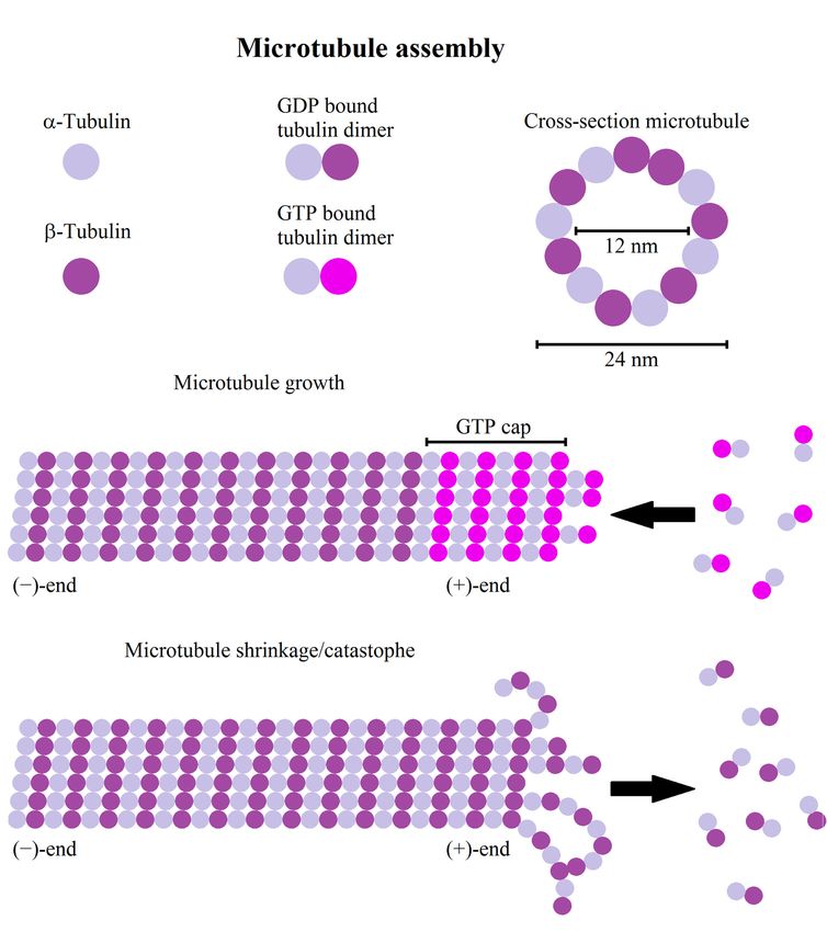
Cells 2019, 8, 362                                                                                          2 of 55

    Here we provide an overview of different cytoskeletal filaments, including actin, microtubules,
and intermediate filaments, their (dis-)assembly, interactions, and function in motility and shape
changes of healthy cells. Afterwards, cytoskeletal alterations in glioma and their impact on their
migratory behavior are discussed.

2. Actin Regulation and Structure
     In cells, actin occurs in two distinct states, as follows: The monomeric G-actin and filamentous
F-actin. The modulation of the actin cytoskeleton is regulated by the balance of globular G- and
polymeric F-actin and by actin associated proteins [2]. The actin cytoskeleton forms a network consisting
of polarized filaments that are mostly associated with force generation necessary for movement, focal
adhesion, and shape changes. In the following we describe the building blocks of actin filaments,
the assembly and disassembly of filaments, their kinetics, regulation, as well as filament bundles,
network structures, and their mechanical properties. A summary of all mentioned actin binding or
associated proteins and their function can be found in Table 1.

          Table 1. Summary of mentioned actin associated proteins and their direct or indirect functions.

                     Actin Associated Proteins                        Function
                             Arp2/3                               Polymerization factor
                            Ena/VASP                  Polymerization factor, anti-capping function
                             FMNL2                                Polymerization factor
                              mDia1                               Polymerization factor
                              mDia2                               Polymerization factor
                             Profilin                        Inhibits actin polymerization
                           ADF/Cofilin                                Actin severing
                              Arpin                                  Inhibits Arp2/3
                            Myosin II                      Cell/actin contractility, cross linker
                               RLC                                 Activates myosin II
                              MLCK                                 Activates myosin II
                             MHCK                              Inhibits myosin II activity
                               PKC                             Inhibits myosin II activity
                               CKII                            Inhibits myosin II activity
                              Scruin                                   Cross linker
                              Fascin                                   Cross linker
                            α-actinin                                  Cross linker
                             Filamin                                   Cross linker
                             Fimbrin                                   Cross linker
                             Paladin                                   Cross linker
                              Ezrin                             Membrane-cortex linker
                             Radixin                            Membrane-cortex linker
                             Moesin                             Membrane-cortex linker
                              Cdc42              Signaling molecule, activates mDia2, WAVE, N-WASP
                               Rac1                Signaling molecule, activates WASP/WAVE, arpin
                              RhoA                Signaling molecule, activates ROCK, mDia1, LIMK
                              ROCK                      Signaling molecule, activates myosin II
                           WASP/WAVE                      Signaling molecule, activates Arp2/3
                            N-WASP                        Signaling molecule, activates Arp2/3
                              LIMK                      Signaling molecule, inhibits ADF/cofilin

2.1. Actin Filaments
     Actin filaments are, in contrast to intermediate filaments and microtubules, semi-flexible filaments,
forming dendritic or cross-linked structures. Semi-flexible means that the persistence length of a single
filament is in the order of the filament length, where the persistence length is the length scale on which the
correlation between two tangents along the filament drops to 1/e [3]. As a semi-flexible polymer, actin
filaments are actively bent by thermal fluctuations, thus generating additional resistance to forces stretching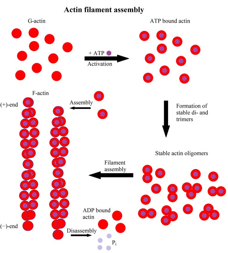
Cells 2019, 8, 362                                                                                           3 of 55

the filament. Actin itself is considered the most dynamic of the three cytoskeletal proteins capable of strong
structural changes in the time scale of minutes, thus determining the shape of a cell. A single actin filament
consists of actin monomers, called globular actin (G-actin). Under nearly physiological conditions G-actin
polymerizes to asymmetric helical structures, filamentous actin (F-actin), with a typical length of 6–7 µm in
in vitro studies [4]. The nucleation kinetics is mostly limited by the generation of dimers and trimers [5].
Having reached the trimer state, filament nucleation increases rapidly, but in dependence of the available
G-actin pool [6] (Figure 1). The resulting actin filaments have a right handed helical structure. G-actin is
polarized, therefore F-actin is polarized as well, with the less dynamic side termed as the (−)-end and the
more dynamic (+)-end having a ten times higher polymerization rate than the (−)-end [6]. As actin is an
ATPase the (+)- and (−)-ends can also be distinguished by their ATP/ADP status, especially if the growth at
theCells
    (−)-end    is362
         2019, 8, inhibited further. Thus the (+)-end contains higher amounts of ATP bound actin while      the
                                                                                                         4 of 58
(−)-end contains more ADP bound actin.

      Figure 1. Scheme of actin filament formation. First G-actin binds to ATP. Afterwards, it forms stable di-
         Figure 1. Scheme of actin filament formation. First G-actin binds to ATP. Afterwards, it forms stable
      or trimers and, finally, filaments elongate by addition of monomers. Hydrolysis of ATP to ADP leads to
         di- or trimers and, finally, filaments elongate by addition of monomers. Hydrolysis of ATP to ADP
      a distinction between the fast growing (+)-end and the slower growing or dissociating (−)-end.
         leads to a distinction between the fast growing (+)-end and the slower growing or dissociating
         (−)-end.

   2.2. Profilin
        As F-actin is capable of forming spontaneously above a certain critical concentration of G-actin
   (≈0.1 µM) a precise cofactor-driven control of actin polymerization is necessary. One such control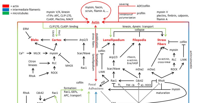
Cells 2019, 8, 362                                                                                    4 of 55

2.2. Profilin
      As F-actin is capable of forming spontaneously above a certain critical concentration of G-actin
(≈0.1 µM) a precise cofactor-driven control of actin polymerization is necessary. One such control
element is profilin. Profilin is an actin binding protein that regulates actin homeostasis [7,8] by
inhibiting the spontaneous formation of actin di- and trimers, but it also catalyzes the transition from
ADP- to ATP-actin [7]. Profilin bound G-actin can be used for the construction of actin filaments
if nucleation factors like the Arp2/3 (actin-related-proteins 2/3) complex or formins are present [9].
Interestingly, if formins and profilin are present, free actin elongation can be increased by a factor of up
to 9 [10,11]. Additionally, profilin was shown to inhibit polymerization at the (−)-end of actin [6].

2.3. Dendritic Actin Networks
     Besides the quasi-linear actin filaments there are dendritic actin networks, formed by the
Arp2/3 complex [12]. These networks are usually formed at the cell front on a short time scale [13] and,
thus, its regulation is of high importance. The generation of a dendritic actin network starts from a so
called primer, an existing actin filament at which Arp2/3 binds to its side [9,14]. Arp2/3 is, amongst
others, activated by members of the WASP family [6,15]. For the generation of a dense dendritic
network not only nucleation factors, but also capping proteins are needed to restrict the elongation
of the actin (+)-ends [14,16–18]. If capping proteins are present the Arp2/3 complex can generate
multiple networks originating from different actin filaments that are able to merge and generate forces
near the cell membrane [14,17]. The number of nodes is important for the mechanical properties of
the generated network and consequently the elastic modulus scales with the mesh size M by 1/M4 .
In general the dendritic network behaves visco-elastic, meaning it is mainly elastic on small time scales
(10 min) [14,17].

2.4. Non-Muscle Myosin
     Another important molecule class is the myosin family. Here we will focus exclusively on the
non-muscle myosin. Myosin is responsible for the contractility of anti-parallel actin structures using
ATP hydrolysis as the energy supply [19,20]. These contractile structures are mainly responsible for the
retraction of the cell rear for productive movement, but also for transmitting forces to the surrounding
extra-cellular matrix.
     Interestingly, myosin II motor activity alone is insufficient to produce contractility. Single myosin
II hexamers are unipolar and thus ineffective in generating contractile forces [21], but when assembled
into bipolar mini-filaments they are highly processive and capable of generating forces by pulling on
anti-parallel actin filaments [22]. Myosin II can be activated via phosphorylation of the regulatory light
chain (RLC) or activation of myosin light chain kinases (MLCK). RLC is activated via Rho-associated
protein kinase (ROCK) or citron kinases (both activated by RhoA) and MLCK by Ca2+ [23]. After RLC
phosphorylation myosin is capable of generating contractile forces [24,25]. Another type of regulation
works via the phosphorylation of the myosin heavy chain, utilizing myosin heavy chain kinases
(MHCK), casein kinase II (CKII), or protein kinase C (PKC), inhibiting mini-filament assembly or
dissociating existing mini-filaments [26–29]. The switch between those two activation states influences
the contractility of the respective acto-myosin network. Consequently, regulation of myosin strongly
impacts organization and properties of contractile actin structures, as discussed below.

2.5. Cross-Linked Actin Networks and Actin Bundles
     Despite the already mentioned dendritic actin structures, there are actin bundles and networks
linked together by cross-linkers. Cross-linkers are molecules that connect single actin filaments either
transiently or non-transiently and are either passive (e.g., scruin, fascin, α-actinin, filamin, or fimbrin)
or active (myosin). Cross-linked actin bundles and networks largely control shape, mechanical integrity,
and contractility of a cell [30–32]. Generally, cross-linkers do not influence actin assembly (except
Cells 2019, 8, 362                                                                                     5 of 55

for Arp2/3) [32–35]. Cross-linkers bind actin filaments based on their own size and the position
of their binding-sites in different distances, ranging from 10 nm for fimbrin to 160 nm for filamin,
and thus determine the density of the resulting actin structure [36,37]. Additionally, the speed of
actin polymerization influences the presence of cross-linkers in the resulting network, supposedly by
crowding effects, thus excluding larger cross-linkers like α-actinin in quickly polymerizing filaments [33].
If the formed actin structures are subject to a force that acts on a longer time scale than the binding
time of the cross-linkers itself, a reorganization of cross-linkers and a subsequent shape change of the
bundle occurs [38]. This time scale depends on the type of cross-linker and its binding and unbinding
time, which can be in the order of seconds [39,40]. Consequently, cross-linked actin is elastic on short
and viscous on long time scales [38] and the presence of cross-linkers generally increases the elastic
part of the visco-elastic answer to external stress.
      If actin filaments are bundled by cross-linkers the filaments inside the bundle can either be oriented
in parallel or anti-parallel, meaning that (+)-ends of neighboring filaments are pointing in the same
or the opposite direction. Parallel actin bundles are found amongst others in filopodia [18,41], while
anti-parallel bundles are mostly found in stress fibers.
      Two mechanisms were proposed to explain the generation of parallel actin bundles. The first
mechanism involves the Arp2/3 complex-dependent elongation of filaments in the absence of capping
proteins, so that free growing (+)-ends transition into bundles via electro-static interactions between
filaments [42]. Here, the geometric constraints and the angle at which filaments make contact to
each other determine whether parallel or anti-parallel bundles are formed [42]. Notably, geometric
constraints refer to all spatial limitations, such as the available free space, steric effects, etc., that
potentially affect the final organization. Parallel bundles are then stabilized via cross-linkers, such
as fascin. The second mechanism is formin dependent [43]. Thereby the FH1 (formin homology)
domain of formin functions as a ring structure capturing profilin bound actin molecules, while the
FH2 domain interacts with the (+)-end of the filament [44,45]. Some members of the formin family also
move from the end of the filament into the middle, additionally functioning as cross-linkers to stabilize
the generated structure [10,46,47]. As formins do not necessarily bundle filaments, further proteins,
such as cross-linkers or Ena/VASP, are also involved in formin dependent bundle formation. Ena/VASP
is a protein family associated with anti-capping function and elongation, but has no nucleation activity
on its own [48].
      In contrast to parallel bundles, anti-parallel bundles are mostly connected with classical
cross-linkers and the motor-protein myosin, which has the ability to actively move antiparallel fibers
relative to each other. As with parallel bundles, anti-parallel bundles are stabilized by cross-linkers
favoring this configuration, like α-actinin or fimbrin [49–51]. Through the activation of myosin
anti-parallel bundles are pre-stressed, leading to either a contraction or dissociation of the bundle [52,53].
Without further cross-linkers, anti-parallel bundles containing myosin first contract strongly and later
disassemble [54].
      The mechanical properties of (anti-)parallel bundles depends on the type and density of
cross-linkers and, thus, on the compactness of the bundle and whether the bundle allows the
sliding of single filaments. For non-cross-linked bundles the persistence length scales with the number
of filaments while for cross-linked bundles that allow no filament sliding it scales with the number of
filaments squared.

2.6. ADF/Cofilin Induced Actin Disassembly
     As most of the actin structures are stable in time, cells need a mechanism to induce actin
disassembly to adapt to environmental cues. One such mechanism is governed by the actin binding
ADF/cofilin protein family, capable of disassembling and fragmenting actin filaments, but incapable
of altering the polymerization rate [55,56]. The efficiency of ADF/cofilin depends on its binding
state to actin filaments. Filaments that are fully decorated with ADF/cofilin are stabilized while
partially decorated filaments fragment faster [57–59]. The induction of fragmentation is likely caused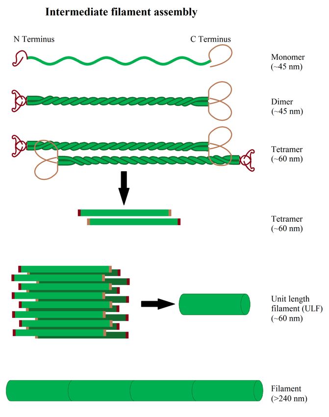
Cells 2019, 8, 362                                                                                                     6 of 55

by a reduced persistence length of the filament, that can drop to ≈20% of its initial value through
ADF/cofilin binding, locally generating increased mechanical stress [58–60]. ADF/cofilin preferentially
binds to the (−)-end of actin filaments (ADP bound actin) [61], but binding to ATP bound actin
favors its transition from ATP to ADP bound actin and, thus, accelerates filament dissociation [62].
The preference for older ADP bound actin implies that ADF/cofilin disassembly mostly affects inactive
compartments of the actin network [55,56,63,64]. As ADF/cofilin cannot bind to the free (+)-end it can
only fully saturate actin filaments if the (+)-end is bound by capping proteins and is not capable of
further elongation [65]. With the exception of fascin, cross-linkers and tension reduce the efficiency of
ADF/cofilin fragmentation [34,66–68]. Furthermore, ADF/cofilin is present in high concentrations at
the
 Cellsleading    edge in dendritic networks, fragmenting links generated by Arp2/3 and the actin filaments
        2019, 8, 362                                                                                                    7 of 58
itself, generating more free (+)-ends [69].
 2.7. Actin Structures Inside the Cell
2.7. Actin Structures Inside the Cell
         Looking at a motile cell the net movement is the result of multiple, mostly actin-dependent,
 processes, as at
        Looking          a motile
                      follows:       cell the net
                                  Formation         movement in
                                               of protrusions     is the   resultof
                                                                      direction   ofmotion,
                                                                                     multiple,    mostly actin-dependent,
                                                                                               subsequent   adhesion to the
processes,
 substrate and  as follows:      Formation
                       loss of adhesion     onofthe
                                                  protrusions
                                                    rear of thein    direction
                                                                 cell,  followed of by
                                                                                    motion,  subsequent(reviewed
                                                                                       rear-contraction    adhesion in to [70–
                                                                                                                          the
substrate     and   loss   of adhesion    on the  rear of the cell, followed    by rear-contraction
 73]). These processes are governed by sub-cellular structures, like filopodia and the lamellipodium (reviewed    in [70–73]).
These    processes
 governing         cellaremotion,
                             governed   by sub-cellular
                                      while                structures,
                                               stress fibers    and the like filopodia  and themechanical
                                                                              cortex secure      lamellipodium     governing
                                                                                                              stability   and
cell  motion, while
 contractility.            stresstypes
                     Further      fibersof
                                         and  the cortex secure
                                            protrusions    are so mechanical
                                                                    called blebs,stability
                                                                                    whichandarecontractility. Further types
                                                                                                 capable of regulating     cell
of protrusions          are  so called   blebs,  which    are capable     of regulating   cell
 movement independently of filopodia and the lamellipodium. The interactions of actin discussed movement    independently
of filopodia
 here    and itsand       the lamellipodium.
                    structural    and functionalThe     interactions
                                                     integration    withofmicrotubules
                                                                            actin discussedandhere  and its structural
                                                                                                intermediate   filamentsandare
functional
 summarized     integration
                     in Figurewith2. microtubules and intermediate filaments are summarized in Figure 2.

      Figure 2. Illustration of actin, microtubule, and intermediate filament signaling, with focus on migration
       Figure 2. Illustration of actin, microtubule, and intermediate filament signaling, with focus on
      associated structures and signaling cascades.
       migration associated structures and signaling cascades.

 2.7.1. The Lamellipodium
      The lamellipodium (Figure 3) is a flat structure mainly associated with cell movement, formed
 by the polymerization of actin at the cell front [13,74], while it is depolymerized at the back of the
 lamellipodium by ADF/cofilin refilling the G-actin pool [75]. The continuing (de-)polymerization of
 the whole network creates a treadmilling effect and a retrograde actin flow in the cell [76,77], which
Cells 2019, 8, 362 7 of 55
2.7.1. The Lamellipodium
The lamellipodium (Figure 3) is a flat structure mainly associated with cell movement, formed
by the polymerization of actin at the cell front [13,74], while it is depolymerized at the back of the
lamellipodium by ADF/cofilin refilling the G-actin pool [75]. The continuing (de-)polymerization of
the whole network creates a treadmilling effect and a retrograde actin flow in the cell [76,77], which is
enhanced in some cell types by myosin induced depolymerization at the back of the lamellipodium [78].
Any flow originating from the contraction of the rear via stress fibers generates a flow of opposite
direction [77]. The forces generated by actin polymerization in the lamellipodium are up to a
few hundred pN/µm [79]. The most important factor for the generation of the lamellipodium is the
intrinsically inactive Arp2/3 complex that becomes activated by the Scar/WAVE complex in an activation
process by the small Rho GTPase Rac1 [80]. Arp2/3 nucleates a new actin filament at the site of existing
filaments [80]. For a three dimensional environment, N-WASP (and not WAVE) was shown to activate
Arp2/3 and Rac1 was not found to be strongly localized at the cell front [81,82]. Actin growth is further
promoted by the presence of members of the Ena/VASP family accumulating at the lamellipodial tip,
promoting further actin elongation and preventing capping [83,84]. Despite the active Arp2/3 complex,
a capping protein is needed as well to limit the elongation of single filaments [16,85] so they remain
productive and do not form bundles with other uncapped filaments or buckle under the load [86].
For the generation of a stable dendritic network, it is cross-linked by proteins such as cortactin [87].
As the described regulation by Rac1 would result in a constant growth of the lamellipodium, it has to
be restricted by a negative feedback loop. One possible mechanism is via the protein arpin, which
inhibits Arp2/3 activity in the lamellipodium [88]. It has been postulated that arpin is recruited by
Rac1 [88]. Thus, it seems possible that Rac1 activation initiates lamellipodium growth via quick
Arp2/3 recruitment and successive actin polymerization and later inhibits its growth via recruitment
of arpin. A high turnover rate of arpin or significantly higher concentration might be necessary to
inactivate Arp2/3 [89]. A proof for this kind of hypothesis is yet lacking. Despite the actin dynamics,
the lamellipodium is also influenced by the cell membrane and its surface tension [90]. A higher
membrane surface tension led to a more oriented actin filament polymerization while a lower tension
resulted in more protrusions [90], probably related to the finite forces generated by the lamellipodium.
Regarding the mechanical properties of the lamellipodium, it has to be noted that myosin was observed
to be present at the rear of the lamellipodium, explaining why the lamellipodium is elastic on short
and viscous on long time scales [78,91].
Due to Arp2/3 the actin in the lamellipodium is connected to a dendritic structure [92].
Interestingly, an analysis of cell speed relative to the actin orientation in the lamellipodium could
demonstrate that faster cells have filaments that are not exactly oriented in the direction of movement
and the parallel orientation of filaments is associated with slower movement [93].Cells 2019, 8, 362 9 of 58
Cells 2019, 8, 362 8 of 55
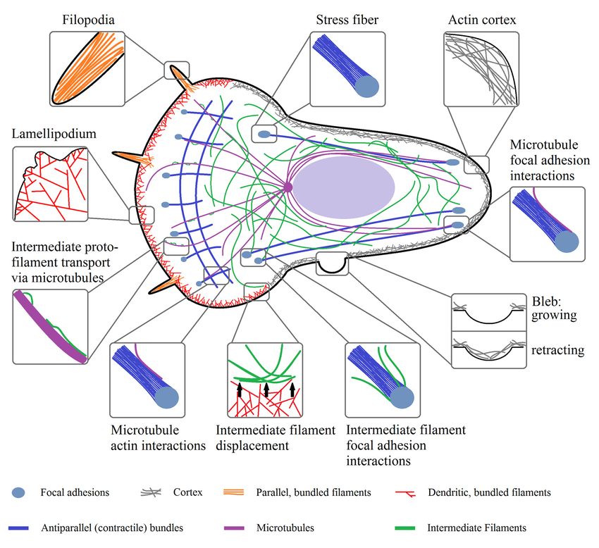
Figure 3. Organizational structures of actin, microtubules, and intermediate filaments inside of a cell
and their 3.
Figure physical interactions.
Organizational Notably,
structures of all three
actin, cytoskeletal and
microtubules, proteins interact directly
intermediate with
filaments each of
inside other.
a cell
and their physical interactions. Notably, all three cytoskeletal proteins interact directly with each
2.7.2. Filopodia
other.
Further structures associated with cell motility are filopodia (Figure 3). Filopodia are associated
2.7.2.
with Filopodia
a sensory function in neurons [94], but do not seem necessary for migration, as the fast moving
corneal keratocytes do not possess filopodia in two dimensions and forces generated by filopodia
Further structures associated with cell motility are filopodia (Figure 3). Filopodia are associated
are significantly smaller than those generated by the lamellipodium [95]. In other systems there
with a sensory function in neurons [94], but do not seem necessary for migration, as the fast moving
might be a role for filopodia in migration, e.g., in three dimensional systems [96]. Filopodia form
corneal keratocytes do not possess filopodia in two dimensions and forces generated by filopodia
a structure consisting of parallel actin bundles, with their (+)-ends pointing in direction of the cell
are significantly smaller than those generated by the lamellipodium [95]. In other systems there
membrane [97]. This orientation is established via formins (e.g., FMNL2) and Ena/VASP, both being
might be a role for filopodia in migration, e.g., in three dimensional systems [96]. Filopodia form a
capable of maintaining a prolonged actin polymerization [98]. Some of these formins, like mDia2, can be
structure consisting of parallel actin bundles, with their (+)-ends pointing in direction of the cell
activated by the small GTPase Cdc42 [99]. Cdc42 is also capable of activating N-WASP and thus Arp2/3,
membrane [97]. This orientation is established via formins (e.g., FMNL2) and Ena/VASP, both being
leading to filopodia formation [100]. A common model for filopodia initiation suggests that actin
capable of maintaining a prolonged actin polymerization [98]. Some of these formins, like mDia2,
polymerization occurs in the presence of activated Arp2/3 and without capping proteins forming actin
can be activated by the small GTPase Cdc42 [99]. Cdc42 is also capable of activating N-WASP and
bundles [98]. Nevertheless, Arp2/3 does not seem to be necessary for filopodia initiation in adherent
thus Arp2/3, leading to filopodia formation [100]. A common model for filopodia initiation suggests
cells [101]. A further model of filopodia initiation states that filopodia are initiated by clusters of
that actin polymerization occurs in the presence of activated Arp2/3 and without capping proteins
activated formins near the plasma membrane, nucleating and elongating actin filaments [98]. For both
forming actin bundles [98]. Nevertheless, Arp2/3 does not seem to be necessary for filopodia
models, subsequent further elongation via formins (e.g., mDia2) and Ena/VASP and stabilization
initiation in adherent cells [101]. A further model of filopodia initiation states that filopodia are
and bundling with cross-linkers, like fascin, generates “mature” filopodia [102]. Besides their role
initiated by clusters of activated formins near the plasma membrane, nucleating and elongating actin
in cell movement, filopodia initiate cell-cell contacts, transmit cell-cell-signals, and respond to the
filaments [98]. For both models, subsequent further elongation via formins (e.g., mDia2) and
mechanical properties of their surroundings [103]. Interestingly, when filopodia are retracted to the
Ena/VASP and stabilization and bundling with cross-linkers, like fascin, generates “mature”Cells 2019, 8, 362 9 of 55
cell the myosins II,V, and VI are not involved in this process [104]. This leads to the idea that only actin
(de-)polymerization and changes in the cortex are responsible for filopodia dynamics.
2.7.3. Stress Fibers
Another type of actin related structures are stress fibers (Figure 3) that are neither present in
filopodia nor in the lamellipodium. Stress fibers are formed from bundles of anti-parallel actin filaments
containing myosin II or parallel filaments [105]. Stress fibers are assembled bundles of 10–30 actin
filaments [106], cross-linked by α-actinin in a bipolar fashion, and linked to focal adhesions [105,107].
Focal adhesions are binding sites that connect the cell to the substrate. Contractile stress fibers are one of
the main contributors to cell contractility in many animal cells. As the contractility of these stress fibers is
regulated by myosin [108], regulation of stress fiber contractility is in many ways similar to the regulation
of myosin activity discussed before. In non-motile cells, stress fibers are usually thick and comparably
stable, while motile cells typically contain fewer less pronounced fibers with a higher dynamic [109].
Actin and myosin are the two principal constituents of contractile stress fibers, while non-contractile
ones do not contain myosin [110]. Despite these components, stress fibers contain actin binding
proteins and focal adhesion-associated proteins binding and unbinding in quick succession [110–113].
The molecules found in stress fibers include cross-linkers, such as α-actinin [114], which does not
only function to stabilize the bundle but is also associated with kinases and signaling proteins and,
thus, functioning as a signaling mediator [115,116]. Stress fibers can contain further cross-linkers,
like fascin, filamin, and paladin, but their precise role despite bundling remains elusive [117–119].
One hypothesis states that these proteins function as basis for regulation of cytoskeletal dynamics as,
for example, paladin interacts with profilin and VASP [120,121]. Further molecules of e.g., the calponin,
tropomyosin, caldesmon family, and others, are found in stress fibers and are all suggested to be
part of the cytoskeletal and/or stress fiber regulation [114,122–125]. Generally speaking, stress fiber
formation has been directly associated with an activation of the formin mDia1 and the small Rho
GTPase RhoA, activating ROCK [126,127]. The formin favors prolonged actin polymerization of
parallel filaments important for dorsal stress fibers [110,128]. In contrast ROCK activates the LIM
kinase (LIMK), which inhibits ADF/cofilin induced filament severing [129], and additionally, ROCK
activates myosin, favoring stress fiber formation [23,105]. Nevertheless, both the ROCK and formin
mechanisms are necessary for the formation of contractile stress fibers [109]. Two other Rho GTPases,
Cdc42 and Rac1, act in more indirect ways via the induction of lamellipodial growth via Arp2/3 (Rac1)
and filopodia formation via the formin mDia2 (Cdc42) [99,130–132]. Collapse of both filament types
can function as seeds for either transversal or ventral stress fibers.
Since stress fibers vary in their morphology, molecular signature, and association with focal
adhesions, the four following types of stress fibers can be distinguished: The perinuclear actin cap,
transverse arcs/stress fibers, and dorsal and ventral stress fibers.
The three classes of contractile stress fibers are the ventral and transverse stress fibers and
the perinuclear actin cap, all characterized by the presence of myosin II along the fibers. Even so,
the myosin II spacing can change over time, indicating that contractile stress fibers are dynamic
structures with non-uniform mechanical properties [133]. Measurements indicate that stress fibers
have a stiffness of roughly 12 kPa, constant for strains up to 0.12 [134]. Perturbation of myosin in stress
fibers reduces the elastic modulus to 8 kPa, indicating the importance of myosin II in contractile stress
fibers [134]. Not surprisingly, the tensile elastic modulus increases from approximately 1.5 MPa to
104 MPa for strains approximating 2 [135]. Ventral stress fibers are oriented parallel to the direction of
cell motion and connect adhesion sites of the cell, while transverse fibers are oriented perpendicular to
the ventral fibers and are not directly connected to focal adhesions. Even so, transversal stress fibers
can contribute to the overall contractility through their connection to dorsal stress fibers [110,136].
The formation of transversal stress fibers is dependent on Arp2/3 and myosin [110] and possess a
periodic α-actinin-myosin pattern [137]. Transversal stress fibers form when the dendritic network
collapses and is restructured by myosin [138,139]. Notably, simulations on the capability of myosin toCells 2019, 8, 362 10 of 55
generate contractile structures suggest that the presence of myosin and actin is sufficient to generate
anti-parallel/contractile bundles, as these were found to be energetically favorable [140]. A further
origin of both transversal and ventral fibers is the collapse of filopodia, which functions as a seed for
stress fibers [141]. Additionally, ventral stress fibers can be formed from existing dorsal stress fibers
and the attached transverse stress fibers, as well as by the fusion of two dorsal stress fibers [110,142].
Ventral stress fibers are also contractile actin-myosin bundles attached to focal adhesions at both ends,
thus being directly part of the contractile machinery [143]. Due to their location at the rear of the cell
and an orientation that is roughly in the direction of motion, they are part of the rear contraction,
and thus associated with cell motility [73,144]. The third type of contractile stress fibers is the so-called
perinuclear cap, consisting of stress fibers positioned above the nucleus, regulating the shape of the
nucleus. Additionally, they are proposed to serve as a mechanical connection between the nucleus and
the rest of the cell [145]. All of these contractile stress fiber types have in common that they are highly
dependent on presence and activity of myosin and, thus, on tension. Consequently, myosin inhibition
leads to the disassembly of these stress fibers [146].
In contrast to the other stress fiber types, dorsal stress fibers do not contain myosin II [110,111]
and are anchored at focal adhesions at their distal ends [110,136]. The lack of myosin directly leads
to the lack of contractility of dorsal stress fibers. It is proposed that these fibers consist of fast
growing (+)-ends that face the cell periphery and more distant parts consisting of mixed polarity actin
filaments [106,109]. Furthermore, paladin and Rac1 are seemingly essential for the formation of dorsal
stress fibers. Paladin promotes fiber assembly via VASP recruitment [147,148]. Functionally, these
stress fibers seem to be an anchor point for the assembly of the other stress fiber types and a link to
focal adhesions [110,111]. It is supposed that dorsal stress fibers are generated via actin polymerization
at emerging focal adhesions [110] and stabilized during retraction phases of the lamellipodium via
coupling to emerging transverse stress fibers [111,138,149].
2.7.4. Actin Cortex and Blebs
The last cytoplasmic structure described here is the actin cortex (Figure 3), which forms a contractile
actin structure at the boarder to the plasma membrane. The cortex is a few hundred nanometer thick
layer, consisting of a mixture of filament bundles and cross-linked filaments, with a mesh-size of
approximately ≈50–150 nm [150], a thickness of 50–100 nm [151,152], and a distance to the cell membrane
of less than 20 nm [151]. The cortex meshwork appears to be mainly isotropic, oriented in parallel
to the plasma membrane, but some filaments are also oriented perpendicular to the membrane [153].
Despite actin filaments, the cortex contains a number of cross-linkers (e.g., fascin, actinin, filamin, etc.),
myosin, proteins that control actin turnover (like profilin, cofilin), capping proteins, proteins of the
ERM (ezrin, radixin, moesin) family, nucleating factors (like Arp2/3, the formin mDia1), and signaling
molecules such as RhoGTPases, RhoGEFs (guanine exchange factors), and RhoGAPs (GTPase activating
proteins) [154,155]. The two mentioned nucleating factors Arp2/3 and mDia1 were also found to be
responsible for the majority of cortical F-actin generation [155,156], while the ERM proteins link the
cortex to the membrane and can therewith transmit forces acting on the membrane and determine
the cell shape [157–159]. Depletion of cofilin-1 or capping proteins in HeLa cells increased cortex
thickness but reduced tension, implying a role for actin regulating proteins in cortical tension [160].
Mechanical properties of the cortex determine how the cell deforms in response to external forces.
On time scales smaller than the remodulation time of the cortex it behaves elastic [161], with a cell
type-dependent elastic modulus in the order of a few hundred to thousands of pascals [162,163].
On long time scales (>1min) the cortex behaves viscous because of the adaption to external forces
via actin modulation, dissociation, and (un-)binding of cross-linkers [161]. If myosin is activated
the cortex turnover times can be even lower [164,165], perhaps via direct disassembly or enhanced
actin breakage [52,78]. Generally speaking, the behavior of the cortex is similar to that of glassy
materials [166] and consistent with relaxations of three dimensional in vitro actin networks [167].Cells 2019, 8, 362 11 of 55
One of the main global and local properties of the cortex is its tension, which regulates the
cell shape of single cells and tissues [168]. Several studies demonstrated that the cortex tension
depends on the myosin activity and actin polymerization, with higher myosin activity and lower
actin polymerization leading to an increased cortex tension [169]. A lower cortex tension is further
associated with an increased protrusive activity of the cell, thus indirectly regulating cell motility [170].
Interestingly, local drops in cortex tension or cortex-membrane adhesion and local ruptures of the cortex
can be the origin of so called blebs (Figure 3), a special, initially actin free, membrane protrusion [171].
Blebs can be initiated by any type of cortex weakening or loss of cortex-membrane adhesion if a
given internal hydrostatic pressure threshold is reached [172,173]. Localized myosin contractions,
promoting either cortex tearing or increasing local intracellular pressure, are some of the main sources
of blebbing [174–176], but others are also discussed [177]. Thus, the activation of myosin via the
already described activation by ROCK or MLCK are sufficient to induce bleb formation [178–180].
The progress of a bleb can be divided in three steps, as follows: Initiation, growth, and retraction.
Initially, the growing bleb does not contain actin, but over time, when the bleb expands further, the actin
cortex reassembles at the plasma membrane, stalling the bleb growth [157] up to the point of a full
restoration where the generated contractile forces retract the bleb [181]. It has to be noted that bleb
retraction does not always occur and in some motile cells blebs are stabilized and used as an alternative
or additional mode of migration [178–180,182]. The expansion of blebs by actomyosin contraction
induced pressures lasts 5–30 s, accompanied by a flow of cytosol into the bleb and a concomitant
increase in surface area. The surface area is increased by a flow of lipids through the tearing of the
membrane from the actin cortex [183]. The maximal bleb size is determined by the initial growth rate
and the cortex re-polymerization time [184], both being dependent on cortex tension. The concept of
tension inhibiting bleb expansion is further supported by the idea that the needed membrane unfolding
is effectively resisting bleb expansion and, thus, slowing down the growth [182,185]. After full
maturation the cortex is reconstructed and if the bleb is not stabilized via adhesions it is retracted by
the re-established cortex via a myosin induced contraction [178,186].
2.7.5. Nuclear Actin
For completeness it has to be mentioned that actin is not only present in the cytoplasm of eukaryotic
cells, but also in the nucleus. As in the cytoplasm, nuclear actin exists in a monomeric and polymeric
form [187]. Nuclear G-actin was shown to associate with all three RNA polymerases, participating
in transcription initiation and elongation [188]. The exact function of G-actin in the transcription
complex remains unclear, but G-actin levels need to be precisely tuned for normal translation [189] and
cofilin is required for its elongation [190]. Contrary, stable actin filaments inhibit transcription [191].
Furthermore, actin is implied to affect the nuclear structure. During nuclear expansion at the mitotic
exit, chromatin reorganizes depending on the transient formation of polymeric actin, in a seemingly
cofilin dependent manner [192]. It is probable that nuclear actin regulates the structure of the nuclear
envelope and the nucleus via interactions with the nuclear intermediate filaments lamin [193].
As mentioned, cell nuclei contain polymeric actin that can be generated inside the nucleus via
actin nucleation factors [194]. These actin assembly factors include, amongst others, mDia1, Spire1/2,
Fmn2, and Arp2/3 [195]. The presence of assembly factors underlines a functional role of polymeric
actin and actin binding proteins. Initiation of DNA replication was demonstrated to require formin
dependent nuclear actin polymerization [196]. Additionally, the loss of Spire1/2 and Fmn2 resulted in
a less efficient clearance of DNA double strand breaks [197]. This is in agreement with two studies
demonstrating an association of actin and Arp2/3 with sites of DNA damage and decreased damage
repair after reduced Arp2/3 dependent actin nucleation [198,199]. For more information on the role of
actin in the nucleus the interested reader is referred to other reviews [188,200].Cells 2019, 8, 362 12 of 55
3. Microtubules
Microtubules consist of α- and β-tubulin heterodimers forming hollow filaments, usually consisting
of 13 protofilaments [201]. The microtubules are seeded by the microtubule organization center (MTOC),
generally centrosomal, but in some types of differentiated cell non-radial microtubules are assembled
by non-centrosomal MTOCs, for example at the Golgi apparatus [202–204]. MTOC, amongst others,
contains γ-tubulin as a microtubule nucleator, as well as anchoring and adaptor proteins for attachment
of microtubules [205]. Heterodimers interact at the MTOC with the γ-TURC, thus microtubules are
nucleated and anchored with their (−)-ends to the MTOC [205]. Microtubules show a behavior called
dynamic instability, characterized by a sudden switch from growing to growth arrest and/or quick
depolymerization (termed catastrophe), followed by a new growth cycle [201] (Figure 4). A possible
explanation for this behavior can be derived from the formation process of single protofilaments.
During polymerization, GTP-bound tubulin heterodimers are bound to the (+)-end and normally
hydrolyzed shortly after, but sometimes older parts of microtubules still contain GTP-bound tubulin
heterodimers [206]. This leads to the following model: If, due to stochastic fluctuations or other
perturbations, the (+)-end does not contain the more stable GTP-bound tubulin, it depolymerizes and
is stabilized at older GTP-bound tubulin sites [206]. Stochastic fluctuations in the rate of microtubule
growth and the stochastic nature of GTP hydrolysis lead to a dynamic GTP cap size [207], with
the consequence that faster polymerizing microtubules have a larger GTP cap, resulting in less
frequent catastrophe events [208]. The dynamic behavior of microtubules can be regulated by both
intrinsic and extrinsic factors (see Table 2 for summary). Microtubule interacting proteins are either
microtubule (+)-end-binding proteins (+TIP) or structural microtubule-associated proteins (MAP)
interacting with microtubules along their length. These protein classes can have stabilizing or
destabilizing effects, changing polymerization dynamics or severing microtubules. Important proteins
belonging to the family of the +TIPs are CLASPs (cytoplasmic linker associated protein) and APC
(adenomatous polyposis coli) [209,210] that suppress microtubule catastrophe events and promote
rescue after catastrophe [211]. Part of the stabilizing effects of CLASPs arises from their capability
to modulate interactions between microtubules and the cell cortex [212]. Further important families
of +TIPs are the spectraplakins, binding both microtubules and actin [213] and EBs (end binding
proteins). EBs are supposed to be a master regulator of +TIP recruitment (e.g., CLASP [214], APC,
MACF1 (microtubule-actin crosslinking factor) [215]) and complex assembly [211], generally promoting
persistent microtubule growth [216]. EBs are generally associated with an increased polymerization
rate and reduced catastrophe number [217,218]. Despite these molecules that mainly (de-)stabilize
microtubules, there is a bunch of proteins that sever microtubules, like spastin [219] or katanin [220],
or influence depolymerization and polymerization, e.g., stathmin (favors depolymerization) [221]
or XMPA215 (increases polymerization rate) [222]. Additionally, there are structural MAPs, like tau
protein, MAP2, or DCX (doublecortin), that interact with the filament at its whole length and stabilize
it [223,224] by reducing shrinkage speed, promoting filament growth, and reducing catastrophe
frequency [223,224]. The effect of structural MAPs can also inhibit the effect of other microtubule
associated proteins, as, for example, tau protein can inhibit the katanin induced severing [225]. A further
important class of MAPs are the motor proteins kinesin and dynein, both serving as cargo transporters,
exploiting the microtubule meshwork [226,227]. In general, kinesin motor proteins transport cargo to
the (+)-end, while dynein moves to the (−)-end of microtubules [228,229], transporting diverse cargo
types, including membrane components, signaling molecules, such as the small GTPases Rac and
Cdc42 [230,231], but also intermediate filaments and their precursors [232–234], β-actin coding mRNA,
and sub-units of the Arp2/3 complex [235,236]. Motor proteins possess not only a transport function
but can stabilize or destabilize microtubules. For example, members of the kinesin-8, kinesin-13 family,
or KinI kinesins can induce depolymerization, likely via the destabilization of the GTP cap [237]
or the induction of kinks [238]. Despite their transport function and regulatory role in microtubule
dynamics, some kinesins organize the microtubule network via the bridging of microtubules, thus
favoring the generation of parallel arrays. For example, in neurons kinesin-5 and kinesin-12 areCells 2019, 8, 362 13 of 55
necessary for axonal outgrowth because of their cross-linking ability and the concomitant focus on
the extension of microtubule arrays [239,240]. Additionally, kinesin-1 may be involved in this process
by sliding filaments alongside each other [241]. A further important aspect regulating microtubule
dynamics are the post-translational modifications modifying microtubule properties and affinities of
MAPs [242]. Important modifications are, amongst others, tyrosination, glutamylation, and acetylation.
Acetylation protects microtubules against repeated mechanical stress via an increased flexibility, but does
not protect against depolymerization [243]. Additionally, some severing proteins, such as katanin,
preferentially interact with acetylated tubulin [244]. Tyrosination affects the recruitment of microtubule
interacting proteins, such as CLIP-170 or kinesin-1, that prefer detyrosinated microtubules, probably
facilitating directional transport [245,246]. In contrast, spastin favors cleavage of detyrosinated
microtubules [247]. Similar to tyrosination, glutamylation can also affect the interaction with
microtubule associated proteins. Map2, tau, and kinesin-1 were reported to preferentially interact with
those microtubules with up to three glutamates on their tail [245]. Similarly, both microtubule severing
proteins, katanin and spastin, show an increased affinity for glutamylated microtubules [248,249].
Taken together, this data suggests that post translational modifications are important and finely tunable
regulators of microtubule dynamics and, consequently, of cell behavior.
For regulation of MAPs, and microtubules in general, the family of Rho GTPases is of major
significance. An important example is the stabilizing effect of RhoA, but not Rac1 and Cdc42,
on microtubules via the RhoA effector mDia. mDia is capable of interacting with EB1 and APC, leading
to a stabilization of microtubules via e.g., Kif4 [250,251]. Furthermore, active mDia induces the alignment
of actin and microtubules [251,252]. Interestingly, stathmin action seems to negatively regulate the
RhoA/ROCK activity [253], complementing the observation that Cdc42 and Rac1 phosphorylate
stathmin via an activation of PAK (p21-activated kinases) [254,255]. In fact, Rac1 activation was
demonstrated to decrease catastrophe events and increase microtubule growth time in cells via
PAK [255,256]. Furthermore, both Rac1 and Cdc42 activate IQGAP1, interacting with Clip-170,
likely providing a stabilization site for microtubule (+)-ends near the cortex [257]. The interactions
of microtubules discussed here and their interactions with actin and intermediate filaments are
summarized in Figures 2 and 3.
Microtubules typically play a role as tracks for transport, as already mentioned, in spindle
positioning during mitosis, migration (discussed below), and in cell shape control [258–260]. At the
current point, the concept that microtubules control the balance between RhoA and Rac1 and thus
influence cell shape and migration is favored over a direct mechanical participation for most cell
types [261]. Even so, microtubules are relatively stiff polymers, when compared to actin [262], capable of
generating forces of up to 3–4 pN during polymerization [263–265]. As a result, microtubules can deform
membranes and resist compressional forces in such a way that they act as load bearing fibers in living
cells via transversal re-enforcement by other cytoskeletal components [266,267]. Notably, if multiple
microtubule filaments grow as a bundle the generated forces increase linearly with the number of
microtubules per bundle [268]. Complementary to these observations, the load bearing capacities of
microtubules is limited because compressional loads can induce catastrophe events [265,268], in line
with the fact that most catastrophes are indeed induced at the cell edge [269,270] and the observed
short-wavelength buckles near the boundary of adherent cells [170,271]. A further mechanism of force
generation by microtubules is during the shrinkage phase. To actually transmit a force during shrinkage,
the (+)-end needs to stay attached to its cargo. When the GTP cap is lost the microtubule protofilaments
lose their lateral connection with neighboring protofilaments, bending backwards and forming ring-like
shapes [272,273] (Figure 4). If cargo stays attached during this process, a single microtubule can exert
forces of up to 30–65 pN [272,273], a magnitude larger than the pushing force [263].
Microtubules also play a key role during the separation of chromosomes during cell division [274].
Depolymerization of microtubules is believed to generate the needed forces to separate the sister
chromatids [275]. This aspect will not be discussed in more detail here, but the interested reader may
be referred to the following reviews: [274–276].Cells 2019, 8, 362 15 of 58
Cells 2019, 8, 362 14 of 55
Figure 4. Scheme of microtubule formation and dynamic instability. Microtubules consist of α- and
Figure 4. Scheme
β-heterodimers, of microtubule
forming a hollow formation and dynamic
tube elongating instability.
by the addition Microtubules
of heterodimers, consistaof
forming α- and
GTP-cap
β-heterodimers,
at the (+)-end of forming a hollowprotecting
the microtubule, tube elongating by thefrom
microtubules addition of heterodimers,
shrinkage. If the (+)-endforming
loses itsa
GTP-cap itatinduces
GTP-cap the (+)-end of the microtubule,
microtubule shrinkage. protecting microtubules from shrinkage. If the (+)-end
loses its GTP-cap it induces microtubule shrinkage.
Furthermore, microtubules and actin are linked regarding their functional dynamics and structural
Table 2. Summary
organization. On the oneof hand
mentioned
there ismicrotubule
an indirectassociated proteins
co-regulation, and their direct
as microtubules areor indirect
able to locally
functions.
regulate and are regulated by RhoGTPases and focal adhesions (see also chapter 5.2), but on the
other hand, there are molecules
Microtubule Associated interacting
Proteins with microtubules Function
and actin. One such molecule is APC,
which stabilizes microtubules
Stathmin and nucleates actin filaments,Depolymerization
with actin nucleation additionally favored
by the formin mDia1 [209,277].
XMPA215 A further formin mDia2 that is capablefactor
Polymerization of nucleating actin can also
stabilize microtubules, independent
EB of its nucleation function
Polymerization, [278]. There
Stabilization, are additional
Recruitment cross-linkers
of proteins
DCX
connecting actin and microtubule Polymerization
filaments, such as MACF1 and Argfactor, Stabilization
[279,280]. Consequently, the actin
CLASP cannot fully be regarded as decoupled
and microtubule cytoskeleton Stabilization
systems, as they are not regulated
APC
independently and can even be connected physically. Stabilization
mDia1 StabilizationCells 2019, 8, 362 15 of 55
Table 2. Summary of mentioned microtubule associated proteins and their direct or indirect functions.
Microtubule Associated Proteins Function
Stathmin Depolymerization
XMPA215 Polymerization factor
EB Polymerization, Stabilization, Recruitment of proteins
DCX Polymerization factor, Stabilization
CLASP Stabilization
APC Stabilization
mDia1 Stabilization
mDia2 Stabilization
Tau Stabilization
MAP2 Stabilization
Spastin Microtubule severing
Katanin Microtubule severing
Kinesin Cargo transport
Dynein Cargo transport
MACF1 Actin-Microtubule interactions
Cdc42 Signaling molecule, activates PAK
Rac1 Signaling molecule, activates PAK
RhoA Signaling molecule, mDia1
PAK Signaling molecule, inhibits stathmin
4. Intermediate Filaments
Intermediate filament-forming proteins are a large protein class, encoded by at least 70 genes,
organizing filaments with a diameter of 10 nm. Intermediate filaments are grouped in 5 classes
according to their structure and sequence homology. Thereby, the first four classes represent cytoplasmic
intermediate filaments, while type V are nuclear filaments, so called lamins (lamin A/C, B1, B2). Type I
and II are acidic and basic keratins, forming heteropolymers consisting of a mixture of the 54 different
type I and II keratins, expressed in dependence of cell type and differentiation status [281]. In contrast,
type III intermediate filaments are homopolymers of vimentin, desmin, peripherin, or glial fibrillary
acidic protein (GFAP). Vimentin is mainly expressed in fibroblasts, endothelial cells, astrocytes;
peripherin in neurons of the peripheral nervous system and desmin in muscle cells and GFAP mainly
in astrocytes. Type IV intermediate filaments contain three neurofilament heteropolymers (NF-L/M/H),
internexin, synemin, and nestin, mainly expressed in the cells of the nervous system. Nestin and
synemin cannot form filaments on their own, but only in conjunction with other intermediate filament
proteins [282,283]. Two further intermediate filaments, called filensin and phakinin, cannot be grouped
into the mentioned five types. They are expressed in the lens epithelium, forming heteropolymers [284].
All cytoplasmic intermediate filaments have a similar monomer structure, consisting of a central
α-helix with a non-helical structure at both of its ends [285]. Two monomers spiral around each
other, forming a so-called “coiled-coil” dimer [286] and, subsequently, these dimers form unpolarized
tetramers via antiparallel association and 8 tetramers form a cylindrical unit-filament [287]. The unit
filaments aggregate further with other unit filaments at the time scale of minutes to form intermediate
filaments [288] (Figure 5). After aggregation, the filaments undergo a compaction step during which
the filament diameter shrinks to its final size of approximately 10 nm [289–291]. For nucleation and
polymerization of intermediate filaments co-factors are not needed [292]. Intermediate filaments show
a constant, but slow, subunit exchange along the whole filament, occurring at a rate of approximately
1 per 200 tetramers per hour in vitro for vimentin [293].You can also read