TRAILblazing Strategies for Cancer Treatment - MDPI
←
→
Page content transcription
If your browser does not render page correctly, please read the page content below
cancers
Review
TRAILblazing Strategies for Cancer Treatment
Anna-Laura Kretz 1 , Anna Trauzold 2,3 , Andreas Hillenbrand 1 , Uwe Knippschild 1 ,
Doris Henne-Bruns 1 , Silvia von Karstedt 4,5 and Johannes Lemke 1, *
 1   Department of General and Visceral Surgery, Ulm University Hospital, Albert-Einstein-Allee 23, 89081 Ulm,
     Germany; anna-laura.kretz@uni-ulm.de (A.-L.K.); andreas.hillenbrand@uniklinik-ulm.de (A.H.);
     uwe.knippschild@uniklinik-ulm.de (U.K.); doris.henne-bruns@uniklinik-ulm.de (D.H.-B.)
 2   Institute for Experimental Cancer Research, University of Kiel, 24105 Kiel, Germany;
     atrauzold@email.uni-kiel.de
 3   Clinic for General Surgery, Visceral, Thoracic, Transplantation and Pediatric Surgery, University Hospital
     Schleswig-Holstein, 24105 Kiel, Germany
 4   Department of Translational Genomics, University Hospital Cologne, Weyertal 115b, 50931 Cologne,
     Germany; s.vonkarstedt@uni-koeln.de
 5   Cologne Excellence Cluster on Cellular Stress Response in Aging-Associated Diseases (CECAD),
     University of Cologne, Joseph-Stelzmann Straße 26, 50931 Cologne, Germany
 *   Correspondence: johannes.lemke@uniklinik-ulm.de

 Received: 11 March 2019; Accepted: 26 March 2019; Published: 30 March 2019

 Abstract: In the late 1990s, tumor necrosis factor (TNF)-related apoptosis-inducing ligand (TRAIL),
 a member of the TNF-family, started receiving much attention for its potential in cancer therapy,
 due to its capacity to induce apoptosis selectively in tumour cells in vivo. TRAIL binds to its
 membrane-bound death receptors TRAIL-R1 (DR4) and TRAIL-R2 (DR5) inducing the formation of a
 death-inducing signalling complex (DISC) thereby activating the apoptotic cascade. The ability of
 TRAIL to also induce apoptosis independently of p53 makes TRAIL a promising anticancer agent,
 especially in p53-mutated tumour entities. Thus, several so-called TRAIL receptor agonists (TRAs)
 were developed. Unfortunately, clinical testing of these TRAs did not reveal any significant anticancer
 activity, presumably due to inherent or acquired TRAIL resistance of most primary tumour cells.
 Since the potential power of TRAIL-based therapies still lies in TRAIL’s explicit cancer cell-selectivity,
 a desirable approach going forward for TRAIL-based cancer therapy is the identification of substances
 that sensitise tumour cells for TRAIL-induced apoptosis while sparing normal cells. Numerous of
 such TRAIL-sensitising strategies have been identified within the last decades. However, many of
 these approaches have not been verified in animal models, and therefore potential toxicity of these
 approaches has not been taken into consideration. Here, we critically summarise and discuss the
 status quo of TRAIL signalling in cancer cells and strategies to force tumour cells into undergoing
 apoptosis triggered by TRAIL as a cancer therapeutic approach. Moreover, we provide an overview
 and outlook on innovative and promising future TRAIL-based therapeutic strategies.

 Keywords: TRAIL signalling; TRAIL sensitising; TRAIL-induced apoptosis; TRAIL in cancer

1. Introduction
     Pharmacotherapy for cancer uses one or more chemotherapeutic drugs with either a curative
aim or to prolong life and manage symptoms. Chemotherapy targets rapidly dividing cells to slow
or stop their growth, conversely not distinguishing between transformed and non-transformed cells
which leads to the commonly observed strong side effects in most scenarios. The last decade has seen
the development and testing of targeted therapies such as small-molecule inhibitors or monoclonal
antibodies. The clinical use of a substance with the ability to selectively kill tumour cells while

Cancers 2019, 11, 456; doi:10.3390/cancers11040456                               www.mdpi.com/journal/cancers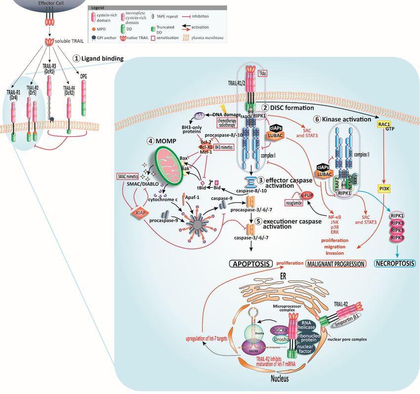
Cancers 2019, 11, 456                                                                              2 of 30

sparing healthy cells thereby reducing severe side effects is urgently needed in pharmacological cancer
therapy. This “silver bullet” against cancer seemed close at hand in 1995 when tumor necrosis factor
(TNF)-related apoptosis-inducing ligand (TRAIL/Apo2L) was introduced [1,2] and later shown to
selectively induce apoptosis in tumour cells in vivo [3,4]. After much testing, this selectivity holds to
this day, however, its efficacy to induce significant levels of apoptosis in some resistant cancers will
have to be helped through additional means.

2. The TRAIL System
      TRAIL is a type II transmembrane protein with apoptosis-inducing capabilities, which belongs
to the TNF-superfamily (TNF-SF) [1,2]. In humans, apoptosis is induced by binding of TRAIL to the
death receptors (DR) TRAIL-R1/DR4 (also called APO-2 or TNFRSF10A) [5] and TRAIL-R2/DR5
(TNFRSF10B, TRICK2, KILLER) [6]. TRAIL is active as a trimer, analogous to other TNF-SF members,
and consequently binds three receptor molecules [7,8]. Besides TRAIL-R1 and TRAIL-R2, TRAIL
engages TRAIL-R3 (DcR1, TNFRSF10C, TRID, LIT) and TRAIL-R4 (DcR2, DcR2, TNFRSF10D,
TRUNDD) also designated as decoy receptors (DcR), and the soluble receptor osteoprotegerin
(OPG, TNFRSF11B). These latter three receptors are thought to potentially negatively regulate
apoptosis through TRAIL-R1 and -R2 by scavenging TRAIL [9–14]. TRAIL-R1 and TRAIL-R2 mediate
apoptosis via a cytoplasmic death domain (DD), which is missing in TRAIL-R3 and is truncated in
TRAIL-R4 [6,12,15–17]. In a physiological setting, the affinity of TRAIL to OPG is notably weaker
compared to the membrane-bound receptors suggesting a subordinate role of the soluble receptor
in TRAIL signalling [18,19]. Noteworthy, when being presented at low concentrations and under
physiological conditions, TRAIL favours interaction with TRAIL-R2 [20]. Eventually, TRAIL-R2 was
presumed as the preferential inducer of apoptosis. Though, this assumption has been debunked since,
for instance, in lymphocytic leukaemia and a variety of pancreatic cancer cells, apoptosis is induced
via TRAIL-R1 although TRAIL-R2 is present and functioning. Consequently, the precedence for one
receptor seems to be tumour entity-dependent [21–24].
      In mice, only one death-inducing TRAIL-R is expressed, mTRAIL-R (MK) sharing 43% and 49%
sequence homology with human TRAIL-R1 and TRAIL-R2, respectively [25]. The murine decoy
counterparts mDcTRAIL-R1 (TNFRS23) and mDcTRAIL-R2 (TNFRS22) lack a DD and differ widely
from the amino acid sequence of human TRAIL-R3 and -R4 [26].
      Despite a research history of more than 20 years, it is still puzzling why humans have evolved to
express two death-inducing TRAIL-Rs [5,27]. In addition, TRAIl-R2 is expressed as short and long
splice variant with a difference of an additional 29 amino acid stretch present in the extracellular
domain of the long TRAIL-R2 isoform. However, a higher incidence of expression of the long isoform
is described, whereas the ratio of the variants differs tissue-dependently [28].

3. The Apoptotic ‘TRAIL’
     The induction of apoptosis can be subdivided into either intrinsic apoptosis, triggered by p53 in
response to cellular injuries [29] or extrinsic apoptosis induced upon death ligand binding to a death
receptor. Caspases orchestrate both apoptosis pathways via cleavage of target proteins (reviewed
in [30–33]). Conventional chemotherapy triggers intrinsic apoptosis through p53 in response to cellular
damage. Nevertheless, many tumour entities contain mutations in p53 leading to its inactivation
and failure of chemotherapy. In stark contrast to this, TRAIL-induced apoptosis remains possible
in numerous cases despite non-functional p53 [34–37]. In Figure 1, the TRAIL-induced apoptotic
signalling is illustrated.
Cancers 2019, 11, 456 3 of 30
Cancers 2019, 11, x 3 of 28
Figure1.1. TRAIL
Figure signalling
TRAIL and sensitisation.
signalling Human tumour-necrosis-factor-related
and sensitisation. apoptosis-inducing
Human tumour-necrosis-factor-related
ligand (TRAIL) can bind to four membrane-bound TRAIL receptors
apoptosis-inducing ligand (TRAIL) can bind to four membrane-bound TRAIL receptors (TRAIL-Rs) and one soluble receptor
(TRAIL-Rs) and
①. TRAIL-R1 and TRAIL-R2 can trigger apoptotic signals while the other receptors
one soluble receptor 1 . TRAIL-R1 and TRAIL-R2 can trigger apoptotic signals while the other receptors might serve as
decoys
might andasregulate
serve decoysapoptosis
and regulatenegatively. Cysteine-rich
apoptosis negatively.domains of the receptors
Cysteine-rich domains areof
crucial for ligandare
the receptors
binding. Following TRAIL binding to TRAIL-R1 or TRAIL-R2, the death-inducing
crucial for ligand binding. Following TRAIL binding to TRAIL-R1 or TRAIL-R2, the death-inducing signalling complex
(DISC) is assembled ②. In type I cells, the signal of DISC-released caspase-8 is sufficient for activation of
signalling complex (DISC) is assembled 2 . In type I cells, the signal of DISC-released caspase-8
further downstream caspases and apoptosis induction ③, whereas the DISC signal is amplified via
is sufficient for activation of further downstream caspases and apoptosis induction 3 , whereas
mitochondria in type II cells ④. Cleaved and thereby truncated Bid (tBid) activates the mitochondria-
the DISC signal is amplified via mitochondria in type II cells 4 . Cleaved and thereby truncated
associated B-cell CLL/lymphoma 2 (Bcl-2)-family members Bcl-2-associated X protein (Bax) and Bcl-2
Bidantagonist
(tBid) activates the mitochondria-associated B-cell CLL/lymphoma 2 (Bcl-2)-family members
or killer (Bak) resulting in mitochondrial outer membrane permeabilisation (MOMP) and
Bcl-2-associated
finally cytochrome c and(Bax)
X protein andmitochondrial
second Bcl-2 antagonist or killer
activator (Bak) resultinginhibitor
of caspases/direct in mitochondrial outer
of apoptosis-
membrane permeabilisation
binding protein (MOMP)point
with low isoelectric and finally cytochrome c and
(pI) (SMAC/DIABLO) second
release. Themitochondrial activator of
apoptosome comprising
caspases/direct inhibitor
apoptotic protease of apoptosis-binding
activating protein
factor-1 (Apaf-1), with low
cytochrome isoelectric
c, and point
caspase-9, (pI) (SMAC/DIABLO)
presents the activation
release. Thefor
platform apoptosome
caspase-9 whichcomprising apoptotic to
leads eventually protease activating
the activation factor-1 (Apaf-1),
of executioner caspasescytochrome
⑤ . p53 is c,
andactivated
caspase-9, presentstothe
in response activation
stress signals platform for caspase-9
such as DNA damage and which leadsBcl-2
promotes eventually
homology to the activation
(BH)3-only
proteins resulting
of executioner caspasesin MOMP.
5 . p53 TRAIL-receptor-interaction
is activated in response tocan stressprovoke
signalsthe formation
such as DNAofdamage
a second and
cytosolic complex ⑥, retaining Fas-associated protein with death domain (FADD)
promotes Bcl-2 homology (BH)3-only proteins resulting in MOMP. TRAIL-receptor-interaction can and caspase-8 and
recruiting
provoke the receptor-interacting
formation of a second serine/threonine-protein
cytosolic complex kinase 1 (RIPK1),
6 , retaining TNF receptor-associated
Fas-associated protein with factor
death
2 (TRAF2), and nuclear factor kappa-light-chain-enhancer of activated B cells (NF-κB)
domain (FADD) and caspase-8 and recruiting receptor-interacting serine/threonine-protein kinase 1 essential modifier
(NEMO) [38–40]. TRAF2 recruits cellular inhibitor of apoptosis protein 1/2 (cIAP1/2) which in turn
(RIPK1), TNF receptor-associated factor 2 (TRAF2), and nuclear factor kappa-light-chain-enhancer
trigger the ubiquitination of RIPK1 and therefore recruitment of linear ubiquitin chain assembly complex
of activated B cells (NF-κB) essential modifier (NEMO) [38–40]. TRAF2 recruits cellular inhibitor
of apoptosis protein 1/2 (cIAP1/2) which in turn trigger the ubiquitination of RIPK1 and therefore
recruitment of linear ubiquitin chain assembly complex (LUBAC) [41]. LUBAC poly-ubiquitinylatesCancers 2019, 11, 456 4 of 30
RIPK1. RIPK1 is the stimulus for tyrosine-protein kinase Src and signal transducer and activator
of transcription 3 (STAT3) promoting migration and invasion [42]. Complex I and complex II
induce NF-κB, p38 mitogen-activated protein kinase (p38 MAPK), c-JUN N-terminal kinase (JNK)
and extracellular signal-regulated kinase (ERK) pathways. LUBAC is present in both complexes,
responsible for caspase-8 activation and recruitment of the inhibitor of κB (IκB) kinase (IKK) complex,
and consequently activation of NF-κB [41]. In case of blocked caspase-8, the necrosome is formed by the
interaction of RIPK1 and RIPK3. Independently of FADD and complex I and II, the membrane-proximal
domain (MPD) of TRAIL-R2 can activate Ras-related C3 botulinum toxin substrate 1 (Rac1) to
promote migration and invasion [43]. TRAIL-R2 can also occur in the nucleus where it interacts
with ribonucleoprotein complexes involved in the maturation of microRNAs (miRNAs) of the let-7
family. These miRNAs interact with and constrain mRNAs of several regulators of mitogenic pathways
such as Ras and c-Myc thereby encouraging proliferation of tumour cells [44]. Further abbreviations:
glycosylphosphatidylinositol (GPI), osteoprotegerin (OPG), death domain (DD).
3.1. TRAIL-Induced Extrinsic Apoptosis
TRAIL binding to TRAIL-R1 and TRAIL-R2, respectively, initiates extrinsic apoptosis through
receptor trimerisation and assembly of the death-inducing signalling complex (DISC) platform at their
cytoplasmatic domains [45–47]. Caspase-8 and cellular FLICE-like IL-1β-converting enzyme-inhibitory
protein long/short (c-FLIPL/S ), are recruited to TRAIL-Rs via a homotypic interaction between
the death domains (DDs) within the receptor and the adapter molecule Fas-associated death
domain (FADD) [48,49]. The exact stoichiometric composition of the DISC unveiled three TRAIL-Rs
recruiting one FADD molecule, its death effector domain (DED) in turn recruiting DED-only proteins
(procaspase-8/procaspase-10 molecules or isoforms of c-FLIP [46,49,50]. The fact that procaspase-8
contains two DEDs has led to modelling of caspase-8 recruitment as a growing chain wherein merely
the first procaspase-8 molecule directly binds to FADD [49,50]. Eventually, DISC formation allows
procaspase-8/-10 dimerisation, its activation, and further activation of downstream caspases [13,48–50].
The caspase-8 homolog c-FLIP fulfils a particular function in regulating caspase-8 activity at
the DISC [50–55]. Depending on the isoform, c-FLIP can act on caspase-8 in an antiapoptotic or
proapoptotic manner [54]. An early, simpler concept of a negative regulation was based on the
observation that c-FLIP could compete with caspase-8 for FADD binding [55–57]. Isoform-wise, c-FLIP
long (c-FLIPL ) is highly homologous to procaspase-8, but lacks a catalytic cysteine residue in the
active site and therefore proteolytic activity. c-FLIP short (c-FLIPS ) represents a shortened variant
containing only tandem DEDs [58,59]. c-FLIPS seems to perform anti-apoptotically, whereas c-FLIPL ’s
regulatory function is more complex [60]. Both splice variants, c-FLIPS and c-FLIPL can inhibit the
activation of caspase-8 at the DISC when overexpressed [58,59,61]. Conversely, in an alternative
scenario, c-FLIPL and procaspase-8 can form an active heterodimer, which has been shown to promote
apoptosis induction, but also to inhibit RIPK3-dependent necroptosis [62–65]. Recently, the prior
model of procaspase-8 and c-FLIP competing for FADD banding has been revised at it was shown
in vitro that c-FLIP recruitment to the DISC follows sequentially after an interaction of procaspase-8
and FADD has been established [54].
3.2. TRAIL-Induced Cross-Signalling to Mitochondria
Soon after the discovery of TRAIL/TRAIL-Rs, it became evident that not all cells follow a similar
linear pathway of TRAIL-induced extrinsic apoptosis. Some cells’ sensitivity depended on the ratio and
expression levels of mitochondria-associated B-cell CLL/lymphoma 2 (Bcl-2) family proteins [66,67].
We now know that two cell types can be distinguished regarding their requirement to cross-signal
to the mitochondrial apoptotic machinery [68]. In type I cells, DISC activation is sufficient to effectively
trigger the full caspase cascade. Conversely, in type II cells, DISC activation does not result in efficient
downstream caspase-3 activation and, hence, requires mitochondrial outer membrane permeabilisationCancers 2019, 11, 456 5 of 30
(MOMP) to release the second mitochondrial activator of caspases/direct inhibitor of apoptosis-binding
protein with low isoelectric point (pI) (SMAC/DIABLO) for counteracting protein X-linked inhibitor
of apoptosis protein (XIAP) [68,69], a blocker of caspase-3, -7 and -9 [70–72]. Additionally, the
level of XIAP is decisive in whether a cell undergoes type I or type II apoptosis [69]. MOMP is
enabled by caspase-8-mediated cleavage of the Bcl-2 homology (BH)3-only protein (Bid) generating
truncated Bid (tBid) [73], which translocates to the mitochondria and eventually activates the
mitochondrial cascade (intrinsic apoptosis) by generating an oligomerisation [74,75] of Bcl-2-associated
X protein (Bax) and Bcl-2 antagonist or killer (Bak) [73,76]. These Bcl-2 proteins permeabilise the
mitochondrial outer membrane (MOM) leading to the release of cytochrome c into the cytosol. The
cytochrome c concentration in the cytosol stimulates protease activating factor-1 (Apaf-1), which in
turn mediates the assemblage of the apoptosome, the intracellular activation platform for procaspase-9.
Active procaspase-9 promotes further activation of caspases-3, -6, and -7, which eventually execute
apoptosis [33,77].
3.3. Checkpoints for TRAIL-Induced Apoptosis
Maintenance of the balance between death receptor-induced cell death and survival in
non-malignant cells is achieved by numerous manifold control instruments [78], although not yet
fully described, an interesting recent publication has revealed that many adult tissues downregulate
pro-apoptotic proteins after birth, making them highly resistant against apoptotic stimuli [79]. The
expression of decoy receptors can prevent apoptosis induction [80–82]. At the DISC, the inhibitor of
caspase-8, c-FLIP, is upregulated following the induction of nuclear factor kappa light chain enhancer
of activated B cells (NF-κB), thereby blocking apoptosis [83,84]. In type II cells, MOMP can be
prevented when Bax and Bak are converted to their inactive form by binding of Bcl-2, Bcl-xL or
induced myeloid leukaemia cell differentiation protein (Mcl-1) [84]. The release of cytochrome c upon
MOMP is accompanied by SMAC/DIABLO [33,85,86]. SMAC/DIABLO binds and inactivates XIAP
enabling effective activation of the effector caspase cascade and execution of cell death [66,84].
4. On the TRAIL for Targeted Cancer Therapy
In 1999, two independent groups demonstrated tumour regression in xenografts after systemic
treatment with recombinant variants of human TRAIL (rhTRAIL) [3,4]. The consistent tumour
cell-selective, apoptosis-inducing capabilities of TRAIL in preclinical research encouraged the
development of clinical TRAIL-R agonists (TRAs). Two sets of TRAs used for clinical testing can be
distinguished: (i) recombinant forms of human TRAIL and (ii) agonistic antibodies targeting TRAIL-R1
or TRAIL-R2 [14,87,88]. However, in order to achieve higher killing activity and considering the
tissue-dependent preference of one specific TRAIL-R to induce apoptosis, an active agent targeting
both apoptosis-inducing TRAIL-Rs is preferable [21–24].
4.1. TRAs in Clinical Studies—Can Failure Still Lead to Success?
First hopes in targeting a death receptor for cancer therapy became disillusioning when fulminant
toxicity occurred in clinical trials of phase I and phase II testing recombinant TNF-α [89,90]. The
TNF-SF system gained attention again when non-toxic and tumour cell-selective killing could be
observed using TRAIL instead. Currently, only one recombinant form of human TRAIL, dulanermin
(APO2L.0, AMG-951) (Figure 2), has reached clinical trials. The substance is untagged and comprises
amino acids 114–281 of the extracellular portion of TRAIL and binds both death-inducing TRAIL-Rs [4].
Clinical phase I studies established tolerability and safety of TRAIL without dose-limiting toxicity
(DLT), either as single-agent or in combination with conventional chemotherapeutic drugs for advanced
non-small-cell lung cancer (NSCLC), non-Hodgkin lymphoma, colorectal cancer, advanced cancers,
and B-cell lymphoma [91–97]. A maximum tolerated dose (MTD) could not be reached in the
dose escalation study [96]. Nonetheless, the antitumor effects from preclinical models could not
be confirmed in a phase II study paired with paclitaxel, carboplatin, and bevacizumab in advancedCancers 2019, 11, 456 6 of 30
NSCLC [98]. The efficacy of dulanermin was eminently limited by its serum half-life of only 30–60 min
restricting its ability to reach and kill cancer cells [99], and to achieve a steady-state concentration
without continuous parenteral administration. Due to the weak response to dulanermin, it was not
practicable to find biomarkers to predict the activity of the substance and patients’ response [100].
Following this initial failure, in 2017 the results of a phase III study of dulanermin combined with
the semi-synthetic vinca-alkaloid vinorelbine [101] and the platinum-based cisplatin [102] for patients
with NSCLC have been published. The objective response rate (ORR) was 46.78% in the treated group
versus 30.00% in the placebo group. The progression-free survival (PFS) could be doubled in the
treatment arm, however, a significant effect on the overall survival (OS) was not detectable [103].
It is thus doubtful, whether the response of the treatment was generated from the combination of
dulanermin with the cytostatics or merely from the established drugs alone.
Agonistic TRAIL-R-specific antibodies also entered clinical trials. These are mapatumumab
targeting TRAIL-R1 and conatumumab, lexatumumab, tigatuzumab, drozitumab, and LBY-135 which
are all directed against TRAIL-R2 [87]. An advantage of these antibodies is their prolonged half-life
in serum, usually prolonged from days to weeks. Just like dulanermin, these agonistic antibodies
achieved favourable results in preclinical experiments [104,105] and were well tolerable and safe in
first launched in-human studies [96,106–111]. Despite improved half-lives, their anticancer activities
have been very limited.
Given its positive preclinical performance, TRAIL has been downgraded from a ‘non-plus
ultra’ agent for cancer treatment to a ‘plus ultra’ agent. Noteworthy, anti-TRAIL-R antibodies
have been primarily evaluated in solid tumours. Blood perfusion of solid tumours is significantly
restricted to provide a hypoxic, pro-tumorigenic surrounding. Poor perfusion within solid tumours
might have hampered TRAIL’s efficacy in these entities [112]. Moreover, solid tumours are often
protected by a complex immune- and non-immune microenvironment [113]. Another shortcoming
of TRAIL-R targeting antibodies might lie in their Y-structure providing only two TRAIL-R-binding
epitopes whereas efficient TRAIL-R-triggering requires cross-linking of receptor trimers [114,115].
Although, TRAIL-R-targeting antibodies failed in most patients, a small percentage responded
to monotherapy. Therefore, predictive biomarkers would offer the potential to identify patients
who might benefit. In 2007, a first such biomarker was published. The study found polypeptide
N-acetylgalactosaminyltransferase 14 (GALNT14) expression to correlate with the cells’ sensitivity to
TRAIL [116]. However, this biomarker of response could not be confirmed in patients, so far.
Nevertheless, high demand for targeting strategies while sparing healthy cells led to further
development of TRAIL-R-targeting antibodies. TAS266 (Figure 2), a TRAIL-R2-specific, tetravalent
nanobody was evaluated in clinical studies [117]. The small size of nanobodies is advantageous for
distribution and tumour targeting. Moreover, nanobodies are highly stable, soluble, and specific and
can be easily cloned [118]. TAS266 comprises four humanised high-affinity heavy chain domain (VHH)
antibody fragments which enable clustering of four TRAIL-R2s [117]. The substance is an example
for the pitfalls which come with excessively potent molecules since fulminant liver toxicity obliged
the termination of the phase I clinical trial. Although the underlying mechanism causing the toxicity
is not fully understood, the high potency of the molecule, potential immunogenicity and amplified
TRAIL-R2 expression on liver cells may have caused an increased clustering of the receptors. In three
patients suffering from the DLT pre-existing antibodies were detected able to bind to TAS266 [119].Cancers 2019, 11, 456 7 of 30
Cancers 2019, 11, x 7 of 28
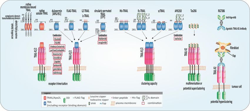
Figure
Figure 2.2. Highly
Highlypotent TRAIL
potent TRAILreceptor agonists
receptor (TRAs)(TRAs)
agonists and combinatorial approaches.approaches.
and combinatorial Schematic illustration of native
Schematic tumour-necrosis-factor-related
illustration apoptosis-inducing
of native tumour-necrosis-factor-related
ligand (TRAIL) andligand
apoptosis-inducing potent(TRAIL)
TRAs evaluated in clinical
and potent TRAsstudies or with
evaluated promising
in clinical resultsorinwith
studies preclinical experiments
promising results and their combination
in preclinical with anticancer
experiments and theirapproaches:
combination
native membrane-bound TRAIL is a protein of 281 amino acids (aa) comprising a cytoplasmic domain (aa 1–17), a transmembrane domain
with anticancer approaches: native membrane-bound TRAIL is a protein of 281 amino acids (aa) comprising a cytoplasmic domain (aa 1–17), a transmembrane domain(aa 18–38), and an extracellular
(aadomain
18–38), (aa
and39–281). The receptor-binding domain (aa 114–281) is located in the extracellular portion [1] which is cleaved from the cell surface to form bell-shaped homo-
an extracellular domain (aa 39–281). The receptor-binding domain (aa 114–281) is located in the extracellular portion [1] which is cleaved from the cell
trimers. Recombinant human TRAIL/Apo2L (dulanermin) is based on the amino acids 114-281 of the extracellular portion of TRAIL [4]. The patients’ benefit in clinical
surface to form bell-shaped homo-trimers. Recombinant human TRAIL/Apo2L (dulanermin) is based on the amino acids 114–281 of the extracellular portion of
trials was merely marginal even combined with chemotherapeutic regimens such as FOLFIRI (folinic acid, fluorouracil, irinotecan) [93,94]. Tags at the N-terminus of the
TRAIL [4]. The patients’ benefit in clinical trials was merely marginal even combined with chemotherapeutic regimens such as FOLFIRI (folinic acid, fluorouracil,
extracellular domain of TRAIL such as FLAG and poly-histidine (His) aid purification of the ligand and leucine zipper (LZ) and isoleucine zipper (iz) stabilize the trimers
irinotecan) [93,94]. Tags at the N-terminus of the extracellular domain of TRAIL such as FLAG and poly-histidine (His) aid purification of the ligand and leucine
[3,120]. Circularly permuted TRAIL (CPT) consists of the N-terminal amino acids 121-135 linked to the C-terminal amino acids 135-281 of TRAIL via a flexible linker [121].
zipper (LZ) and isoleucine zipper (iz) stabilize the trimers [3,120]. Circularly permuted TRAIL (CPT) consists of the N-terminal amino acids 121–135 linked to the
To achieve the fragment crystallizable region (Fc)-TRAIL fusion protein, the Fc portion of human immunoglobulin G1 (IgG1) was fused to the N-terminus of human
C-terminal amino acids 135-281 of TRAIL via a flexible linker [121]. To achieve the fragment crystallizable region (Fc)-TRAIL fusion protein, the Fc portion of human
TRAIL (aa 95 to 281) [122]. Single-chain TRAIL (scTRAIL) is assembled by 3 extracellular domains of TRAIL that are covalently connected by 2 short peptide sequences
immunoglobulin
[123]. APG350 was G1 (IgG1) wasby
developed fused to the of
the fusion N-terminus
scTRAIL and of human TRAIL
the Fc-part of a (aa 95 toIgG1
human 281)leading
[122]. Single-chain
to six receptorTRAIL
binding (scTRAIL) is assembled
sites per drug moleculeby 3 extracellular
[124]. domains
TAS266 comprises
of TRAIL that are covalently connected by 2 short peptide sequences [123]. APG350 was developed by the fusion of scTRAIL and the Fc-part
four humanised high-affinity heavy chain domain (VHH) antibody fragments which enable clustering of four TRAIL-R2s [117]. The substance caused severe hepatoxicity of a human IgG1 leading
to six
in clinical phase I studies. The tetravalent fibroblast-activation protein (FAP)-TRAIL-R2 antibody RG7386, targets cancer-associated fibroblasts in the tumour stroma enable
receptor binding sites per drug molecule [124]. TAS266 comprises four humanised high-affinity heavy chain domain (VHH) antibody fragments which and
clustering
TRAIL-R2 of on
four TRAIL-R2s
tumour [117]. The substance
cells simultaneously therebycaused
inducingsevere hepatoxicity
higher-order in clinical
clustering phase I studies.
and apoptosis inductionThe tetravalent
in vitro fibroblast-activation
and animal models [125]. protein (FAP)-TRAIL-R2
antibody RG7386, targets cancer-associated fibroblasts in the tumour stroma and TRAIL-R2 on tumour cells simultaneously thereby inducing higher-order clustering
and apoptosis induction in vitro and animal models [125].Cancers 2019, 11, 456 8 of 30
Circularly permuted TRAIL (CPT) (Figure 2) is a novel form of recombinant human Apo2L/TRAIL
with ongoing clinical evaluation for multiple myeloma (MM) and other hematologic cancers. It consists
of the N-terminal amino acids 121–135 linked to the C-terminal amino acids 135–281 of TRAIL via a
flexible linker. CPT’s pharmacokinetic is superior to wild-type TRAIL due to an enhanced stability and
half-live [121]. In a single dose-escalation study adverse effects such as fever, fatigue, leucopenia, and
vomiting remained below 10% incidence, and no immune responses were traceable [126]. To further
evaluate the safety and efficacy of CPT as single agent, a phase II open-label study has been initiated.
The monotherapy was well tolerated with 20–30% partially- or better-responding patients. No DLT and
MTD could be observed [127]. A further dose-finding phase II study enrolled thalidomide-resistant
relapsed refractory multiple myeloma (RRMM) patients and investigated the combination of CPT with
the MM-approved drug thalidomide. The drug has been initially distributed under the trade name
Contergan® (Chemie-Grünenthal, Aachen, Germany) as sleeping pill, known from the Contergan
disaster [128]. The ORR was 22%, however, treatment-related adverse effects (TRAE) occurred more
often in the combinatorial treatment versus the single-agent treatment. The rate of higher-grade
adverse effects had been certainly lower compared to the treatment with first-line therapy for MM
such as bortezomib [129] demonstrating a comparatively low toxicity of the treatment regimen. An
antitumour effect could be denoted, while no DLT occurred [130]. A phase II study for RRMM was
conducted to compare the safety and efficacy of CPT plus thalidomide combined with dexamethasone
(CPT + TD arm) and thalidomide plus dexamethasone (TD arm) [131]. The trial wanted to harness the
antiproliferative effect of the glucocorticoid dexamethasone on melanoma cells [132]. The ORR of the
CPT + TD arm was 38.3% compared to the ORR of the TD arm with 25%. Occurring TRAEs were in
large part of the lower grades, whereas around 20% of patients suffered from severe side effects [131].
Currently, CPT presents the exception considering the antitumour potential amongst the TRAs. These
findings provided the rationale for prospective confirmatory studies.
ABBV-621 is designed for higher-order clustering of TRAIL-Rs by fusing an immunoglobulin
G1 (IgG1)-fragment crystallizable region (Fc) portion to a single chain trimer of TRAIL subunits.
In this way, six TRAIL-R binding sites are available upon dimerization of Fc portions. As with
other TRAs, the preclinical data were promising. Apoptosis-inducing efficiency was observed in
hematologic malignancies alone and in combination with the sensitisers navitoclax and venetoclax
(see Section 5.4. BH3 and SMAC mimetics plus TRAIL) [133]. Currently, pre-treated patients are
recruited for a dose-escalation study in solid tumours and hematologic malignancies using ABBV-621
(ClinicalTrials.gov Identifier: NCT03082209). The results are expected in February 2021.
Although CPT is the exception amongst TRAs boasting attributable anti-tumour action in clinical
settings, a notable patient number showed no responses to the treatment. The main reason for the
disappointing outcome of the majority of TRAs can be found in the inherent insensitivity of tumour
cells to TRAIL monotherapy or acquired resistance [134–136]. To overcome this resistance, more
profound understanding of the underlying mechanisms is vitally important.
4.2. TRAIL Resistance Mechanisms in Cancer
One hallmark of cancer is an intrinsic or acquired resistance to apoptotic signals [137]. The
TRAIL-induced apoptosis pathway provides manifold steps at which sensitivity can be modified
starting with regulation of TRAIL-R levels and stretching all the way to mitochondrial anti-apoptotic
proteins (Figure 1) [138–140]. While the big picture of TRAIL resistance is still not completely
understood, numerous details and steps of the mechanism have been revealed [13,14,87].
4.2.1. TRAIL Receptors
At the receptor level, mutations in TRAIL-R1 and TRAIL-R2 [141–143], and expression of the decoy
receptors TRAIL-R3 and TRAIL-R4 lacking the functional intracellular DD may play a regulatory role
in TRAIL-induced apoptosis [9,16,82]. DcRs, particularly TRAIL-R4, might compete for TRAIL binding
or may disrupt trimerisation of the death-inducing receptors -R1 and -R2 [9,144,145] when co-expressedCancers 2019, 11, 456 9 of 30
on the same cell. Indeed, TRAIL-R4 was shown to directly interact with the death-inducing
receptors directly thereby disrupting efficient DISC formation [82,145], and to signal alternative,
tumour-promoting pathways such as NF-κB. Intriguingly, DcRs expressed by stromal cells have been
demonstrated to negatively influence TRAIL sensitivity of tumour cells [146]. There is mounting
evidencethat in contrast with its name, TRAIL seems to generate versatile effects, while enhanced
tumour progression and metastasis could be observed upon treatment with exogenous TRAIL and
activation of alternative pathways [147]. Moreover, in Kirsten rat sarcoma (KRAS)-mutated cancer
upregulated TRAIL-R2 and endogenous TRAIL act in a tumour-supportive manner [43], a concept
which does not seem to be limited to KRAS-mutated cancers and also engages tumour-favourable
polarisation of immune cells [148].
Post-translational modifications of TRAIL-Rs have gained increasing interest over the last
couple of years [149]. O-Glycosylation of TRAIL-R2 was found to control the cells’ sensitivity
to TRAIL (described previously as a possible biomarker (see Section 4.1 above TRAs in Clinical
Studies—Can Failure still lead to success?). A whole genome-profiling approach identified a correlation
of increased GALNT14 and TRAIL resistance. Cells could be sensitised to apoptosis when GALNT14
was silenced [116]. In line with the idea that posttranslational modifications of the receptors have a
considerable effect on TRAIL binding and killing, N-glycosylation of TRAIL-R1 increased its apoptotic
efficacy [150,151].
Besides their membrane localisation, TRAIL-Rs have been observed also in intracellular
compartments and in the nucleus [152,153]. The cellular localisation of TRAIL-Rs seems to play
a growing role in TRAIL non-apoptotic signalling. Recent experiments revealed a correlation between
increased levels of nuclear TRAIL-R2 and shortened patient survival, indicating a tumour-promoting
role of the nuclear variant [44].
4.2.2. The DISC
At the DISC level, FADD was shown to be indispensable for TRAIL-triggered apoptosis
and loss of function led to resistance to TRAIL [48,154]. c-FLIP overexpression was detected in
TRAIL-resistant cancer [155,156]. This antiapoptotic regulator and caspase-8 seem to be critical
factors and unique molecules in death-receptor apoptosis signalling [48,55,139,157–159]. Recent
discoveries identified a novel deubiquitylase of c-FLIPL , ubiquitin-specific peptidase 8 (USP8), directly
interacting with the long isoform leading to its stabilisation and thereby inhibition of DR-induced
apoptosis. Interestingly, increased levels of USP8 were found in melanoma and cervical cancer [160].
Changes in c-FLIP [155,161,162] and caspase-8 ratio [163] can trigger cell death-independent cellular
responses via a cytosolic signalling complex termed complex II [164]. This complex II includes,
besides the DISC components FADD and caspase-8, receptor-interacting protein kinase (RIPK1),
TNF receptor-associated factor-2 (TRAF2) and NF-κB essential modifier (NEMO)/inhibitor of κB
(IκB) kinase (IKK) [38–40]. TRAF2 recruits cellular inhibitor of apoptosis protein 1/2 (cIAP1/2) that
in turn promote the ubiquitination of RIPK1 [165] thereby presenting the recruitment platform for
linear ubiquitin chain assembly complex (LUBAC) [41]. LUBAC adds linear poly-ubiquitin chains to
RIPK1 and completes tasks in complex I and complex II, enabling caspase-8 cleavage and IKK complex
assembly, and accordingly activation of NF-κB [27,35,41,166–172]. Triggering the NF-κB pathway is one
example of TRAIL’s non-canonical signalling routes occurring at the DISC level and conveying TRAIL
resistance (Figure 1). Moreover, activation of mitogen-activated protein kinase (MAPK) [173,174] and
tyrosine kinase Src and phosphoinositide 3-kinase (PI3K) pathways [175] through this complex II have
been described [14].
4.2.3. Bcl-2 Family
TRAIL resistance can also be attributed to expression patterns of the Bcl-2 family [13,87]: If cells
undergo type II extrinsic apoptosis, death signals need the amplification via the mitochondrial pathway.
Here, the ratio of the proapoptotic proteins Bax and Bak [176] and their antiapoptotic antagonistsCancers 2019, 11, 456 10 of 30
Bcl-2, Bcl-xL, and Mcl-1 [177–182] govern cell fate [78]. The mitochondrial release of SMAC/DIABLO
ensuring activation of effector caspases, for example, is inhibited by Bcl-2 overexpression [183].
4.2.4. IAPs
As indicated by their name, inhibitors of apoptosis proteins (IAPs) can block programmed cell
death [184] and mediate TRAIL resistance [180,181,185–187] by regulating effector caspase activity
such as caspase-3 or by inhibiting caspase-9 and apoptosome activity. An upregulation of IAPs has
been described as a common feature of cancer [188–190]. The human IAP family comprises eight
members so far, which have at least one baculovirus IAP repeat (BIR) domain in common: neuronal
apoptosis inhibitory protein (NAIP; BIRC1), cIAP1, cIAP2 (BIRC2 and BIRC3), XIAP (BIRC4), survivin
(BIRC5), BRUCE (Apollon; BIRC6), livin (BIRC7), and testis-specific IAP (Ts-IAP; BIRC8). Caspases of
the apoptotic cascade are inhibited by XIAP, cIAP1, c-IAP2, and survivin [191,192].
4.2.5. Autophagy—The Self-Consuming TRAIL
Besides its apoptosis-inducing capabilities, TRAIL is associated with autophagy. Autophagy
(or autophagocytosis) is an essential cellular degradation mechanism to remove damaged or redundant
proteins and cell organelles [193]. Since the 1950s, the mechanism has enthralled cell scientists.
The cell biologist Yoshinori Ohsumi was rewarded with the 2016 Nobel Prize in Physiology or
Medicine for the early description of the autophagy machinery [194], while other aspects are still
obscure. The mechanism is involved in growth regulation and ageing processes and a diversity of
other cellular functions such as differentiation and pathogen defence [195–197]. During periods of
increased nutritional stress, the cellular components are recycled to maintain cell survival. Apoptosis
and autophagy’s elements have various points of contact indicating the multifaceted cross-talk
between the pathways. Usually, autophagy blocks apoptosis induction, and caspase activation in
the apoptotic cascade shuts off autophagy signalling. One of the crucial controllers of autophagy
is the mechanistic target of rapamycin (mTOR) kinase controlled by starvation, growth factors, and
cellular stressors [197,198]. Upon DNA damage, p53 mediates the activation of AMP-activated protein
kinase (AMPK) and thereby the blockage of mTOR and autophagy [199]. However, collected data over
the last decade describes that p53 can also act in an autophagy-activating manner [200,201]. Beclin 1
(autophagy-related gene 6, ATG6) holds a vital role in the surveillance of autophagic actions [202,203]
and the crosstalk between apoptosis and autophagy [198]. The Bcl-2 family members Bcl-2, Bcl-xL,
and Mcl-1 are regulators of Beclin 1 by directly interacting with its BH3 domain. Noteworthy, the
underlying mechanism of how these proteins either inhibit or stimulate autophagy and apoptosis is
poorly understood [204].
Apoptosis and autophagy are both stimulated in response to metabolic stressors, however,
in tumour cells, a different picture emerges, and the dialogue between autophagy and apoptosis
is disrupted [193,205,206]. Generally, the nature of autophagy in cancer is bivalent, most likely
describable as dynamic, demonstrated by either tumour-supportive or -suppressive actions [206–208].
Due to this interplay of autophagy and apoptosis, studies have been focused on the link between
TRAIL resistance and autophagy. The connection between the mechanisms has become tangible
when TRAIL has been shown to induce both, apoptosis and autophagy, in a series of cancer cell
lines [209–211]. For cytoprotective TRAIL-induced autophagy, the activation of TRAF2- and
RIPK1-mediated MAPK8/c-Jun N-terminal kinases (JNK) signalling [212] and AMPK signalling [213]
was reported. In a nutshell, these results suggest that several pathways and regulators work together
in a concerted manner to mediate TRAIL-induced apoptosis and autophagy. There are accumulating
results in recent years identifying further regulators which maintain the crosslinking of apoptosis and
autophagy. Notable, caspase-9 as such regulator facilitates the early events leading to autophagosome
formation by complexing with ATG proteins thereby suppressing the apoptotic character of the
caspase [214].Cancers 2019, 11, 456 11 of 30
Autophagy controls the timing and efficiency of TRAIL-induced MOMP by the level of
autophagy-releasing p53 upregulated modulator of apoptosis (PUMA, also known as Bcl-2-binding
component 3) [215], a proapoptotic BH3-only Bcl-2 family member and downstream target of
p53. MOMP is inefficient in cells with low PUMA levels, and some of these cells have been
observed to recover and divide again whereas high PUMA levels led to MOMP quickly followed by
apoptosis [216]. Most solid tumours exhibit areas permanently or transiently under hypoxic conditions
resulting from abnormal vascularisation and blood flow [217]. The hypoxia intensifies mitochondrial
autophagy in colorectal cancer cells. This loss of the cell organelles leads eventually to the scarcity of
mitochondria-derived pro-apoptotic molecules such as SMAC thereby disrupting the transmission of
TRAIL-mediated apoptotic signals in type II cells [218].
Recently, the accumulation of autophagic organelles in TRAIL-resistant cancer cell lines has
been described. It was proven that this accretion led to TRAIL resistance in breast cancer cells that
was accompanied by decreased surface expression of TRAIL-R1 and TRAIL-R2 [219]. The influence
of autophagy on TRAIL-induced apoptosis in metastasis was just investigated by use of in vitro
circulating tumour cells (CTCs) models for breast cancer which developed resistance to TRAIL with
transition to a non-adherent state. This resistance was connected with downregulated surface levels
of TRAIL-R2. The CTCs showed a fast autophagic flux detectable by an increase of autophagosome
organelles where TRAIL-R2 was degraded [220]. The data obtained reveal a potential mechanism of
how autophagy contributes to bypass TRAIL-induced apoptosis in CTCs.
Taken together, TRAIL-induced apoptosis and autophagy are controlled by a plethora of pathways
and often shared regulators that are not fully explored. This crosstalk gains in importance, and insights
into this highly complex network in the period ahead contributes to novel treatment approaches in
TRAIL-related cancer therapy including autophagy inhibitors such as beclin 1 inhibitors or inhibitors
of PI3K.
4.2.6. Fractional Killing and Microenvironment
TRAIL resistance can occur during treatment cycles when only a fraction of cancer cells die due
to cell cycle-imposed variations in pro- and anti-apoptotic protein levels. This enigmatic phenomenon
has a non-genetic origin termed “fractional killing” [221]. A hypothesis for fractional killing by
TRAIL is a caspase activation threshold determined by caspase inhibitory protein levels in cells at
the time of TRAIL stimulation [222]. Eventually over time, monotherapy with TRAIL may select for
survival of a resistant cell population and inefficiency of the therapy without any genetic predictors
causing this process of natural selection. In keeping with this notion, cells surviving TRAIL-treatment
within a population have been identified as the primary source for secreted cytokines such as
interleukin 8 (IL-8) [223]. Moreover, overexpression of TRAIL-R1/R2, and engagement of an agonistic
antibody with TRAIL-R2 can induce the release of inflammatory cytokines in an NF-κB-dependent
manner [224]. The cancer microenvironment is permanently governed by these inflammatory factors
described as a hallmark of cancer to protect the tumour and drive its progression [148]. Monotherapy
with TRAIL and acquired resistance or resulting resistance from fractional killing may, therefore,
promote the modulation of the tumour-promoting and -protecting microenvironment. Vice versa, the
microenvironment seems to regulate TRAIL sensitivity of tumour cells via DcRs expressed on stromal
cells [146].
5. Strategies to Regain TRAIL Sensitivity—The Bench-to-Bedside TRAIL
So far, most clinical studies evaluating TRAs have been discontinued due to insufficient
anti-tumour effects. A fundamental reason is the intrinsic or acquired TRAIL resistance of
cancer cells. Fortuitously, the various levels of regulation within the TRAIL-induced apoptotic
pathway which can provoke resistance to apoptotic signals, offer equal opportunities to sensitise
cancer cells to TRAIL-induced apoptosis [13,87]. Whilst mechanisms that mediate resistance to
TRAIL in non-malignant cells are not entirely uncovered, it is questionable and unpredictable ifCancers 2019, 11, 456 12 of 30
TRAIL-sensitising strategies are limited to cancer cells or may also affect healthy cells. In cells that
express both death-inducing TRAIL-Rs, heterocomplexation was observed [225], although it is still
obscure whether this phenomenon has a unique signalling function. Nevertheless, it would be
advantageous to target both, TRAIL-R1 and TRAIL-R2, in tumours considering the tissue-dependent
preference of one receptor. In an effort to overcome TRAIL resistance, the ligand has been combined
with other anti-cancer drugs or with nature-derived products to enhance cell sensitivity, respectively,
and more potent TRAs have been developed. Several TRAs are presented in Figure 2. A variety
of sensitising strategies can be found in the literature and have been reviewed in depth [13,14,87].
Unfortunately, many of these approaches had not been verified in animal models, and therefore
possible toxicity has been ignored. Therefore, in the following section, only sensitising approaches
with a high chance for quick and efficient clinical translation will be discussed.
5.1. Highly Potent TRAs
TRAIL engages three receptors, each at the interface between two of its cysteine-rich domains. The
trimer is stabilised via a zinc atom bound by cysteines that is crucial for its biological activity [226,227].
TRAs lacking the critical central atom were poorly soluble and tended to aggregate, presumably
conveying liver toxicity [228,229]. Aggregated TRAIL can evoke increased immunogenicity when
epitopes are exposed that are usually hidden. An untagged variant including stoichiometric zinc did
not provoke significant hepatotoxicity [228].
Tags at the N-terminus of the extracellular domain of TRAIL, such as FLAG™ (peptide sequence
DYKDDDDK) and poly-histidine (His, His6-Tag) aid purification of the ligand. Leucine zipper (LZ) and
isoleucine zipper (iz) stabilise TRAIL trimers thereby increasing their agonistic potential [3,120]. Since
liver toxicity of His-TRAIL and antibody-crosslinked FLAG-TRAIL has been reported, safety concerns
were noted for these variants and research on non-tagged variants has been intensified [120,228,230].
Presumably, these safety concerns are causative for untagged dulanermin entering clinical trials.
A hepatoxicity of LZ- and iz-TRAIL, however, could be excluded and a safe in vivo application has
been performed [3,120,231].
Due to their bivalent binding mode, agonistic TRAIL-R antibodies are not able to induce higher
crosslinking of TRAIL-Rs leading to insufficient DISC activation and weak apoptotic signals compared
to the tagged variants of TRAIL [114]. The crosslinking capacity is limited by the absence of
adequate Fcγ receptors in the surrounding of cancer cells [114,232]. The aim is therefore to obtain an
antibody multimerisation resulting in TRAIL-R-clustering on cancer cells and amplified apoptosis
induction. For this reason, research into novel, stable TRAs suited for this purpose is pressing
ahead [13,87,233]. A further effort to amplify the DISC signal can be the combined treatment with
rhTRAIL and TRAIL-R-targeting antibodies binding to distinct receptor epitopes, thereby enhancing
DISC formation and apoptosis induction [234].
Prolonged half-life and an increased tendency for oligomerisation was achieved by the fusion of
TRAIL to the Fc portion of human IgG1 (Fc-TRAIL). Although the capacity for apoptosis induction
was higher in vivo and in vitro, no liver toxicity was observed [122].
A recent development is a hexavalent TRA, designed by the fusion of two trimers of the
extracellular part of TRAIL to the Fc portion of human IgG1 (APG350) thereby creating six receptor
binding sites per drug molecule [124]. Recently, the superiority of APG350 over soluble TRAIL has
been studied in pancreatic cancer in vitro and xenograft models [235]. Whilst Bcl-xL overexpression
rendered cells resistant, sensitivity could be re-established when cells were additionally treated
with the BH3 mimetic navitoclax (see Section 5.4 BH3 and SMAC mimetics plus TRAIL) [235,236].
Although APG350 showed encouraging preclinical results, the urgent need for predictive biomarkers
and TRAIL sensitising strategies is highlighted through its evaluation in Bcl-xL expressing cells.
The formation of higher-order complexes induced by APG350 is not sufficient to overcome blocked
mitochondrial apoptosis.Cancers 2019, 11, 456 13 of 30
The tumour microenvironment is known to influence the therapy response negatively [113]. DcRs
on stromal cells influence TRAIL resistance [146], and, in turn, as recently reported, a TRAIL-induced
cancer secretome triggers a tumour-promoting microenvironment [148]. These facts illustrate the
relevance of the tumour microenvironment when considering TRAIL-based therapies. The bispecific,
tetravalent fibroblast-activation protein (FAP)-TRAIL-R2 antibody, RG7386, targets cancer-associated
fibroblasts in the tumour stroma and TRAIL-R2 on tumour cells simultaneously thereby inducing
higher-order clustering and apoptosis induction in vitro and animal models. The approach showed
promise as monotherapy as well as in combination with chemotherapeutics [125].
5.2. Chemo- and Radiotherapy Plus TRAIL
Additive effects of standard chemotherapeutic agents such as gemcitabine, irinotecan,
and cisplatin together with TRAIL have been extensively tested in preclinical models [237].
Furthermore, radiotherapy was proven to synergistically induce apoptosis in combination with
TRAIL [238]. Modified receptor expression, enhanced DISC formation, and influence on pro- and
anti-apoptotic protein expression are amongst the presumed mechanisms leading to the chemo- and
radiotherapy-induced sensitisation. These findings laid the foundation for the combination with TRAs
also in clinical studies, so far with marginal effects. Hitherto CPT is an exception, since the ORR of the
combined treatment arm was increased in comparison to treatment with chemotherapy alone [131].
The 26S proteasome inhibitor bortezomib is used in MM care and is applied also in other
malignancies including breast cancer, colon cancer, and prostate cancer [239]. The exact mode of
action of bortezomib is revealed bit by bit. At the TRAIL-R level, a shift of the apoptotic signals from
TRAIL-R1 to TRAIL-R2 by internalisation and degradation of TRAIL-R1 could be observed [240].
It also seems that a crosstalk of apoptosis and autophagy plays a central role [241–243]. TRAIL’s
part in the regulation of the autophagosome formation is dependent on the expression of the ATG
proteins ATG5, ATG7, and beclin-1. This TRAIL-induced ATG expression was weakened by JNK.
In combination with bortezomib the activation of extracellular signal-regulated kinase (ERK) has
been shown to induce autophagy [244]. Conversely, the inhibitory role of sustained activation
of ERK has also been demonstrated. Bortezomib may block the autophagic flux by induction of
ERK phosphorylation [243]. The evidence-based sensitisation capacity for TRAIL-induced apoptosis
directed the substance to clinical trials combined with the TRAIL-R2 specific TRA mapatumumab.
Again, an additive therapeutic advantage was not detectable mainly due to the failure of the antibody
for to sufficiently crosslink TRAIL-Rs (see Section 5.1 Highly potent TRAs) [87].
5.3. Inhibition of Anti-Apoptotic c-FLIP in Combination with TRAIL
c-FLIP is an essential anti-apoptotic regulator and can suppress TRAIL-mediated apoptosis [245].
Coming back to the clinical studies investigating CPT (see Section 4.1 TRAs in Clinical Studies—Can
Failure still lead to Success?), the therapy was efficient in 20–30% of the patients. The remaining
70% were nevertheless resistant to CPT. Analysing the reasons, elevated expression of c-FLIP in
RRMM patients was striking [246] which may mediate resistance to CPT by inhibiting the DISC.
Prior experiments already demonstrated the sensitisation of human melanoma cells to TRAIL by
suppression of c-FLIP [245]. In a recent study, resistance to CPT was counteracted in MM in vitro and
in vivo by downregulation of c-FLIP by rocaglamide (Figure 1) [246]. Rocaglamides are phytochemicals
with cancer inhibiting abilities [247]. The natural product in combination with TRAIL also generated
impactful in vitro action in additional cancer entities including renal carcinoma [248] and hepatocellular
carcinoma (HCC) [249].
5.4. BH3 and SMAC Mimetics Plus TRAIL
The mitochondrial pathway of apoptosis is an important starting point for TRAIL sensitisation
since high IAP expression correlates with drug resistance and tumour progression [250].
Small-molecule inhibitors have been designed which mimic the XIAP-binding site of SMAC therebyCancers 2019, 11, 456 14 of 30
antagonising multiple members of the IAP family in the mitochondrial apoptosis cascade (Figure 1).
Hitherto, the preclinical evidence [251,252] and first clinical studies [253,254] of these SMAC mimetics
indicate a particular aptitude for anticancer therapy as a single agent or in combination with
chemotherapeutic drugs. Their real capability as TRAIL sensitiser has already been analysed in
a broad range of cancers [192]. Thus, the SMAC mimetic birinapant made it into a clinical trial in
ovarian cancer in combination with the TRAIL-R2 agonist conatumumab. In the phase 1b study the
tolerability of the combinatorial approach was proven [255]. Future studies will reflect the actual
suitability of the substances as a treatment regimen.
Bcl-2 family members govern cell fate in the mitochondrial apoptosis pathway. Overexpression
of the anti-apoptotic relatives Bcl-2, Bcl-xL, and Mcl-1 mediates resistance to apoptosis signals and
is a frequent feature among human cancers. To re-sensitise cells to apoptotic signals, BH3 mimetics
have been established to conquer the anti-apoptotic Bcl-2 family members (Figure 1) [256]. Candidates
that reached clinical phases are ABT-199 (venetoclax) directed against Bcl-2, and ABT-263 (navitoclax)
antagonising Bcl-2 and Bcl-xL [257–261]. The treatment-related effects of BH3 mimetics were striking.
Hence, an orally bioavailable form of venetoclax was approved in 2016 by the United States Food and
Drug Administration (FDA) for chronic lymphocytic leukaemia (CLL) [262]. An effective synergism
of navitoclax and venetoclax together with TRAIL was observed in vitro [263,264]. The clinical TRA
candidate ABBV-621 induced apoptosis alone and enhanced in combination with navitoclax and
venetoclax in haematological tumours [133]. According to these studies, SMAC mimetics, and BH3
mimetics might be a suitable class of sensitisers emerging from experimentation to overcome resistance
to TRAIL-induced apoptosis in patient trials.
6. Conclusions and Future Perspectives
During the last decade, research into novel anti-cancer drugs has especially revolved around the
discovery of specifically-targeted, side effect-reduced approaches. In this context, the discovery of the
tumour selectiveness of apoptosis induction by TRAIL in the late 90s was thought to be a breakthrough
and research into TRAIL-induced signalling pathways has been intensified. Following favourable
preclinical results of tumour cell-selective apoptosis induction, recombinant forms of TRAIL and
TRAIL-R-targeting antibodies have been evaluated in clinical trials, though with sobering outcome
in most cases. The drawbacks have been outlined above as well as strategies to overcome TRAIL
resistance. Novel, highly potent TRAs have been designed. By the use of high-throughput screenings
(HTS), the identification and development of these TRAs can become more efficient. Although the TRAs
with improved agonistic potential may finally boast the desired antitumor effect in clinical settings,
overcoming TRAIL resistance remains a struggle ahead. There is a stable demand for highly-potent
and adverse effect-reduced TRAIL sensitisers without losing sight of possible, fulminant adverse
effects. For more appropriate pre-clinical testing, humanised and patient-derived preclinical mouse
models are warranted. Moreover, predictive biomarkers are urgently needed to identify responding
patients or to follow therapy response more precisely. To date, everything suggests that CPT will
perform superior to conventional chemotherapy in haematological malignancies. In case that this
approach can be validated for other tumour entities, and efficient sensitisers such as SMAC and BH3
mimetics can be clinically validated in combination, targeting this receptor/ligand system might just
turn out to be a “TRAILblazer” in cancer therapy.
Author Contributions: The study was designed by J.L., D.H.-B., and S.v.K. Literature was collected by A.-L.K.
and A.H., figures were devised by A.-L.K. and J.L., A.-L.K., S.v.K., A.T., and U.K. wrote the manuscript. All
authors read and approved the manuscript.
Funding: This work was funded by a research fellowship from the Deutsche Forschungsgemeinschaft (DFG) to
J.L.; S.v.K. is funded through the Max-Eder-Programme of the German Cancer Aid.
Conflicts of Interest: The authors declare no conflict of interest.You can also read