Nonsense Suppression Therapy: New Hypothesis for the Treatment of Inherited Bone Marrow Failure Syndromes - MDPI
←
→
Page content transcription
If your browser does not render page correctly, please read the page content below
International Journal of
            Molecular Sciences

Review
Nonsense Suppression Therapy: New Hypothesis for
the Treatment of Inherited Bone Marrow
Failure Syndromes
Valentino Bezzerri 1 , Martina Api 1 , Marisole Allegri 1 , Benedetta Fabrizzi 1 , Seth J. Corey 2
and Marco Cipolli 3, *
 1    Cystic Fibrosis Center, Azienda Ospedaliero Universitaria Ospedali Riuniti, Via Conca 71, 60126 Ancona,
      Italy; valentino.bezzerri@ospedaliriuniti.marche.it (V.B.); martina.api@ospedaliriuniti.marche.it (M.A.);
      marisole.allegri@ospedaliriuniti.marche.it (M.A.); benedetta.fabrizzi@ospedaliriuniti.marche.it (B.F.)
 2    Department of Pediatric Hematology/Oncology and Stem Cell Transplantation, Cleveland Clinic, Cleveland,
      OH 44195, USA; coreys2@ccf.org
 3    Cystic Fibrosis Center, Azienda Ospedaliera Universitaria Integrata, P.le A. Stefani 1, 37126 Verona, Italy
 *    Correspondence: marco.cipolli@aovr.veneto.it

 Received: 30 May 2020; Accepted: 29 June 2020; Published: 30 June 2020

 Abstract: Inherited bone marrow failure syndromes (IBMFS) are a group of cancer-prone genetic
 diseases characterized by hypocellular bone marrow with impairment in one or more hematopoietic
 lineages. The pathogenesis of IBMFS involves mutations in several genes which encode for proteins
 involved in DNA repair, telomere biology and ribosome biogenesis. The classical IBMFS include
 Shwachman–Diamond syndrome (SDS), Diamond–Blackfan anemia (DBA), Fanconi anemia (FA),
 dyskeratosis congenita (DC), and severe congenital neutropenia (SCN). IBMFS are associated with
 high risk of myelodysplastic syndrome (MDS), acute myeloid leukemia (AML), and solid tumors.
 Unfortunately, no specific pharmacological therapies have been highly effective for IBMFS. Hematopoietic
 stem cell transplantation provides a cure for aplastic or myeloid neoplastic complications. However,
 it does not affect the risk of solid tumors. Since approximately 28% of FA, 24% of SCN, 21% of DBA,
 20% of SDS, and 17% of DC patients harbor nonsense mutations in the respective IBMFS-related genes,
 we discuss the use of the nonsense suppression therapy in these diseases. We recently described the
 beneficial effect of ataluren, a nonsense suppressor drug, in SDS bone marrow hematopoietic cells
 ex vivo. A similar approach could be therefore designed for treating other IBMFS. In this review
 we explain in detail the new generation of nonsense suppressor molecules and their mechanistic
 roles. Furthermore, we will discuss strengths and limitations of these molecules which are emerging
 from preclinical and clinical studies. Finally we discuss the state-of-the-art of preclinical and clinical
 therapeutic studies carried out for IBMFS.

 Keywords: inherited bone marrow failure syndromes; nonsense suppression therapy; nonsense
 mediated decay; ataluren

1. Introduction
     Inherited bone marrow failure syndromes (IBMFS) are characterized by peripheral cytopenia(s)
with a hypocellular bone marrow andimpairment in one or more hematopoietic lineages. They are
also cancer predisposition syndromes. The classical IBMFS are represented by Shwachman–Diamond
syndrome (SDS), Diamond–Blackfan anemia (DBA), Fanconi anemia (FA), dyskeratosis congenita (DC),
and severe congenital neutropenia (SCN). Importantly, 10–15% of bone marrow aplasia and 30% of
pediatric bone marrow failure disorders are caused by IBMFS, with approximately 65 cases per million
live births every year [1]. However, emerging data reveal that IBMFS are underdiagnosed because of

Int. J. Mol. Sci. 2020, 21, 4672; doi:10.3390/ijms21134672                             www.mdpi.com/journal/ijms
Int. J. Mol. Sci. 2020, 21, 4672 2 of 30
decreased recognition or because genetic mutation associated with congenital bone marrow failure are
detected only after a malignancy has arisen [2].
Clinical management of IBMFS requires multidisciplinary care and surveillance to detect early
emergence of malignancies. Early hematopoietic stem cell transplant (HCT) may correct bone marrow
failure and prevent the development of myeloid neoplasia, but it does not affect the risk of solid
tumors. However, post-HCT complications, such as graft-versus-host disease and immune dysfunction,
frequently occur. As an alternative to HCT, androgen administration may be suitable for patients with
FA and DC. Corticosteroids are often effective for patients with DBA, especially for those who lack
a compatible donor or are ineligible for HCT transplantation. In addition, recombinant granulocyte
colony-stimulating factor (G-CSF) is used to ameliorate severe neutropenia in SCN or SDS and prevent
recurrent infections. Long-term use of G-CSF in SCN and SDS has been associated with increased risk
of myelodysplastic syndrome (MDS) and acute myeloid leukemia (AML) [3]. No therapies to reduce
bone marrow failure or cancer risk in IBMFS have been developed so far.
It has been estimated that approximately 12% of human genetic disorders are caused by single-
nucleotide or out-of-frame nonsense mutations, leading to the generation of premature termination
codons (PTC). Correct protein synthesis normally represents the core of biological process for any
living organism. For this reason, protein synthesis is regulated at multiple levels and any disruption
on each of these steps may cause severe diseases. The therapeutic strategy aimed at overcoming
PTC has been defined as “nonsense suppression therapy”. This approach is intended to generate the
readthrough of PTC, restoring a full-length protein synthesis. Alternatively, nonsense suppression
therapy may be designed to reduce the nonsense mediated decay (NMD) induced by the nonsense
mutations. NMD is an evolutionarily conserved defense mechanism of eukaryotic cells that surveys
newly synthesized mRNA and degrades transcripts containing a PTC [4,5].
We have tested the efficacy of a small nonsense suppressor molecule, ataluren (PTC124, PTC
Therapeutics, NJ) [6], in correcting the basic defect of SDS with promising preclinical results. We are
planning a clinical trial of ataluren for SDS. Our preclinical studies might serve as proof of concept
for the development of nonsense suppression therapy for other IBMFS. This review will discuss the
possible use of nonsense suppression strategy in IBMFS as a novel therapeutic hypothesis.
2. Inherited Bone Marrow Failure Syndromes
Bone marrow failure syndromes (BMFS) cluster different disorders characterized by impaired
hematopoiesis which lead to selective or global cytopenia. BMFS may be acquired, such as aplastic
anemia (AA) and paroxysmal nocturnal hemoglobinuria (PHN), or congenital. IBMFS are classified
into those that result in pancytopenia and those limited to deficiency of one or more hematopoietic
lineages. FA and DC are characterized by progressive peripheral pancytopenia. Patients with DBA
exhibit anemia, whereas SDS and SCN are mostly associated with severe neutropenia. Some of the
IBMFS can be characterized by physical anomalies and failure to thrive. IBMFS are all associated with
increased risk for the development of MDS/AML and solid tumors.
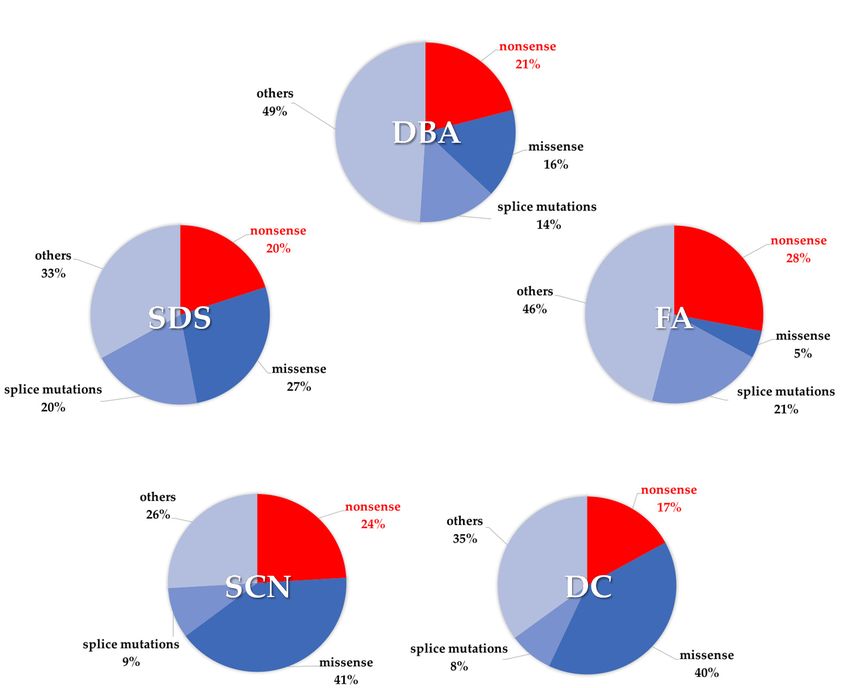
Approximately 80 different genes have been associated with different IBMFS [7]. Notably, 28% of
FA, 24% of SCN, 21% of DBA, 20% of SDS, and 17% of DC mutated alleles show nonsense pathogenic
variants (Figure 1). Classes of genes correlate with specific IBMFS, such as genes which encode
for proteins involved in DNA repair (FA), telomere biology (DC), and ribosome biogenesis (DBA).
Many cancers have been associated with somatic mutations in genes encoding ribosomal proteins [8].
Importantly, defective ribosomal proteins may induce the upregulation of the tumor suppressor gene
TP53 which encode the tumor suppressor protein p53 [9,10]. In this regard, p53 over-activation has
been implicated in the pathogenesis of DBA, SDS, and DC [11–14].Int.
Int. J.
J. Mol. Sci. 2020,
Mol. Sci. 2020, 21,
21, 4672
x FOR PEER REVIEW 33 of
of 30
31
Figure 1.
Figure 1. Incidence
Incidence of nonsense mutations
of nonsense mutations in inherited bone
in inherited bone marrow
marrow failure
failure syndromes
syndromes (IBMFS).
(IBMFS).
Percentages have been calculated on the basis of ClinVar database [15]. Only pathogenic and likely
Percentages have been calculated on the basis of ClinVar database [15]. Only pathogenic and likely
pathogenic variants
pathogenic variantshave
havebeen
beentaken
takeninto
intoaccount.
account.
2.1.
2.1. Shwachman–Diamond
Shwachman–Diamond Syndrome
Syndrome
SDS
SDSisisoneoneofofthe
themost
most common
common IBMFS,
IBMFS, first described
first described in 1964 [16],[16],
in 1964 withwith
an incidence of 1:76,000
an incidence [17].
of 1:76000
It is characterized
[17]. by neutropenia,
It is characterized pancreatic
by neutropenia, exocrine
pancreatic insufficiency,
exocrine and bone
insufficiency, andmalformations. Almost
bone malformations.
all
Almost all patients with SDS exhibit some degree of a hypocellular bone marrow specimen andfifth
patients with SDS exhibit some degree of a hypocellular bone marrow specimen and one one
of patients
fifth developdevelop
of patients pancytopenia. MyeloidMyeloid
pancytopenia. lineage maturation is severelyisimpaired
lineage maturation severelyand neutrophil
impaired and
maturation
neutrophil ismaturation
blocked at myelocyte-metamyelocyte
is blocked at myelocyte-metamyelocytestage [18]. Mild or severe stage neutropenia
[18]. Mild isor exhibited
severe
by most SDS patients. Thrombocytopenia and anemia are less frequent.
neutropenia is exhibited by most SDS patients. Thrombocytopenia and anemia are less frequent. SDS SDS patients exhibit a
propensity to develop
patients exhibit clonal cytogenetic
a propensity to developchanges
clonal in the bone marrow,
cytogenetic changesespecially
in the bone the marrow,
interstitialespecially
deletion
of
thethe long armdeletion
interstitial of chromosome 20, del(20)(q)
of the long and chromosome
arm of chromosome 7 anomalies
20, del(20)(q) [19–22].
and chromosome 7 anomalies
About 90% of SDS is caused by mutations affecting the Shwachman–Bodian–Diamond syndrome
[19–22].
(SBDS) gene,90%
About which ofencodes
SDS isa caused
protein involved
by mutations in ribosome biogenesis.
affecting Biallelic mutations in SBDS
the Shwachman–Bodian–Diamond
have been found in 90% of SDS patients [23,24]. Most
syndrome (SBDS) gene, which encodes a protein involved in ribosome importantly, 56% of these patients share the
biogenesis. same
Biallelic
nonsense mutation c.183-184TA>CT (K62X) in one allele [23]. Nevertheless,
mutations in SBDS have been found in 90% of SDS patients [23,24]. Most importantly, 56% of these the splicing mutation
c.258+2T>A,
patients sharewhich the sameaffects the donor
nonsense splice c.183-184TA>CT
mutation site of intron 2, is displayed
(K62X) in one inallele
the second allele of the
[23]. Nevertheless,
almost totality of SBDS-mutated patients [23,25]. It turns out that the
the splicing mutation c.258+2T>A, which affects the donor splice site of intron 2, is displayedincidence of nonsense mutations
in the
second alleleinofSBDS
recognized gene is
the almost 20% (Table
totality 1). The apparent
of SBDS-mutated patientsinconsistency
[23,25]. It turns between
out thatthetheincidence
incidence of of
nonsense
nonsense mutations
mutations in the SDS population
recognized in SBDS gene (56%) and (Table
is 20% the allelic incidence
1). The apparent of this type of pathogenic
inconsistency between
variation
the incidence(20%)ofisnonsense
notable because
mutationsno patients
in the SDS homozygous
populationfor the c.183-184TA>CT
(56%) mutationofhave
and the allelic incidence this
been
type recognized
of pathogenic so far.variation
This is probably
(20%) isdue to the severity
notable because ofnothepatients
homozygous conditionfor
homozygous whichthe
critically impairs the embryonic development. Other genes have been
c.183-184TA>CT mutation have been recognized so far. This is probably due to the severity of theassociated with SDS during the
last few years,condition
homozygous the DNAJC21,
including which criticallyElongation
impairs the Factor Like GTPase
embryonic (EFL)-1 and
development. Other Signal
genesRecognition
have been
Particle
associated (SRP)-54
with [26–29].
SDS during DNAJC21,
the lastEFL1few and SRP54
years, are involved
including in ribosomeElongation
the DNAJC21, formation and peptide
Factor Like
processing [24]. DNAJC21 is a co-factor for the pre-60S ribosomal
GTPase (EFL)-1 and Signal Recognition Particle (SRP)-54 [26–29]. DNAJC21, EFL1 and SRP54 aresubunit; EFL1 promotes the release
involved in ribosome formation and peptide processing [24]. DNAJC21 is a co-factor for the pre-60S
ribosomal subunit; EFL1promotes the release of the eukaryotic initiation factor 6 (eIF6) duringInt. J. Mol. Sci. 2020, 21, 4672 4 of 30
of the eukaryotic initiation factor 6 (eIF6) during ribosome assembly; and SRP54 supports nascent
polypeptide trafficking from the ribosome. Impaired ribosome assembly may lead in turn to reduced
number of ribosomes and deficient translation, activating the tumor suppressor protein p53 pathway [9].
Not surprisingly, TP53 expression is upregulated in SDS [11,12] as well as in other ribosomopathies
such as DBA and DC.
Approximately 15–20% of SDS patients develop MDS or AML, with a progression rate of almost
1% per year [30]. A study conducted on 102 patients from the French SDS registry showed a cumulative
risk of MDS/AML of 36% at 30 years of age [31]. Importantly, mutations on TP53 in patients with SDS
have been associated with early leukemogenesis [32]. Overall cancer risk in SDS has been calculated
with an observed over expected ratio of 8.5 [9].
Table 1. IBMFS-associated genes mostly affected by nonsense mutations.
Incidence of
Disease Gene Nonsense Gene Function Ref
Mutations
RPL5 33%
RPL11 7%
RPL35A 22%
Diamond–Blackfan RPS10 17% Pre-rRNA processing of the 18 rRNA,
[33–53]
anemia RPS17 25% formation of the 40S or 60S ribosome subunit
RPS19 20%
RPS24 25%
RPS26 20%
Shwachman–Diamond
SBDS 20% 60S ribosome assembly [23–25,54]
syndrome
CTC1 28% Telomere protection
PARN 21% Poly(A)-specific ribonuclease
Dyskeratosis
RTEL1 26% Stability and elongation of telomeres [55–62]
Congenita
TERT 27% Telomerase reverse transcriptase
TINF2 18% Stability and elongation of telomeres
FANCA 14%
FANCB 16%
FANCC 33%
FANCD1 59%
FANCE 43%
Fanconi anemia FANCF 25% DNA cross-link repair, chromosome stability [33,37,63–83]
FANCG 24%
FANCI 29%
FANCJ 30%
FANCL 14%
FANCN 37%
CSF3R 40% G-CSF receptor
ELANE 8% Neutrophil elastase
Severe congenital G6PC3 33% Hydrolysis of glucose 6-phosphate
[33,84–96]
neutropenia HAX1 29% Apoptosis control, cytoskeletal development,
myeloid regulator
JAGN1 14% Neutrophil differentiation and survival
Data from ClinVar database [15]. Percentages have been calculated taking into account only pathogenic and likely
pathogenic genetic variations.
2.2. Diamond–Blackfan Anemia
Diamond–Blackfan anemia (DBA) is an IBMFS which affects 5 to 10 cases per million newborns
in Europe. Almost 55% of DBA patients exhibit mutations in one of 19 different ribosomal protein
(RP) genes, resulting in RP haploinsufficiency [53,97]. A plethora of pathogenic variants have been
reported to affect genes encoding either for the small ribosomal subunit (RPS) or the large ribosomal
subunit (RPL), including RPS7, RPS10, RPS15A, RPS17, RPS19, RPS24, RPS26, RPS27, RPS28, RPS29,
RPL5, RPL35, RPL11, RPL15, RPL18, RPL26, RPL27, RPL31, and RPL35A [98]. In addition, mutations
in GATA1, a master transcription factor that is fundamental for normal erythropoiesis [99], and in
RPS26 chaperone protein TSR2 gene [49], have been identified as non-RP mutations in DBA. Of note,
GATA1 mRNA translation deficiency is associated with reduced levels of RPS19, RPL5, and RPL11
proteins [99]. RPS19 was first linked to DBA. RPS19 mutations account for almost 25% of all DBAInt. J. Mol. Sci. 2020, 21, 4672 5 of 30
cases. Other RP genes most frequently implicated in DBA are RPL5, RPS26, and RPL11. Currently,
130 different variants in RPS19 have been reported [100]. On the basis of ClinVar database [15],
missense mutations are the most common (25%), followed by nonsense mutations, which occur in
20% of RPS19 mutated-patients (Table 1). In particular, p.Arg94X is the most frequent pathogenic
variant recurring in unrelated families carrying RPS19 mutations [101]. Most nonsense mutations,
large deletions and frameshift which generate a PTC, are expected to result in haploinsufficiency [102].
In this regard, it has been suggested that incomplete or aberrant transcripts resulting from PTC located
up to 50–55 nucleotides upstream the last exon–exon junction, may be more instable and may induce a
rapid transcript turnover through the NMD [36].
Although most DBA patients (55–60%) have nofamily history for this disorder, DBA is typically
inherited with an autosomal dominant pattern (40–45%) or, less frequently, with an autosomal recessive
pattern (10% of patients) [13].
Originally, DBA was called “hypoplastic anemia”, since anemia is the main feature of this disease.
In fact, anemia is reported in 25% of DBA patients at birth. Furthermore, 95% of DBA patients
early develop macrocytic anemia, reticulocytopenia and limited cytopenia. Short stature is frequent
among DBA patients, similarly to other IBMFS such as SDS [103]. Other clinical manifestations
include cranio-facial abnormalities, including cleft/lip palate (4%), eye malformations (5%), abnormal
facies (3%), and microcephaly (2%). Upper-limb malformations have been described, especially
regarding thumbs (8%). Urogenital (3%), gonadic (3%), cardiac (3%), and neurological anomalies have
been also described together with and developmental issues [104]. Bone marrow biopsies collected
from DBA patients show a markedly decrease of erythroid progenitors, which are often sparsely
distributed in a few small clusters. Such clusters exhibit delayed or absent maturation of erythroid
cells, probably because of excessive tendency to apoptosis or incapability to correctly differentiate [105].
Less frequently, granulopoiesis and megakaryopoiesis result further compromised. Many reports
suggest that the limited amount of available ribosomes can specifically impair erythroid differentiation,
although the molecular mechanisms that may underlie this deficiency remain poorly clarified. In vivo
experiments conducted in zebrafish provided some clues about the link between ribogenesis and
anemia. Zebrafish models defective for RPS19 or RPL11 displayed decreased globin protein translation
in erythroid cells [106]. Ribosome machinery is particularly essential during erythroid differentiation,
as the highest rate of protein synthesis occur in bone marrow progenitors undergoing erythroid lineage
commitment [107].
Additionally, many RPs show extra-ribosomal functions, such as the induction of p53-dependent
cell cycle arrest and control of apoptosis. Loss of a RP is often found in the presence of concurrent p53
mutations in several forms of cancer [8]. In fact, likeother IBMFS, DBA patients have a predisposition
to develop solid tumors and hematological malignancies. The rate of any solid tumor or leukemia is
5.4-fold higher in DBA patients than in healthy population [108]. According to the Diamond–Blackfan
Anemia Registry (DBAR), the risk for a solid tumor including colon adenocarcinoma, osteosarcoma,
lung cancer, Wilms tumor, osteogenic sarcoma, and female genital cancer, increasesby at 30 years of
age. AML generally occurs only after the fourth decade of life [109].
2.3. Fanconi Anemia
Fanconi anemia is an autosomal recessive or X-linked recessive IBMFS caused by mutations in
almost 22 genes [110]. FA patients show variable hematological and non-hematological manifestations.
FA patients may exhibit short stature (40%) and developmental delay (10%), typical skin pigmentation
like “café au lait” spots (Int. J. Mol. Sci. 2020, 21, 4672 6 of 30
hematological malignancy, mostly featured by myelodysplastic syndromes (MDS) and acute myeloid
leukemia (AML) [112]. In this regard, it has been estimated that FA patients have a 785-fold increased
risk of AML evolution from MDS compared to general population [113]. BMF and AML onset in FA
begins in childhood [114]. In addition to their hematological condition, patients with FA also have a
higher risk of solid cancers. In particular, some evidences suggest that FA patients have an almost
700-fold greater risk of developing carcinomas affecting head, neck and anogenital tissue than the
general population by the age of 50 years [115]. According to the Italian RIAF registry, 11% of the
Italian FA patients were diagnosed with solid cancers. Although that analysis was limited by the
small number of events, large part (44%) of those cancer was represented by head and neck squamous
carcinomas, 11% by liver carcinoma, whereas thyroid, breast, and genital tract carcinomas accounted
for 7% of solid tumors, each [112]. Similar incidence was observed by the National Cancer Institute’s
registry (NCI) prospective longitudinal cohort study, which enrolled 130 FA families [116].
FA mutated genes affect Fanconi anemia complementation group (FANC) proteins which are involved
in DNA crosslinks repair (Table 1). Accordingly, FA cells exhibit spontaneous and induced chromosome
instability, showing chromatid gaps and breaks, interchanges, radial figures, endoreduplication,
and chromosome gain or loss [117,118]. Several genes have been associated with FA, including FANCA,
FANCC, FANCD1, FANCD2, FANCE, FANCF, FANCG, FANCI, FANCJ, FANCL, FANCN, FANCP, FANCQ,
FANCT, FANCU, FANCV, and FANCW [119]. However, almost 66% of all FA patients harbors mutations
in FANCA gene, 14% in FANCC and 9% in FANCG [65]. Other genes are accounting only for 0.1–4%
of cases. FANCB is the only one located in the X-chromosome. Homozygous mutations in FANCD1
(also known as BRCA2) lead to increased susceptibility to breast, ovarian, and pancreatic cancer [120].
On the basis of ClinVar database [15], 955 genetic variants of FANCA have been registered, 301 of
whose have been identified as pathogenicor likely-pathogenic. The higher mutation rate observed in
FANCA may be explained by FANCA gene huge size (43 exons) as well as by the presence of repetitive
sequence elements, which may promote unequal recombination and ultimately originate deletions.
Thus, the instability of FANCA could partially justify why its variations account for about two-thirds
of all FA patients [68]. Most FANCA mutations are rare, inherited exclusively in few families, with
the exception of some founder mutations, as the c.295C>T (Q99X) nonsense mutation, which has
been largely spread within the Spanish Gipsy individuals [70]. Recently, an international cohort of
159 FANCA-mutated families has been characterized, outlining the extensive heterogeneity of FANCA
pathogenic variants [65]. Nonsense mutations in FANCA account for almost 14%. Similarly, FANCC and
FANCG nonsense mutations account for 33% and 24% of FA pathogenic cause, respectively (Figure 1).
Data from the Italian RIAF registry showed that the incidence of nonsense mutations due to SNPs in
the Italian cohort (24%) [63] is in line with which we calculated, although deletions remain the most
common genetic abnormalities, accounting for 29%.
2.4. Dyskeratosis Congenita
Dyskeratosiscongenita (DC) is a cancer-predisposition IBMFS caused by mutations involving genes
that regulate telomere maintenance (Table 1). DC is associated with a spectrum of clinical conditions
that arise from short and dysfunctional telomeres, termed telomere biology disorders. DC can be
inherited via X-linked or autosomal dominant, rarely autosomal recessive, patterns. The rare autosomal
recessive conditions are due to variants in NOP10 and NHP2 ribonucleoproteins, poly(A)-specific
ribonuclease (PARN) and repeat containing antisense to TP53 (WRAP53 WD). Most patients exhibit
mutations in DKC1, which is associated with X-linked inheritance. Conversely, TERC, TERT, TINF2,
RTEL1, ACD, and CTC1 genes are identified to segregate with an autosomal dominant pattern [121].
A wide range of features are associated with DC, including developmental delay, hematological defects,
liver or lung fibrosis, and physical abnormalities [122]. The most common physical alterations are
lacrimal duct abnormalities, esophageal and urethral stenosis, early grey hair and eyebrows, osteopenia,
and poor dentition [104]. Moreover, 90% of DC patients develop cytopenia by the fourth decade of life
in at least one lineage, as well as aplastic anemia and immunodeficiency. Clinical manifestations, ageInt. J. Mol. Sci. 2020, 21, 4672 7 of 30
of onset and severity of symptoms depend on which gene is mutated and vary among DC patients due
to incomplete penetrance and genetic anticipation phenomenon.
The pathogenesis of DC is incompletely understood. Performing fluorescence in situ hybridization
(FISH) experiments, Alter and colleagues have shown that aging is associated with telomere shortening.
The reduction of age-adjusted value of telomere length is remarkably associated with hematologic
status, providing a quantitative measure of disease severity [123]. Thus, it has been proposed that
telomere shortening can lead to stem cell pool exhaustion, especially in tissues characterized by high
cellular turnover such as blood and epithelium. The balance between the attrition due to the replication
process and telomerase-mediated repeat addition is an important determinant for stem cell self-renewal
ability and tissue regeneration. Defects in telomere length may provide an intuitive explanation
about the link with carcinogenesis, but the exact mechanism remains poorly clarified. It has been
suggested that shortened telomeres cause chromosome instability with subsequent cell senescence due
to the activation of p53-dependent signaling pathways [124]. Telomere erosion accumulation among
successive generations is responsible for precocious onset of the disease and generally leads to severe
phenotypes [125]. Mutations in TERT gene, which encodes telomerase reverse transcriptase protein,
occur in 1% of DC patients and are associated with mild to severe anemia, liver disease, and often with
pulmonary fibrosis. Germline mutations of TERT were detected in almost all sample from an adult
patients’ cohort suffering from idiopathic pulmonary fibrosis [126]. Interestingly, large deletions or
nonsense mutations resulting in total loss of functional proteins are rarely identified in DC patients.
According to ClinVar database [15], the analysis of pathogenic and likely pathogenic variations revealed
that nonsense mutations account for 17% (Figure 1). Despite the rarity of nonsense mutations in DC,
patients carrying this type of genetic variation show very short telomeres with subsequent severe
phenotype [58]. Almost 10–20% of DC patients carry TINF2 mutations. These patients are characterized
by early severe manifestations, including bone marrow failure, very short telomeres and dramatically
poor life expectancy. Carcinogenesis is more common in DC patients carrying mutations in TERT and
TERC compared to TINF2. Unfortunately, TINF2-mutated patients generally display poor outcome due
to the severity of the disease, regardless of cancer [124]. RTEL1 mutations account for 5% of DC cases
and are associated with heterogeneous clinical manifestations ranging from mild hypocellular bone
marrow with B/NK cell lymphopenia to early, very severe cellular deficiency [57]. In addition, other
pathological manifestations may be displayed by patients carrying nonsense mutations in RTEL1 gene
such as early onset of thrombocytopenia, anemia, microcephaly, developmental delay, and cerebellar
hypoplasia [62].
DC patients are prone to develop solid tumors (typically squamous cell carcinomas of the head,
neck and anogenital tract) and hematological malignancies (mostly, non-Hodgkin lymphoma, MDS
and AML). At least 4% of DC patients suffer from multiple cancers. According to data reported by
Alter and colleagues, the cumulative incidence of all cancers in patients with DC enrolled in National
Cancer Institute (NCI) IBMFS cohort is 20% by the age of 50 years. In particular, the risk of MDS
and AML are, respectively, increased almost 600 and 73-fold compared with general population [116].
The cumulative risk of AML progression is 10% by age of 70 years in patients who had not received an
HSCT. Mortality in non-transplanted patients with DC is mainly due to bone marrow failure, aplastic
anemia, infections, and hematological malignancies. Unfortunately, bone marrow transplantation
increases the risk of solid tumors because of chronic immunosuppressive therapies [116].
2.5. Severe Congenital Neutropenia
Congenital neutropenia (CN) encompasses a variety of inherited bone marrow disorders
characterized by the arrest of cellular maturation process within granulocytopoiesis, leading to
susceptibility to infections and high risk of leukemic transformation. Congenital neutropenias
arise from pathogenic mutations affecting different genes implied in granulocyte differentiation.
As a result, granulocyte maturation is generally arrested at promyelocyte stage. Severe neutropenia
is defined as an absolute neutrophil granulocyte counts (ANC) less than 0.5 × 10⁹ /L (500/µL) [84].Int. J. Mol. Sci. 2020, 21, 4672 8 of 30
Frequently, other hematological abnormalities are associated with neutropenia, including monocytosis,
hypereosinophilia, and polyclonal hypergammaglobulinemia [127].
Severe congenital neutropenia (SCN) was firstly reported in 1956 by Rolf Kostmann, a Swedish
physician who analyzed 14 children from an inbreed family, all affected by chronic neutropenia.
For that reason, SCN was originally named “Kostmann disease” [128]. Subsequently, several subtypes
of congenital neutropenia have been described. Currently, seven different genes have been associated
with SCN. Some of these genes leads to autosomal dominant inheritance, such as elastase, neutrophil
expressed (ELANE) [129], growth factor independent 1 transcriptional repressor (GFI1) [130] and
T cell immune regulator 1, (TCIRG1) [131] and colony stimulating factor 3 receptor (CSF3R) for
G-CSF [93]. Conversely, HCLS1 associated protein X-1 (HAX1), jagunal homolog 1 (JAGN1) [132] and
glucose-6-phosphatase catalytic subunit 3 (G6PC3) [133] segregate with autosomal recessive pattern.
Other genes are involved in the pathogenesis of cyclic or intermittent neutropenias, such as solute
carrier family 37 member 4 (SLC37A4) [134], vacuolar protein sorting 45 homolog (VPS45) [135,136],
C-X-C motif chemokine receptor 4 (CXCR4) [137], CXCR2 [138] serine/threonine kinase 4 (STK4) [139],
GATA binding protein 2 (GATA2) [140], and WASP actin nucleation promoting factor (WAS) [141].
Among all SCN clinical subtypes, SCN1 is the most common, since it has been reported to affect
60–80% of SCN patients. SCN1 is caused by mutations in ELANE gene encoding for neutrophil
elastase, a cytotoxic serine protease which hydrolyzes multiple protein substrates, including G-CSF
receptor, VCAM, c-kit and CXCR4 proteins. According to clinical data collected from the Severe
Chronic Neutropenia International Registry, some patients carrying ELANE mutations can show cyclic
neutropenia with a low risk of evolution to AML, whereas other patients exhibit severe neutropenia
with high risk of AML [142,143]. ELANE-mutated patients commonly show recurrent fever, skin and
oropharyngeal inflammation including ulcers, gingivitis, sinusitis, pharyngitis, and omphalitis which
early occur after birth. Untreated children are prone to suffer from diarrhea, pneumonia, and deep
abscesses in liver and lungs which are quite common within the first year of life. Nonsense mutations
account for 8% of ELANE mutations (Table 1) and are often localized inthe fifth or in the final exon with
the exception of c.364C>T (p.Q122X) and c.580C>T (p.Q194X), which are identified in the fourth exon.
Reasonably, mutations in the final exon should not be a target of NMD since they are likely to yield
a stable transcript resulting in translation of a shortened protein (last exon rule). Conversely, a PTC
localized in the initial exon is likely to result in the complete loss of protein synthesis. Intriguingly,
some patients with SCN harbor digenic mutations in SCN-associated genes. Rare combinations
of ELANE with G6PC3 or HAX1 mutations have been reported, eventually associated with severe
neutropenia [144].
The current Kostmann disease (K-SCN) is also recognized as SCN3 and is caused by mutations in
HAX1 gene, encoding HCLS1-associated protein X-1, a mitochondrial protein. Generally, mutations
affecting HAX1 are extremely rare (A (p.W44X) nonsense mutation is the most common cause of congenital neutropenia in those
populations [146]. Conversely, p.R86X and p.Q190X nonsense mutations are frequently detected in
HAX1-mutated patients originating from Japan and Sweden [90,145]. In particular, p.Q190X variant is
associated with early, severe neutropenia (Int. J. Mol. Sci. 2020, 21, 4672 9 of 30
deficient protein folding within the endoplasmic reticulum [92]. Moreover, SCN4 is characterizedby
several non-hematological abnormalities including structural cardiac and urogenital defects, enlarged
liver, facial dysmorphism, severe primary pulmonary hypertension, respiratory failure, intermittent
thrombocytopenia, and growth and developmental delays.
Antibiotic administrations, along with G-CSF treatment, are the therapy of choice for SCN. Data
collected from the International Severe Chronic Neutropenia Registry, based on a follow-up survey of
3,590 person-years, showed that after 10 years of treatment with G-CSF, the annual risk of MDS/AML
was 2.3%. Nevertheless, this risk increases up to 25% after 15 years [147]. In fact, patients who required
higher doses of the growth factor due to the lack of response to the treatment, exhibit increased risk of
developing myeloid transformation [87].
Similarly to other IBMFS, SCNis considered a pre-leukemic condition. In fact, the rate of
hematological malignancy in SCN, regardless of genetic subtype, is far higher than that observed in the
general population (10–60% in SCN compared to 1/10,000 inhabitants in the general population) [148].
3. The Nonsense Suppression Therapy
The termination of eukaryotic translation process requires the recognition of a stop codon into the A
(aminoacyl) site of ribosome by specific aminoacyl-tRNA bounded to eukaryotic translation termination
factor 1 (eRF1) and GTP. Rarely, translational mistakes, defined as mispairing, could occur when a
near-cognate aminoacyl-tRNA, whose anticodon is complementary just for two of the three nucleotides
of a stop codon, improperly binds the stop codon. This process, defined “readthrough”, leads to the
incorporation of an amino acid into the nascent polypeptide chain preventing the normal termination
of translation. It has been estimated that the endogenous readthrough take place in 0.001% to 0.1% of
total tranlsation processes [149] and 0.01% to 1% generally occurs at the PTC [150–152]. It follows that
PTC may be endogenously inhibited by the natural readthrough leading to a random substitution of
the eRF1 with a near-cognate (nc)-tRNA [153,154]. Several factors can affect the readthrough process,
including the sequence of nucleotides upstream and downstream the stop codon. It has been observed
that the nucleotide which immediately follows the termination codon in the 30 direction (position +4,
considering the first nucleotide of stop codon as +1) is involved in the interactions between mRNA and
the translational machinery [155–157]. For instance, studies conducted in yeasts have suggested that
cytosine at position +4 negatively affect the recognition of eRF1 on the stop codon [158]. Additionally,
nucleotides located at positions +5, +6 and +9 can influence the translational readthrough. The relative
abundance of various near-cognate aminoacyl-tRNAs is another important aspect [159].
3.1. Aminoglycoside Compounds
Aminoglycosides are a class of natural or semisynthetic antibiotics derived from actinomycetes
generally used in the treatment of aerobic gram-negative bacilli infections, even though they have also
shown antibiotic capabilities against other bacteria including Staphylococcus sp. and Mycobacterium
tuberculosis [160]. Commonly, aminoglycosides share a dibasic aminocyclitol 2-deoxystreptamine
(2-DOS) characterized by a core structure of amino sugars connected via glycosidic linkages [161].
Based on components of aminocyclitol moiety and variation of amino and hydroxyl substitutions,
different subclasses of aminoglycosides could affect the mechanism of action and susceptibility to
various aminoglycoside-modifying enzymes [162,163].
The common feature of all aminoglycosides is the bactericidal effect due to the perturbation
of peptide elongation at the 30S procariotic ribosomal subunit, which leads to altered protein
biosynthesis [161]. Aminoglycosides can bind the A-site on the prokaryotic 16S ribosomal RNA
inside the 30S ribosome, modifying its conformation which in turn leads to codon misreading by
aminoacyl tRNA and mistranslation [164,165]. Reduced aminoglycoside affinity for the eukaryotic
decoding region is due to a key difference in two nucleotides in the eukaryotic ribosomal rRNA
sequence compared with prokaryotic rRNA. This affinity for procariotic ribosome allows their clinical
use as antibiotics. However, aminoglycosides can partially target the translational machinery ofInt. J. Mol. Sci. 2020, 21, 4672 10 of 30
eukaryotic cells.They may induce toxicity affecting mitochondrial translational system, which is similar
to the prokaryotic one.
In eukaryotic cells, aminoglycosides may promote the binding of a near-cognate tRNA to a PTC,
displacing class 1 releasing factor, therefore resulting in nonsense codon suppression. PTC avoids
the normal translation of human transcripts, except for selenoprotein genes. In those cases, a UGA
stop codon can sometimes be translated as a selenocysteine in ribosomes. This process is driven
by a quartenary complex consisting of a specialised selenocysteine tRNAsec, a specific elongation
factor, a specific RNA secondary structure named SECIS, and GTP. Interestingly, aminoglycosides
have been proposed to promote also SECIS-mediated translation [166]. X-ray crystallography and
single-molecule FRET analysis revealed that several aminoglycosides such as G418 and gentamicin
can directly interact with the 80S eukaryotic ribosome at multiple sites in the large and small subunits.
In particular, the 6’-hydroxyl substitution in ring I plays a key role in the binding of the canonical
eukaryotic ribosomal decoding center. Therefore, the chemical structure of each aminoglycoside
defines the affinity for ribosome interaction and may influence the PTC readthrough efficiency [167].
The first proof of concept concerningPTC-readthrough inducing was represented by geneticin
(G418), initially investigated in cystic fibrosis (CF) cell models harboring nonsense mutated CFTR
gene [168]. Further clinical studies demonstrated the efficacy of aminoglycosides G418 and gentamicin
in restoring a significant amount of functional CFTR and dystrophin proteins in CF and Duchenne
muscular dystrophy (DMD), respectively (Table 2). But despite this, almost 50% of CF and much fewer
DMD patients exhibited the functional rescue of the protein [149].
Severe adverse effects caused by prolonged treatments with aminoglycosides, including auditory
and vestibular toxicities have been reported [169]. These side effects limit the widespread clinical
use of aminoglycosides for nonsense suppression therapy, even though the addition of antioxidants
including D-methionine and melatonin can mitigate the toxic effects sustained by aminoglycosides.
Mechanistically, D-methionine and melatonin were shown to reduce ROS production caused by the
administration of aminoglycosides such as gentamicin and tobramycin in vitro [159].
Table 2. State-of-the-art of the nonsense suppression therapy.
Molecule Function Clinical Trials Ref
G418 (geneticin) Readthrough inducer None [152,168,170–172]
Gentamicin Readthrough inducer Phase II in CF (NCT00376428) [168,171,173–179]
NB30 Readthrough inducer None [176,180]
NB54 Readthrough inducer None [171,176,180–183]
NB84 Readthrough inducer None [176,184]
Phase II in NephrophaticCystinosis (NCT04069260)
ELX-02 Readthrough inducer [185,186]
Phase II in CF (NCT04126473)
Pyramycin (TC007) Readthrough inducer None [187,188]
Phase IIa in HA and HB (NCT00947193)
Phase IIa in methylmalonic academia (NCT01141075)
Phase II in Dravet syndrome (NCT02758626)
Phase II in aniridia (NCT02647359)
Phase II in CF (NCT00351078; NCT00237380; NCT00458341;
Ataluren (PTC124) Readthrough inducer NCT00234663) [189–200]
Phase II in DMD (NCT00264888)
Phase III in CF (NCT02139306; NCT02107859; NCT02456103)
Phase III in DMD(NCT02456103; NCT02139306; NCT00803205;
NCT01140451; NCT02107859)
Phase IV in CF (NCT03256968; NCT03256799)
NV2445 Readthrough inducer None [201]
CDX3, CDX4, CDX5,
Readthrough enhancer None [202,203]
CDX10, CDX11
Poly-L-aspartic acid Readthrough enhancer None [204]
NMD inhibitor/
Amlexanox None [205,206]
Readthrough inducer
Caffeine NMD inhibitor None [179,207,208]
Wortmannin NMD inhibitor None [209]Int. J. Mol. Sci. 2020, 21, 4672 11 of 30
3.2. Aminoglycoside Derivatives
Several approaches aimed at modifying the chemical structure of aminoglycosides were carried
out in the last two decades (Table 2). G418, gentamicin, neomycin and kanamycin analogs were
designed and synthesized to improve nonsense suppression capability and to reduce toxicity, finally
increasing their possible therapeutic effect as nonsense mutation suppressors [149,181,210]. Since it
has been proposed that nonsense suppression effect and toxicity are separate functions within the
aminoglycoside chemical structure, the moieties responsible for the cytoplasmic binding of the
molecules were re-designed and improved, whereas the structures responsible for mitochondrial
damage were made less detrimental. For instance, paromomycin derivative NB30 [182] led to the
subsequent development of second generation of aminoglycoside derivatives, termed NB54 and NB84.
These molecules have shown reduced toxicity and improved read-through capability compared with
gentamicin, in CF, DMD lysosomal storage disease, mucopolysaccharidosis I-Hurler, Rett syndrome
and Usher syndrome in vitro and in vivo models [171,176,180,182,183].
Another aminglycoside derivative of neomycin, pyranmycin (TC007) has been identified as a
potential PTC-readthrough inducer compound for spinal muscular atrophy (SMA). TC007 restored
the full-length survival motor neuron (SMN) protein in human fibroblasts from patients affected by
SMA [211]. Moreover, TC007 injection into the central nervous system in a murine model of SMA
resulted in longer survival of the motor neurons and increased lifespan of mice [187,188].
Mechanistically, it has been reported that 80S eukaryotic ribosomes contain multiple sites for
the binding of paromomycin and TC007 within the ribosomal decoding center [167], similar to other
classical aminoglycosides such as G418 and gentamicin.
Recently, the synthetic eukaryotic ribosome-selective glycoside ELX-02 6’-(R)-methyl-5-O-
(5-amino-5,6-dideoxyα-L-talofuranosyl)-paromaminesulfate, also known as NB124, has been reported
as promising PTC-readthrough inducer compound [185]. ELX-02 can restore the expression of nonsense
mutated CFTR in CF models by interfering with the NMD process and/or stabilizing the mRNA. ELX-02
exerts its nonsense suppressor activity with improved efficacy and a 100-fold lower antibiotic activity
compared with first generation aminoglycosides [186,212]. ELX-02 showed a ten-fold improved
PTC-readthrough efficacy compared with gentamicin in restoring nonsense mutated CFTR gene
in vitro and in vivo [213]. A clinical safety study was conducted in healthy human subjects using
increasing doses (from 0.3 to 7.5 mg/kg) of ELX-02 establishing its low renal toxicity and ototoxicity,
supporting a further optimization of ELX-02 for therapeutic applications [185]. Importantly, two Phase
II clinical trials for ELX-02 in patients with cystic fibrosis (NCT04135495) and nephrophatic cystinosis
(NCT04069260) are currently ongoing.
3.3. Readthrough Enhancer Molecules
A new class of compounds has been reported to enhance PTC-readthrough sustained by
aminoglycosides and their derivatives. These enhancer molecules have been found by high-throughput
screening of a huge library of chemical compounds. Five novel compounds, termed CDX3, CDX4, CDX5,
CDX10, and CDX11 (Table 2), emerged as enhancers of the nonsense suppressor activity in combination
with aminoglycosides, although the use of these compounds alone showed very poor readthrough
efficiency [202]. The most promising compound was CDX5. Itincreased up to 180-fold the readthrough
activity sustained by G418 at all three PTC sequences (UGA, UAA, UAG). The administration of
G418 in combination with CDX5 in patients bearing nonsense mutated tripeptidyl-peptidase-1 (TPP1),
dystrophin and SWI/SNF-related matrix-associated actin-dependent regulator of chromatin subfamily
A-like protein 1 (SMARCAL1) genes efficiently induced PTC readthrough, justifying the hypothesis of
a clinical development of the combination of PTC readtrough inducers plus enhancers [202].
Poly-L-aspartic acid, a polyanion with protective effects against alterations induced by aminoglycosides
in cultured human kidney proximal tubule cells, showed synergic effect with aminoglycosides.
This molecule significanlty increased (up to 40%) the readthrough effect of gentamicin in a murine
model of CF [204].Int. J. Mol. Sci. 2020, 21, 4672 12 of 30
3.4. Ataluren and Analogues
In 2007 a new small molecule drug with readthrough activity of premature codons without
antibiotic propriety, termed ataluren (also known as PTC124) was launched by PTC Therapeutics [6].
Ataluren, 3-(5-(2-fluorophenyl)-1,2,4-oxadiazol-3-yl)-benzoic acid, is structurally different from
aminoglycosides [195]. Compared with classical aminoglycosides, ataluren may promote a more
selective readthrough of PTC without affecting endogenous stop codons. Furthermore, ataluren has
shown less toxicity and better safety tolerability than aminoglycosides [212,214]. This last feature arises
from the fact that ataluren displays its therapeutic activity at concentration much lower (i.e., 3 µM)
than gentamicin (i.e., 1 mM) and other aminoglycosides [5,178,197]. It has been demonstrated that the
readthrough efficacy of ataluren depends on the sequence of the premature termination codon (PTC)
(UAAInt. J. Mol. Sci. 2020, 21, 4672 13 of 30
functions will be evaluated upon twelve weeks of treatment. However, a recent publication by
Landsberger’s group casts doubt on the final effectiveness of that clinical trial, because preclinical
results showed that both ataluren and another non-aminoglycoside drug, termed GJ072, failed to
induce PTC-readthrough on nonsense mutated CDKL5 gene in vitro [172].
Of note, almost 50% of patients suffering from aniridia, an inherited disorder causing defect
in iris development, optic nerve hypoplasia, cataract, glaucoma, and progressive corneal opacity,
exhibit in-frame nonsense mutations. For that reason, ataluren has been proposed as an hopeful
therapeutic hypothesis also in this case. More than 600 mutations in paired box (PAX)-6 gene cause
aniridia. Ataluren has been tested in a semi-dominant Pax6Sey/+ mouse model of aniridia carrying
a nonsense mutation (G194X) [198]. Results indicated that ataluren treatment can improve PAX6
expression in vivo. In addition, the expression of a downstream effector of PAX6, namely the matrix
metalloprotease (MMP)-9, which is required for the maintenance and repair of the corneal epithelium,
was also increased. Interestingly, since the sequence of Pax6Sey/+ mouse PTC is UGA, Wang and
colleagues postulated that the normal Gly194 would most likely be substituted with Trp, leading to a
tolerated change within the linker region where Gly194 is normally located. Accordingly with these
findings, a recent study reported that ataluren can improve PAX6 expression by inducing 30–40%
of translational readthrough in primary lymphocytes isolated from patients with aniridia carrying
nonsense mutations [200]. These findings provide rationale for the ongoing Phase II clinical trial
aimed at assessing the safety and efficacy of ataluren for the treatment of aniridia caused by nonsense
mutations (NCT02647359). The study has been designed to carry out a 144-week treatment with an
optional 96-week open label extension sub-study and will soon beterminated.
In 2010 ataluren has been clinically tested for the treatment of methylmalonic acidemia, an
inherited polygenic disease caused by mutations in genes encoding the mitochondrial enzyme
methylmalonyl-CoA mutase (MCM) or for adenosylcobalamin (AdoCbl), also known as coenzyme
B12. Loss of expression of MCM or AdoCbl causes the release of elevated levels of methylmalonic acid
(MMacid) in blood, urine, and other tissues. Since approximately 5–20% of patients with mutations in
the MCM gene, and 20–50% of patients with mutations in AdoCbl genes carry nonsense mutations,
ataluren was proposed as a possible therapeutic option. A Phase IIa trial (NCT01141075) evaluating the
safety and efficacy of ataluren on methylmalonic acidemia was carried in pediatric patients (age 2 years
and older), aimed at observing a decrease of MMacid levels in blood and urine. Although the study
was concluded in 2012, no results have been published so far.
In August 2019 a further open label Phase I-II clinical trial investigating the efficacy and
the safety of the combination of ataluren with pembrolizumab for the treatment of metastatic
mismatch repair deficient and proficient colorectal adenocarcinoma and metastatic mismatch repair
deficient endometrial carcinoma was sponsored by the University of Amsterdam (NCT04014530).
Pembrolizumab (commercially available as Keytruda® ) is a clinically approved humanized monoclonal
antibody able to block the interaction between the programmed cell death protein (PD)-1 and its
ligands which is currently used for the treatment of non-small-cell lung carcinoma (NSCLC) and
other solid tumors [223]. Ataluren combination with Pembrolizumab was hypothesized because many
deficient metastatic mismatch repair cancers often possessout-of-frame PTCs. PTC readthrough of this
code was proposed to generate new target peptides which might be detected by the immune system.
This should enhance the effect of pembrolizumab’s anti-PD1 therapy.
In addition, we recently studied the effect of ataluren on the nonsense mutated SBDS gene in
different hematological and non-hematological cells obtained from 13 patients with SDS [197]. In those
experiments, ataluren restored full-length SBDS protein expression in bone marrow hematopoietic
progenitors, mesenchymal stromal cells, and lymphoblasts. SDS bone marrow failure is mainly
dominated by defective myeloid differentiation in bone marrow precursor cells. Restoration of
SBDS protein synthesis was associated with a significant improvement of myeloid differentiation
as determined by hematopoietic colony assays. We had reported that SDS is characterized by
hyper-phosphorylation of the mammalian target of rapamycin (mTOR) and signal transducer andInt. J. Mol. Sci. 2020, 21, 4672 14 of 30
activator of transcription (STAT)-3 proteins [224]. Several reports showed that SDS hematopoietic cells
exhibit increased apoptosis rate [225,226]. Interestingly, ataluren reduced both mTOR and apoptotic
rate in SDS cells [197]. More than half of SDS patients exhibit a unique nonsense mutation, namely the
c.183-184-TA>CT, unlike other inherited diseases, including other IBMFS. Thus, our cohort of patients
was genetically homogeneous with the same stop codon, UGA, and possibly avoiding fluctuation of
ataluren efficacy due to different PTC sequences. Interestingly, this stop codon has the same sequence
reported for the Pax6Sey/+ aniridia mouse model discussed above, and similar positive preclinical
results in full-length protein expression were obtained. Despite this, almost 23% of ex vivo experiments
showed no effect upon ataluren treatment, sometimes in terms of restoration of SBDS protein synthesis,
in some cases in terms of functional effect (myeloid differentiation). Another limitation of this study
was that no other functional aspects related to restored SBDS expression, such as decreased TP53
expression and STAT3 phosphorylation, was tested due to the paucity of primary cells collected from
bone marrow aspirates. Further analyses should be carried out in order to clarify these additional
evidence. Finally, the assessment of the effect of ataluren on other tissues affected by SDS, such as
bones (e.g. osteoblasts, chondrocytes), pancreatic epithelial cells and cells of the nervous system would
be helpful to better clarify ataluren preclinical efficacy. These observations provide a rationale for
ataluren as a potential treatment for SDS.
However, some critical limitations emerged, as stated above. Ataluren can compete with other
aminoglycoside antibiotics, therefore reducing its efficacy. These observations were already reported
in the clinical studies conducted on patients with CF, who receive aminoglycoside antibiotics such as
tobramycin [193]. To obtain new compounds with improved efficacy compared to ataluren, Pibiri’s
research group has designed and synthesized novel derivatives of ataluren through the modification of
the oxadiozole heterocyclic core or its lateral portion. Pibiri and colleages modified the aromatic ring
of ataluren allowing a different electron distribution within the molecule, which leads to an improved
interaction with lipids, including the biological membrane, therefore influencing the absorption of new
derivatives and ameliorating their pharmacokinetic [227,228]. These ataluren analogues displayed a
reduced cytotoxicity in vitro, compared to G418. It has been hypothesized that these new compounds
may not share the same biological target of aminoglycosides [227]. In particular, the 1,3,4-oxadiazole
termed NV2445 showed promising results in terms of readthrough efficacy in vitro, proving to be more
effective than ataluren [201]. However, further studies on these molecules are needed to clarify the
possible mechanism of action and the toxicology in vivo.
3.5. NMD Inhibitors
The success of nonsense suppression therapy depends on both the readthrough activity of a
specific compound and the nonsense mediated decay which often take place in response to nonsense
mutations and can limit the efficiency of the PTC-readthrough sustained by nonsense suppressor
molecules. NMD is an evolutionarily conserved process that surveys newly synthesized mRNAs
and degrades those that present a PTC. Since truncated polypeptides can damage the normal cellular
functions, eukaryotic cells have developed a NMD, a defense mechanism by which the mutated
transcripts containing a PTC are rapidly subject to degradation [4,229].
NMD takes place when the PTC is recognized by the protein complex consisting of UPF1, UPF2,
UPF3, suppressor of morphogenetic effect on genitalia 1 (SMG1), SMG8, SMG9, DEAH box polypeptide 34
(DHX34), and the Exon–Exon Junction protein Complex. UPF2 binds to UPF1 amino-terminal domain,
causing the release of this domain from the UPF1 central core. Subsequently, UPF1 is phosphorylated by
SMG1, leading to mRNA degradation due to the recruitment of proteins which trigger endonucleolytic
cleavage, normally sustained by SMG6, or by deadenylation and decapping [229]. However, sometimes
PTC may not trigger NMD. For instance, PTC located at less than 50 nucleotides upstream of the
last exon–exon junction typically do not trigger NMD (this is also known as the 50nt rule) as well as
PTCs in the last exon of a gene also do not trigger NMD (last exon rule). Moreover, PTCs locatedInt. J. Mol. Sci. 2020, 21, 4672 15 of 30
150 nucleotides downstream the start codon typically fail to trigger NMD (the start-proximal rule),
probably because of translation re-initiation [229].
Interestingly, some inhibitors of UPF1 including the serine/threonine protein kinase SMG1,
wortmannin and caffeine can inhibit NMD in vitro. Studies conducted in Caenorhabditis elegans revealed
that SMG1 mediates in vivo NMD by phosphorylating UPF1. SMG1 is one of the components of the
so-called SURF complex, which is composed of other SMGs kinases, UPF1 and eukaryotic release
factors [230]. It has recently been reported that antisense oligonucleotides designed to interfere with
SMG1 can inhibit NMD restoring CFTR protein synthesis in a model of cystic fibrosis carrying the
W1282X nonsense mutation in vitro [231]. Moreover, knockdown of SMG-8, a subunit of the SMG-1
complex, restored collagen type VI α 2 mRNA and protein expression in Ullrich congenital muscular
dystrophy fibroblasts carrying homozygous frameshift mutation generating a PTC [232]. Similarly
results were found knocking down SMG-8 in a cerebral autosomal recessive arteriopathy model
obtained from a patient who carried nonsense mutations in HtrA serine peptidase 1 gene [232].
The methylxanthine alkaloid, caffeine, and the covalent inhibitor of phosphoinositide 3-kinases
(PI3K), wortmannin, represent other UPF1 inhibitors that can reduce NMD, as reported in Ullrich
congenital muscular dystrophy models [209]. Both caffeine and wortmannin can inhibit UPF1
phosphorylation through the inhibition of the SMG1 kinase [233,234].
Caffeine prevented NMD in HEK293 cells expressing a construct plasmid encoding for the R577X
nonsense mutated ACTN3 gene, involved in skeletal muscle fast fiber contraction [179]. Furthermore,
caffeine-mediated NMD inhibition was shown to restore c.715C>T nonsense mutated CHM mRNA
expression near to healthy control levels in a model of x-linked recessive chorioretinal dystrophy,
Choroideremia [208]. Interestingly, NMD inhibition sustained by caffeine enhanced ataluren-mediated
readthrough in a nonsense mutated cell model of cystic fibrosis, strengthening the hypothesis of a
combinatory therapy of NMD inhibitors and readthrough inducers [207].
One particular case concerning NMD inhibitors is represented by the anti-inflammatory drug
amlexanox (commercialized as Aphthasol® ), already approved for the treatment of aphthous ulcers.
In 2012 amlexanox emerged from a tethering-screening system of a large chemical library as a putative
NMD inhibitor [205]. In addition to its UPF-1-mediated NMD inhibitor capability [206], amlexanox
surprisingly produced PTC-readthrough in cell models of cystic fibrosis and Duchenne muscular
dystrophy. More recently, these data were confirmed in cells obtained by patients affected by recessive
dystrophic epidermolysis bullosa, where amlexanox successfully restored nonsense mutated Collagen
type VIIα1 chain (COL7A1) gene both in terms of mRNA and full length protein [206].
3.6. Modified t-RNA
Suppressor (sup)-tRNA are modified aminoacylated-tRNA that compete with translation
termination factors, forcing the incorporation of an amino acid in place of PTC. The advantage
of sup-tRNA consists of the specific amino acid substitution, avoiding the introduction of missense
mutations due to the randomness of classical readthrough. Sup-tRNA have been already tested for
the restoration of several nonsense mutated genes in cell models of β-thalassemia [235], xeroderma
pigmentosum [236] and Ulrich disease [237]. Although sup-tRNA have shown high stop codon
specificity and higher readthrough capability compared with other nonsense mutation suppressor
molecules, sup-tRNA have shown also several limitations, including very low in vivo efficacy due
to poor cellular uptake [238]. More recently, a new generation of anticodon engineered (ACE)-tRNA
displayed higher PTC-suppression potency both in vitro and in vivo, reporting the restoration of
expression of multiple genes including CFTR [239].
4. Discussion and Perspectives
No pharmacological therapies designed specifically against an IBMFS have been developed yet.
The application of gene therapy and induced pluripotent stem cells (iPS) sounds promising. At least
two lentiviral gene-based trials are recruiting for the treatment of Fanconi anemia (NCT01331018You can also read