CHRONIC KIDNEY DISEASE AND TYPE 2 DIABETES
←
→
Page content transcription
If your browser does not render page correctly, please read the page content below
CHRONIC
KIDNEY
DISEASE
AND TYPE 2
DIABETES
2021
CONTRIBUTING AUTHORS
MATTHEW R. WEIR, MD, Editor
RAJIV AGARWAL, MD, MS
PETER ROSSING, MD, DMSc
MUHAMMAD SHARIQ USMAN, MD
MUHAMMAD SHAHZEB KHAN, MD, MSc
JAVED BUTLER, MD, MPH, MBA
KEITH C. NORRIS, MD, PhD
SAM DAGOGO-JACK, MD, DSc
SANDRA C. NAAMAN, MD, PhD
GEORGE L. BAKRIS, MD, MA

                  This publication has been supported by unrestricted educational grants
                  to the American Diabetes Association from AstraZeneca and Bayer.
“Chronic Kidney Disease and Type 2 Diabetes” is published
by the American Diabetes Association, 2451 Crystal Drive,
Arlington, VA 22202. Contact: 1-800-DIABETES, https://
professional.diabetes.org. TABLE OF CONTENTS
The opinions expressed are those of the authors and do not
necessarily reflect those of AstraZeneca, Bayer, or the American
Diabetes Association. The content was developed by the authors 1 | Introduction
and does not represent the policy or position of the American
Diabetes Association, any of its boards or committees, or any of Matthew R. Weir, MD
its journals or their editors or editorial boards.
©2021 by American Diabetes Association. All rights reserved.
None of the contents may be reproduced without the written 2 | Pathogenesis of Diabetic Nephropathy
permission of the American Diabetes Association.
Rajiv Agarwal, MD, MS
Acknowledgments
Editorial and project management services were provided by
Debbie Kendall of Kendall Editorial in Richmond, VA. M.R.W.
8 | Risk Factors, Symptoms, Biomarkers, and Stages
is supported by National Institutes of Health grants R01 of Chronic Kidney Disease
HL-127422, U01 DK-16095-01, U01 DK-106102, and R01
DK-120886. R.A. is supported by National Heart Lung and Blood Peter Rossing, MD, DMSc
Institute grant R01 HL126903 and U.S. Veterans Administration
grant I01 CX001753. P.R. is supported by Novo Nordisk
Foundation grant PROTON Personalized Treatment of Diabetic 13 | The Interplay Between Diabetes, Cardiovascular
Nephropathy (NNF14OC0013659).
Disease, and Kidney Disease
Dualities of Interest
Muhammad Shariq Usman, MD, Muhammad Shahzeb Khan,
M.R.W. has served on a clinical trial steering committee for Vifor
and on advisory boards for AstraZeneca, Boehringer Ingelheim, MD, MSc, and Javed Butler, MD, MPH, MBA
Janssen, Merck, Novo Nordisk, and Vifor.
R.A. has received consulting fees from Akebia, AstraZeneca,
Bayer, Boehringer Ingelheim, Chinook, Diamedica, Merck, Reata,
19 | Socioeconomic and Racial Disparities Related to
Relypsa, and Sanofi and has received royalties from UpToDate. Chronic Kidney Disease and Type 2 Diabetes
P.R.’s institution has received honoraria from his teaching
and consultancy activities from Astellas, AstraZeneca, Bayer, Keith C. Norris, MD, PhD
Boehringer Ingelheim, Eli Lilly, Gilead, Merck, Merck, Sharp, &
Dohme, Mundipharma, Novo Nordisk, Sanofi, and Vifor.
J.B. serves as a consultant for Abbott, Adrenomed, Amgen, Array,
23 | Screening, Monitoring, Prevention, and Treatment
AstraZeneca, Bayer, Boehringer Ingelheim, Bristol Myers Squib,
CVRx, G3 Pharmaceutical, Impulse Dynamics, Innolife, Janssen,
Strategies for Chronic Kidney Disease in Patients
LivaNova, Luitpold, Medtronic, Merck, Novartis, Novo Nordisk, with Type 2 Diabetes
Relypsa, Roche, V-Wave Limited, and Vifor.
Sam Dagogo-Jack, MD, DSc
K.C.N. has been a consultant to Atlantis Health Care, served
on an advisory board for ESRD Network 3, and been a board
member of the Forum of ESRD Networks.
28 | Slowing Diabetic Kidney Disease Progression:
S.D.-J. has received research grants from the National Institutes
of Health, and his institution has received funding for clinical Where Do We Stand Today?
trials in which he was an investigator from AstraZeneca,
Boehringer Ingelheim, and Novo Nordisk; he has served as a Sandra C. Naaman, MD, PhD, and George L. Bakris, MD, MA
consultant or advisory board member for AstraZeneca, Bayer,
33 | Conclusion
Boehringer Ingelheim, Janssen, Merck, and Sanofi; and he is a
stock shareholder in Aerami Therapeutics and Jana Care.
G.L.B. has been a consultant to Alnylam, AstraZeneca, Bayer, Matthew R. Weir, MD
Ionis, KBP Biosciences, Merck, and Vifor and has served on
clinical trial steering committees for Bayer, Novo Nordisk, and
Vascular Dynamics.
34 | References
No other potential conflicts of interest relevant to this
compendium were reported.
Author Contributions
All authors researched and wrote their respective sections.
Lead author M.R.W. reviewed all content and is the guarantor
of this work.
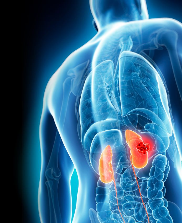
About the Covers
Male kidneys. Credit: Science Photo Library / Sebastian
Kaulitzki / Getty Images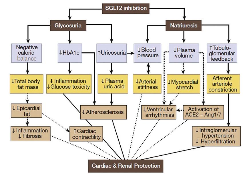
Introduction
Matthew R. Weir, MD

Diabetic kidney disease (DKD) remains an important             mechanisms underlying the progression of diabetic
clinical problem with substantial medical comorbidity          vascular disease and target organ damage so that newer
despite many recent medical advances (1,2). More               and traditional therapeutic options can be used together
focus on the earlier identification of patients with type      most efficiently to improve clinical outcomes. We need
2 diabetes who are at risk for developing chronic kidney       to consider the therapeutic index of these treatments
disease (CKD) is needed, especially with regard to             and appreciate the massive amount of pharmacopeia
biomarkers, genetics, and high-risk phenotypes. Another        that patients with diabetes and CKD consume on a daily
key area of opportunity is the need for better clinical care   basis. Thus, to enhance the precision of therapy, we
models to eliminate socioeconomic and racial disparities.      need more knowledge of the mechanisms of kidney and
    Fortunately, in the past few years, new therapeutic        cardiovascular disease progression in type 2 diabetes.
opportunities have been discovered, and more are                   The results of newer clinical trials are another
being considered, for possible use in improving clinical       important area for discussion, as well as trials that
outcomes. Angiotensin receptor blockers were the last          are planned or are currently underway. The newer
major advance for the treatment of DKD, in 2001 (3,4).         clinical trials have been conducted in patients who are
The serendipitous observations of improved cardiovas-          already on optimal medical therapy, including improved
cular and renal outcomes with sodium–glucose cotrans-          blood pressure control, highest tolerated doses of
porter 2 (SGLT2) inhibitors and glucagon-like peptide 1        renin-angiotensin system blockers, and lipid-lowering
receptor agonists in cardiovascular outcomes trials were       therapy.
a major surprise (5–7). These observations were followed           Ultimately, we need more precision in guiding pharma-
by the improved cardiorenal outcomes in two large renal        cotherapy given the many new therapeutic options
protection trials in patients with DKD: the CREDENCE           available. This compendium will provide an updated
(Canagliflozin and Renal Events in Diabetes with Estab-        opportunity to gauge our progress in the efforts underway
lished Nephropathy Clinical Evaluation) trial (8) using        to improve longer-term outcomes for patients who have
the SGLT2 inhibitor canagliflozin and the FIDELIO-DKD          diabetes and CKD.
(Finerenone in Reducing Kidney Failure and Disease
Progression in Diabetic Kidney Disease) study (9) using        See references starting on p. 34.
the novel and not-yet-approved selective nonsteroidal
mineralocorticoid receptor antagonist finerenone.              Dr. Weir is a professor and chief of nephrology in the Division of
    As more therapeutic opportunities become estab-            Nephrology, Department of Medicine, at the University of Maryland
lished, we need an improved understanding of the               School of Medicine in Baltimore.

                                                                              CHRONIC KIDNEY DISEASE AND TYPE 2 DIABETES        1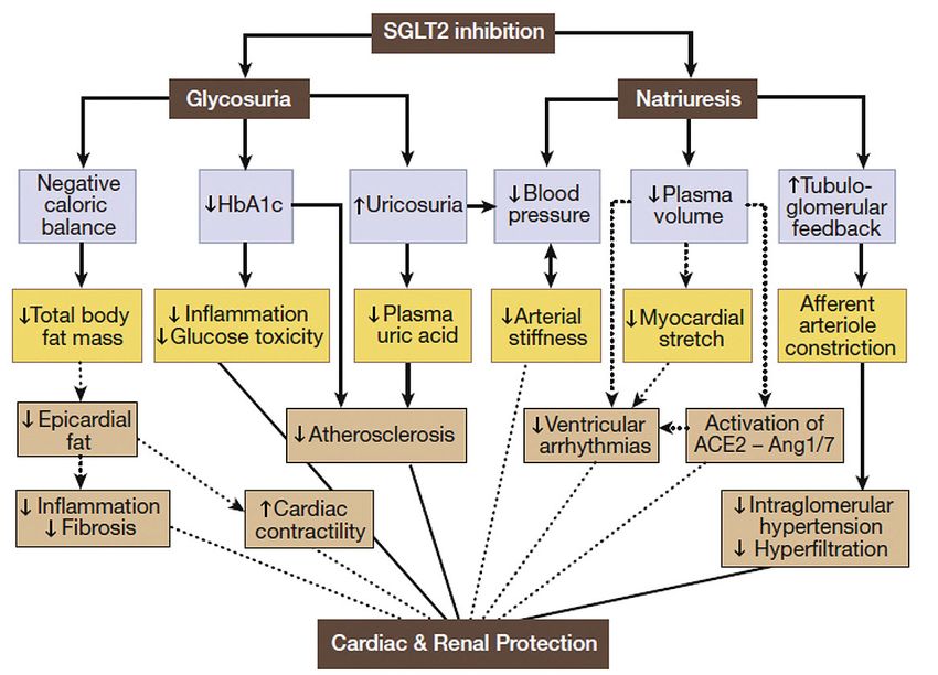
Pathogenesis of Diabetic Nephropathy
Rajiv Agarwal, MD, MS

Our understanding of the natural history of diabetic nephropathy                 extracellular matrix (ECM), glomerulosclerosis, vascular hyalinosis,
has emerged largely from patients with type 1 diabetes. However,                 interstitial fibrosis and tubular atrophy, and loss of function
histological manifestations among those with type 2 diabetes are                 culminating in end-stage renal disease (ESRD).
similar (10). Both the clinical manifestations and the histological
appearances of kidney disease associated with diabetes have                      Metabolic Factors
been well characterized. The pathogenesis, however, is less well                 The earliest changes are triggered by metabolic factors, namely
understood, and there are gaps in our understanding of how                       hyperglycemia. Damage resulting from hyperglycemia can occur
various causal factors relate to the histological manifestations of              by alteration of tissues or can be induced by products of glucose
diabetes; in part, this is because of a paucity of kidney biopsies               metabolism (11). An overview of the deranged metabolic pathways
and longitudinal data. Here, we will focus on the pathogenesis,                  that mediate the pathogenesis of nephropathy in people with
summarizing our current understanding of the histological and                    diabetes is shown in Figure 2.
clinical correlates and pointing out remaining controversies in the
context of pathogenesis.                                                         Glycation of Tissues
     The pathogenesis of diabetic nephropathy is initiated and                   Hyperglycemia through a nonenzymatic mechanism can lead to
maintained by four causal factors, which can be classified broadly               production of advanced glycation end products (AGEs), which by
into metabolic, hemodynamic, growth, and proinflammatory or                      glycation of various tissue constituents such as proteins, collagen,
profibrotic factors (Figure 1). Although there is both a substantial             lipids, and ECM can provoke organ dysfunction. This process is
overlap among these factors and variability in their relative contri-            likened to that of accelerated aging through browning of tissues or
bution among individuals and over time, for ease of discussion, we               the Maillard reaction (11).
will describe the pathogenesis as if each factor played an isolated                   Glycation of molecules provokes downstream injury by several
role. These pathogenetic factors produce lesions in various kidney               mechanisms that can be broadly classified into receptor-mediated
compartments: glomeruli, tubuli, interstitium, and vasculature. A                and non–receptor-mediated categories (12).
complex series of molecules, receptors, enzymes, and transcription                    Glycation leads to activation of receptors on cells—the best
factors participate in the process that drives the earliest stages of            characterized of which is the receptor of advanced glycation end
kidney disease to an enlarged kidney with hypertrophy, expanded                  products (RAGE)—that trigger the synthesis and release of nuclear

FIGURE 1 Overview of pathogenic factors in diabetic nephropathy. The key drivers of diabetic nephropathy can be broadly classified as metabolic,
hemodynamic, growth, and proinflammatory or profibrotic factors.

                                                       Receptor-mediated
                                  AGEs                                                                          VEGF
                                                     Non–receptor-mediated
     Metabolic                                                                        Growth
      factors                                                                         factors
                                                         Polyol pathway
                                Glucose
                               metabolism             Hexosamine pathway                                    Angiopoietins
                               byproducts
                                                          PKC pathway                                                              Endothelial cell activation

                                                                                                            Macrophage                    Tissue injury
                                                                                                             activation                MR overactivation

                                Systemic                                                                       Innate                 TLR
                              hypertension                                                                    immunity                NOD
                                                                                 Proinflammatory
    Hemodynamic                                                                   and profibrotic
      factors                                                                         factors               Complement                MBL
                                                                                                             activation             H-ficolin
                             Intraglomerular
                               hypertension
                                                                                                              Recurrent            Myeloid MR
                                                                                                                AKI                  Others

2       CHRONIC KIDNEY DISEASE AND TYPE 2 DIABETES
FIGURE 2 Metabolic pathways of diabetic nephropathy. Hyperglycemia provokes the accumulation of AGEs and other products of glucose metabolism.
Activation of each of these pathways can injure the kidney. AGEs can produce cell injury by receptor and non-receptor pathways. Outside the cells, they can
cause tissue damage by glycating molecules such as collagen that can reduce tissue compliance through crosslinking. Increased glucose flux can result in
activation of pathways such as polyol, hexosamine, and PKC that can result in cellular injury and organ dysfunction.

                                                                                          NFκB                  Cell growth, inflammation,
                                                                                                                angiogenesis, endothelial
                                                                   RAGE                   ROS                  dysfunction, ECM production
                                 Receptor-mediated
            AGEs                                                Within cells          ↓ NO bioavailability
                               Non–receptor-mediated
                                                                Outside cells             ECM                Collagen crosslinking

                                                                                                              ↑ Oxidative stress
                                   Polyol pathway           ↑ Aldose reductase          ↓ NADPH
         Glucose                                                                                               ↑ CTGF
        metabolism              Hexosamine pathway                ↑ GFAT                ↑ TGFβ1                                         Tissue fibrosis
        byproducts                                                                                            ↓ MMPs
                                    PKC pathway                   ↑ DAG                  ↑ PKC                                       Vascular dysfunction

factor κB (NFκB) and the generation of reactive oxygen species                     important microvascular complication of diabetes—eye disease—in
(ROS). These molecules, although transcription factors, initiate and               a randomized trial (13).
maintain kidney damage by several processes (12), including cell                   Hexosamine Pathway
growth and hypertrophy, inflammation, angiogenesis, endothelial
                                                                                   The hexosamine pathway is important for the synthesis of proteo-
dysfunction, and ECM production.
                                                                                   glycans, glycolipids, and glycoproteins (14). The synthesis of these
    Within the cells, AGEs can produce cellular dysfunction without
                                                                                   molecules requires an amino sugar substrate called UDP-N-acetyl-
binding to a receptor. For example, glycation of cytosolic proteins
                                                                                   glucosamine, which is the final product of the hexosamine
can reduce nitric oxide (NO) bioavailability and provoke oxidative
                                                                                   pathway. The rate-limiting enzyme of the hexosamine pathway is
stress (12). Similarly, outside the cells, AGEs can provoke tissue
                                                                                   glutamine:fructose-6-phosphate-amidotransferase (GFAT), which
dysfunction without binding to a receptor. For example, glycation
                                                                                   catalyzes the reaction between fructose-6-phosphate and the
of connective tissue constituents such as collagen can crosslink
                                                                                   amine-donor glutamine to produce glucosamine-6-phosphate
molecules in the ECM and cause dysfunction (12).
                                                                                   (14). In cultured mesangial cells, high glucose levels provoke
    Histological manifestations of AGE accumulation include
                                                                                   production of transforming growth factor β1 (TGF-β1); this effect is
basement membrane thickening, reduced protein degradation
                                                                                   eliminated by inhibition of GFAT. In contrast, stable overexpression
that results in an increase in mesangial matrix, and an increase in
                                                                                   of GFAT increases TGF-β1 production. Furthermore, the effects
interstitial extracellular volume.
                                                                                   appear to be transduced by PKC. In humans, GFAT is absent in
Damage Induced by Products of Glucose Metabolism                                   glomerular cells. However, in patients with diabetic nephropathy,
Glucose can induce damage in cells independent of glycation such                   GFAT is expressed in the glomerulus, suggesting that it may play a
as by the activation of the polyol pathway, hexosamine pathway, or                 pathophysiological role (14).
protein kinase C (PKC) pathway or through the generation of ROS.                   PKC Pathway
Polyol Pathway                                                                     PKC is a family of enzymes that are critical intracellular signaling
The polyol pathway involves the activation of the enzyme aldose                    molecules and are important for vascular function. In the physi-
reductase within cells when intracellular concentrations of                        ological state, receptor-mediated activation of PKC releases
glucose rise to hyperglycemic levels (11). This depletes the                       intracellular calcium ions and diacylglycerol (DAG) and activates
cellular nicotinamide adenine dinucleotide phosphate hydrogen                      these enzymes. In pathological states such as in diabetes,
(NADPH) concentration and alters the redox ratio, which can reduce                 DAG production can be abnormally increased and can lead to
NO bioavailability and alter enzyme function. Although aldose                      activation of PKC. In diabetes, DAG production is increased
reductase inhibitors were found to be effective in rodent models                   by increased glycolysis and an elevated level of intracellular
of diabetes, human trials have failed to reveal protection from an                 glyceraldehyde-3-phosphate and glycerol-3-phosphate. PKC

                                                                                                    CHRONIC KIDNEY DISEASE AND TYPE 2 DIABETES                3
can also be activated by ROS and AGEs. An inhibitor of PKC-β—                    tubuloglomerular feedback; the afferent arteriole dilates, and the
ruboxistaurin—has been tested in a phase 2 randomized clinical                   efferent arteriole constricts (12). An increase in insulin by itself can
trial in patients with type 2 diabetes and persistent albuminuria                increase sodium and glucose transport in the proximal tubule and
(albumin-to-creatinine ratio [ACR] 200–2,000 mg/g creatinine)                    provoke tubuloglomerular feedback. Insulin, as noted above, can
despite therapy with renin-angiotensin system inhibitors (15).                   also reduce afferent arteriolar tone directly. Thus, insulin can both
Compared to placebo, the reduction in ACR at 1 year—the primary                  directly and indirectly cause hyperfiltration.
endpoint of the study—was not significant.
                                                                                 Growth Factors
Hemodynamic Factors                                                              It has long been recognized that microangiopathy such as that
The increases in glomerular capillary pressure increase the single               occurs in the eye also associates with kidney disease. Therefore,
nephron glomerular filtration rate—hyperfiltration—and this occurs               investigators have explored the relation between vascular prolifer-
early in the course of diabetes. An increase in intraglomerular                  ation and endothelial permeability—factors known to be important
pressure is the result of an increase in efferent arteriolar tone and            in the pathogenesis of diabetic eye disease—with the occurrence of
a reduction in afferent arteriolar tone (Figure 3) (16). How this                diabetic nephropathy. Vascular endothelial growth factor (VEGF) is
process occurs is not settled, but two theories have emerged.                    activated early and leads to vascular expansion, which can provoke
     One group believes that hyperfiltration is mediated by circulating          hyaline arteriosclerosis and hypertensive changes in the kidney (18).
molecules that primarily operate within the glomerulus (17). Several             Similarly, angiopoietins can cause vascular proliferation and have
mediators have been proposed to increase intraglomerular pressure                been implicated in the pathogenesis of diabetic nephropathy (19).
via increasing efferent arteriolar tone and reducing afferent arteriolar
tone. Increase in efferent arteriolar resistance can result from an              Proinflammatory and Profibrotic Factors
increase in the concentration of angiotensin II, thromboxane A2                  Inflammation and fibrosis are important causes of diabetic
(TxA2), endothelin 1 (ET-1), and ROS (16). Reduction in afferent                 nephropathy (20). Whether this is causal or in response to injury
arteriolar resistance can be provoked by reduction in NO oxide                   remains a matter of debate. However, there is a strong relation
bioavailability; increased cyclooxygenase-2 (COX-2) prostanoids;                 between the degree of infiltration of macrophages and subsequent
activation of the kallikrein-kinin system, atrial natriuretic peptide,           occurrence of tubular interstitial fibrosis and progression of
and angiotensin 1-7; and an increase in insulin (16).                            diabetic kidney disease (21,22).
     However, another group proposes that tubular mechanisms                         Macrophages are attracted to the kidney by a variety of
remain the primary driver of the intraglomerular hypertension                    mechanisms (23). Endothelial cell dysfunction, activation, and
(12). The activation of glucose transporting pathways in the                     injury all stimulate the production of adhesion molecules on the
proximal tubule early in the course of diabetes stimulates the                   endothelial surface that facilitate transendothelial migration of
reabsorption of both glucose and sodium in the proximal nephron                  macrophages. Injury and activation of resident kidney cells such
(12). Sodium delivery to the distal nephron is reduced. This triggers            as podocytes, mesangial cells, and tubular cells result in secretion
                                                                                 of chemokines that facilitate intrarenal macrophage infiltration.
FIGURE 3 Mechanisms of intraglomerular hypertension. Intraglomerular             Macrophages are activated to the proinflammatory (M1) phenotype
pressure can increase as a result of either an increase in efferent arteriolar   by ROS, angiotensin II, and the activation of mineralocorticoid
tone or a reduction in afferent arteriolar tone. The mediators of these          receptors (MRs). That by itself can damage podocytes, endothelial
alterations are shown.
                                                                                 cells, mesangial cells, and tubular cells. Activated macrophages,
                                                                                 by releasing profibrotic cytokines, can increase cell proliferation
                                     ↑                                           and matrix volume expansion and provoke fibrosis. Fibrosis at a
                              Intraglomerular
                                  pressure
                                                                                 molecular level is mediated in part because of activation of TGFβ1,
                                                                                 which has two synergistic effects: activation of connective tissue
                                                                                 growth factor (CTGF) and a reduction in matrix metalloproteinases
                                                                                 (MMPs). In contrast, MR antagonists can coax macrophages to
     ↑Efferent arteriolar tone           ↓Afferent arteriolar tone               the antiinflammatory (M2) phenotype and be protective (24).
        ⊲   Angiotensin II                 ⊲   NO bioavailability                Thus, macrophages play an important role in the pathogenesis of
        ⊲   TxA2                           ⊲   ↑COX-2 prostanoids                diabetic nephropathy (23).
        ⊲   ET-1                           ⊲   ↑Kallikrein-kinin system
        ⊲   ROS                            ⊲   Atrial natriuretic peptide        Acute Kidney Injury, Inflammation, Chronic Kidney Disease,
                                           ⊲   Angiotensin 1–7                   and the Role of MRs
                                           ⊲   Insulin
                                                                                 Inflammation and fibrosis may also be important promoters of
                                           ⊲   Tubuloglomerular feedback
                                                                                 progression of chronic kidney disease (CKD) in patients with

4        CHRONIC KIDNEY DISEASE AND TYPE 2 DIABETES
diabetes, and this may be the result of acute kidney injury (AKI).
                                                                                 TABLE 1 MR Blockade and Kidney Protection in Diabetes
It is increasingly being recognized that single or repeated bouts
of AKI on a background of CKD in diabetes may play a vital role in
the progression of CKD to ESRD (25). Macrophage infiltration is                    ⊲   Reduced maladaptive response
commonly seen in AKI, and depletion of macrophages in preclinical                  ⊲   Reduced ROS
                                                                                   ⊲   Improved endothelial function
models can protect from AKI (26). In two different rodent models                   ⊲   Shift in macrophage phenotype from proinflammatory (M1)
of AKI, bilateral ischemia reperfusion (IR) pretreatment with the                      to antiinflammatory (M2)
nonsteroidal MR antagonist finerenone prevented the development                    ⊲   Better blood pressure control
of AKI (27). In a separate set of experiments, unilateral IR injury
was also associated with reduced fibrosis when animals were
pretreated with finerenone (27). Furthermore, in a pig model of IR               Innate Immunity, Complement Activation, and Diabetic
AKI, the administration of the MR antagonist potassium canrenoate                Nephropathy
prevented the progression of AKI to CKD at 90 days (27).                         Activation of the innate immune system through pattern recognition
     The relative contributions of the knockout of MRs in smooth                 receptors such as membrane-bound toll-like receptors (TLR) and
muscle cells versus their knockout in myeloid cells have been                    nucleotide-binding oligomerization domain (NOD)-like receptors may
investigated in mouse models (Figure 4) (27). With MR knockout                   play an important role in the pathogenesis of diabetic nephropathy
in smooth muscle cells, IR models demonstrated that the                          (30). The complement system, in addition to fighting infections,
short-term elevation of serum creatinine and blood urea nitrogen                 facilitates the removal of damaged cells by antibodies and phagocytic
was prevented. However, at 30 days, there was no difference                      cells. The activation of the complement component C3 generates
between wild-type and smooth muscle cell MR knockouts. In                        the membrane attack complex (MAC) that lyses, damages, or
contrast to MR knockout in smooth muscle cells, among myeloid                    activates target cells. Mannose-binding lectin (MBL) activates the
MR knockout mice, there was no immediate protection from AKI.                    lectin pathway; pattern recognition molecules called ficolins can
However, at 30 days, there was a marked improvement in renal                     also activate the lectin pathway. The lectin pathway is activated after
function and markers of inflammation. Furthermore, there was                     binding of ficolins to glycated proteins. Glycation of complement
a shift in the polarization of macrophages infiltrating the kidney.              regulatory proteins such as CD59 might by itself activate complement;
Although the total number of macrophages in wild-type and                        this is so because CD59 normally inhibits MAC (30).
myeloid MR knockouts were similar, there was a shift in the nature                    A causal relation between MBL activation and diabetic
of macrophages such that the M2 macrophages associated with                      nephropathy is firmly established in animals. For example,
an antiinflammatory response were increased in relation to the M1                compared to wild-type mice with streptozotocin-induced diabetes,
macrophages, which are proinflammatory (27).                                     MBL knockout mice have less kidney damage, less kidney hyper-
     Although these studies were done in animals without diabetes,               trophy, lower urine albumin excretion, and less type IV collagen
the experiments demonstrate the importance of inflammation and                   expression (31).
MRs in mediating CKD after AKI; similar mechanisms likely operate                     Several lines of evidence in humans suggest the important role
in patients with CKD resulting from diabetes (Table 1) (28,29).                  of complement activation in CKD progression. As examples, 1) in
                                                                                 patients with type 1 diabetes, concentrations of MBL associate
FIGURE 4 Short- and long-term effects of MRs are location dependent. In          with progression of kidney disease from macroalbuminuria to
smooth muscle cells, MRs protect from short-term AKI. In contrast, MRs in
                                                                                 ESRD (32); 2) in a prospective cohort study of 270 patients with
myeloid cells have no short-term effects but prevent long-term inflammation
and fibrosis. These experiments are helpful in understanding the long-term       newly diagnosed type 1 diabetes, H-ficolin was associated with
consequences of repeated AKI in the progression of kidney disease in diabetes.   an increased risk of worsening of albuminuria (33); and 3) MAC
                                                                                 detected by antibodies directed against the C9 component of MAC
                                                                                 localize it to the glomerular basement membrane (GBM), tubules,
                                                                                 and Bowman capsule in patients with type 1 diabetes (34–36).
                                                                                      Taken together, these data point out the important role of the
                                                                                 complement system and its components in the pathogenesis of
                                                                                 diabetic nephropathy.

                                                                                 Interrelations Among Pathogenic Factors in Diabetic
                                                                                 Nephropathy
                                                                                 The interplay of metabolic, hemodynamic, growth, and profibrotic
                                                                                 factors is illustrated by consideration of the following preclinical
                                                                                 experiments (37). Cultured mesangial cells exposed to CTGF

                                                                                                CHRONIC KIDNEY DISEASE AND TYPE 2 DIABETES              5
increase production of profibrotic molecules such as fibronectin light microscopy shows minimal, non-specific, or no changes.
and collagen type I (37). Although the baseline production of CTGF Thickening of the GBM does not directly correlate with clinical
by mesangial cells is low, exposure of mesangial cells to increased injury. Patients may have such thickening but have no increase in
glucose concentration (a metabolic factor) or cyclic metabolic urine albumin excretion rate or impairment of glomerular filtration
strain (a hemodynamic factor) increases the production of CTGF rate (39,40). Although an increase in diastolic blood pressure (40)
(a growth factor). The induction of CTGF protein by a high glucose or nocturnal blood pressure (39) is correlated with GBM thickening,
concentration is blocked by TGFβ1-neutralizing antibody. This the causal relation is not established because of a lack of longitu-
suggests that another growth factor—TGFβ1—mediates the effect dinal data and interventional studies. GBM thickening occurs as a
of high glucose concentration to provoke CTGF production. In vivo result of either an increased rate of deposition or a reduced rate of
studies in obese db/db diabetic mice demonstrate that CTGF removal of connective tissue. Target molecules include collagen IV
transcription was increased 28-fold after ~3.5 months of diabetes and VI, fibronectin, and laminin (35,41).
(37). At 3.5 months of diabetes, mesangial expansion was mild,
and interstitial disease and proteinuria were absent. Furthermore, Class II Diabetic Nephropathy
rather than being diffusely increased throughout the kidney, the Among the earliest manifestations on kidney histology that
CTGF production was limited to the glomerular compartment. These correlate with kidney damage is an increase in mesangial matrix,
experiments demonstrate the interplay of all the pathogenic factors as seen in class II diabetic nephropathy. Class II is further
discussed above and underscore the complex interrelations of subclassified based on the degree of mesangial expansion;
these factors, over time and at different locations in the kidney, in class IIa is characterized by ≤25% mesangial expansion, and
producing the histological manifestations of diabetic nephropathy. class IIb involves >25% of the mesangial expansion. An increase
in mesangial matrix, glomeruli, and kidney volume is clinically
Pathological Classification of Diabetic Nephropathy manifested as kidney enlargement; kidneys are often 11 cm or
According to an international consensus conference, the larger on kidney ultrasound. Urine albumin excretion is often
histological manifestations of diabetic nephropathy follow four increased in these patients.
progressive classes (Table 2) (38). The classification acknowl-
Class III Diabetic Nephropathy
edges lesions in the glomeruli, tubuli, and vessels, but the root
An increase in mesangial matrix is followed by mesangial
of the classification system is based on the appearance of the
sclerosis. The hallmark lesion on a kidney biopsy is nodular
glomerulus. According to this classification system, diabetic
glomerulosclerosis, or Kimmelstiel-Wilson nodules. The presence
nephropathy progresses from thickening of the GBM, to
of Kimmelstiel-Wilson nodules on kidney biopsy correlates with
mesangial expansion, Kimmelstiel–Wilson lesions, and global
the occurrence of diabetic retinopathy, suggesting activation of
glomerulosclerosis, which is reflected in the four classes, as
common pathogenetic pathways such as VEGF.
discussed further below. Although this system has not been
validated with clinical outcomes, it serves as an important Class IV Diabetic Nephropathy
clinical and research tool to classify the severity of diabetic Advanced, or class IV, diabetic nephropathy is characterized by
nephropathy lesions. sclerosis in >50% of the glomeruli. These patients often have a loss
of kidney function at the time of biopsy.
Class I Diabetic Nephropathy An enlargement of glomeruli is often seen along with thickening
On ultrastructural evaluation of the kidney histology, among the
of the walls of the glomerular capillaries. Arteriolar hyalinosis of
earliest change that occurs in the kidney is thickening of the GBM;
both the afferent and efferent arteriole should alert health care
professionals to the possibility of diabetic nephropathy. The
TABLE 2 Pathological Classification of Diabetic Nephropathy proximal tubules can contain protein resorption droplets. In the
setting of severe persistent hyperglycemia, glycogen deposits may
⊲ GMB thickening on electron microscopy; minimal, be seen rarely in the proximal tubules (i.e., Armanni Ebstein lesion).
Class I Interstitial fibrosis and tubular atrophy (IFTA) and interstitial inflam-
non-specific, or no changes on light microscopy
Class II ⊲ Increase in mesangial matrix mation are often seen. Despite tubular atrophy, the basement
Class IIa ⊲ Mesangial expansion ≤25%
membranes are often thickened in patients with diabetes.
Class IIb ⊲ Mesangial expansion >25% The Heterogeneity of Kidney Injury in Type 2 Diabetes: A
⊲ Nodular glomerulosclerosis: Kimmelstiel-Wilson Pathogenetic Explanation
Class III
lesion
Although kidney disease is histologically similar in type 1 and
⊲ Advanced glomerulosclerosis; >50% glomeruli type 2 diabetes, the relative contributions of causes of kidney
Class IV
sclerotic
damage differ in these two conditions. Compared to patients
6 CHRONIC KIDNEY DISEASE AND TYPE 2 DIABETESwith type 1 diabetes, those with type 2 diabetes are older, Conclusion
have a greater BMI, and are more likely to have dyslipidemia, The pathogenesis of diabetic nephropathy is similar in type 1 and
hypertension, and other cardiovascular risk factors and, type 2 diabetes. Diabetic nephropathy is classified histologically by
consequently, atherosclerosis and arteriosclerosis. Thus, the the appearance of the glomerulus on kidney biopsy. It progresses
nature of kidney injury in patients with type 2 diabetes may be from GBM thickening, to mesangial expansion, nodular glomerulo-
modified by environmental factors and genetic background. sclerosis, and global glomerulosclerosis. Glomerulomegaly, vascular
This heterogeneity in environmental and genetic factors in lesions, IFTA, and tubular resorption droplets are all commonly
patients with type 2 diabetes may explain the distinct kidney seen. The pathogenesis of diabetic nephropathy involves metabolic,
injury phenotypes. hemodynamic, growth, and inflammatory and fibrotic factors. The
As an example, consideration of an animal experiment relative contributions of these factors vary among patients, over
provides evidence for interplay between genetics and time, and even in different compartments of the kidney, and genetic
environment with regard to kidney injury phenotype (42). and environmental factors can modify the appearance of the kidney
Progeny of rats with one parent with heart failure and another lesions. AKI plays an important role in the progression of kidney
with obesity were fed a diet either high in carbohydrate or high disease in patients with diabetes. MR activation, particularly in the
in fat; all progeny had diabetes (42). Compared to animals fed myeloid cells, may be important in mediating inflammation and
a high-carbohydrate diet, animals fed a high-fat diet demon- fibrosis in CKD and after AKI in individuals with type 2 diabetes, and
strated a greater preponderance of tubulointerstitial injury MR antagonist therapy may be protective.
and non-nodular glomerulosclerosis. There was evidence of
lipid peroxidation and increased kidney TGFβ1 that correlated See references starting on p. 34.
with kidney injury. Furthermore, injury in animals fed a high-fat
diet was seen in the arterial wall and renal microcirculation. In Dr. Agarwal is a professor of medicine in the Division of
contrast, animals fed a high-carbohydrate diet had increased Nephrology, Department of Medicine, at the Indiana University
glycoxidation stress biomarkers, but these did not correlate School of Medicine and a staff physician at Richard L. Roudebush
with kidney injury (42). Veterans Administration Medical Center, in Indianapolis, IN
CHRONIC KIDNEY DISEASE AND TYPE 2 DIABETES 7Risk Factors, Symptoms, Biomarkers, and Stages of
Chronic Kidney Disease
Peter Rossing, MD, DMSc
Whereas the symptoms of chronic kidney disease (CKD) in diabetes Modified Release Controlled Evaluation) trial, in which 11,140
are few, there are many risk factors and biomarkers that can be patients with type 2 diabetes were followed and a 21% reduction
used to identify individuals at high risk for development of this (95% CI 7–34%) in development of nephropathy was seen in
complication, and many of these are targets for intervention to patients randomly assigned to strict glycemic control (49). Even
prevent or delay the disease. This article describes the risk factors end-stage renal disease (ESRD) was reduced in the ADVANCE trial,
and other markers of CKD and the various stages of the disease. although it was a very rare event (50).
Overall, it has been difficult to demonstrate the benefit of
Risk Factors for CKD in Diabetes improving glycemic control on established CKD in type 2 diabetes,
Many factors are associated with CKD in diabetes (Figure 1). in contrast to the benefit on development of CKD. Recent studies
Associations may be with both albuminuria and glomerular filtration with glucose-lowering agents such as glucagon-like peptide 1
rate (GFR) or with one variable only. Some factors influence initial receptor agonists found reduced progression of albuminuria and
development of kidney disease and others progression of the loss of kidney function (51,52). Sodium–glucose cotransporter
disease. Duration of diabetes is one of the strongest risk factors for 2 (SGLT2) inhibitors in particular have demonstrated benefit
diabetic nephropathy, but because type 2 diabetes is often silent, on progression of albuminuria, decline in kidney function, and
CKD may be present at diagnosis of diabetes. development of kidney failure; but, although the mechanisms are
not clear, the reduction in glucose is probably of minor importance
Hyperglycemia (8,53). Thus, SGLT2 inhibitors are even beneficial in people with
Several studies demonstrate the importance of hyperglycemia in CKD who do not have diabetes (53).
the development and progression of CKD in diabetes (or diabetic
kidney disease [DKD]) (43,44). The UK Prospective Diabetes Study Blood Pressure
documented a progressive beneficial effect of intensive metabolic Blood pressure is crucial to the development and progression of
control on the development of microalbuminuria and overt CKD in diabetes (44,54,55). The excess prevalence of hypertension
proteinuria (45), and a 10-year post-study follow-up demonstrated in type 1 diabetes is confined to patients with nephropathy (56).
long-lasting benefit, which was termed a “legacy effect” (46). Once severely increased albuminuria is present, frank hypertension
Greater variability in A1C is associated independently with is present in 80% of individuals and is almost universal in those
albuminuria and diabetic nephropathy (47,48). The beneficial with ESRD. In type 2 diabetes, the link between hypertension and
effect of improved glycemic control was confirmed in the ADVANCE kidney disease is less striking because hypertension is so common.
(Action in Diabetes and Vascular Disease: Preterax and Diamicron Almost all patients with moderately elevated or worse albuminuria
have hypertension. In people with diabetic nephropathy, variability
in systolic blood pressure is independently associated with the
FIGURE 1 Putative promoters of CKD progression in diabetes. development of ESRD (57).
Treatment of blood pressure, particularly with inhibitors of
Metabolic syndrome the renin angiotensin system (RAS), has been a standard of care
Protein intake for both prevention and treatment of CKD in diabetes based on
Obesity
studies with angiotensin II receptor blockers in moderately elevated
Smoking albuminuria (microalbuminuria) and type 2 diabetes (58), as
Albuminuria
well as in established proteinuria in type 2 diabetes (3,4). Even
Genetics
Glycemic control prevention of CKD has been suggested, at least in hypertensive
Fibrosis type 2 diabetes, when treated with RAS-blocking agents (59).
Dyslipidemia
Inflammation Renin-Angiotensin-Aldosterone System
RAAS Several components of the renin-angiotensin-aldosterone
Oxidative stress
system (RAAS) are elevated and considered to contribute to
Systemic and intraglomerular
the progression of diabetic nephropathy. Accordingly, blocking
blood pressure the RAAS has been demonstrated to be kidney protective.
8 CHRONIC KIDNEY DISEASE AND TYPE 2 DIABETESExperimental studies have suggested that succinate, formed by the Multiple genes, either protective or deleterious, are involved. Different
tricarboxylic acid cycle, provides a direct link between high glucose loci may influence albuminuria and GFR separately (84). Epigenetic
and renin release in the kidney (60). Focus was initially on the modification may also be important (85).
damaging effect of angiotensin II.
As discussed for blood pressure, RAS-blocking agents have Ethnicity
been a standard of care in CKD in 20 years. Aldosterone represents Albuminuria and CKD stages 4 and 5 are more common in U.K.
another component of the RAAS that should be considered Afro-Caribbean and South Asian individuals than White European
important in the pathophysiology of diabetic nephropathy. people (86,87). The prevalence of early CKD (defined as moderately
Aldosterone is a hormone that, in addition to regulating electrolyte elevated or greater albuminuria and eGFR40 kg/m2) enhances ESRD risk sevenfold (62). Individuals who develop type 2 diabetes at a young age have a high
Even a BMI >25 kg/m2 was found to increase ESRD risk (62). This prevalence of hypertension and moderately elevated albuminuria
effect is independent of the effects of hypertension and diabetes, (92). ESRD and death are particularly common in young people
the prevalence of which are increased in individuals with obesity. from ethnic minorities (93–95). However, in some of these
An effect of obesity on renal hemodynamics leading to increased populations, there is a high prevalence of kidney disease unrelated
glomerular pressure and hyperfiltration has been suggested as the to diabetes (96).
mechanism (63), and adiponectin was suggested to link obesity
to podocyte damage (64). Weight reduction from bariatric surgery
Albminuria and eGFR
Baseline albuminuria and eGFR independently influence the
(65) or pharmacological treatment (66) has been associated with
development and rate of progression of CKD (97,98). Baseline
improved renal outcomes, although large weight reductions will
albuminuria strongly predicts ESRD (99). Higher levels of normo-
improve estimated GFR (eGFR) and not true GFR because of loss of
albuminuria (100) and lower eGFR (101) predict a faster decline
muscle mass and then decline in serum creatinine (67).
in eGFR. Conversely, a short-term reduction in albuminuria with
Other Metabolic Factors intervention is associated with reduced progression of kidney and
Blood lipids, including triglycerides (68,69), contribute to the devel- cardiovascular complications (102,103).
opment and progression of CKD, although the lipid phenotype alters
as kidney disease progresses (70–72). Insulin resistance increases
Other Risk Factors
Other risk factors for nephropathy include smoking (98),
the risk of albuminuria in type 2 diabetes (73). Individuals with type
pre-eclampsia (104), periodontitis (105), obstructive sleep
1 or type 2 diabetes and CKD are more likely to have the metabolic
apnea (106), and nonalcoholic fatty liver disease, all of which are
syndrome (74–76). Multifactorial intervention targeting lifestyle,
glucose, blood pressure, and lipids has a beneficial impact on both independently associated with diabetic nephropathy (107,108).
cardiovascular and kidney outcomes (77).
Symptoms of CKD
Genetic Factors Whereas albuminuria is often an early sign of CKD, there is a
Genetic factors influence susceptibility to CKD in both type 1 paucity of symptoms related to CKD in diabetes until late stages,
and type 2 diabetes (78,79). If one sibling with type 1 diabetes making systematic screening mandatory to detect CKD as early as
has nephropathy, the risk to a second sibling is increased four- to possible. Edema is often the first symptom, followed by fatigue and
eightfold compared to sibling sets in which neither has nephropathy other uremic symptoms with pruritus, and then nausea, but this
(80). Similar familial clustering has been described in type 2 usually does not occur until CKD stage 4 or 5 (Figure 2) (109).
diabetes (81). Despite these findings, strong and clinically useful Other symptoms relate to complications, including angina
genes for CKD in diabetes are still lacking. from ischemic heart disease, dyspnea resulting from heart
The clustering of conventional cardiovascular risk factors and failure, aching from painful neuropathy, or typical symptoms
cardiovascular disease (CVD) in people with diabetes and CKD also of urinary tract infection. Although these complications are
occurs in their parents (82,83). This finding suggests that the genetic frequent, the symptoms may be atypical or weak because of the
susceptibility to nephropathy also influences the associated CVD. presence of neuropathy.
CHRONIC KIDNEY DISEASE AND TYPE 2 DIABETES 9FIGURE 2 Stages and prognosis of CKD based on albuminuria and GFR from the KDIGO (Kidney Disease: Improving Global Outcomes) 2012 clinical practice
guideline (109). The GFR and albuminuria grid depicts the risk of progression, morbidity, and mortality by color, from best to worst. Green indicates low risk (if no
other markers of kidney disease and no CKD), yellow indicates moderately increased risk, orange indicates high risk, and red indicates very high risk. Reprinted
with permission from Kidney Disease: Improving Global Outcomes (KDIGO) CKD Work Group. Kidney Int Suppl 2013;3:1–150.
Persistent Albuminuria Categories,
Description and Range
A1 A2 A3
Normal to mildly Moderately Severely
increased increased increased
300 mg/g
G1 Normal or high >90
G2 Mildly decreased 60–89
GFR Categories Mildly to moderately
G3a 45–59
(mL/min/1.73 m2), decreased
Description Moderately to
and Range G3b 30–44
severely decreased
G4 Severely decreased 15–29
G5 Kidney failure 30% (HR 4.81, 95% CI 1.92–12.01). Interestingly, the signature was improved with the anti-inflammatory
Applying urinary proteomic analysis with capillary electrophoresis agent baricitinib, but not with RAS blockade (119).
10 CHRONIC KIDNEY DISEASE AND TYPE 2 DIABETESFIGURE 3 Pathways and biomarkers of CKD. BNP, brain natriuretic peptide; KRIS, kidney risk inflammatory signature; U-CAD238, urinary proteome-based
classifer for coronary artery disease 238 ; U-CKD273, urinary proteome-based classifer for chronic kidney disease 273. Adapted from Rossing P, Persson F,
Frimodt-Moller M, Hansen TW. Diabetes 2021;70:39–50.
⊲ Blood glucose
⊲ Metabolic
⊲ Blood pressure, BNP
⊲ Hyperglycemia ⊲ Hemodynamic
Risk ⊲ Obesity
Pathway to ⊲ Inflammation
Related ⊲ KRIS
factors damage biomarkers ⊲ U-CKD273,
⊲ Hypertension ⊲ Fibrosis
U-CAD238, PRO-C6
⊲ Oxidative stress ⊲ 8-oxoGuo
Oxidative Stress Metabolomics
It has been proposed that elevated levels of uric acid induce Metabolites have been investigated in blood and urine using
vascular and kidney damage, hypertension, and atheroscle- platforms that capture hundreds or even thousands of metab-
rosis due to inflammation and oxidative stress. Elevated uric olites. So far, there have only been a few studies in people with
acid levels were associated with cardiovascular events and type 2 diabetes and CKD. Pena et al. (127) demonstrated that
progression of kidney disease in type 1 diabetes (120). The PERL a few metabolites in serum and urine could improve prediction
(Prevention of Early Renal Function Loss) study (121) tested of progression in albuminuria status in type 2 diabetes,
whether lowering uric acid with allopurinol in people with type and Solini et al. (128) demonstrated in patients with type 2
1 diabetes and early CKD with albuminuria or declining eGFR diabetes that serum, but not urine, metabolites could improve
could prevent loss of measured GFR over 3 years. Mean serum prediction of progression of albuminuria and decline in GFR.
urate level decreased from 6.1 to 3.9 mg/dL with allopurinol Sharma et al. (129) described a signature of 13 metabolites
and remained at 6.1 mg/dL with placebo. Despite this lowering, in urine that pointed toward mitochondrial dysfunction as a
the trial found no evidence of a kidney protective effect on key feature in progression of CKD in diabetes. Niewczas et al.
albuminuria or decline in GFR. These results suggest that uric (130) demonstrated that uremic solutes were associated with
acid is not a target, in line with a Mendelian randomization the development of ESRD in people with type 2 diabetes. Both
study in type 1 diabetes (122). However, a study was presented the metabolome and lipidome were recently studied in type 1
in 2019 with greater reduction of uric acid in a small group of diabetes (72,131). A number of markers of progression of CKD
people with type 2 diabetes who were followed for 24 weeks were identified but await confirmation, which is often a problem,
taking the urate reabsorption inhibitor verinurad and the as different studies use diverse platforms.
xanthine oxidase inhibitor feboxustat in combination, resulting
in a 49% reduction in urine albumin-to-creatinine ratio (ACR)
Stages of CKD
compared to placebo (123).
CKD in diabetes is defined as the presence of persistently
Other markers of oxidative stress are oxidatively modified
elevated albuminuria of >30 mg/24 hour or a urinary ACR >30
guanine nucleosides 8-oxo-7,8-dihydro-2’-deoxyguanosine
mg/g creatinine, confirmed in at least two out of three samples
(8-oxodG) and 8-oxo-7,8-dihydroguanosine (8-oxoGuo) excreted in
(132). As such, its diagnosis is clinical, requiring little more than
the urine. The level of 8-oxoGuo was associated with mortality and
basic clinical and laboratory evaluations. The normal range for
CVD in type 2 diabetes (124).
albuminuria is 15,000
suggested the importance of the Janus kinase/signal transducers people with type 2 diabetes suggested that patients with elevated
and activators of transcription (JAK-STAT) pathway as a key pathway albuminuria display the typical microvascular phenotype, whereas
in DKD. A clinical study in diabetes intervening with a JAK-STAT nonalbuminuric subjects with impaired kidney function had a more
inhibitor subsequently demonstrated reduced albuminuria (126). cardiovascular or macrovascular phenotype.
CHRONIC KIDNEY DISEASE AND TYPE 2 DIABETES 11For CKD in general, including in people with diabetes, it has right person may seem complicated and costly initially but has
been recommended to stage the severity using a combination of the potential to save both patients and the health care system
etiology (if known), level of urinary albumin excretion, and eGFR considerable costs (137). Integrating multiple “-omics” platforms
(Figure 2) (109). may lead to a much deeper understanding of the disease.
Hopefully, such an approach will help to prevent CKD in diabetes
Conclusion and improve kidney outcomes in the future. For now, much can
Advances in diagnosis and treatment have provided new options already be achieved if we ensure full integration of the use of simple
and potential for better outcomes for CKD in diabetes. As treatment biomarkers such as albuminuria and eGFR (138).
opportunities continue to expand, biomarkers and, most likely, See references starting on p. 34.
combinations of biomarkers will help us select the optimal
treatment or combination of treatments for each patient. This Dr. Rossing is a professor of endocrinology and head of compli-
ability will ensure better outcomes and reduce adverse events cations research at the Steno Diabetes Center Copenhagen and
and unnecessary polypharmacy. A more detailed approach Department of Clinical Medicine at the University of Copenhagen
applying multiple biomarkers to select the right treatment for the in Denmark.
12 CHRONIC KIDNEY DISEASE AND TYPE 2 DIABETESThe Interplay Between Diabetes, Cardiovascular Disease,
and Kidney Disease
Muhammad Shariq Usman, MD, Muhammad Shahzeb Khan, MD, MSc, and Javed Butler, MD, MPH, MBA
Burden of Diabetes and Associated Cardiorenal Advanced Glycation End Products
Disorders Oxidative stress and hyperglycemia drive a nonenzymatic reaction
The Global Burden of Disease Study estimates that there are that causes excessive covalent binding between glucose and
currently 476 million patients with diabetes worldwide, the large substrates such as proteins, lipids, and nucleic acid, a process
majority of whom suffer from type 2 diabetes. In the United States, known as nonenzymatic glycation. The resulting compounds
the prevalence of type 2 diabetes is 32.6 million, or ~1 in 10 are termed advanced glycation end products (AGEs). AGEs can
people. These numbers are expected to continue to rise (139). increase the production of ROS, causing increased intracellular
The metabolic system is closely interrelated with the cardiac oxidative stress. This increased oxidative stress, in turn, promotes
and renal systems, and these three systems share a symbiotic the formation of more AGEs, thus resulting in a vicious cycle. AGEs
relationship that helps maintain homeostasis. The heart is one and associated oxidative stress can result in inflammation, cellular
of the most metabolically demanding organs and is sensitive to dysfunction, and cell death. In the context of CVD and CKD, the effect
changes in energy and volume status. Thus, it relies on the liver, of AGEs on the endothelium of blood vessels is important (143).
pancreas, and fat for optimal energy metabolism and on the
kidneys for volume maintenance. Similarly, the kidneys rely on Endothelial Dysfunction
the heart for adequate perfusion and on the metabolic system Endothelial dysfunction in patients with type 2 diabetes results
for the appropriate hormonal milieu, both of which are necessary from nonenzymatic glycation of the endothelium and oxidative
to maintain their function. The metabolic system depends on damage. Endothelial dysfunction subsequently drives the devel-
functioning heart and kidneys to prevent neurohormonal activation, opment of microvascular and macrovascular disease. Hyper-
which keeps metabolic derangements such as insulin resistance, tension, a common comorbidity in patients with type 2 diabetes, is
glucose dysregulation, and dyslipidemias at bay (140). also a potent risk factor for endothelial dysfunction (143).
Given the close-knit physiology of the metabolic, cardiac, and
renal systems, it is not surprising that type 2 diabetes frequently Hypercoagulability
coexists with cardiovascular disease (CVD) and chronic kidney The first line of defense against a thrombotic event is an intact
disease (CKD). A 2018 study of >500,000 adults living with type and functioning vascular endothelium. The endothelium releases
2 diabetes in the United States demonstrated thatpromote atherosclerosis. Dysfunctional endothelial cells within prevalence of diabetes ranging from 40–50% in patients with
large arteries are a fertile ground for the initiation of atherosclerosis HF. Moreover, in patients with HF, mortality is higher in those with
(143). Dyslipidemia is prevalent in ~80% of patients with type 2 versus those without concomitant diabetes (148).
diabetes and is associated with atherosclerosis. Insulin deficiency HF with preserved ejection fraction (HFpEF) is emerging as an
and insulin resistance activate the enzyme hormone-sensitive especially significant problem among patients with type 2 diabetes.
lipase, which releases free fatty acids (FFAs) into the blood. This Many of these patients have asymptomatic diastolic dysfunction,
release leads to increased lipoprotein generation and release by and HFpEF, a disease without known mortality-modifying therapies,
the liver and, ultimately, increased circulating levels of triglycerides is the predominant form of HF in type 2 diabetes (143,149). It is
and LDL cholesterol. Lipoprotein lipase, the enzyme that clears LDL important to note that type 2 diabetes has distinct myocardial
cholesterol, is downregulated, which aggravates dyslipidemia. HDL effects in HFpEF and in patients with HF and reduced ejection
cholesterol levels are decreased in diabetes (143). fraction (HFrEF), with different biomarker profiles. In HFpEF,
Diabetes also affects the microvasculature. Microvascular the systemic inflammation is associated with higher serum
damage can lead to complications such as nephropathy, levels of inflammatory biomarkers such as soluble interleukin-1
retinopathy, and neuropathy. Microvascular damage is often receptor-like 1 and C-reactive protein; biomarkers of myocardial
initiated by nonenzymatic glycation of endothelial cells. This injury and stretch such as troponins and natriuretic peptides are
process leads to formation of glycated proteins that trigger a higher in HFrEF than in HFpEF.
range of effects on surrounding tissues, the most prominent ones In patients with type 2 diabetes, HF can occur as a result of
being, 1) thickening of endothelium and collagen, leading to ischemia or a thrombotic event secondary to CAD. In many cases,
local ischemia; 2) overproduction of endothelial growth factors however, pathophysiological factors unrelated to CAD are at play.
and pathologic angiogenesis; and 3) vascular inflammation and Cardiac disease in patients with type 2 diabetes that is not be
generation of ROS (146). In tandem, these changes increase the attributed to any other known CVD such as CAD or hypertension
risk of endothelial cell apoptosis, vascular remodeling, capillary is sometimes labeled as “diabetic cardiomyopathy,” although
blockage, capillary hemorrhage, and formation of microthrombosis the exact mechanism and identity of this entity is not fully under-
(146). Depending on the site of involvement, these changes can stood (150). The mechanism behind diabetic cardiomyopathy
lead to organ dysfunction and failure. The vascular remodeling and is attributed to two-pronged abnormalities involving metabolic
endothelial cell damage increase arterial stiffness and also lead derangements and microvascular injury (143).
to the loss of local nitric oxide, a potent vasodilator released by Analysis of the UK Prospective Diabetes Study demonstrated
the endothelium (143), leaving the vasculature in a predominantly that every 1% increase in A1C was associated with a 12% increase
constricted state. Type 2 diabetes contributes to the development in the risk of HF (43). In states of chronic hyperglycemia and
of hypertension by this major mechanism. Damage to microvas- insulin deficiency/insulin resistance, cardiac glucose metab-
culature of the kidney can lead to CKD. Hypervolemia secondary olism is impaired, and the heart in patients with type 2 diabetes
to CKD is also an important mechanism by which type 2 diabetes switches to FFA oxidation. As discussed earlier, hyperglycemia
leads to hypertension. also induces generation of ROS. FFA oxidation also contributes to
Damage to microvasculature of autonomic nerves (vasa oxidative stress. Increased ROS-mediated cell death may drive
nervorum) is responsible for the characteristic autonomic cardiac remodeling and subsequent morphological and functional
neuropathy of type 2 diabetes. Autonomic neuropathy further abnormalities. In addition, hyperglycemia-induced nonenzymatic
impairs autoregulation of blood flow in the vascular beds of a glycation of cardiac tissue is another factor that can contribute to
variety of organs, including the heart. Patients with diabetic myocardial cell damage and remodeling (143).
autonomic neuropathy lack the normal cardiac flow reserve Hyperinsulinemia plays a role in the development of HF (151).
recruited in conditions that require increased myocardial perfusion. Animal studies show that excessive insulin signaling exacerbates
This could, in part, explain the increased rates of sudden cardiac cardiac dysfunction. Insulin use has also been shown to be
death and overall cardiovascular mortality seen in patients with independently associated with development of HF (151). Moreover,
diabetic autonomic neuropathy (143). Autonomic neuropathy use of drugs that promote insulin signaling (e.g., thiazolidine-
also predisposes patients with diabetes to fatal arrhythmias and diones) and those that increase insulin secretion is associated with
sudden cardiac death (147). increased risk of HF. In contrast, drugs that ameliorate hyperinsu-
linemia such as SGLT2 inhibitors and metformin demonstrate a
Heart Failure reduced risk of HF (143,151).
The prevalence of heart failure (HF) in patients with diabetes is Microvascular injury, particularly hyaline arteriolosclerosis
~15–20%, which is multiple-fold higher than the prevalence in and angiopathy of the small blood vessels, is a common finding in
age- and sex-matched control subjects without type 2 diabetes the myocardium of patients with type 2 diabetes. Microvascular
(4.5%) (148). The converse is concerning as well, with the disease results in local ischemia and subsequent morphological
14 CHRONIC KIDNEY DISEASE AND TYPE 2 DIABETESYou can also read