Lithium and Therapeutic Targeting of GSK-3 - MDPI
←
→
Page content transcription
If your browser does not render page correctly, please read the page content below
cells
Review
Lithium and Therapeutic Targeting of GSK-3
Melinda E. Snitow † , Rahul S. Bhansali †                   and Peter S. Klein *

                                          Department of Medicine, Perelman School of Medicine, University of Pennsylvania, 3400 Spruce St., Philadelphia,
                                          PA 19104, USA; snitow@pennmedicine.upenn.edu (M.E.S.); Rahul.Bhansali@Pennmedicine.upenn.edu (R.S.B.)
                                          * Correspondence: pklein@pennmedicine.upenn.edu; Tel.: +1-215-898-2179
                                          † These authors contributed equally.

                                          Abstract: Lithium salts have been in the therapeutic toolbox for better or worse since the
                                          19th century, with purported benefit in gout, hangover, insomnia, and early suggestions that lithium
                                          improved psychiatric disorders. However, the remarkable effects of lithium reported by John Cade
                                          and subsequently by Mogens Schou revolutionized the treatment of bipolar disorder. The known
                                          molecular targets of lithium are surprisingly few and include the signaling kinase glycogen syn-
                                          thase kinase-3 (GSK-3), a group of structurally related phosphomonoesterases that includes inositol
                                          monophosphatases, and phosphoglucomutase. Here we present a brief history of the therapeutic
                                          uses of lithium and then focus on GSK-3 as a therapeutic target in diverse diseases, including bipolar
                                          disorder, cancer, and coronavirus infections.

                                          Keywords: lithium; GSK-3; Wnt; bipolar disorder; cancer; coronavirus; severe acute respiratory
                                          syndrome (SARS); nucleocapsid


                                   1. History of Lithium Treatment
Citation: Snitow, M.E; Bhansali, R.S;           Lithium was discovered as a new element (atomic number 3) in 1817. Lithium is
Klein, P.S Lithium and Therapeutic        an alkali metal, the lightest solid element, and a monovalent cation in solution, used
Targeting of GSK-3. Cells 2021, 10,       therapeutically as lithium carbonate (most common), acetate, citrate, chloride, or sulfate
255. https://doi.org/10.3390/             salts. Lithium salts first gained pharmacological popularity in the 19th century as treatment
cells10020255                             for gout and other “gouty” conditions thought to be caused by excessive uric acid, as
                                          lithium carbonate dissolved uric acid in vitro. By 1859, Sir Alfred Baring Garrod’s work on
Academic Editor:                          gout included his recommendation for lithium to treat “brain gout”, including mania. In
Hagit Eldar-Finkelman                     1871, lithium bromide was used to treat acute mania by William Hammond at Bellevue
Received: 5 January 2021                  Hospital. Danish psychiatrists Carl and Frederik Lange recommended lithium carbonate
Accepted: 25 January 2021
                                          as maintenance therapy to treat periodic depression, which they believed to be caused by
Published: 28 January 2021
                                          uric acid. The “uric acid diathesis” hypothesis of disease was largely abandoned in the
                                          early 1900s [1–4].
Publisher’s Note: MDPI stays neutral
                                                Lithia water and lithium tablets were popular patent medicines and health supple-
with regard to jurisdictional claims in
                                          ments in the late 19th and early 20th centuries purported to prevent or treat uric acid-
published maps and institutional affil-
                                          induced ailments. The carbonated beverage now known as 7Up was originally sold as
iations.
                                          “Bib-Label Lithiated Lemon-Lime Soda” and was marketed as a hangover cure. Lithium
                                          toxicity was described in several studies, but lithium salts remained popular in the United
                                          States until patent medicines were restricted by the FDA. In 1948, lithium chloride (LiCl)
                                          was marketed as a table salt substitute for low-sodium diets, resulting in multiple reports
Copyright: © 2021 by the authors.         of lithium toxicity, including fatal cases by 1949 [2,5,6].
Licensee MDPI, Basel, Switzerland.
                                                In 1949, Australian psychiatrist John Cade reported successfully using lithium citrate
This article is an open access article
                                          to treat patients institutionalized with mania after observing a sedative effect of lithium
distributed under the terms and
                                          on guinea pigs in uric acid experiments and after assessing the safety of lithium by self-
conditions of the Creative Commons
                                          administration [7]. Cade demonstrated remarkable improvement in multiple patients who
Attribution (CC BY) license (https://
                                          had been institutionalized for years and also showed that patients reverted to mania when
creativecommons.org/licenses/by/
                                          lithium was discontinued. While some have questioned whether Cade was aware of prior
4.0/).

Cells 2021, 10, 255. https://doi.org/10.3390/cells10020255                                                       https://www.mdpi.com/journal/cells
Cells 2021, 10, 255                                                                                            2 of 24

                      work with lithium in bipolar disorder patients [8], his landmark paper was confirmed in
                      randomized controlled clinical trial led by Mogens Schou in Denmark in 1954 [9] and by
                      others. Cade’s work therefore catalyzed the modern age of lithium therapy for bipolar
                      disorder and stands as one of the earliest papers on rational medicinal therapy of major
                      psychiatric disorders. Longitudinal studies by Schou and colleagues also demonstrated
                      the prophylactic effect of lithium as a mood stabilizer to prevent recurrent manic and
                      depressive episodes. The development of a simple assay for plasma lithium levels made
                      it possible to establish a therapeutic range for lithium. This revealed that lithium has a
                      narrow therapeutic window. The US FDA approved lithium for the treatment of mania in
                      1970, becoming the 50th country to do so, and approved lithium for maintenance therapy
                      in 1974 [2,10,11].

                      2. Direct Targets of Lithium
                            The direct targets of lithium in psychiatry are unknown, but basic science research
                      gives rise to several viable candidate targets. While lithium induces wide-ranging physio-
                      logical and developmental effects, lithium directly inhibits few known targets. Establishing
                      which of these targets is responsible for a given effect of lithium can be challenging, es-
                      pecially for the therapeutic effects in humans. Criteria for validating direct targets of
                      lithium have been previously discussed [12,13]. Briefly, evidence to support a putative
                      target of lithium includes (1) inhibition of the target enzyme at therapeutically relevant
                      concentrations of lithium in vitro and in vivo, (2) structurally distinct inhibitors of the
                      proposed target mimic the effect of lithium, (3) genetic loss-of-function of the proposed
                      target mimics the effect of lithium, and (4) the effect of lithium is reversed by restoring
                      target function or a downstream effector.
                            Direct targets of lithium include inositol monophosphatase [14] and structurally re-
                      lated, magnesium-dependent phosphomonoesterases. These share a metal ion binding
                      consensus sequence and include inositol polyphosphate 1-phosphatase (IPPase), fructose
                      1,6-bisphosphatase (FBPase), and bisphosphate 30 nucleotidases (BPntase) [12,15,16]. Phos-
                      phoglucomutase (PGM), which catalyzes the interconversion of glucose-1-phosphate (G1P)
                      to glucose-6-phosphate (G6P), is structurally unrelated to these phosphomonoesterases
                      but is also magnesium-dependent and directly inhibited by lithium [17]. Additionally,
                      glycogen synthase kinase-3 (GSK-3), a CMGC protein kinase family member most closely
                      related to cyclin dependent kinases (CDKs), is also inhibited by lithium [18].
                            While these three classes of lithium targets are structurally distinct, all are magnesium
                      dependent, and in each case, lithium, which has an ionic radius similar to magnesium,
                      competes for a magnesium binding site [15,16,19,20]. However, most magnesium depen-
                      dent enzymes, including almost all protein kinases that have been tested, are not sensitive
                      to lithium [18,21,22]. Furthermore, it is curious that all of the known targets of lithium are
                      closely linked to the regulation of glucose or phospho-glucose levels. PGM isomerizes G6P
                      to G1P; FBPase is required for gluconeogenesis; IMP is generated from isomerization of
                      G6P; and GSK-3 suppresses incorporation of glucose into glycogen. The sensitivity of these
                      enzymes to lithium is observed in organisms from protozoans to mammals.

                      Evidence Supporting GSK-3 as a Lithium Target
                            Lithium salts have been widely used experimentally to disrupt cell fate specification
                      in diverse organisms from protozoans to vertebrates. Development of the slime mold
                      Dictyostelium discoideum is sensitive to lithium: Upon starvation these free living amoeboid
                      cells form multicellular aggregates comprising stalk and spore cells. Lithium exposure
                      prior to aggregation inhibits spore cell fate and promotes stalk cell fate [23,24]. Lithium
                      also alters cell fate in sea urchin embryos, diverting animal cells to a vegetal fate in a
                      process referred to as vegetalization [25,26]. Lithium also dorsalizes vertebrate embryos
                      including Xenopus and zebrafish, expanding dorsal mesoderm and inducing a second
                      dorsal axis [27–29]. Lithium also alters cell fate in the developing mouse kidney [30] and
                      affects the development of multiple other organisms.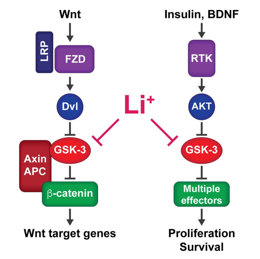
Cells 2021, 10, 255                                                                                           3 of 24

                            Lithium also stimulates glycogen synthesis in muscle, hepatocytes, and adipocytes by
                      increasing the activity of Glycogen Synthase (GS), enhancing glucose uptake and storage
                      and mimicking insulin action, although the direct target for these effects was not identified
                      in those early studies [31–35].
                            Each of these developmental and metabolic effects of lithium is mimicked by inhibition
                      or genetic loss of glycogen synthase kinase-3 (GSK-3). GSK-3 was so named because
                      it phosphorylates and inhibits GS; thus the effects of lithium on insulin signaling and
                      glycogen synthesis can be explained by inhibition of GSK-3, as demonstrated later [34].
                      Similarly, inhibition of GSK-3 offered a compelling explanation for the developmental
                      effects of lithium. Knockout of gska in Dictyostelium caused expansion of stalk cell fate
                      at the expense of spore cells [36] that was remarkably similar to the effect of lithium
                      described by Maeda [24]. Furthermore, dominant negative forms of Gsk3b mimic the effect
                      of lithium in Xenopus [37–39]. In the context of vertebrate development, GSK-3 functions
                      as an antagonist of canonical Wnt signaling [40–42], and therefore lithium, as a GSK-3
                      inhibitor, is predicted to activate Wnt signaling. Indeed, Wnt signaling had recently been
                      shown to recapitulate lithium phenotypes in Xenopus embryos, where Wnt activation also
                      induces axis duplication [43].
                            Recognition that the metabolic and developmental effects of lithium parallel the effects
                      of GSK-3 inhibition in these settings led to the hypothesis that lithium acts through direct
                      inhibition of GSK-3, and this was demonstrated through in vitro assays using mammalian
                      GSK-3β [18]. Subsequent work confirmed that lithium inhibits GSK-3 from mammals,
                      amphibians, Drosophila, and Dictyostelium [22,44]. Lithium competes for a low Km mag-
                      nesium binding site in GSK-3 with a Ki ~1–2 mM [19,20]. Other monovalent cations do
                      not mimic this effect on GSK-3 activity, and the effect of lithium is independent of the
                      anion species [18–20,22]. Furthermore, lithium does not significantly inhibit multiple other
                      protein kinases, demonstrating relative selectivity for GSK-3 [18,21].

                      3. GSK-3 Function
                           GSK-3 is a serine/threonine kinase expressed in most vertebrates as two highly similar
                      and mostly redundant isoforms, GSK3A and GSKB, which encode the proteins GSK-3α and
                      GSK-3β [45,46] (for simplicity, we will refer to both as GSK-3 unless otherwise specified).
                      GSK-3 is ubiquitously expressed in mammalian tissue, and is evolutionarily conserved
                      across slime molds, yeast, plants, and animals [12,47,48]. GSK-3 is constitutively active and
                      is negatively regulated by phosphorylation on serine-21 (GSK-3α) and serine-9 (GSK-3β)
                      primarily through activation of Akt (aka PKB), but also through growth factor stimulation
                      of mitogen activated protein kinases (MAPKs), mechanistic target of rapamycin (mTOR),
                      protein kinase A (PKA), and protein kinase C (PKC) [40,49]. GSK-3 is also constitutively
                      phosphorylated at a conserved tyrosine within the activation loop (tyrosine-279 in GSK-3α
                      and tyr-216 in GSK-3β), which may facilitate substrate binding [50–52]. GSK-3 is inhibited
                      by Wnt signaling independently of GSK-3 Ser21/9 phosphorylation [53–55] (Figure 1).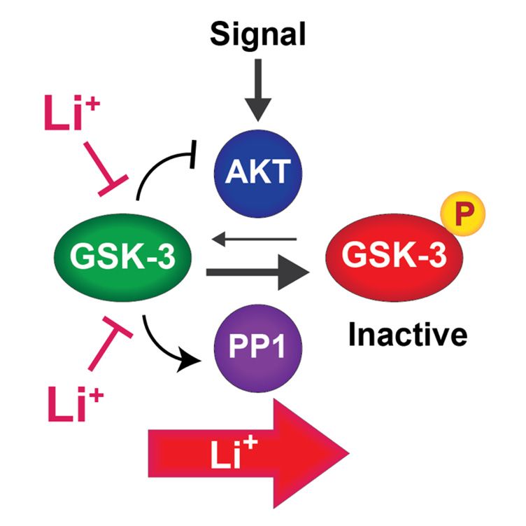
Cells 2021, 10, x FOR PEER REVIEW
Cells 2021, 10, 255 4 of 24
Figure 1. Signaling pathways that inhibit GSK-3: Wnt signaling (left) inhibits GSK-3 through an
Figure 1. Signaling pathways that inhibit GSK-3: Wnt signaling (left) inhibits GSK-3 through
allosteric mechanism to stabilize β-catenin and activate Wnt target genes. Insulin, BDNF, and other
allosteric mechanism to stabilize β-catenin and activate Wnt target genes. Insulin, BDNF, and
receptor tyrosine kinase (RTK) ligands that act through PI3 kinase to activate AKT inhibit GSK-3
other receptor tyrosine kinase (RTK) ligands that act through PI3 kinase to activate AKT inhib
through N-terminal phosphorylation to modulate diverse downstream pathways. Lithium mimics
GSK-3 through N-terminal phosphorylation to modulate diverse downstream pathways. Lith
both types of signaling pathways by directly inhibiting GSK-3.
mimics both types of signaling pathways by directly inhibiting GSK-3.
GSK-3 was first identified among several protein kinases that phosphorylate glyco-
genGSK-3
synthase was [46],
firstbutidentified
since then among
many other directprotein
several targets of GSK-3 that
kinases have phosphorylate
been iden- g
tified, including the Wnt effector protein β-catenin, other transcription factors, mul-
gen synthase [46], but since then many other direct targets of GSK-3 have been ident
tiple RNA splicing factors, regulators of translation, protein kinases, and cytoskeletal
including the WntGSK-3
proteins [40,56,57]. effector protein
target β-catenin,
phosphorylation other transcription
frequently factors,
impairs the activity multiple
or stabil-
splicing
ity of itsfactors, regulators
targets, including of translation,
glycogen protein [56],
synthase and β-catenin kinases,
although and
GSK-3cytoskeletal
can also pro
stabilize substrates such as the nuclear hormone receptor Rev-erbα
[40,56,57]. GSK-3 target phosphorylation frequently impairs the activity or stability [58] and enhance the
function
targets, of targets glycogen
including including the tuberousand
synthase sclerosis complex
β-catenin 2 protein
[56], [59]. GSK-3 can also sta
although
Multisite phosphorylation of GS by GSK-3 established a paradigm for many GSK-3
substrates such as the nuclear hormone receptor Rev-erbα [58] and enhance the fun
substrates in which GSK-3 depends on prior phosphorylation at the
of +4
targets including
position [60,61]. Thisthe“priming”
tuberous sclerosis complex
phosphorylation enhances2 protein [59].
GSK-3 phosphorylation of a
Multisite
serine phosphorylation
or threonine 4 residues to theof N GS by GSK-3
terminal established
side of the priming site.a paradigm for many G
GSK-3 substrates
such as GSinand
substrates β-catenin
which GSK-3 frequently
dependshave on
multiple
priorserines or threonines spaced
phosphorylation at the4 residues
+4 position [6
apart, allowing processive phosphorylation in the C to N terminal direction. Phosphoryla-
This “priming” phosphorylation enhances GSK-3 phosphorylation of a serine or threo
tion of N terminal serines in GSK-3 itself creates a pseudosubstrate that inhibits activity
4 residues
toward primedto thesubstrates
N terminal [61].side of themany
However, priming
GSK-3site. GSK-3including
substrates, substrates such as GS a
Inhibitor-2
catenin
(I-2) and Tau protein, do not contain +4 priming sites [56,57], and the mechanismsapart,
frequently have multiple serines or threonines spaced 4 residues of allo
substrate recognition
processive phosphorylation in these cases areCless
in the to well characterized.
N terminal direction. Phosphorylation of N t
GSK-3 inhibition by Wnt signaling does not involve N-terminal phosphorylation [53–55].
nal serines in GSK-3 itself creates a pseudosubstrate that inhibits activity toward pr
Several non-exclusive mechanisms for Wnt-mediated inhibition of GSK-3 have been proposed,
substrates
including[61]. However,
inhibition by the many GSK-3 substrates,
phosphorylated C-terminus including
of the LRP5/6 Inhibitor-2
co-receptor(I-2)
[62]and Tau
tein,
and do not contain
dissociation +4 priming
of APC, sites [56,57],
which enhances and thefrom
GSK-3 activity, mechanisms of substrate
the Axin scaffold [63–65]. recogn
in these cases are less well characterized.
On a longer time scale, Wnt signaling drives sequestration of GSK-3 in the Axin complex
into distinctinhibition
GSK-3 intracellularby pools
Wnt termed multivesicular
signaling does not bodies,
involvewhich protects β-catenin
N-terminal and
phosphorylation
other GSK-3 substrates from phosphorylation and degradation [66]. These mechanisms are not
55].mutually
Several non-exclusive mechanisms for Wnt-mediated inhibition of GSK-3 have
exclusive but they are independent of Akt signaling and N-terminal phosphorylation.
proposed, including inhibition by the phosphorylated C-terminus of the LRP5/6 co-r
tor [62] and dissociation of APC, which enhances GSK-3 activity, from the Axin sca
[63–65]. On a longer time scale, Wnt signaling drives sequestration of GSK-3 in the
complex into distinct intracellular pools termed multivesicular bodies, which prote
catenin and other GSK-3 substrates from phosphorylation and degradation [66]. T
mechanisms are not mutually exclusive but they are independent of Akt signaling an
Cells 2021, 10, 255                                                                                             5 of 24

                      4. GSK-3 and Human Disease
                           As outlined above, GSK-3 phosphorylates multiple substrates involved in critical
                      cellular processes aside from its classical role in glycogen metabolism [40,56,57,67]. Many
                      of these processes, such as autophagy, cell survival/differentiation, and cell cycle regulation,
                      have clear implications in the pathogenesis of human disease. This has consequently led
                      to substantial interest in better understanding the role of GSK-3 and related pathways as
                      therapeutic targets in these disease models. Below, we will review known roles of GSK-3 in
                      bipolar disorder, cancer, neurodegenerative diseases, and virally mediated diseases with
                      an emphasis on targetable pathways.

                      4.1. Bipolar Disorder and Lithium
                            Bipolar disorder is a major psychiatric disorder associated with episodes of hyperac-
                      tivity, elevated mood, and psychosis (mania and hypomania), periods of major depression,
                      and a substantial risk of suicide [68]. Type I bipolar disorder is defined by at least one
                      episode of mania and type II bipolar disorder is defined by episodes of both hypomania
                      and major depression. The incidence of bipolar disorder is estimated to be 1–2% worldwide.
                      Lithium is a first-line therapy for bipolar disorder, but the therapeutic window between
                      the effective and toxic doses is narrow [68]. In addition to functioning as a mood stabilizer,
                      lithium also reduces the risk of suicide in affective disorder patients, either as a monother-
                      apy or adjunctive therapy. Lithium is more effective at suicide prevention than other mood
                      stabilizers and antidepressants, although the mechanism is unknown [10,69,70]. Lithium
                      is also an effective adjunctive therapy for treatment resistant depression, especially in
                      combination with tricyclic antidepressants [71,72].
                            In addition to directly inhibiting GSK-3 enzymatic action [18,22], lithium also inhibits
                      GSK-3 indirectly by increasing inhibitory Ser21/9 phosphorylation [73,74]. Another mood
                      stabilizer (valproic acid), antidepressants (including the rapid-acting antidepressant ke-
                      tamine), and antipsychotics also inhibit GSK-3 indirectly through Ser21/9 phosphorylation,
                      suggesting a common mechanism involving GSK-3 [74–78].
                            The hypothesis that GSK-3 is the therapeutic target of lithium is supported by animal
                      behavior studies using genetic manipulation of GSK-3. Gsk3b haploinsufficiency mimics
                      lithium treatment in mouse behavioral tests that are responsive to lithium [79], while Gsk3b
                      overexpression in the CNS blunts the response to lithium without affecting the animal’s
                      health or activity levels [13], demonstrating a specific role for GSK-3 in lithium-responsive
                      behavior. Furthermore, diverse small molecule inhibitors of GSK-3 also mimic the behav-
                      ioral effects of lithium in mice [79–83], providing compelling support for GSK-3 as a critical
                      lithium target in mouse behaviors. Conversely, a competing hypothesis that lithium acts
                      therapeutically through IMPase inhibition to deplete inositol is not supported by animal
                      behavior in genetic models of inositol depletion. Genetically reducing inositol levels to
                      a greater extent than lithium did not phenocopy lithium treatment (or Gsk3 haploinsuffi-
                      ciency) in mouse behavior [84], nor did depletion of inositol affect phosphatidylinositol
                      abundance, arguing against this hypothesis [85–87]. Therefore, GSK-3 remains the best-
                      supported direct target of lithium action in behavior.
                            While GSK-3 is a promising target of mood stabilizers and other psychotropic drugs,
                      it is unclear how its inhibition can encompass anti-manic, antidepressant, and antipsy-
                      chotic effects. We propose a model in which mood stabilizers function through GSK-3
                      inhibition to re-sensitize cells to endogenous extracellular signals (Figure 2). Extracellular
                      signals transduced through Akt (e.g., neurotrophins and neurotransmitters) lead to phos-
                      phorylation and inactivation GSK-3. However, GSK-3 opposes its own phosphorylation
                      through at least two mechanisms. (1) GSK-3 inhibits Akt by binding to and stabilizing a
                      β-arrestin complex that brings Akt into contact with its deactivating phosphatase PP2A.
                      Inactivation of Akt thereby prevents phosphorylation and inhibition of GSK-3. Lithium
                      or other GSK-3 inhibitors disrupt this complex [13,88], allowing Akt to remain active and
                      phosphorylate GSK-3. (2) GSK-3 also maintains its own dephosphorylation by activating
                      protein phosphatase 1 (PP1), which is inhibited by I-2; GSK-3 phosphorylation of I-2 causes
Cells 2021, 10, 255 6 of 24
it to dissociate from PP1, which then dephosphorylates GSK-3, restoring GSK-3 activity.
Cells 2021, 10, x FOR PEER REVIEW 6 of 24
Lithium or other GSK-3 inhibitors prevent PP1 from dephosphorylating GSK-3 in an I-2
dependent manner [73].
(a) (b)
Figure 2. Positive feedback circuits regulate GSK-3: (a) GSK-3 is phosphorylated and inhibited by AKT in response to
Figure 2. Positive feedback circuits regulate GSK-3: (a) GSK-3 is phosphorylated and inhibited by AKT in response to
upstream signals. PP1 dephosphorylates and activates GSK-3. GSK-3 inhibits AKT and activates PP1, thereby enhancing
upstream signals. PP1 dephosphorylates and activates GSK-3. GSK-3 inhibits AKT and activates PP1, thereby enhancing its
its own activity; (b) lithium inhibits GSK-3 directly and disrupts both feedback circuits. Disruption of these feedback cir-
own activity;
cuits (b)may
by lithium lithium inhibits
enhance theGSK-3 directly
response and disrupts
to endogenous both feedback
ligands that signalcircuits.
through Disruption
AKT. of these feedback circuits
by lithium may enhance the response to endogenous ligands that signal through AKT.
Therefore, pharmacological GSK-3 inhibition interrupts both autoregulatory feed-
Therefore, pharmacological GSK-3 inhibition interrupts both autoregulatory feedback
back loops that drive GSK-3 to maintain an active state [13,73]. We hypothesize that phar-
loops that drive GSK-3 to maintain an active state [13,73]. We hypothesize that pharma-
macological inhibition of GSK-3 lowers the threshold for endogenous signals to inactivate
cological inhibition of GSK-3 lowers the threshold for endogenous signals to inactivate
cellular pools of GSK-3, allowing weak or transient signals to effect a stable response
cellular pools of GSK-3, allowing weak or transient signals to effect a stable response
through amplification of GSK-3 inactivation. This mechanism would allow lithium to sen-
through amplification of GSK-3 inactivation. This mechanism would allow lithium to
sitize cells to endogenous neurotransmitters or other signals that may be reduced in bipo-
sensitize cells to endogenous neurotransmitters or other signals that may be reduced in
lar disorder
bipolar and could
disorder explain
and could why lithium
explain is also effective
why lithium in treatment
is also effective resistantresistant
in treatment depres-
sion when used as an adjunct to antidepressants.
depression when used as an adjunct to antidepressants.
4.2.
4.2. Neurodegenerative
Neurodegenerative Disease
Disease
GSK-3 has well documented
GSK-3 has well documented rolesroles in neurodegenerative
in neurodegenerative diseases,
diseases, established
established through
through
extensive mechanistic studies of GSK-3 in neurological models. Below, we providewe
extensive mechanistic studies of GSK-3 in neurological models. Below, pro-
a brief
vide
overview of the literature regarding GSK-3, neurodegenerative disease, and
a brief overview of the literature regarding GSK-3, neurodegenerative disease,
therapeutic
therapeutic insights.
4.2.1. Alzheimer’s
4.2.1. Alzheimer’s Disease
Disease and
and Related
Related Tauopathies
Tauopathies
Alzheimer’s disease (AD)
Alzheimer’s disease (AD) is the is the most common form of neurodegenerative disease
increasing prevalence
with increasing prevalence inin an
an aging
aging population
population [89].[89]. Two
Two pathologic
pathologic findings
findings charac-
charac-
terize AD,
terize AD,neurofibrillary
neurofibrillarytangles
tangles(NFTs)
(NFTs)andandamyloid
amyloid plaques,
plaques, andand
bothboth
areare promoted
promoted by
by GSK-3.
GSK-3. NFTs NFTs
are are intracellular
intracellular deposits
deposits of protein,
of tau tau protein, a microtubule
a microtubule stabilizer
stabilizer that that is
is reg-
regulated by phosphorylation; pathologic hyperphosphorylation
ulated by phosphorylation; pathologic hyperphosphorylation of tau disrupts microtu- of tau disrupts micro-
tubules
bules and
and promotes
promotes development
development of of NFTs
NFTs [90].
[90]. GSK-3
GSK-3 phosphorylates
phosphorylates tautau in vitro
in vitro andandin
in vivo at several residues, which can accelerate aberrant tau aggregation
vivo at several residues, which can accelerate aberrant tau aggregation and resultant neu- and resultant
neurodegenerative
rodegenerative phenotypes
phenotypes [91–98].
[91–98]. Second,
Second, β-amyloid
β-amyloid (Aβ)(Aβ) peptides
peptides (primarily
(primarily Aβ40Aβand40
and42)Aβ
Aβ 42 ) accumulate
accumulate in extracellular
in extracellular amyloid amyloid
plaques.plaques.
Plaque Plaque formation
formation precedes precedes tau
tau pathol-
pathology
ogy in AD in ADoccurs
and and occurs
throughthrough cleavage
cleavage of theofamyloid
the amyloid precursor
precursor protein
protein (APP)(APP)
intointo
Aβ
by β- and γ-secretases [99]. APP processing is also regulated by GSK-3. Both APPAPP
Aβ by β- and γ-secretases [99]. APP processing is also regulated by GSK-3. Both and
and presenilin-1
presenilin-1 (PS1),(PS1),
whichwhich
is partisof
part
theof the γ-secretase
γ-secretase complex,complex, are mutated
are mutated in familial
in familial forms
forms of AD, leading to increased accumulation of the more pathogenic
of AD, leading to increased accumulation of the more pathogenic Aβ42 peptide.42Both Aβ peptide.
APP
and PS1 are GSK-3 substrates [100–105] and inhibition or knockdown of GSK-3 impairs
APP processing, reducing generation of Aβ40/42 in mouse brain and cell culture models
[94,105–111]. The requirement for GSK-3 in APP processing was challenged by a group
that knocked out either Gsk3a or Gsk3b in mice and observed no effect on Aβ levels [112].Cells 2021, 10, 255 7 of 24
Both APP and PS1 are GSK-3 substrates [100–105] and inhibition or knockdown of GSK-3
impairs APP processing, reducing generation of Aβ40/42 in mouse brain and cell culture
models [94,105–111]. The requirement for GSK-3 in APP processing was challenged by
a group that knocked out either Gsk3a or Gsk3b in mice and observed no effect on Aβ
levels [112]. However, this report involved single gene knockouts only, overlooking the
important and well established observation that the two genes are redundant in most
functions [53], so that a single gene knockout may not affect APP processing in cells that
express both genes equally. Furthermore, multiple structurally diverse GSK-3 inhibitors,
including lithium, Tideglusib, kenpaullone, bisindolylmaleimide-I, FRAT peptide, and
kinase-dead GSK-3 also impair APP processing [94,105–111], providing compelling sup-
port that GSK-3 facilitates APP processing. Moreover, Aβ aggregates activate GSK-3 and
cause tau hyperphosphorylation [113–115]. This may form a feedback loop by which Aβ
activates GSK-3, which then alters APP cleavage, resulting in more Aβ formation. These
data suggest GSK-3 inhibition may have a role in slowing progression of AD, which is
further supported by clinical studies described below.
As GSK-3 plays roles in both NFT formation and APP processing, GSK-3 has been a
focus of extensive study for pharmacological interventions in AD. Inhibition with lithium
and other agents in preclinical studies reduces Aβ mediated neurotoxicity, improves be-
havioral phenotypes, and rescues neuronal loss [105,116–119]. In addition, retrospective
studies, meta-analyses, and a randomized controlled trial with patients with mild cogni-
tive impairment showed that lithium prevented cognitive decline in patients with mild
cognitive impairment when compared to matched patients not taking lithium [120–122].
Unfortunately, a phase II trial with the allosteric GSK-3 inhibitor Tideglusib did not achieve
study endpoints for slowing cognitive decline [123] and studies with the GSK-3 inhibitors
AZD2558 and AZD1080 were halted for intolerable safety profiles [124]. Nonetheless,
additional clinical trials (NCT03185208) are assessing the utility of lithium in preventing
AD in elderly patients with mild cognitive impairment.
4.2.2. Parkinson’s Disease
Parkinson’s disease (PD) is the second-most common neurodegenerative disorder and,
like AD, tends to occur in older populations. PD is caused by neuronal loss in the substantia
nigra, leading to dopamine deficiency, and intracellular accumulation of α-synuclein, called
Lewy Bodies (LBs) [125]. Tau pathologies have also been implicated in PD [126]. Early
studies demonstrated the localization of GSK-3 in LBs [127–129] and noted that GSK-3
polymorphisms, which alter its transcription and splicing, are associated with PD [130].
GSK-3 phosphorylates α-synuclein and tau in PD, which leads to the development of the
neurotoxic aggregates that drive the disease [128,131–133]. Several inhibitors of GSK-3,
including lithium [134,135], indirubin 30 -oxime [136,137], and AR-A014418 [137], have
been used in vitro and in animal models to slow neurodegeneration and increase the
concentration of dopamine. However, other studies have demonstrated conflicting efficacy
of lithium and instead show that it decreases neuronal dopamine levels [138,139]. This
underscores a caveat of lithium use in human studies of PD, which is the adverse effect
of inducing parkinsonian features [140,141]. This may indicate that the efficacy of GSK-3
inhibition in PD is dose dependent; consistent with this hypothesis, preclinical studies have
shown that higher doses of Tideglusib are more neuroprotective [142], though this has not
yet been fully recapitulated in human studies of other parkinsonian syndromes [143,144].
Moreover, an ongoing trial (NCT04273932) is evaluating dose titration of lithium in PD.
4.3. GSK-3 in Cancer
GSK-3 cannot be strictly characterized as a tumor suppressor or as a proto-oncogene.
For example, GSK-3 inhibits the canonical Wnt signaling pathway, functioning in a complex
with the classical tumor suppressor adenomatous polyposis coli (APC) to phosphorylate
and degrade β-catenin. Loss of APC or GSK3 therefore activates Wnt target genes, such as
MYC (c-Myc) and CCND1 (cyclin D1), and promotes tumor growth, as described in greaterCells 2021, 10, 255 8 of 24
Cells 2021, 10, x FOR PEER REVIEW 8 of 24
detail below. GSK-3 also suppresses the epithelial-to-mesenchymal transition (EMT), a
(EMT), a critical process in tumor invasion and metastasis (reviewed in [145]) by phos-
critical process in tumor invasion and metastasis (reviewed in [145]) by phosphorylating
phorylating Snail family transcription factors and targeting them for degradation [146–
Snail family transcription factors and targeting them for degradation [146–149]. While
149]. While these
these observations observations are consistent
are consistent withsuppressor-like
with a tumor a tumor suppressor-like role for
role for GSK-3, GSK-also
GSK-3
3, GSK-3 also activates NFκB signaling [150–153], which has a pro-tumorigenic
activates NFκB signaling [150–153], which has a pro-tumorigenic role in the context role in the of
context of inflammatory
inflammatory microenvironments
microenvironments of several
of several cancers cancers
(reviewed (reviewed
in [154]).inThis
[154]). This to
appears
appears to be through GSK-3 mediated phosphorylation and subsequent
be through GSK-3 mediated phosphorylation and subsequent stabilization of the adaptor stabilization of
the protein
adaptorNEMO protein[155],
NEMO [155], which then promotes
which then promotes NFκB signaling. NFκB signaling.
Similarly,
Similarly,GSK-3 phosphorylates
GSK-3 phosphorylatesand destabilizes c-Myc;c-Myc;
and destabilizes inhibition of GSK-3
inhibition of and
GSK-3 sta-and
bilization of c-Myc can be oncogenic or pro-apoptotic depending on
stabilization of c-Myc can be oncogenic or pro-apoptotic depending on whether c-Myc whether c-Myc pro-
teinprotein
accumulates at intermediate
accumulates or high
at intermediate levels,
or high respectively
levels, respectively[156].[156].
Ultimately, it seems
Ultimately, it seems
thatthat
GSK-3GSK-3is tightly regulated
is tightly in the
regulated in context of different
the context cell cell
of different types, developmental
types, developmental stages,
stages,
andand
in concert with other signaling molecules; when this regulation is disrupted,
in concert with other signaling molecules; when this regulation is disrupted, it can it can
inhibit or promote
inhibit or promote malignancy
malignancy depending
depending on that context
on that (Figure
context 3). The
(Figure involvement
3). The involvement of of
GSK-3 in cancer has recently been reviewed in depth [157,158]; therefore,
GSK-3 in cancer has recently been reviewed in depth [157,158]; therefore, we will providewe will provide
a detailed
a detailedmechanistic
mechanisticoverview
overviewof GSK-3
of GSK-3involvement
involvement in certain solid
in certain andand
solid hematologic
hematologic
malignancies
malignancies with significant
with therapeutic
significant potential.
therapeutic potential.
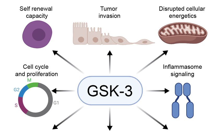
Figure 3. GSK-3 regulates diverse pathways in cancer: GSK-3 can promote or inhibit tumorigenesis
Figure 3. GSK-3 regulates
by regulating diverse pathways
several hallmarks in These
of cancer. cancer:roles
GSK-3 can
vary inpromote or inhibit
the specific tumorigene-
contexts of cell type and
sis by regulating several hallmarks of cancer. These roles vary in the specific contexts of cell type
developmental stage.
and developmental stage.
4.3.1. Solid Malignancies
4.3.1. Solid Malignancies
Glioblastoma multiforme (GBM) is the most common malignant brain neoplasm in
Glioblastoma
adults multiforme
and carries (GBM) isPI3K/AKT
a high mortality. the most common malignant
signaling, brain neoplasm
which inhibits in
GSK-3 activity
adults and carries
(reviewed a high
in [159]), is mortality.
critical in PI3K/AKT signaling,
the pathogenesis which[160,161].
of GBM inhibits GSK-3 activity
However, GSK-3
(reviewed
remainsinhighly
[159]),expressed
is critical in
andthe pathogenesisactivated
constitutively of GBM [160,161]. However,
in GBM cells GSK-3
[162–164], re-
paradoxi-
mains highly expressed and constitutively activated in GBM cells [162–164], paradoxically
cally suggesting an oncogenic role for GSK-3 in GBM. Indeed, GSK-3 inhibition through
suggesting an oncogenic role for GSK-3 in GBM. Indeed, GSK-3 inhibition through TDZD-Cells 2021, 10, 255 9 of 24
TDZD-8 [165], an allosteric inhibitor of GSK-3 [166], decreased GBM growth and stemness,
reflected by impaired neurosphere formation and reduced Nestin levels. Furthermore,
both GSK-3 knockdown and its pharmacologic inhibition with LiCl, Kenpaullone, and
Enzastaurin impaired GBM proliferation and survival in vitro and in vivo through several
mechanisms [167]. First, loss of GSK-3 activity led to transactivation of c-Myc, thereby
promoting the expression of pro-apoptotic molecules, including Bim, DR4/DR5, and
TRAIL. Second, GSK-3 inhibition altered glucose metabolism and destabilized the mito-
chondrial membrane, increasing levels of the pro-apoptotic molecule Bax. Third, GSK-3
inhibition impaired NFκB activity, leading to abrogation of pro-survival signals. Although
these studies suggest that both GSK-3α and GSK-3β isoforms are oncogenic in GBM, a
tumor suppressor role for GSK-3α has also been proposed in GBM by which it promotes
apoptotic alternative splice events [168], though this remains incompletely defined and
warrants further investigation. Finally, hallmarks of GBM that increase mortality include
its invasive nature and propensity for treatment resistance. GSK-3 has been implicated
in both of these mechanisms in GBM [169–173]. Therefore, GSK-3 inhibitors have been
exploited in pre-clinical studies to improve sensitivity to common GBM treatments, such
as temozolomide [174,175], offering a potential therapeutic opportunity.
Colorectal cancer (CRC) is one of the most common cancers in adults. Our under-
standing of the pathophysiology of CRC has drastically improved survival since it has
led to the use of widespread early screening. CRC commonly begins as a precancerous
polyp, followed by sequential accumulation of somatic mutations that ultimately lead to
malignant transformation; this adenoma-to-carcinoma sequence [176] classically begins
with a mutation in the tumor suppressor APC, followed by a mutation in the oncogene
KRAS, and culminates with a mutation of TP53 [177]. APC is of particular significance in
the context of GSK-3 as both are core components of the Axin complex that promotes pro-
teasomal degradation of β-catenin in the absence of Wnt signaling [178]. Loss of APC leads
to the robust activation of the Wnt pathway frequently observed in CRC [179,180]. APC
has several proposed roles in the Axin complex that may contribute to inhibition of Wnt
signaling and tumor suppression, including recruitment of β-catenin into the destruction
complex, facilitation of ubiquitination and degradation, and direct enhancement of GSK-3
enzymatic activity toward β-catenin [181]. APC enhances GSK-3 activity in vitro and
in vivo [181,182], and dissociation of APC from the Axin complex upon Wnt stimulation
may explain how Wnt signaling inhibits β-catenin phosphorylation [63–65]. Loss of APC
and the consequent reduction in GSK-3 activity leads to activation of several downstream
targets in addition to β-catenin, including mTOR signaling, MAPK/ERK signaling, and
BMP signaling [64].
Despite these compelling data that GSK-3 serves as a tumor suppressor in CRC, the
exact role of GSK-3 in this malignancy remains controversial. For example, a conflicting
report on the role of GSK-3 in KRAS dependent CRC demonstrated that GSK-3 inhibition
decreased cell survival through an increase in β-catenin and c-Myc [183]. Additionally,
multiple studies have demonstrated that inhibition of GSK-3 activity may still have thera-
peutic potential in the management of CRC through the induction of P53 dependent and
independent apoptosis [184–189], downregulating EMT [190–193], re-establishing Hedge-
hog signaling [194], and disrupting malignant cytoskeletal dependencies [190,195,196].
Interestingly, Dow et al. observed that restoration of Apc in murine models of CRC trig-
gered intestinal differentiation and tumor regression in spite of activating mutations in
Kras or inactivating mutations in Tp53 [197]. However, differentiation occurred prior to
full Apc expression and did not induce apoptosis. While the authors did not specifically
comment on the activity of GSK-3 in this study, their findings underscore the dynamic
nature of cell survival and differentiation in CRC; in this regard, a better understanding of
how GSK-3 is involved at different stages of tumorigenesis in CRC may help evaluate its
efficacy as a therapeutic target.
Despite advances in therapies for other cancers, survival in pancreatic ductal ade-
nocarcinoma (PDAC) remains poor, with incremental improvements through the use ofCells 2021, 10, 255 10 of 24
escalating cytotoxic chemotherapy in addition to surgery when possible. Therefore, the
possibility of targeted therapy has been of great interest. KRAS mutations [198], which
occur in nearly 90% of PDAC patients, promote an inflammatory microenvironment that
is quintessential to the development of PDAC. In murine models, this occurs through
mutant Kras induction of IL-1α, which ultimately leads to NFκB activation [199]. This
inflammatory state, characterized by high levels of NFκB signaling, can be seen in other
preceding risk factors for PDAC, such as chronic pancreatitis (reviewed in [200]). Therefore,
GSK-3, which has established relationships with both KRAS and NFκB in other tumors,
may have a significant role in the pathogenesis and treatment of PDAC. GSK-3β is highly
active in PDAC cells, and both pharmacologic inhibition and genetic ablation of GSK-3
decreased PDAC survival and proliferation [151]. Loss of GSK-3 activity in this model also
decreased expression of NFκB target genes involved in proliferation and survival, such as
CCND1, BCL2, BCL2L1, and XIAP. Subsequent studies have confirmed that sensitivity of
PDAC cells to GSK-3 inhibition is in part through loss of GSK-3 dependent NFκB signal-
ing [152,201]. However, GSK-3 inhibition may also impair PDAC survival through other
mechanisms such as TRAIL-dependent synthetic lethality [202–204] and interruption of
pro-survival transcription pathways [205–207]. Given the prevalence of chemoresistance
in PDAC, several preclinical studies have investigated whether targeting GSK-3 could
improve sensitivity to gemcitabine, which is commonly used in PDAC treatment, and
have shown promising results, especially in combination with HDAC inhibition [208–210].
These findings indicate that GSK-3 inhibitors may ameliorate chemoresistance in PDAC.
Clinically, these findings have prompted several trials of GSK-3 inhibitors in ad-
vanced solid tumors. LY2090314 is an ATP-competitive inhibitor of GSK-3 that demon-
strated a tolerable safety profile in combination with pemetrexed and carboplatin for
advanced/metastatic solid tumors, though a randomized-control trial will be needed to
assess the efficacy of this protocol [211]. A Phase I/II trial (NCT016323306) for LY2090314 in
metastatic PDAC was unfortunately terminated due to lack of enrollment. Ongoing trials
are being performed with agents such as 9-ING-41 in combination with other chemotherapy
in adults (NCT03678883) and children (NCT04239092), CHIR99021 to expand NK cells for
infusion in patients with various solid tumors (NCT03213964, NCT03319459), and CLOVA
(cimetidine, lithium, olanzapine, valproic acid) in combination with gemcitabine for PDAC
(UMIN000005095) or temozolomide in GBM (UMIN000005111).
4.3.2. Normal and Malignant Hematopoiesis
Normal hematopoiesis is characterized by a hierarchical evolution of hematopoietic
precursors, which undergo several rounds of proliferation followed by either quiescence or
lineage determination. GSK-3 has multiple roles in HSC maintenance, as demonstrated by
the effects of GSK-3 inhibitors or loss of function in mouse hematopoietic cells. Inhibition
of GSK-3 activates Wnt/β-catenin signaling to support HSC self-renewal but also activates
mTOR complex 1 (mTORC1) signaling leading to HSC activation and an increase in multi-
potent progenitors at the expense of self-renewing HSCs [212]. Consistent with the latter
findings, activation of mTORC1 [213] and loss of LKB1 [214] promote HSC pool depletion
through proliferation of committed progenitors through improved nutrient sensing. Fur-
thermore, combined inhibition of GSK-3 and mTORC1 allows ex vivo maintenance of long
term, self-renewing HSCs in a cytokine-independent manner [215]; under these conditions,
Wnt activation maintains HSC fate while mTORC1 inhibition prevents differentiation and
loss of stemness. HSCs were maintained without loss of long-term renewal capacity for up
to one week in the absence of cytokines or stromal cells.
Acute myeloid leukemia (AML), the most common acute leukemia in adults, results
from clonal proliferation and maturation arrest of early myeloid progenitors. Progenitors
with self-renewal capability have been defined functionally as leukemic stem cells (LSCs) or
leukemia initiating cells (LICs) [216]. Wnt/β-catenin signaling has proven to be indispens-
able for the development of LSCs in AML [217], but several studies have also demonstrated
GSK-3 as an important regulator of LSC development and maintenance. LSCs expressingCells 2021, 10, 255 11 of 24
fusion proteins containing Mixed Lineage Leukemia (MLL) have been useful models to
study the role of GSK-3 in the development of AML. Wang et al. initially demonstrated
that GSK-3 can paradoxically maintain clonal proliferation of MLL-transformed cells and is
required for transformation into leukemic cells in murine models through destabilization of
p27Kip1 [218]. A subsequent study by the same group further elucidated a model in which
GSK-3 maintains the association of CREB with MEIS to maintain gene expression profiles
associated with HOX mediated leukemic transformation [219]. Although these studies
provide strong support for the role of GSK-3 in maintaining LSCs and allowing for trans-
formation, they do not readily explain the paradox of how both GSK-3 and β-catenin are
required for LSC development and survival despite the former canonically downregulating
the latter. Yeung et al. proposed a model by which β-catenin is progressively activated in
the transformation of MLL pre-LSCs to LSCs, and deletion of β-catenin in either subset
completely abolished leukemogenicity of MLL-transformed cells [220]. MLL LSCs with
high levels of nuclear β-catenin were insensitive to GSK-3 inhibition, while suppression
of β-catenin expression re-sensitized cells to GSK-3 inhibition. Therefore, while GSK-3
inhibition may be a suitable treatment in AML or early in the development of LSCs, it
may not completely abolish the LSC pool. Moreover, it is likely that GSK-3 inhibition
is still effective in treating AML cells through mechanisms outside of maintaining stem-
ness such as NFκB signaling [221,222], modulation of cytotoxic chemosensitivity [223,224],
and alterations in apoptotic signaling [225]. It should be noted, however, that inhibition
of GSK-3 can also enhance the ex vivo survival of primary leukemic cells from patients
with AML [226].
Chronic myeloid leukemia (CML) LSCs also require active Wnt/β-catenin signal-
ing [227]. The Armstrong group hypothesized that this may offer an approach to target
minimal residual disease with tyrosine kinase inhibitor (TKI) resistance [228]. In their
study, genetic deletion of β-catenin combined with imatinib drastically reduced CML LSC
survival, though bulk CML cells persisted. Interestingly, increased β-catenin expression
may be a result of aberrant splicing of GSK3B, as demonstrated in a study of CML patients,
in which CML cells from four of seven patients in blast crisis and one of four in chronic
phase CML showed mis-splicing of GSK3B, yielding a form that did not interact with AXIN
and was unable to phosphorylate β-catenin [229]. However, a clear role for GSK-3 and its
inhibition in CML will require further investigation.
The role of GSK-3 in acute lymphoblastic leukemia (ALL), chronic lymphocytic
leukemia (CLL), and lymphomas remains poorly understood with primarily correlative
pathophysiology driving studies of GSK-3 inhibition in these models. In other tumors,
GSK-3 regulates FOXO proteins, PI3K/AKT signaling, NOTCH signaling, and NFAT sig-
naling (reviewed in [157]), all of which are critical in lymphocyte development (reviewed
in [230,231]). Despite the lack of in depth mechanistic studies of GSK-3 in normal and
malignant lymphopoiesis, preclinical evaluation of GSK-3 inhibitors in ALL [232–234],
CLL [235], and lymphoma [156,236,237] have been promising, and further investigation is
warranted.
In regard to clinical trials, a phase I trial investigating lithium and tretinoin in non-
promyelocytic AML demonstrated an acceptable safety profile with observed on-target
effects, which led to AML cell differentiation [238]. However, a recent phase II trial in
AML found that LY2090314 did not lead to complete or partial remissions despite on-target
effects [239]. Therefore, further evaluation of other GSK-3 inhibitors, combination therapies,
and an improved understanding of GSK-3 mediated targetable pathways are necessary.
Ongoing trials are being performed with CHIR99021 to stimulate NK cell production
for infusion in patients with AML (NCT03081780) and with 9-ING-41 in patients with
lymphoma (NCT03678883).Cells 2021, 10, 255 12 of 24
5. Lithium, GSK-3, and Coronaviruses
5.1. Antiviral Activity of Lithium
Potential antiviral activity of lithium has been investigated for over 40 years, as
comprehensively reviewed recently [240]. Lithium reportedly antagonizes replication of
multiple DNA and RNA viruses in cell culture assays, and in some cases lithium therapy
has been associated with modest antiviral activity in patients. While the clinical benefit
of lithium and other GSK-3 inhibitors in viral infections is uncertain, the in vitro effects
may provide new insights into the roles of host proteins during virus infection. Although
inhibition of inositol turnover may also mediate antiviral effects of lithium [240], we will
focus here on the potential role of GSK-3 as an antiviral target of lithium, with special
attention to the role of GSK-3 in coronavirus infections.
Lithium was reported to have antiviral activity against herpes simplex in the 1970s.
These studies included in vitro demonstration that lithium impairs HSV replication in cell
culture [240–242] and culminated in a series of both retrospective and small prospective
clinical trials with lithium carbonate to assess duration and rate of recurrence of labial
herpes infections [240,243]. As reviewed by Murru et al., the retrospective study reported
a reduced rate of recurrence for labial HSV for patients on lithium carbonate (LiCO3)
compared to patients treated for depression, and the protective effect was higher in patients
with higher serum lithium levels. Two randomized, placebo-controlled trials with LiCO3
for HSV found a trend towards a modest effect of lithium in reduction of HSV recurrence,
but both trials were small and underpowered [240,244].
Lithium has also been reported to have antiviral activity against diverse DNA and
RNA viruses in vitro, including hepatitis C, adenoviruses, Dengue virus-2, pseudorabies
and vaccinia viruses, parvoviruses, foot-and-mouth disease virus (FMDV), and feline
calicivirus (FCV) [240]. Limited studies in patients have also supported modest antiviral
effects against HIV and influenza. Prospective studies on HIV were small but reported a
transient reduction in viral transcription in HIV infected patients [245]. A retrospective
study on 177 patients taking LiCO3 for bipolar disorder also showed a modest but signif-
icant reduction in flu-like infections when comparing the incidence in the same patient
cohort before and during lithium therapy [246]. There was no effect of treatment in the
smaller control group of patients taking antidepressants.
While lithium has direct antiviral activity (described in more detail below for coron-
aviruses), it may also function indirectly by enhancing host anti-inflammatory responses.
The literature on lithium effects on the immune system is complex, with evidence for both
pro-inflammatory and anti-inflammatory effects that may contribute to host responses
to viral infections, as more fully reviewed in [240]. The effects of lithium and GSK-3 on
inflammation are also comprehensively reviewed in [247,248].
5.2. Lithium and GSK-3 in Coronaviruses
Perhaps for these reasons, lithium salts have been tested for activity against a number
of coronaviruses in cell culture models. LiCl impairs replication and infectivity of diverse
coronaviruses, including infectious bronchitis virus (IBV), porcine epidemic diarrhea virus
(PEDV), transmissible gastroenteritis virus (TGEV), and the β-coronavirus responsible
for severe acute respiratory syndrome (SARS-CoV) [249–256]. These observations have
led to recent calls to use lithium to treat COVID19. However, the effects of lithium on
these coronaviruses were observed at concentrations well above clinically tolerable levels.
Furthermore, experimental evidence to support an inhibitory effect of lithium on SARS-
CoV-2, the cause of COVID19, has not yet been reported.
As the IC50 for LiCl inhibition of GSK-3 [19,20], as well as the optimal therapeutic
plasma concentration in bipolar disorder [20], is ~1 mM, the high concentrations needed
to inhibit coronaviruses (at least 5–10 mM) also raise the possibility that lithium may be
acting through another target. However, considerable data now support GSK-3 as the
direct target responsible for lithium effects on diverse coronaviruses. Initial studies using
multiple protein kinase inhibitors hinted that GSK-3 may phosphorylate the nucleocapsidCells 2021, 10, 255 13 of 24
protein [257], and subsequent work from several labs including Yeh and colleagues pro-
vided strong support for a critical role for GSK-3 in phosphorylating and enhancing the
function of nucleocapsid protein [250,258,259].
Coronaviruses encode a nucleocapsid (N) protein that binds viral genomic RNA
(gRNA) and is essential for viral replication, transcription, and virion
assembly [250,251,259–261]. N is phosphorylated within an arginine-serine (RS) domain
that is conserved in coronaviruses [250,257–259,262–264]. As pointed out by Wu et al., the
RS domains in N protein from the JMHV strain of mouse hepatitis virus and SARS-CoV
includes the canonical SXXXS motif found in many GSK-3 substrates [250]. N from other
coronaviruses diverge in overall sequence but retain RS domains and tandem SXXXS motifs.
Evidence that GSK-3 phosphorylates N from JHMV and SARS-CoV includes: (1) GSK-3
binds to N and phosphorylates sites within the SXXXS motifs [250,257,258]; (2) GSK-3
dependent phosphorylation of N from SARS-CoV and JHMV has been documented by mass
spectrometry and phospho-specific antibodies [250]; (3) mutation of the +4 priming serine
to alanine prevents phosphorylation by GSK-3, blocks N functions, and interferes with
viral transcription and replication; (4) siRNA mediated knockdown of both GSK3 mRNAs
(GSK3A and GSK3B) impairs viral replication and infectivity; (5) inhibition of GSK-3 with
Kenpaullone or LiCl impairs replication and infectivity of SARS-CoV and JHMV.
Mechanistically, N phosphorylation plays multiple roles. Immunoprecipitation of
N followed by mass spectroscopy shows that, in addition to GSK-3α and GSK-3β, N
binds multiple RNA binding proteins involved in splicing and translation [258,259,265].
N phosphorylation is required for the recruitment of the RNA helicase DDX1 to form
a complex that mediates transcription of long mRNAs and for replication of genomic
RNA [259]; knockdown of DDX1 or inhibition of GSK-3 impairs recruitment of DDX1,
binding of N and DDX1 to viral mRNAs, and viral replication.
Taken together, these observations strongly support a critical role for GSK-3 in N pro-
tein function and in coronavirus infections. While the concentrations of lithium required to
inhibit these functions of GSK-3 may be too high to treat infected patients, a role for other,
clinically well tolerated GSK-3 inhibitors remains a potential option worth further explo-
ration. As GSK-3 is a host kinase that may be required for multiple coronaviruses, targeting
it may be effective against diverse pathogenic coronaviruses and this approach may be less
susceptible to mutation and evolution of the viral genome. Clinically well tolerated GSK-3
inhibitors include Tideglusib (phase I–II) and Enzastaurin (phase I–III), a PKC inhibitor that
also has activity against GSK-3 at a clinically well tolerated concentration [167,266–269].
Other protein kinases have also been proposed to phosphorylate the RS domain of
N, including SR domain protein kinases (SRPK). Like GSK-3, SRPKs tend to processively
phosphorylate multiple serines within a local domain. SRPK inhibitors have demonstrated
efficacy against Hepatitis C, Sindbis virus, HIV, and cytomegalovirus [270,271] and have
been proposed to inhibit coronavirus N protein phosphorylation as well [262].
6. Concluding Remarks
Despite the extensive literature on lithium effects, few direct targets have been identi-
fied, which is surprising given the simplicity of lithium and its mechanism of inhibition.
Thus, the complexity of lithium effects has less to do with the number of targets and
more do to with the wide-ranging functions of the known targets, especially GSK-3 and
IMPase, which both play fundamental roles in diverse signaling pathways. Here we have
focused on GSK-3, which has well over 100 substrates and functions in multiple signaling
pathways, including RTK, GPCR, Wnt, and nuclear hormone signaling. GSK-3 manages to
be a complicated enzyme even without the many inputs and outputs, as it employs several
autofeedback circuits to regulate its own activity. These feedback circuits could play a key
role in modulating sensitivity to diverse extracellular signals, for example in postsynaptic
neurons. As these regulatory circuits are sensitive to lithium, they may also explain why
lithium can ameliorate markedly distinct, pathological mood states without perturbing
euthymic mood.You can also read