Delivery of cancer therapies by synthetic and bio-inspired nanovectors
←
→
Page content transcription
If your browser does not render page correctly, please read the page content below
Briolay et al. Molecular Cancer    (2021) 20:55
https://doi.org/10.1186/s12943-021-01346-2

 REVIEW                                                                                                                                             Open Access

Delivery of cancer therapies by synthetic
and bio-inspired nanovectors
Tina Briolay†, Tacien Petithomme†, Morgane Fouet, Nelly Nguyen-Pham, Christophe Blanquart and
Nicolas Boisgerault*

  Abstract
  Background: As a complement to the clinical development of new anticancer molecules, innovations in
  therapeutic vectorization aim at solving issues related to tumor specificity and associated toxicities. Nanomedicine is
  a rapidly evolving field that offers various solutions to increase clinical efficacy and safety.
  Main: Here are presented the recent advances for different types of nanovectors of chemical and biological nature,
  to identify the best suited for translational research projects. These nanovectors include different types of
  chemically engineered nanoparticles that now come in many different flavors of ‘smart’ drug delivery systems.
  Alternatives with enhanced biocompatibility and a better adaptability to new types of therapeutic molecules are
  the cell-derived extracellular vesicles and micro-organism-derived oncolytic viruses, virus-like particles and bacterial
  minicells. In the first part of the review, we describe their main physical, chemical and biological properties and
  their potential for personalized modifications. The second part focuses on presenting the recent literature on the
  use of the different families of nanovectors to deliver anticancer molecules for chemotherapy, radiotherapy, nucleic
  acid-based therapy, modulation of the tumor microenvironment and immunotherapy.
  Conclusion: This review will help the readers to better appreciate the complexity of available nanovectors and to
  identify the most fitting “type” for efficient and specific delivery of diverse anticancer therapies.
  Keywords: Cancer therapy, Vectorization, Nanomedicine, Drug delivery, Targeting, Virus, Nanoparticle, Vesicle

Introduction                                                                           molecules such as hydrophobic drugs, radioisotopes, toxins
Cancer causes approximately 10 million deaths per                                      or nucleic acids cannot be injected systemically to patients
year worldwide for around 18 million new cases [1].                                    because of their instability or of extensive off-target effects.
Advanced understanding of cancer biology and continuous                                These limitations can be overcome through vectorization
improvement of treatments such as radiotherapy, chemo-                                 using nanocarriers that will increase drug solubility and
therapy and more recently immunotherapy have steadily                                  bioavailability, improve the targeting of the cancer micro-
ameliorated patient survival over the years. In many cases,                            environment, augment local drug concentration in tumors
these treatments remain associated with adverse effects                                and potentiate the efficacy of therapeutic combinations
and limited efficacy due to a lack of tumor specificity.                               [2, 3] (Fig. 1).
Resistances to single treatments are commonly addressed                                  Specific targeting, which is key to increase treatment
by combination therapies that can further increase the risks                           efficacy while reducing detrimental off-target effects,
of life-threatening toxicities. Moreover, some categories of                           remains a major scientific challenge in multiple areas of
                                                                                       therapeutic research. In cancer therapy, vectorization
                                                                                       approaches have recently diversified with the development
* Correspondence: nicolas.boisgerault@inserm.fr
†
 Tina Briolay and Tacien Petithomme contributed equally to this work.
                                                                                       of new families of nanovectors (1 to 1,000 nm) created by
Université de Nantes, Inserm, CRCINA, F-44000 Nantes, France                           chemical engineering (e.g. nanoparticles) [3] or derived
                                        © The Author(s). 2021 Open Access This article is licensed under a Creative Commons Attribution 4.0 International License,
                                        which permits use, sharing, adaptation, distribution and reproduction in any medium or format, as long as you give
                                        appropriate credit to the original author(s) and the source, provide a link to the Creative Commons licence, and indicate if
                                        changes were made. The images or other third party material in this article are included in the article's Creative Commons
                                        licence, unless indicated otherwise in a credit line to the material. If material is not included in the article's Creative Commons
                                        licence and your intended use is not permitted by statutory regulation or exceeds the permitted use, you will need to obtain
                                        permission directly from the copyright holder. To view a copy of this licence, visit http://creativecommons.org/licenses/by/4.0/.
                                        The Creative Commons Public Domain Dedication waiver (http://creativecommons.org/publicdomain/zero/1.0/) applies to the
                                        data made available in this article, unless otherwise stated in a credit line to the data.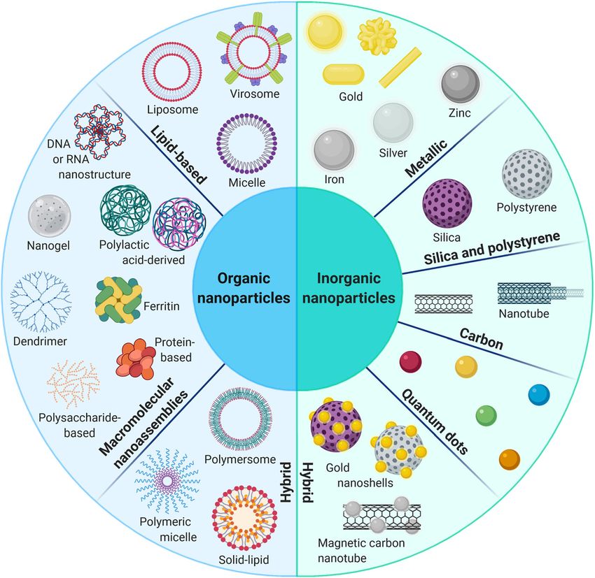
Briolay et al. Molecular Cancer        (2021) 20:55                                                                                                 Page 2 of 24

 Fig. 1 Advantages of vectorization for delivering cancer therapies. The clinical efficacy of therapeutic molecules (e.g. chemotherapeutic drugs,
 radionuclides, nucleic acids, antibodies) relies on efficient tumor delivery and limited off-targeting. Nanovectors of different natures (e.g. nanoparticles,
 extracellular vesicles, viruses) can improve the transport of these molecules in the bloodstream by increasing their solubility, half-life and bioavailability,
 and by helping the crossing of biological barriers. Tumor delivery is also enhanced by improved targeting of the tumor microenvironment, leading to
 the accumulation of the therapeutic molecules in the tumors and thus potentiating the use of combination therapies

from the biological world (e.g. bacteria, viruses, extracellu-                     radioisotopes, proteins, nucleic acids) and make them
lar vesicles) [4]. Although this adds to the complexity of                         adapted to different biological and clinical situations.
drug development, efficient vectorization appears as                               A clear understanding of the advantages and limitations of
essential to further improve the safety and efficacy of both                       each of these nanovectors (Table 1) to transport different
current and future cancer therapies. In this review, we                            therapeutic agents (Table 2) and of their evolving potential
chose to focus on nanovectors that are able to protect and                         will help developing better vectorization approaches in the
to carry therapeutic payloads to tumors following a sys-                           future.
temic injection. This does not include antibody-mediated
vectorization [5], cancer vaccination strategies [6] or                            Types of nanovectors
vectorization for imaging [7] – for instance for guided sur-                       Nanoparticles
gery – which have been reviewed elsewhere. We first                                Chemically engineered nanoparticles form a vast class of
introduce the various families of nanovectors available                            nanovectors with a wide variety of structures, sizes and
today, including the different subtypes of organic and in-                         compositions [8, 9] (Fig. 2). Among the inorganic family,
organic nanoparticles (Fig. 2), cell-derived extracellular                         the most studied are metallic (e.g. gold, iron oxide)
vesicles (EVs), virus-like particles (VLPs) (e.g. plant and                        nanoparticles that display unique optical and electronic
animal viruses, bacteriophages), oncolytic viruses (OVs)                           properties particularly favorable for biomedical imaging
and bacterial minicells (Figs. 3 and 4). These vectors                             [10]. Because of their solid core, drug functionalization
display different physical and structural properties that                          consists in surface bonding and exposes conjugated
dictate their abilities to be coupled to different types                           drugs to both degradation and exchange dynamics in the
of therapeutic molecules (e.g. chemotherapeutic drugs,                             bloodstream. Their use in therapy is also limited by a
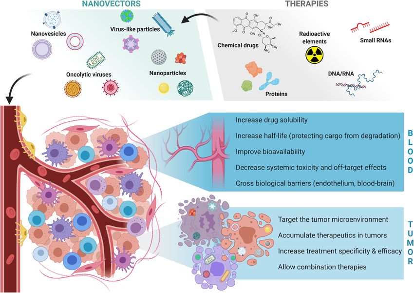
Briolay et al. Molecular Cancer        (2021) 20:55                                                                                                 Page 3 of 24

 Fig. 2 Chemically engineered nanoparticles for cancer therapy. This class of nanovectors is commonly divided between inorganic and organic
 nanoparticles. Inorganic nanoparticles (e.g. metallic, silica, carbon, quantum dots) are characterized by a high stability, a low biodegradability and intrinsic
 electronical and optical properties suitable for cancer imaging and theranostics. Because of their solid core, therapeutic molecules are generally conjugated
 on their surface and may be exposed to rapid degradation in vivo. Organic nanoparticles (e.g. lipid-based, macromolecular assemblies) exhibit a lower
 stability but a good biocompatibility and multiple possibilities of drug functionalization on their surface or their inner space. Hybrid nanoparticles combine
 the advantages of both inorganic and organic families to improve the biocompatibility and the stability of the nanovector

low biodegradability. Mesoporous inorganic nanoparticles                           their unmatched biocompatibility [8, 14, 15]. They basic-
– mostly biodegradable, silica-based – constitute an alter-                        ally consist in lipid monolayered (i.e. micelles) or bilayered
native to protect drugs within a porous structure but their                        (i.e. liposomes) nanovesicles and can vectorize a broad
safety profile still needs characterization [11, 12]. On the                       range of molecules with distinct physicochemical proper-
other hand, the organic nanoparticle family exhibits better                        ties; hydrophobic drugs can be embedded within the lipid
biocompatibility and biodegradability, making those more                           bilayer of liposomes or loaded in the core of micelles while
suitable for therapeutic applications. The first organic                           hydrophilic drugs are either entrapped in the aqueous
subfamily encompasses natural (e.g. protein- and                                   core of liposomes or displayed on their surface [16, 17].
polysaccharide-based) and synthetic (e.g. polylactic acid                          However, lipid-based nanoparticles still face several limita-
derivatives, dendrimers, fluorescent organic nanoparticles)                        tions among which a low loading capacity and a relative
macromolecular nanoassemblies (also improperly called                              lack of stability leading to drug leakage. New hybrid nano-
polymeric nanoparticles) that possess a good stability and                         particles have recently been developed to combine the
display numerous free functional groups endowing them                              respective advantages of the different subfamilies, namely
with a high loading capacity [8, 13]. These properties                             solid-lipid, hybrid polymer-lipid [18] and hybrid organic-
explain the growing interest for such nanoassemblies in                            inorganic nanoparticles [19].
cancer therapy even if the in vivo characterization of each                           Nanoparticular vectorization is traditionally believed
of their subunits remains challenging. The second organic                          to take advantage of the enhanced permeability and
subfamily contains lipid-based nanoparticles that are the                          retention (EPR) effect that results from the abnormal
most represented in preclinical and clinical studies due to                        tumor vasculature causing preferential extravasation and
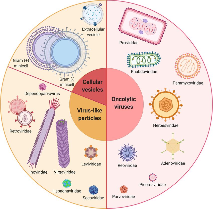
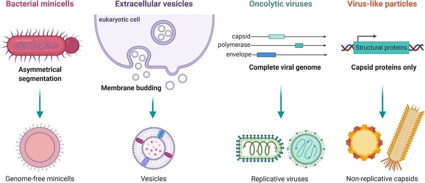
Briolay et al. Molecular Cancer     (2021) 20:55                                                                                         Page 4 of 24

 Fig. 3 Biological and bio-inspired nanovectors for cancer therapy. These nanovectors have been derived from different types of organisms and
 exhibit high biocompatibility and extensive engineering possibilities. Extracellular vesicles derive from eukaryotic cell membranes and naturally
 transport different types of biomolecules (e.g. proteins, RNA). Bacterial minicells are achromosomal 400-nanometer vesicles that can be generated
 by genetic engineering of bacteria and have been recently used to vectorize various types of therapeutic molecules. Virus-like particles are
 basically viruses (e.g. bacteriophages, plant viruses, eukaryotic viruses) stripped of their replicative capacity; they exist as naked or enveloped
 capsids and sometimes require a non-replicative template genome for their assembly. On the contrary, oncolytic viruses are tumor-specific, live-
 replicating viruses with intrinsic cytotoxic and immunoactivating properties; they can equally be naked or enveloped and may be modified by
 genetic engineering to transport therapeutic transgenes that will be expressed exclusively by infected malignant cells

increased concentration of nanoparticles in tumors                           both the tumor neovasculature and some malignant
[9, 20, 21]. Recent evidence also supports the exist-                        cells – was also reported to improve the specific
ence of an additional active uptake process through                          extravasation of nanoparticles in tumors [23, 27].
endothelial cells [22]. However, even though the                               Overall, nanoparticles act as multimodal platforms
global biodistribution of nanoparticles seems to rely                        that can be extensively engineered to improve both
mostly on these mechanisms, only actively targeted                           tumor targeting and the delivery of combined treat-
nanoparticles efficiently infiltrate tumors and enter                        ments to malignant cells; they are perfectly suited to in-
malignant cells [2, 23]. This requires coupling nano-                        crease both the half-life of therapeutic molecules in the
particles to targeting molecules – directed against                          bloodstream and their concentration in tumors while
surface antigens overexpressed on tumor cells – in-                          lowering their systemic toxicity [3]. Nevertheless, they
cluding but not limited to proteins (e.g. antibodies                         face several biological barriers that have limited their
[24, 25]), aptamers [26], peptides [27] or polysaccha-                       clinical use so far (Fig. 5). These hurdles can however
rides [28]. An emerging alternative modality of active                       be overcome by rational engineering [3, 9]. As such,
tumor targeting is the external magnetic guidance of                         clearance by the mononuclear phagocytic system is
metallic nanoparticles to promote preferential tumor                         usually diminished by functionalizing nanoparticles
extravasation [29]. Their coupling to iRGD peptides                          with non-immunogenic hydrophilic polymers such as
– recognized by the αvβ3 integrin overexpressed on                           polyethylene glycol (PEG) or zwitterionic ligands [30];
Briolay et al. Molecular Cancer      (2021) 20:55                                                                                                 Page 5 of 24

 Fig. 4 Biogenesis of biological nanovectors. Biological nanovectors are either derived from prokaryotic (bacterial minicells) or eukaryotic
 (extracellular vesicles) cells, or from viruses (oncolytic viruses and virus-like particles). Bacterial minicells are achromosomal vesicles obtained upon
 genetic engineering (deletion of the Min operon) from ectopic septation of Gram-positive or Gram-negative bacteria. Extracellular vesicles are
 produced by all eukaryotic cells by outward budding of the plasma membrane (microvesicles) or through inward budding and exocytosis
 (exosomes). Regarding viruses, whereas live-attenuated oncolytic viruses carry a complete genome and thus retain a replicative capacity specific
 for transformed cells, virus-like-particles are only constituted of structural proteins and are consequently not competent for replication

this prevents interactions with immune cells – thereby                          to study the effect of the protein corona formation
enhancing their half-life in blood – but can also de-                           around nanoparticles, as it can drastically impact their
crease internalization by tumor cells. Of note, PEG can                         stealthiness and tumor uptake [33–35]. Tunable drug
also be recognized by-anti-PEG antibodies that will im-                         release solutions have also been created to promote a
pair vectorization efficacy and may generate immune-                            specific delivery of packaged drugs exclusively in tu-
related adverse effects [31]. To improve the cellular in-                       mors. Hence, so-called ‘smart’ drug delivery systems
take of PEGylated nanoparticles within tumors, stealth                          enclose pH-, enzyme-, heat- or photo-sensitive mole-
polymer coatings that specifically dissolve in the tumor                        cules which conformations change in tumors to specif-
microenvironment (TME) have been developed [32].                                ically destabilize the nanoparticle structure and release
Stealthiness can also be improved by entrapping nano-                           the therapeutic cargo [9, 36]. To improve nanoparticle tis-
particles into cellular membranes to mimic biological                           sue penetration and diffusion through the dense extracel-
vesicles [19]. A lot of work has been performed lately                          lular matrix (ECM) in tumors, several combinations of

Table 1 Main properties of the different families of nanovectors
Nanovector      Biocompatibility Stealth       Immunogenicity Ease of     Systemic            Frequent Replicative Stability Standardized                Cost
family                                                        retargeting injection           off-targets                    production
Inorganic     Very low              Good       Low                 High           Possible    Liver,        No             Good       Adapted            $$$
nanoparticles                                                                                 spleen
Organic       Good                  Good       Low                 High           Adapted     Liver,        No             Medium Feasible               $$
nanoparticles                                                                                 spleen
Extracellular   High                High       None                Low            Adapted     Liver         No             Low        No                 $$$
vesicles
Bacterial       High                Low        Medium              Medium         Adapted     Liver         No             Medium Feasible               $
minicells
Virus-like      High                Medium Medium                  High           Adapted     Liver         No             Medium Feasible               $$$
particles
Oncolytic       High                Medium High                    Low            Possible    Depends       Yes            Low        Difficult          $$$
viruses                                                                                       on virus
                                                                                              tropism
Briolay et al. Molecular Cancer (2021) 20:55 Page 6 of 24
Table 2 Suitability of the different families of nanovectors for the vectorization of anti-cancer therapeutics
Nanovector family Chemotherapy Radiotherapy Gene therapy RNA interference TME modification Immunotherapy
Nanoparticles +++ ++ + + + +
Extracellular vesicles + - + ++ NT +
*
Bacterial minicells ++ NT NT ++ NT NT
Virus-like particles + NT +++ ++ NT +
Oncolytic viruses - + +++ ++ +++ +++
+++: optimal; ++: adapted; +: feasible; -: not adapted.
NT: never tested, TME: tumor microenvironment.
*
expected to be similar to RNA interference
ECM-modifying molecules and nanoparticles are also cur- Biological and bio-inspired nanovesicles
rently under investigation [37]. Finally, a major pitfall for The biological world provides attractive alternatives to
vectorization with nanoparticles is their trapping in endo- artificial lipid-based nanoparticles. Extracellular vesicles
lysosomes after endocytosis, which exposes the thera- (EVs) are naturally occurring vesicles produced by
peutic cargo to degradation. Available solutions include eukaryotic cells and play important roles in intercellular
coupling nanoparticles to endosomal escape domains or communications [39]. They naturally package a broad
proton sponges to destabilize endosomes and promote range of cargos, from nucleic acids to proteins or lipids.
drug release toward the cytoplasm [38]. There are two main types of EVs at the nanometer scale,
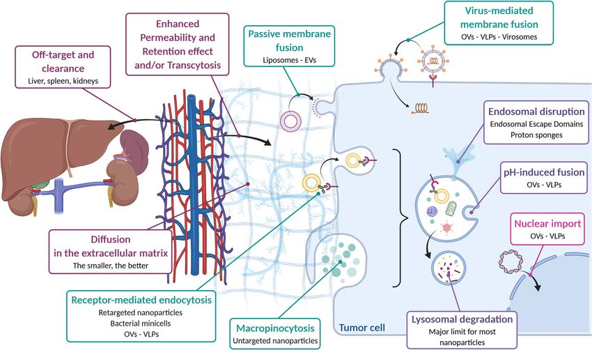
Fig. 5 From the blood to the tumor cell: the difficult journey of nanovectors. Systemically injected nanovectors face several biological barriers to reach
the tumor microenvironment and exert their therapeutic effect in malignant cells. First, filtering organs such as the liver (for nanovectors > 5 nm) or
the kidneys (for nanovectors < 5 nm) eliminate an important fraction of the injected nanovectors. Nanovectors then extravasate from the bloodstream
to the tumor either because of an increased vascular permeability (Enhanced Permeability and Retention effect) or by active transcytosis through
endothelial cells. The nanovectors have to overcome the interstitial pressure and to diffuse in the extracellular matrix to reach tumor cells. This can be
partially improved by active targeting strategies through nanovector engineering. Once reaching the cancer cells, nanovectors can be internalized by
several mechanisms (e.g. passive or virus-mediated fusion, endocytosis, macropinocytosis) depending on their origin, size, composition and
functionalization. The final difficulty consists in delivering the therapeutic cargo in the appropriate cellular compartment – generally the cytoplasm – to
achieve optimal therapeutic efficacy. This usually requires further vector engineering (e.g. endosomal escape domains, pH-sensitive moieties), in
particular for non-biological nanoparticles.EVs: Extracellular Vesicles; VLPs: Virus-Like Particles; OVs: Oncolytic VirusesBriolay et al. Molecular Cancer (2021) 20:55 Page 7 of 24 namely microvesicles (50 nm to 1 μm) and exosomes with different structures, charges and solubilities in an (50 to 150 nm) that differ by their biogenesis and easier way than with lipid-based nanoparticles [55, 59]. composition. Microvesicles directly bud outward of the They display a high loading capacity – up to 1,000 times plasma membrane while exosomes are generated from higher than liposomes – following simple drug import- the inward budding of endosomal membranes and are ation through the outer membrane via the non-specific released in the extracellular environment by exocytosis FadL or OmpW channels. To confirm their interest in (Fig. 4). Because of their low immunogenicity and their cancer therapy [60, 61], comprehensive studies are still efficient intake by cells [40], EVs have been investigated needed to better characterize their properties, among as drug nanocarriers for cancer therapy [41]. Therapeutic which their immunogenic profile. Their safety however drugs can be loaded either directly into pre-formed pleads for further developments, as was demonstrated in vesicles or through modification of the EV-producing cells three recent phase I clinical trials that tested Epidermal (e.g. drug exposure, transfection) to entrap the cargo into Growth Factor Receptor (EGFR)-targeted minicells loaded EVs during their formation [42, 43]. Although still contro- with either paclitaxel [62], doxorubicin [63] or miRNA versial [44], EVs are suspected to possess inherent target- mimics [64] in patients with end-stage solid cancers, ing capacities depending on their progenitor cell type [45]; glioblastoma or mesothelioma, respectively. tumor cell-derived exosomes thus appear to preferentially home to their cell types of origin in vitro compared with Virus-like particles untargeted liposomes [46]. As for liposomes, the surface Viruses are extensively studied in therapeutic vectorization of EVs can be modified with targeting molecules or PEG due to their active cell entry mechanisms, biocompatibility [47]. Nevertheless, the lack of content standardization and and well-characterized structures. Virus-like particles of large-scale production methods still hinders their clin- (VLPs) were developed to mimic animal, plant or bacteria ical use; the development of EV-like nanovesicles, which viruses without retaining the ability to replicate in human are basically liposomes enriched with membrane proteins cells [65] (Figs. 3 and 4). They are viral capsids with an to enhance cellular intake, is expected to help overcoming icosahedral or filamentous structure composed of self- some of these limitations [48]. A derivative from this idea assembled proteins. Their diameters range from 25 (e.g. are “virosomes” (150 to 500 nm) that are composed of a parvoviridae) to several hundred (e.g. herpesviridae) nano- synthetic lipid bilayer containing viral or parasitic meters and they can contain a non-infectious genome fusogenic glycoproteins [49, 50]. Those take advantage of composed of single- or double-stranded RNA or DNA the ability of viral envelopes to recognize the targeted cells [66]. Icosahedral VLPs can be used as genome-free parti- and to promote direct fusion with the plasma membrane, cles such as the ones derived from the MS2 bacteriophage hence skipping the potential degradation of the encapsu- [67], which spontaneously assemble during protein lated cargo into late endosomes after endocytosis (Fig. 5). production in bacteria, or from the cowpea mosaic virus Other strategies use cell-derived nanovesicles to camou- (CPMV) [68]. On the contrary, filamentous VLPs derived flage other types of vectors (e.g. nanoparticles, viruses) to from plant viruses and bacteriophages generally require a take advantage of their intrinsic properties and to escape template genome for capsid proteins to assemble around it neutralizing antibodies [51–54]. and form a rigid or flexible tube which length and width The trend to exploit bio-derived nanostructures for are determined by the capsid protein and the genome size. cancer therapy extends to different families of patho- In addition, some viruses (e.g. retroviridae) present an gens. Bacterial minicells (200 to 400 nm) are achromoso- envelope composed of an external lipidic membrane mal vesicles produced by bacteria upon ectopic septation acquired while budding from the host cell surface [69]. As [55] (Fig. 4), an asymmetric division obtained by deleting VLPs contain non-self proteins and potential pathogen- the Min operon [56]. Minicells can be produced from associated molecular patterns, they can be immunogenic Gram-positive and Gram-negative bacteria and contain and were mostly assessed as anti-cancer immuno- all the molecular components of the parent cell except stimulatory treatments [70]. Their use as vaccines showed for the chromosome. Because of their vesicular struc- a good safety profile that makes them suitable for future ture, they are an alternative to lipid-based nanoparticles use as nanovectors. Nevertheless, repeated treatments for cancer therapy (Fig. 3). Although Gram-positive could promote the generation of antibodies and clearance minicells are negative for lipopolysaccharides (LPS) and by immune cells resulting in decreased tumor delivery. may be ultimately more adapted for clinical use, most Capsid PEGylation or elimination of immuno-dominant studies have used Gram-negative minicells that can be epitopes can however limit these issues [71]. easily redirected to cancer-specific receptors (e.g. HER2/ Because of their viral nature, VLPs are perfectly neu) with bispecific antibodies targeting both the LPS adapted to the delivery of therapeutic nucleic acids [72] O-antigen on minicells and a tumor marker [57, 58]. but empty capsids can also be modified to transport Bacterial minicells can package a wide variety of molecules other types of molecules. As such, the fixed structures of
Briolay et al. Molecular Cancer   (2021) 20:55                                                                    Page 8 of 24

VLPs allow for extensive genetic and chemical engineer-        a diversity of immune cell types involved in the anti-
ing. Examples include tobacco mosaic virus VLPs that           tumor responses [86, 92]. After two decades, more than a
can be loaded by simple infusion and ionic interactions        hundred trials and few regulatory approvals for clinical
with their inner surface [73], the hepatitis B virus capsid    use [93–95], they have demonstrated a very good safety
that can be disassembled and re-assembled to capture a         profile but a somewhat modest therapeutic efficacy in
compound [74], or the functionalization of MS2 VLPs            humans.
by inserting genetically a cystein residue in the capsid          To improve their intrinsic anti-cancer properties, OVs
[75]. Interestingly, filamentous VLPs show a natural           are commonly armed to vectorize therapeutic transgenes
biodistribution to tumors after systemic injection, which      that will be expressed by infected malignant cells in the
could be mediated by their physical behavior in the            TME, thereby making them bona fide nanovectors [96].
tumor microvasculature [76, 77]. Non-human virus-              Viruses have evolved to deliver efficiently their genome
based VLPs did not evolve to recognize human cell re-          in host cells and are thus perfectly designed to vectorize
ceptors; they produce less off-target effects but require      nucleic acids (Fig. 5). The first OV to be approved by
genetic or chemical retargeting to malignant cells. Com-       the US and EU regulatory agencies in 2015 was the
mon modifications involve the retargeting of VLPs with         recombinant herpesvirus Talimogene laherparepvec (T-
cancer-specific peptides [78], aptamers [75] or other          VEC) that encodes the Granulocyte-Macrophage Colony-
molecules [72, 79], or the pseudotyping of enveloped           Stimulating Factor to enhance its immunostimulatory
VLPs with exogenous proteins. Similarly, twelve                properties [94, 97]. The transgene capacity of viruses is
serotypes of adeno-associated viruses (AAVs) have been         however limited by the fitness cost – the longer the gen-
identified so far [80] and could be used to target differ-     ome, the longer it takes to replicate – and the size limit of
ent types of cancers. In addition, VLPs from plant or          the viral particle; DNA viruses generally exhibit a higher
bacteria viruses cannot easily escape human endo-              transgene capacity than RNA viruses. OV replication cap-
lysosomes and display lower transfer efficacy, even after      acity allows both spreading of the transgene in the tumor
retargeting [81–84]. Strategies similar to the ones used       and its sustained expression over time [98]. As with VLPs,
with nanoparticles for endosomal escape and cargo              surface molecular coupling is theoretically possible – es-
delivery are being tested to overcome these limitations        pecially for non-enveloped viruses – to enable intracellular
[78]. On the opposite, VLPs derived from human patho-          delivery of drugs in specific cells.
gens benefit from coevolution to achieve efficient gene           The current standard for OV treatment is intratumoral
transfer inside human cancer cells (Fig. 5).                   injection with the limit that only reachable tumors can be
                                                               treated, but recent evidence of viral replication in tumors
Oncolytic viruses                                              following intravenous administration in patients have been
Contrary to VLPs for which the non-replicative nature is       reported [99–103]. Despite pre-existing immunity having
a major determinant of their clinical safety and inter-        no measurable effect on the therapeutic outcome after
mediate immunogenicity, oncolytic viruses (OVs) display        intratumoral injection, innate and adaptive immune re-
all the properties of natural viruses except that their        sponses against circulating viruses may restrict their efficacy
replication is restricted to malignant cells [85] (Fig. 4).    after intravenous administration [104, 105]. PEGylation of
The diversity of OVs has been reviewed extensively             OVs [106, 107] or switching OV species during the course
elsewhere [86] and is summarized in Fig. 3. OVs are            of treatment [108, 109] can improve stealthiness and
either naturally attenuated viral strains or genetically       enhance treatment efficacy. Enveloped viruses can also be
engineered viruses that harness cancer hallmarks such as       pseudotyped with different viral envelops [110–112], while
altered metabolism, immunosuppression or resistance to         changing the serotype of non-enveloped viruses could
cell death that make tumors more sensitive than healthy        evade the immune response [113–115]. Finally, the titration
tissues to viral infections. Tumor cells also commonly         of OVs by healthy cells after non-specific entry – distinct
overexpress surface proteins that are used by some             from their tumor-specific replication and killing – can be
viruses for cell entry [87, 88]. For many oncolytic RNA        answered by retargeting OVs to tumor-specific surface anti-
viruses, tumor specificity mainly depends on defects in        gens through genetic engineering. Advances made in the
the innate antiviral pathways commonly acquired by             field of nanoparticles for chemical modifications are also
malignant cells during tumor evolution [89, 90], while         expected to lead to alternative solutions [107].
DNA viruses can be modified with tumor-specific
promoters [91]. Contrary to other nanovectors, the tumor       Applications in cancer therapy
specificity of OVs thus mostly relies on post-entry restric-   Chemotherapy
tion rather than selective entry through specific surface      Cancer chemotherapeutics are a large family of chemical
markers. They also exhibit therapeutic properties on their     drugs [116] that affect highly proliferating malignant
own as they can both directly kill tumor cells and activate    cells and exhibit diverse modes of action from cell cycle
Briolay et al. Molecular Cancer (2021) 20:55 Page 9 of 24
arrest to cell death induction and epigenetic modulation. Other types of nanovectors are currently studied to
These molecules often lack tumor specificity and healthy transport and deliver chemical drugs to tumors (Table
proliferative cells are frequently impacted, thereby 2). The characterization of VLPs at the atomic level
causing different debilitating symptoms. Consequently, allows for precise chemical coupling strategies similar
vectorization of chemotherapeutics is critical to improve to the ones used for nanoparticles. For example,
their tumor specificity and diminish side toxicities. Here, doxorubicin coupling to Physalis Mottle virus icosahe-
we present an overview of how the different families of dral VLPs [81] or to truncated hepatitis B virus core
nanovectors can help bypassing the major limitations of antigen (tHBcAg) VLPs [138] improved both its cellu-
chemotherapies, including their poor aqueous solubility, lar uptake and cytotoxicity against malignant cells.
their lack of tumor specificity and the acquisition of re- Doxorubicin and mitoxantrone were also passively
sistances. The advantageous physical properties of some loaded into CPMV [139] and filamentous plant vi-
nanovectors that can be exploited in combinatorial strat- ruses VLPs [140–142] by exploiting for the latter the
egies with chemotherapies are also discussed. negative charges of the inner side of the particles.
Simple dissociation/association of tHBcAg allows for
passive dual loading of polyacrylic acid (PAA) along
Solving drug insolubility with doxorubicin that will be released at low pH
Chemical drugs for cancer treatment vary widely by their when no longer retained by protonated PAA [79].
structures, charges and solubilities that can limit their EVs on their part display similar vectorization abilities
clinical use, an illustrative example being the high as liposomes. They were shown for instance to deliver
hydrophobicity of taxanes [117]. The nanomedicine doxorubicin [143] or paclitaxel [144] in vitro to breast
field however provides numerous solutions for drug or prostate cancer cells, respectively, or paclitaxel to
vectorization whether they are hydrophobic (e.g. lung cancer cells after systemic administration in mice
paclitaxel, cisplatin) or amphipathic (e.g. doxorubicin, [145]. Packaging of decitabine in erythro-magneto-
5-fluorouracil). As explained above, the diversity of hemagglutinin nanovesicles showed a specific delivery
chemically engineered nanoparticles with variable to prostate cancer xenografts under in vivo magnetic
loading and functionalization possibilities makes them guidance and a significant tumor mass reduction at a
the most suitable for vectorizing chemotherapeutic lower dose than with free decitabine [146]. Among
drugs [9, 118] (Table 2). Hydrophilic drugs can be eas- the bio-inspired nanovectors, bacterial minicells may
ily encapsulated inside liposomes, adsorbed in pores of be the more promising as they can incorporate a wide
silica nanoparticles or conjugated on metallic or poly- variety of chemotherapeutic agents without drug ef-
meric nanoparticles using reactive hydroxyl, carboxyl, flux up to several days [55]. Their encouraging early
amino or thiol groups. Hydrophobic molecules are clinical results in two phase I clinical trials that used
commonly loaded in micelles or solid-lipid nanoparti- EGFR-targeted bacterial minicells containing either
cles or inserted in the lipid bilayer of liposomes. Nano- doxorubicin or paclitaxel to treat patients with ad-
particles are also used to vectorize hydrophobic vanced solid tumors [62, 63] however need to be
epigenetic modulators (e.g. inhibitors of histone deace- confirmed.
tylases or DNA methyltransferases) to improve their
pharmacokinetics and therapeutic efficacy [119–122]. Improving tumor specificity
Macromolecular nanoassemblies and lipid-based nano- The lack of tumor specificity for chemotherapies causes
particles have been used to vectorize almost all types off-target effects and limits clinical efficacy by decreasing
of chemotherapeutics and several nanomedications drug concentration in tumors. For instance, doxorubicin
have either already been approved by the FDA for can- displays elevated hematological and cardiac toxicities as
cer treatment or are currently evaluated in clinical tri- a free molecule [147]. It has been vectorized as early as
als [8, 123] (Table 3). It is interesting to note that the 1990s in the first FDA-approved nanodrug Doxil®,
cancers with very different profiles, from end-stage which is currently approved for the treatment of ovarian
solid tumors to hematological malignancies, can be eli- cancer, multiple myeloma, metastatic breast cancer and
gible to nanovectorization of chemotherapeutics. As an Kaposi’s sarcoma. Doxil® is composed of doxorubicin en-
example, the nab-paclitaxel formulation (Abraxane®) – capsulated in untargeted, PEGylated liposomes that en-
composed of paclitaxel fused to human albumin nano- able a high concentration of doxorubicin in tumors
particles – has demonstrated improved safety and effi- correlated with a higher tolerability compared to free
cacy compared to free paclitaxel [136] and is approved doxorubicin [148]. This formulation was followed by
against non-small cell lung cancer, metastatic pancre- many other combinations of chemotherapeutic drugs
atic cancer and as a second-line treatment for meta- with numerous types of nanoparticles [124]. As with the
static breast cancers [137]. Doxil® liposomal formulation, their tumor specificityTable 3 Representative examples of the advancement of nanovectors in cancer therapy
Nanovector Therapy Drug administration Phase Cancer types Route of References
family administration
Organic Chemotherapy PEGylated liposomal doxorubicin Approved Ovary, Kaposi’s sarcoma, Intravenous [137]
nanoparticles (Doxil®/Caelyx®) (1995) multiple myeloma
Non-PEGylated liposomal doxorubicin Approved Breast Intravenous
(Myocet®) (2000)
Albumin particle-bound paclitaxel Approved NSCLC, breast, pancreas Intravenous
Briolay et al. Molecular Cancer
(Abraxane®) (2005)
PEGylated liposomal irinotecan Approved Pancreas Intravenous
(Onivyde®/MM-398®) (2015)
Non-PEGylated liposomal cytarabine:daunorubicin Approved AML Intravenous
(VYXEOS®/CPX-351®) (2017)
(2021) 20:55
Gene therapy TR-targeted liposomes encapsulating a I/II Pediatric solid tumors, Intravenous NCT02354547, NCT02340117,
p53-encoding plasmid (SGT-53®) glioblastoma, pancreas NCT02340156
RNA Lipid nanoparticles encapsulating interfering I/II Solid tumors, Edwing’s Intravenous [247]
interference RNAs sarcoma, liver, AML
TME Various NPs for CAFs, TAMs, ECs, ECM suppression Preclinical Various cancer models Mostly intravenous [291]
modification or normalization
Immunotherapy Vectorization of various immunomodulators Preclinical Various cancer models Mostly intravenous [291]
Inorganic Hyperthermia Minosilane-coated iron oxide nanoparticles Approved Glioblastoma Intratumoral [307]
nanoparticles (Nanotherm®) (2010)
Radiotherapy Hafnium oxide nanoparticles (NBTXR3®/Hensify®) Approved Squamous cell carcinoma Intratumoral [137]
(2019)
RNA siRNAs adsorbed on gold nanoparticles I Glioblastoma Intravenous [247]
interference
Bacterial minicells Chemotherapy EGFR-targeted, doxorubicin-loaded minicells I/II Glioblastoma Intravenous [63]
RNA EGFR-targeted minicells containing a miRNA I Mesothelioma, NSCLC Intravenous [64]
interference mimics cocktail
Extracellular vesicles Chemotherapy Tumor-derived microvesicles packaging II Lung cancer Intravenous NCT02657460
methotrexate
Gene therapy Tumor-derived exosomes loaded with Proof-of- Heterotopic ovarian cancer model Intravenous [308]
CRISPR-Cas9 against PARP1 concept
RNA MSC-derived exosomes loaded with I Metastatic prostate cancer Intravenous NCT03608631
interference anti-KrasG12D siRNAs
Virus-like particles Chemotherapy Tobacco Mosaic Virus carrying phenanthriplatin Preclinical Heterotopic breast cancer model Intravenous [73]
Gene therapy TP53-encoding non-replicating adenovirus Diverse Solid cancers Mostly intratumoral [208]
M13 phage encoding HSV-TK Preclinical Orthotopic glioblastoma model Intravenous [309]
RNA delivery MS2-derived VLPs carrying siRNAs Proof-of- Hepatocellular carcinoma cell line NA [310]
concept
Page 10 of 24Table 3 Representative examples of the advancement of nanovectors in cancer therapy (Continued)
Nanovector Therapy Drug administration Phase Cancer types Route of References
family administration
Oncolytic viruses Chemotherapy HSV-TK-encoding adenovirus II Triple-negative breast cancer, NSCLC, Intratumoral NCT03004183, [311]
prostate
HSV-TK-encoding vaccinia virus II Solid tumors Intravenous NCT04226066
Radiotherapy NIS-encoding measles virus II Multiple myeloma Intravenous NCT02192775
II Ovarian, fallopian and peritoneal Intraperitoneal NCT02364713
cancers
Briolay et al. Molecular Cancer
Gene therapy TP53-encoding replicating viruses Preclinical Many solid cancers models Intravenous / [208]
Intratumoral
RNA Oncogene silencing with small Preclinical Many solid cancer models NA [312, 313]
interference RNAs-encoding Adenovirus and HSV
TME Hyaluronidase-expressing adenovirus Preclinical Orthotopic glioblastoma model Intratumoral [134]
(2021) 20:55
modification
Immunotherapy GM-CSF-encoding herpes simplex virus Approved Melanoma Intratumoral [94, 97]
(Talimogene laherparepvec) (2015)
AML acute myeloid leukemia, CAF cancer-associated fibroblast, EC endothelial cell, ECM extracellular matrix, EGFR epidermal growth factor receptor, HSV-TK herpesvirus thymidine kinase, NP nanoparticle, NSCLC non-
small cell lung carcinoma, TAM tumor-associated macrophage, TR transferrin receptor, VLP virus-like particle
Page 11 of 24Briolay et al. Molecular Cancer (2021) 20:55 Page 12 of 24
mostly relied on passive targeting due to destabilized and limit efflux, thereby enhancing drug concentration in
tumor vasculature and the resultant EPR effect. Based tumor cells. They can also carry several drugs at the same
on a similar idea, the natural tumor distribution of fila- time to strike cancer cells on different fronts simultan-
mentous VLPs [77, 149] can also be exploited for this eously and prevent therapeutic escape [157]. Such strat-
purpose; PEGylated Potato Virus X (PVX) VLPs pas- egies can combine several chemotherapies [152] or
sively loaded with doxorubicin were indeed shown to different types of treatments such as a combination of a
elicit a better control of breast cancer xenografts in im- chemotherapeutic drug with a siRNA [158]. Doxorubicin-
munodeficient mice than doxorubicin alone [140]. How- coated, multifunctional mesoporous silica nanoparticles
ever, a combination of PVX and doxorubicin was more containing a siRNA against the P-glycoprotein (Pgp) drug
effective than doxorubicin-loaded PVX in an immuno- exporter showed targeted Pgp knockdown and a synergis-
competent melanoma model [141], suggesting that VLPs tic inhibition of resistant breast tumor growth in preclin-
elicit an adjuvant anti-tumor immune response that par- ical models [159]. A similar approach used sequentially (i)
ticipates in the therapeutic effect and pleading for the CD33- or EGFR-targeted bacterial minicells containing a
use of immunocompetent animal models for future plasmid coding for shRNAs against MDR pumps and (ii)
evaluations. chemotherapies [160]; mice bearing drug-resistant colo-
Current studies mostly focus on actively targeted rectal, breast or uterine tumors were efficiently treated
nanodrug formulations to enhance interactions of the without toxicity as a thousand-fold less drug and shRNA
nanoparticles with malignant cells after having reached were used compared to conventional systemic treatment.
the TME [23, 24, 27]. Several strategies have demon- Another way to circumvent tumor resistance is to use
strated increased drug concentration in tumors and highly cytotoxic compounds – such as the PNU-159682
enhanced therapeutic efficacy compared with the metabolite [161] – that cannot be injected systemically be-
corresponding free molecules or untargeted nanovectors cause of their high toxicity. Systemic vectorization of this
[23, 150]. In a preclinical study, paclitaxel-loaded nano- drug in EGFR-targeted bacterial minicells showed signifi-
capsules constituted of a lipid core surrounded by a cant tumor reduction and immune activation with no side
surfactant were targeted to the altered tumor vascular effects in immunocompetent breast and colorectal murine
endothelium with an iRGD peptide [151]. The authors models but also lung and colorectal human cancer
demonstrated that the targeted nanoparticles concen- xenografts [162].
trated in hepatic tumors, induced specific cytotoxicity
and were better tolerated than non-targeted nanoparti- Exploiting intrinsic physical properties
cles. Another recent study showed that hybrid solid-lipid Some chemically engineered nanoparticle families have
nanoparticles decorated with folic acid can significantly intrinsic physical properties that make them suitable for
increase the concentration of carboplatin and paclitaxel combined therapies. As such, gold nanoparticles can be
in tumors cells in a murine cervical cancer model [152]. used for photothermal therapy, which consists in a local
EGFR-targeted, doxorubicin-containing bacterial mini- vibrational heat generation through the absorption of
cells were demonstrated to rapidly locate in spontaneous specific wavelengths of light [163]. Super Paramagnetic
gliomas in dogs, a tumor usually difficult to reach be- Iron Nanoparticles (SPIONs) on the other hand can be
cause of the blood-brain barrier [60]. Another approach used for hyperthermia, a local heat generation under a
for active tumor delivery is to target the hypoxic center magnetic field [164]. Those two phenomena have dem-
and acidic microenvironment of tumors, in particular onstrated a moderate therapeutic efficacy on their own
using the pH (low) insertion peptide (pHLIP) [153]. An but can sensitize cancer cells to chemotherapies loaded
example for this strategy is the use of doxorubicin- in the same nanoparticles [165]. Indeed, hyperthermia
loaded bacterial minicells with a pHLIP added to their and photothermia inhibit the repair of DNA lesions (e.g.
membrane, which successfully invaded the necrotic and double-strand breaks) generated by chemotherapy or
hypoxic regions of orthotopic murine breast cancers and radiotherapy [166]. Several clinical trials involving the
achieved a significant tumor reduction compared to both use of hyperthermia as adjuvant for chemotherapy are
free drug and untargeted minicells [154]. ongoing [167]. An example is the use of a near-infrared-
responsive polypeptide nanocomposites charged with
Fighting resistance doxorubicin and capable of heat generation and heat-
Cancer cells commonly develop resistance against sensitive nitric oxide (NO) gas delivery [168]. This
chemotherapies, for instance by acquiring a multidrug combination of photothermia, NO gas therapy and
resistance (MDR) phenotype. This can result from the chemotherapy achieved complete breast tumor regres-
expression of ATP-dependent transporters that promote sion in mice after a single near-infrared irradiation.
the efflux of drugs outside the cell to escape death induc- Hyperthermia can also be used to release chemothera-
tion [155, 156]. Nanovectors enable drug immobilization peutics enclosed in hybrid delivery systems constitutedBriolay et al. Molecular Cancer (2021) 20:55 Page 13 of 24
of nanoparticles associated with thermosensitive improved treatment efficacy. However, the clinical trans-
molecules [169]. Regarding epigenetic modulation, some lation of these metallic nanoparticles remains challen-
studies suggest that metallic and silica nanoparticles ging because of both their tendency to aggregate after
could directly induce modifications of DNA methylation systemic injection and their long-term toxicity due to
or of histone acetylation and disrupt miRNA expression liver accumulation. An alternative are chemical ROS-
[170, 171], but the significance of these modifications in generating photosensitizers that can be coupled to a
the context of cancer treatment is still to be investigated. wide variety of biocompatible nanoparticles for PDT
The nanovectorization of chemotherapeutic drugs has [177, 178]. Interestingly, some chemical radiosensitizers
been historically dominated by the use of organic are also able to self-assemble to generate nanostructures
nanoparticles (Table 2), supported by their unmatched by themselves [179]. Upconverting nanoparticles were
diversity of structures and compositions (Fig. 2). This recently modified to assemble with a photosensitizer
led to different clinical successes resulting in several in vivo by click chemistry after systemic injection [180].
drug approvals (Table 3). However, the more recent ad- These nanoparticles are able to convert low energy near-
vances in vesicular nanovectors (e.g. bacterial minicells, infrared light into high energy photons that activate the
EVs), provide new solutions with enhanced biocompati- photosensitizer to generate ROS and achieved inhibition
bility (Table 1) that may advantageously replace syn- of tumor growth in an ectopic breast cancer model. A
thetic nanoparticles in some clinical contexts. Studies on recent study used EVs purified from mouse blood and
VLPs are at an earlier stage of development but also surface-loaded with the photosensitizer protoporphyrin
demonstrated interesting properties in preclinical experi- IX (PplX) in a two-stage irradiation protocol to
ments. In the end, hybrid vectorization systems incorp- efficiently deliver PplX and induce apoptosis by PDT in
orating both synthetic and biological moieties may a breast tumor model [181]. The porphyrin photosensi-
constitute a rational compromise between efficacy, tizer has also been effectively vectorized with M13 fila-
biocompatibility and standardized manufacturing even if mentous phage VLPs retargeted to mammary cancer
complex designs may generate additional difficulties for cells by a specific peptide displayed on the pVIII coat
clinical development. protein and demonstrated efficient cancer cell targeting
and sensitization to PDT [182]. The lack of oxygen in
Radiotherapy the tumor hypoxic core can lead to radioresistance,
Half the cancer patients receive radiotherapy – which which can be bypassed by developing nanoparticles with
exploits the low resistance of tumor cells to radiation- O2-elevating abilities or nano-radiosensitizers with
induced DNA damages – during their course of treat- diminished oxygen dependence [183]. As an example,
ment [172]. Overexposure of healthy cells to radiations mesoporous manganese dioxide nanoparticles are able
leads to radiotherapy-related toxicities that could be to catalyze O2 production to actively reverse hypoxia in
partially addressed using appropriate vectorization tumors. These nanoparticles were loaded with the pho-
strategies. For external-beam radiotherapy [173] – or for tosensitizer acridin orange and exhibited enhanced
related photodynamic therapy (PDT) that uses non- radiotherapy efficacy both in vitro and in vivo in a lung
ionizing wavelengths [163] – nanovectors can sensitize cancer xenograft model [184]. Hypoxia-reverting lipo-
tumors to radiations. For internal radiotherapy, nanomedi- somes [185], macromolecular nanoassemblies [186, 187]
cine is an elegant solution to deliver specifically radioele- and other types of nanoparticles [177] have also been
ments to tumors and an alternative to the use of radiolabeled used for their photosensitizing properties.
antibodies in radioimmunotherapy approaches [174]. The radiosensitizer family also encompasses all mole-
cules able to enhance tumor cell sensitivity to radiation
a. Radiosensitization effects by interfering with essential cellular pathways like
DNA repair, apoptosis induction or cell cycle progres-
Radiations not only cause direct damages to biomole- sion. As such, chemotherapeutics are used as radiosensi-
cules but also generate reactive oxygen species (ROS). tizers at the clinical level [175] and their loading on
This phenomenon can be enhanced in tumors by the chemically engineered nanoparticles have demonstrated
vectorization of radiosensitizing molecules that increase radiosensitizing effects [185, 188, 189]. As for chemo-
either ROS production in response to ionizing beams or therapy, SPIONs and gold nanoparticles alone or within
malignant cell sensitivity to both direct and indirect a bigger organic nanoparticle can also mediate tumor
radiation effects [175]. Gold nanoparticles (AuNPs) are radiosensitization through inhibition of DNA repair
well-characterized for their radiosensitizing properties mechanisms by hyperthermia or photothermia, respect-
[176]; their concentration in tumors increases the dose ively [166]. DNA viruses are capable of impairing the
delivered locally during radiotherapy, resulting in ROS DNA damage response [190] and some OVs (e.g. adeno-
production, DNA repair machinery impairment and viridae) naturally downregulate key proteins involved inBriolay et al. Molecular Cancer (2021) 20:55 Page 14 of 24
the response to radiation-induced DNA damages [191], Delivery of nucleic acids
which makes them intrinsically radiosensitizing [192]. Malignant transformation results from gene alterations
SiRNA-mediated gene silencing is another strategy to (e.g. deletions, amplifications, mutations, translocations,
target genes involved in the cellular response to ionizing epigenetic or viral dysregulations) that displace the equi-
radiations [175]. As discussed below, OVs and VLPs are librium between oncogene and tumor-suppressor gene
useful tools for such small RNA vectorization, an ex- expression. These alterations can be corrected or com-
ample being an adenovirus encoding a shRNA against pensated using nucleic acids (DNA or RNA) for gene
the DNA-dependent protein kinase DNA damage editing (over-expression or knock-out), direct induction
response protein for local enhancement of radiotherapy of cell death by expression of toxic genes or by modulat-
in a human colorectal cancer xenograft model [193]. ing gene expression. As free nucleic acids are rapidly
degraded in the bloodstream and do not cross cell
b. Internal radiotherapy membranes, clinical translation of cancer gene therapy
requires proper vectorization [208]. Viruses are par-
Radionuclides have been vectorized for several years ticularly suited for this as they are naturally designed
with various nanovectors like VLPs [194–196], EVs to deliver genes in targeted cells (Table 2). Transgenic
[197], nanoparticles [198, 199] or an oncolytic adeno- viruses are also relatively simple to generate and they
virus [200] for cancer imaging, but for VLPs or EVs this ensure a high level of transgene expression. Many
has yet to be studied in therapeutic protocols. High- studies were conducted with retrovirus-like particles
energy, short-range alpha-emitters have been conjugated (RLPs) [209], non-replicative adenoviruses [210] and
to various types of chemically engineered nanoparticles AAVs [211], whereas other VLPs used for both their
with good therapeutic results but a large majority of capacity to package DNA and their easy retargeting
radionuclides currently used in cancer therapy are low- achieved lower transduction efficacy [68, 82, 212].
energy beta-emitters with a longer path length [201]. Despite several limitations – the main one being the
Iodine 131 ( [143]I) is the most common nanoparticle- cytoplasmic delivery of cargos initially addressed to
coupled radionuclide reported in the literature. Recent the nucleus – nanoparticles (mainly lipid-based) have
examples include PEGylated, nuclei-targeted [143]I- been extensively used for nucleic acid delivery [213–
AuNPs tested in a colorectal cancer model [202] and 215]. Some strategies are developed to increase
[143]I-labeled, human serum albumin-bound manganese nanoparticle-mediated gene expression in tumor cells
dioxide nanoparticles that were capable of significantly [216], for instance by using nuclear localization sig-
inhibiting tumor growth in a breast cancer model with a nals (NLS) or by vectorizing messenger RNAs [217].
potentiating effect of MnO2 on radiotherapy efficacy
[203]. In another study, treatment with PEGylated lipo-
somes enclosing an [143]I-albumin core led to subcuta- Gene therapy
neous breast tumor shrinkage when co-administered The most frequent genetic alterations in cancer being
either with liposomes containing a photosensitizer or p53 mutations, most gene therapies consist in vectoriz-
with an anti-PD-L1 antibody [204]. In a very different ing a wild-type TP53. Restoring wild-type p53 functions
strategy, OVs coding for the human sodium-iodine sym- triggers cell death specifically in highly-dividing tumor
porter (NIS) have been used to enhance the specific in- cells exhibiting genome instability. An example of a
take of [143]I in OV-infected tumor cells [99, 205–207]; nanovector exploiting this mechanism is Gendicin, a
OV-NIS are injected several days before [143]I and in- p53-encoding adenoviral vector that was the first-in-class
directly mediate the vectorization of the radioelement to gene therapy treatment for head and neck cancer
tumors neo-expressing NIS. approved by the China Food and Drug Administration in
As for chemotherapy, the different subfamilies of 2003 [218]. While many years of clinical use demonstrated
nanoparticles have been massively investigated to im- its safety, its efficacy remains limited. However, the co-
prove the efficacy of radiotherapy, but the low biocom- vectorization of other tumor suppressors (e.g. ING4,
patibility and biodegradability of inorganic nanoparticles PTEN) in the same vector demonstrated synergistic
called for the development of alternatives. Successful de- efficacy [83]. The enhanced vectorization potential and
livery of radiosensitizing molecules was achieved with intrinsic tumor cytotoxicity of OVs were also exploited to
organic nanoparticles and bio-inspired vectors such as transiently express tumor suppressors at high levels but
EVs, while engineered VLPs can be chemically coupled still lack clinical assessment [129]. Regarding nanoparti-
to radionuclides. Viruses and other bio-derived vectors cles, liposomes containing p53-encoding plasmids are
are also expected to define original approaches to exploit being evaluated against different types of solid cancers
precise biological mechanisms that are involved for [219, 220], including in phase I/II clinical trials
instance in the cellular response to radiations. (NCT02354547, NCT02340156, NCT02340117).You can also read