Focus on Cdc42 in Breast Cancer: New Insights, Target Therapy Development and Non-Coding RNAs - MDPI
←
→
Page content transcription
If your browser does not render page correctly, please read the page content below
Review

Focus on Cdc42 in Breast Cancer: New Insights,
Target Therapy Development and Non-Coding RNAs
Yu Zhang †, Jun Li †, Xing-Ning Lai, Xue-Qiao Jiao, Jun-Ping Xiong and Li-Xia Xiong *
 Department of Pathophysiology, Jiangxi Province Key Laboratory of Tumor Pathogenesis and Molecular
 Pathology, Medical College, Nanchang University, 461 Bayi Road, Nanchang 330006, China;
 yuzhang0512@foxmail.com (Y.Z.); lj012729@163.com (J.L.); laixingning99@outlook.com (X.-N.L.);
 jiaoxueqiao1550@163.com (X.-Q.J.); xjp13879186822@126.com (J.-P.X.)
 * Correspondence: xionglixia@ncu.edu.cn; Tel.: +86-791-8636-0556
 † These authors contributed equally to this work.

 Received: 30 December 2018; Accepted: 8 February 2019; Published: 11 February 2019

 Abstract: Breast cancer is the most common malignant tumors in females. Although the
 conventional treatment has demonstrated a certain effect, some limitations still exist. The Rho
 guanosine triphosphatase (GTPase) Cdc42 (Cell division control protein 42 homolog) is often
 upregulated by some cell surface receptors and oncogenes in breast cancer. Cdc42 switches from
 inactive guanosine diphosphate (GDP)-bound to active GTP-bound though guanine-nucleotide-
 exchange factors (GEFs), results in activation of signaling cascades that regulate various cellular
 processes such as cytoskeletal changes, proliferation and polarity establishment. Targeting Cdc42
 also provides a strategy for precise breast cancer therapy. In addition, Cdc42 is a potential target for
 several types of non-coding RNAs including microRNAs and lncRNAs. These non-coding RNAs is
 extensively involved in Cdc42-induced tumor processes, while many of them are aberrantly
 expressed. Here, we focus on the role of Cdc42 in cell morphogenesis, proliferation, motility,
 angiogenesis and survival, introduce the Cdc42-targeted non-coding RNAs, as well as present
 current development of effective Cdc42-targeted inhibitors in breast cancer.

 Keywords: breast cancer; Cdc42; cytoskeleton remodeling; tumor progression; targeted therapy;
 non-coding RNAs

1. Introduction
     Breast cancer, by far the most common form of malignant tumor in females, has resulted in a
steady increase in morbidity in recent decades. Even with early stage diagnosis and treatment, many
patients suffer postoperative recurrence after several years. Relapse of breast cancer becomes the
leading cause of death and develops in metastatic niches in bone, lung, brain, liver and other tissues
through lymphatic and hematogenous vessels.
     Breast cancer develops through a complicated cascade involving tumorigenesis, increased
motility, cell survival and colonization. Interactions between cancer cells and their surrounding
microenvironment are also required for tumor progression. Substantial evidence indicates an
important role for Rho GTPase Cdc42 (Cell division control protein 42 homolog), a highly
conservative protein, in the progression of breast cancer. Cdc42 deregulation is reflected in many
aspects of breast cancer processes where its role seems to be highly context dependent.

2. Overview of Cdc42
    Cdc42 is a small G protein of the Rho GTPase family. It acts as a molecular switch cycling
between inactive GDP-bound and active GTP-bound states. It was initially discovered in the actin

Cells 2019, 8, 146; doi:10.3390/cells8020146                                     www.mdpi.com/journal/cells
Cells 2019, 8, 146                                                                                         2 of 26

skeleton of Saccharomyces cerevisiae as an essential protein, which is highly conserved in human,
indicating that Cdc42 may play a fundamental role in mammalian cell biology.
     Tight control of Cdc42 activation is crucial. Three protein groups; GTPase-activating proteins
(GAPs), guanine-nucleotide-exchange factors (GEFs) and guanine nucleotide dissociation inhibitors
(GDIs), have been found to regulate the active status of Cdc42. GAPs transform Cdc42 into an inactive
GDP-bound form by raising its GTPase activity, while GEFs change GDP into GTP resulting in active
GTP-bound Cdc42. GDIs are thought to sequester Cdc42 in an inactive GDP-bound state.
     Although the expression of Cdc42 is upregulated (Table 1) during breast cancer, it is not always
mutated (approximately 0.1–1.7%) [1–3]. In fact, overexpression of Cdc42 in breast cancer is mainly
mediated by cell surface receptors (such as epidermal growth factor receptor (EGFR)) or some
oncogenes [4–6]. These factors activate Cdc42–GEFs and lead to Cdc42 hyper-activation. As a result,
the deregulation of Cdc42 activates pro-tumor processes, thus affecting many aspects of breast
cancer. A myriad of downstream effectors including PAKs (p21 activated kinase and all Group 1
PAKs in this review), MLK (mixed-lineage kinase) and scaffolding proteins like WASP/N-WASP
(Wiskott–Aldrich syndrome protein), partitioning-defective 6 (Par6) and the IQ motif containing
GTPase-activating protein (IQGAP) interact with Cdc42 to regulate these processes. Other Rho
GTPases family proteins like Rac1 and RhoA can achieve a “crosstalk” with Cdc42 when necessary.
In addition, Cdc42 regulation via microRNAs provides new insights and potential approaches for
breast cancer treatment.
     This review focuses on some important aspects of breast cancer processes and discusses the
association between Rho GTPase, Cdc42 and breast cancer.

        Table 1. The rates of Rho GTPase family and activators of Cdc42 overexpression in breast cancer.

                                                         Rate of Overexpression
                     Types
                                        Gene Amplification/mRNA                  Protein
  Rho GTPase                 Rac1              >50% [7]                         61.4% [8]
    family                   Cdc42               ------                      42.5–56.9% [9]
 The activators
                             EGFR              2–37.3% [10–15]                 12.6~84.8% [10,11,14–25]
   of Cdc42

3. Cdc42 in Mammary Epithelial Cells Morphogenesis
     Postnatal development of mammary glands is a complex process. It always begins at three weeks
of age with increasing hormone levels that stimulate terminal end buds (TEBs). Concerted with
mammary epithelial cells (MECs) that express receptors for progesterone and estrogen, TEBs move
into the fat pad and give rise to a branched ductal tree (cap cells of TEBs give rise to basal cells while
body cells form luminal cells) [26].

3.1. Cdc42 Is Essential for MECs Morphogenesis
     During the early stages of development, the precise regulation of Cdc42 is crucial for normal
MECs morphogenesis [27]. When Cdc42 expression is lost, MECs form significantly fewer and
smaller acini that lack lumens. This disorder in acinus formation is mainly due to a break in the
balance during MECs proliferation and apoptosis. Cdc42 deficiency disrupts several physiological
behaviors in MECs and acini, such as cell cycle progression, mitotic spindle orientation and polarity
establishment.
     In terms of the cell cycle, cyclin D1 overexpression is closely related to an increased proliferation
rate in both transformed and non-transformed MECs, in vivo and in vitro [28,29]. Activated Cdc42
can stimulate cyclin D1 expression and then trigger on the G1/S transition. In Cdc42-deficient MECs,
the G1/S transition is blocked resulting in a decreased proliferation rate with defective acini and a
decrease in the level of pHH3—a mitosis marker [30,31]. However, the small acinus size cannot be
restored by cyclin D1 overexpression, suggesting that Cdc42 is also important in other situations of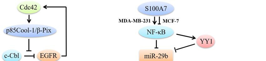
Cells 2019, 8, 146                                                                             3 of 26

cell cycle progression, in addition to G1/S [30]. Cdc42 can also promote the G2/M phase by activating
PAK, whose suppression is known to result in G2/M arrest [32].
      On the other hand, Cdc42 together with Par and protein kinase c (PKC), makes up a polarity
complex. Establishing the Par/PKC/Cdc42 complex contributes to the apical polarity and adherens
junction formation in MECs [27]. MECs-specific Cdc42-knockout can lead to the mis-localization of
PKC and as a result disruption of the complex [30]. This disrupted complex not only fails to form
typical apical polarity but also causes alterations in mitotic spindle orientation in MECs during 3-
Dimensions cultures. Thus, it is believed that the altered mitotic spindle orientation and abnormal
apical polarity contribute to the defective lumen formation in MECs acini [33–35]. During Cdc42
deficiency, the acini also failed to maintain the basal integrin attachment to extracellular matrix
proteins (ECM), demonstrated by the mis-localization of the basal polarity marker α6-integrin [30].

3.2. Deregulation of Cdc42 in Breast Cancer during MECs Morphogenesis
     TEBs are the most immature duct structure in the mammary gland and divide at a high rate to
drive their invasion into fat pad, which make them more susceptible to cancerization [26]. Cdc42
activity in MECs is precisely controlled by multiple mechanisms, such as being maintained by
RhoGDI1 [36]. Once the limitations on Cdc42 activity are released, cell cycle progression and polarity
disorders can proceed to a malignant cell fate. Cdc42 overexpression can disrupt normal TEB
morphogenesis and result in aberrant hyperbranching in association with stromal alterations [37].
Intriguingly, hyperactivated Cdc42-derived hyperbranching does not display a pro-proliferation
phenotype but an increased intracellular contractility and cell motility phenotype that may be
induced by mitogen-activated kinase (MAPK) signaling in MECs [37,38]. Moreover, stromal
alterations driven by Cdc42 overexpression also result in increased ECM remodeling and stromal
deposition, which also affect Cdc42 activity in MECs [39,40].

4. Cdc42 and Breast Cancer Cell Proliferation

4.1. Cdc42 Regulates Breast Cancer Cell Proliferation through MAPK Signaling
      An important characteristic of carcinogenesis is malignant proliferation (Figure 1). EGF and its
receptor EGFR, are the most vital factors during the proliferation process. EGFRs precisely regulate
cell growth under normal conditions, while they exist in excessive amounts in breast cancer cells. The
mechanisms underlying EGFR overexpression are quite complex and cannot be simply ascribed to
gene amplification [41]. Binding of EGF and EGFR mainly activates the classical MAPK pathway and
finally phosphorylates extracellular regulated protein kinase (ERK) to promote breast cancer cell
proliferation.
      Cdc42 mainly functions as an EGFR-signaling regulator in breast cancer cell proliferation. The
termination of EGFR signaling requires the ubiquitin ligase activity of c-Cbl, which triggers EGFR
ubiquitination and subsequent degradation. However, c-Cbl is often compromised in breast cancer
and the upregulation of Cdc42 activity is considered to impair c-Cbl activation, thus inhibiting EGFR
degradation [42]. It is noteworthy to mention that a positive feedback loop exists between EGFR and
Cdc42 and that EGFR is able to stimulate Cdc42 activation [43]. Hyperactivated Cdc42 through its
effector p85Cool-1 (cloned-out-of-library)/β-Pix (PAK-interactive exchange factor), directly impedes
c-Cbl binding to EGFR, which results in EGFRs escape from catalyzing receptor ubiquitination [44].
Moreover, diabetes mellitus (DM), especially type 2 diabetes, has been recently regarded as a risk
factor in breast cancer, due to the fact that high blood–glucose levels can stimulate EGFR activation
and then trigger the EGFR/Cdc42 positive loop [45].
      Activated Cdc42-associated tyrosine kinase (Ack1) is an oncogene encoded by the human TNK2
gene. Its overexpression in cancer cells is induced by Cdc42 through EGFR signaling. Ack1 can
interact with the seven in absentia homolog (SIAH) via estrogen in breast cancer. The SIAH2 gene is
a target of estrogen/estrogen receptor (ER)-signaling and mediates the ubiquitylation of Ack1 [46].
Triple negative breast cancer (TNBC) lacks ER and exhibits a high level of Ack1 [47], which correlates
with high proliferation, migration and colony formation. It has been reported that constitutive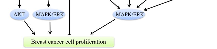
Cells 2019, 8, 146                                                                              4 of 26

activation of Ack1 can trigger the recruitment of PI3K-independent protein kinase B (PKB, also
known as AKT) to the cell membrane and subsequently activate AKT in breast cancer [48], which
may be the underlying mechanism of Ack1-induced tumor progression.
      In addition to interacting with EGFR, Cdc42 has many other means of advancing breast cancer
cell growth. Par6 can cause an EGFR-independent proliferation in normal cells except for its well-
known polarity establishment function [49]. Par6 is also genomically amplified and hyperactivated
in both human precancerous breast lesions and advanced breast cancer [49]. It is functionally required
in breast cancer for the participation of Cdc42 and atypical PKC (aPKC). Interactive with Cdc42 and
aPKC, overexpressed Par6 promotes the MAPK signaling pathway and phosphorylates ERK, even in
absence of EGF/EGFR binding or ligand-independent EGFR phosphorylation [49]. Cdc42 also
promotes proliferation through PAK. It is quite clear that PAK is an important downstream effector
of Cdc42 and that Cdc42 binds to PAK at its N-terminal Cdc42/Rac1 interactive binding (CRIB) site.
Activated PAK directly stimulates anchorage-independent proliferation of breast cancer cells by
phosphorylating MEK1 and ERK1/2 [50,51]. Meanwhile, the scaffold protein IQGAP1 and
procathepsin D can also interact with Cdc42 to enhance breast cancer cell growth and invasion in a
MEK/ERK-dependent manner [52,53].

4.2. Cdc42/p53 Signaling in Breast Cancer Cell Proliferation
      Activated anti-oncogene p53 exhibits multiple anti-proliferative effects including apoptosis and
cell cycle arrest [54]. Studies have shown that Cdc42 can induce p53 ubiquitination to overcome cell
growth inhibition [55]. However, it is notable that although Cdc42 can promote the proliferation of
breast cancer cells through inhibiting p53, its precise role is breast cancer type-dependent. For
example, the inflammatory protein S100A7 (Psoriasin), NF-κB and anti-oncogene miR-29b together
regulate Cdc42/p53 signaling in totally different ways in ER-positive and ER-negative breast cancer
cells. NF-κB can directly or indirectly decrease the levels of miR-29b, by directly binding to the miR-
29b promotor or through transactivating YY1, which binds to the miR-29b promotor [56,57]. In ER-
positive breast cancer, S100A7 inhibits NF-κB and restores its inhibitory effect on miR-29b. Activated
miR-29b, on the one hand, inhibits Cdc42/p53 signaling and directly activates p53 to exert anti-
proliferative effects. However, the opposite is observed in ER-negative breast cancers. The activity of
miR-29b is inhibited by overexpressed NF-κB, which is activated by S100A7 [58].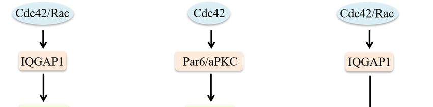
Cells 2019, 8, 146                                                                                      5 of 26

      Figure 1. Cdc42 regulates breast cancer cell proliferation. Hyperactivated Cdc42 through p85Cool-
      1/β-Pix impedes c-Cbl binding to EGFR, results in EGFRs escaping from catalyzing receptor
      ubiquitination. Through EGFR signaling, Cdc42 induces overexpression of Ack1. Constitutive
      activation of Ack1 can recruit AKT to the cell membrane and subsequently activate AKT to promote
      breast cancer progression. A positive feedback loop exists between EGFR and Cdc42 and that EGFR
      is able to stimulate Cdc42 activation. Cdc42 can also interact with aPKC, overexpressed Par6, PAK,
      IQGAP1 and procathepsin D to promote breast cancer cell growth in a MAPK/ERK-dependent
      manner. Besides, Cdc42 induces p53 ubiquitination to overcome cell growth inhibition. In ER-positive
      MCF-7 breast cancer cells, S100A7 inhibits NF-κB. In ER-negative MDA-MB-231 cells, S100A7
      activates NF-κB. NF-κB can decrease the levels of miR-29b directly or through YY1. Decreased miR-
      29b cannot inhibit Cdc42/p53 signaling, thus to promote breast cancer cells proliferation.

5. Cdc42 and Breast Cancer Cell Motility
     Cell migration is a significant phenomenon both in physiological and pathological events, such
as embryogenesis, inflammatory response and wound healing [59], especially in cancer metastasis.
More attention needs to be paid to the relationship between Cdc42 and breast cancer cell metastasis.
Overexpression of Cdc42 usually leads to cancer cell migration and invasion, which are required for
breast cancer spreading into the surrounding tissues and its distant metastasis. In these multifaceted
processes, polarized cells extend motile protrusions (characterized as lamellipodia, filopodia and
invadopodia) in the cell front to interact with the ECM and neighboring cells, then contract the cell
body and detach the cell rear from the matrix to move forward [1]. It is a cyclic process initiated with
the cells response to polarize and extend protrusions in the direction of migration [59]. During this
process, Cdc42 can function as a central regulator via controlling the reorganization of the actin-based
cytoskeleton and cell-cell junctions.

5.1. Cdc42 Is a Key Regulator of Migratory Protrusion Formation
     Breast cancer can move via single cells or collective clusters and both movement forms have
different mechanisms [60]. In the process of collective cell migration, Cdc42 is mainly involved in
regulating the polymerization of actin filaments that drive protrusion formation. Cell filament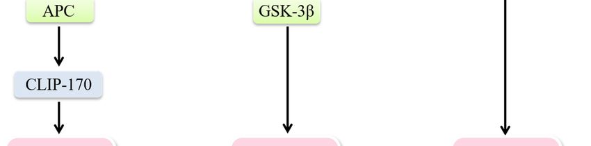
Cells 2019, 8, 146                                                                                 6 of 26

geometry consists of two basic choices; branched filaments that lead to sheet-like protrusions
characterized as lamellipodia and long parallel or bundled filaments that lead to spike-like filopodia
[61].
      The lamellipodia formation can be described as follows: Actin filaments polarize with the
formation of fast-growing “barbed ends” and slow-growing “pointed ends” to drive protrusions [62].
Cofilin can sever pre-existing actin filaments to produce free barbed ends. The Src/FAK (proteins
kinase/focal adhesion kinase) complex activates Paxillin (a scaffolding protein that can recruit several
regulatory and structural proteins to modulate cell adhesions and cytoskeleton reorganization [63])
to recruit Cdc42 and trigger N-WASP activation. The combination of activated N-WASP with actin-
related protein 2/3 (Arp2/3) leads to conformation changes of Arp2/3 and brings actin monomers (G-
actin) to this complex [1]. Subsequently, the Arp2/3 complex mediates actin nucleation of new
filaments at the cofilin-severed barbed ends [64–66] Rac can also modulate actin nucleation. Active
Rac proteins can interact with a WAVE-associated complex of proteins, which in turn activates actin
nucleation via Arp2/3. Furthermore, an extension of the actin filament can also be induced by Rac via
interaction with scaffold protein lamellipodin that binds with WAVE complex [67,68]. Actually, the
Rac-mediated mechanism is predominant during lamellipodia formation. In contrast, Cdc42 or other
Rho GTPases activate formins to extend Arp2/3 complex-induced filaments. Then profilin delivers
G-actin to formins to elongate the linear actin network and to facilitate rapid actin assembly [69] The
renewal of such an actin network largely depends on the regulation of cofilin. Cofilin severs and
depolymerizes older actin filaments in the network, leading to the rapid turnover of actin filaments
[70]. Activated Rac or Cdc42 can activate PAK1 and its downstream effector LIM kinase 1 (LIMK1),
which contributes to the inactivation of cofilin. A lack of its actin filament (F-actin, filamentous state
of actin, can be converted from G-actin [71])-depolymerizing activity leads to the accumulation and
aggregation of actin filaments [72,73]. Due to the inactivation of cofilin, rapid actin filaments turnover
slows down and a relatively stable network is generated. Enhancement of stable actin filaments, in
turn, reduces cell migration [74]. In breast cancer cells, the activation and inactivation of cofilin are
unbalanced, altering protrusions and cell motility [75]. Vasodilator-stimulated phosphoprotein
(VASP), termed as “anti-cappers,” can prevent blockage of actin filaments by capping proteins, thus
promoting the formation of unbranched actin networks in lamellipodia [76]. However, silencing
Cdc42 does not block the formation of lamellipodia in MDA-MB-231 cells [42]. The regulation of
lamellipodia formation is predominantly dependent on the activation of Rac and Cdc42 mainly
functions to modulate the formation of filopodia.
      The formation of filopodia is initiated by IRSp53 (insulin receptor phosphotyrosine 53 kDa
substrate, a multi-domain protein that induces filopodia through its I-BAR domain [77]). IRSp53 can
bend the membrane and recruit Cdc42 and diaphanous-related formin 3 (DRF3 = mDia2), which in
turn mediates actin nucleation. VASP delivers actin monomers to the filopodial tip and G-actin is
provided directly to mDia2 by profilin. Cdc42 and Rif can regulate actin polymerization by targeting
mDia2 and Cdc42 can stimulate N-WASP/Arp2/3-driven polymerization, similar to the mechanism
in lamellipodia [69]. Filopodia is not necessary for cell migration. It is usually considered to be an
environmental sensor that can also contribute to migration by converting to lamellipodia during
growth factor receptor signaling [62,78].
      Another special protrusion called invadopodium is often assembled for cancer cell invasion.
Such protrusions can secrete metalloproteases (MMPs) at the front cells to degrade extracellular
matrix and basement membrane components [79]. Membrane type 1 metalloprotease (MT1-MMP)
and perhaps other MMPs are transported to the tip of invadopodia by microtubule-mediated vesicle
trafficking, which requires ADP ribosylation factor 6 (ARF6) [69]. There are many similarities in the
regulatory mechanisms involved in the formation of filopodia, lamellipodia and invadopodia. The
key difference is that invadopodia can degrade the extracellular matrix; therefore, the delivery of
vesicles containing matrix-degrading proteases, in particular, MT1-MMP is required. These vesicles
target invadopodia through the vesicle-tethering exocyst complex [80]. In highly invasive MDA-MB-
231 human breast carcinoma cells, activated Cdc42 and RhoA can trigger the interaction of IQGAP1
with the exocyst subunits Sec3 and Sec8, which is necessary for invadopodia activity, because the
Cells 2019, 8, 146 7 of 26
deletion of the exocyst-binding site is accompanied by the loss of IQGAP1-induced enhancement of
matrix degradation. Thus, the exocyst and IQGAP1 are required for the accumulation of cell surface
MT1-MMP at invadopodia [81] (Figure 2).
However, the regulation of Cdc42 during pseudopods formation is not specific. Overexpression
of podoplanin in MCF-7 cells induces filopodia formation and cell polarization, leading to the
enhanced β1-integrin-mediated cell spreading and adhesion on the extracellular matrix, thus
increasing cell migration and invasion [82]. Furthermore, Stromal cells such as fibroblasts may also
induce collective cancer cell migration, playing a similar role to MMPs to open a way for trailing cells.
In this model, Cdc42-mediated activation of MRCK is required to allow cancer cell migration behind
leading fibroblasts [83]. Protrusions of the plasma membrane at the front of cell groups drive the
movement of the clusters.
Figure 2. Role of Cdc42 on regulation of migratory protrusions formation. Model of lamellipodia
formation: cofilin severs pre-existing actin filaments to produce free barbed ends. The Src/FAK
complex activates Paxillin to recruit Cdc42 and trigger N-WASP activation. The combination of
activated N-WASP with Arp2/3 leads to actin nucleation of new filaments at the cofilin-severed
barbed ends. Cdc42 activates formins. Profilin delivers G-actin to formins to extend filaments. Rac
interacts with lamellipodin that binds with WAVE to extend actin filaments. Model of filopodia
formation: IRSp53 recruits Cdc42 and mDia2. mDia2 mediates actin nucleation. VASP delivers actin
monomers to the filopodial tip and G-actin is provided directly to mDia2 by duringilin. Cdc42 and
Rif can regulate actin polymerization by targeting mDia2. Cdc42 can also stimulate N-WASP/Arp2/3-
driven polymerization. Model of invadopodia formation: Cdc42 and RhoA trigger the interaction of
IQGAP1 with the exocyst subunits Sec3 and Sec8, which is necessary for invadopodia activity. MT1-
MMP is transported to the tip of invadopodia by microtubule-mediated vesicle trafficking to degrade
the extracellular matrix.
5.2. Cdc42 Modulates the Establishment of Cell Polarity
A polarized morphology is required to form a stable actin network, which is a prerequisite for
directed cell migration. Migrating cells dynamically polarize during the process of movement.Cells 2019, 8, 146 8 of 26
Establishing polarity demands asymmetric distribution of the cytoskeleton, cell-adhesion
molecules and signaling molecules, as well as directed membrane trafficking performed by motor
proteins such as dynein and kinesin. The model consists of several coordinated processes, including;
membrane ruffling and filopodia at the leading edge, capture of microtubule plus-ends near the
leading edge and reorientation of the microtubule-organizing center (MTOC) and the Golgi
apparatus towards the direction of migration [84]. Nuclear repositioning is an initial polarizing event
in migrating cells. The nucleus moves away from the leading edge to reorient the MTOC, while the
MTOC remains stationary, which is coupled with actin retrograde flow and is regulated by a pathway
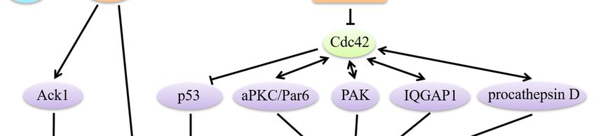
involving Cdc42, MRCK, myosin and actin [85]. Cdc42 participates in regulating the establishment
of single-cell polarity through modulating microtubule-based intracellular vesicle trafficking to the
apical cell surface and orientation of the cell division spindle. Furthermore, Cdc42 plays a role in
maintaining collective cell polarity by strengthening cell–cell junctions [4] (Figure 3). Activated Rac1
and Cdc42 are able to mark spots where IQGAP1 tethers actin filaments. IQGAP1 then acts as a
scaffold linking adenomatous polyposis coli (APC) to actin filaments and captures the plus-ends of
microtubules through the microtubule-binding protein CLIP-170, which directly and/or indirectly
stabilizes microtubules and generates a stable actin meshwork at the leading edge [86]. Cdc42 can
also regulate the reorientation of the MTOC via a Par6–atypical protein kinase C (aPKC) complex,
which induces the phosphorylation of GSK-3β and the interaction of APC with the plus ends of
microtubules [87]. Microtubule-mediated delivery of vesicles and the associated proteins needed are
provided to the membrane at the leading edge [62,88].
Specialized cell–cell adhesion complexes, characterized as E-cadherin-containing adherens
junctions (AJs), are also necessary to help maintain proper barrier function and apical-basolateral
polarity in epithelial cells. Disruption of these normal characteristics in epithelial cells has been
associated with tumor progression, such as epithelial-mesenchymal transition (EMT). A dynamic
equilibrium exists between the E-cadherin–β-catenin–α-catenin and E-cadherin–β-catenin–IQGAP1
complexes at sites of cell–cell contact. The ratio between these two complexes could determine
adhesion strength [86]. Strong adhesion is established when the increasingly active Rac1 and Cdc42
interact with IQGAP1 to crosslink actin filaments and weak adhesion is built under the opposite
conditions. This is due to the fact that free IQGAP1 interacts with β-catenin to dissociate α-catenin
from the cadherin–catenin complex [86]. Such deficient adhesions facilitate EMT.
5.3. Cdc42 Involves the Progression of EMT
During the process of EMT, epithelial cells lose cell-to-cell interactions and cell polarity, tissue
structures become loose and transform from polygonal epithelial cells to a spindle-like fibrocyte-like
morphology. Moreover, apical and basolateral epithelial-specific proteins in cells such as E-cadherin,
catenins and cytokeratins progressively redistribute or downregulate, while mesenchymal molecules
such as vimentin, fibronectin and N-cadherin are re-expressed. These series of changes confer breast
cancer cells with the motility necessary for invasion [82]. It is noteworthy that EMT does not occur in
the case of collective cell migration. Interferon regulatory factor 4 binding protein (IBP, a Rho-family
guanine nucleotide exchange factor for Rho family GTPases, including Rac1, RhoA and Cdc42 [89])
can mediate Rac1, RhoA and Cdc42 activation in breast cancer cells to regulate actin cytoskeleton
rearrangement and MMP production. Meanwhile, IBP also decreases the expression of the epithelial
markers E-cadherin and keratin 18 but increases the expression of mesenchymal markers fibronectin
and N-cadherin to trigger the acquisition of an EMT phenotype [90]. Consequently, IBP may regulate
EMT and the movement of breast cancer cells via Rac1, RhoA and Cdc42 signaling pathways.
5.4. Cdc42 Regulates Breast Cancer Cells Motility via Various Effectors
Various regulators have been reported to target Cdc42 and influence breast cancer movement
due to Cdc42 functions. In T47D mammary epithelial cells, activation of PI3K via chronic activation
of Cdc42 and Rac1 disrupts the normal, polarized organization of these cells and promotes a motile,
invasive phenotype [91]. Melanoma differentiation-associated gene-9 (MDA-9), also known as
syntenin-1 (SDCBP; syndecan binding protein), a member of the PDZ-domain-containing family [92],Cells 2019, 8, 146 9 of 26
modulates the small Rho GTPases RhoA and Cdc42 to enhance invasion and cytoskeletal
rearrangement in MDA-MB-231 and SUM159 breast cancer cells via TGFβ1 [93]. The Kruppel-like
factor 5 (KLF5) transcription factor, highly expressed in high-grade, poorly differentiated and basal-
like triple-negative breast cancer (TNBC [94]), directly binds to the TNFAIP2 gene promoter and
activates its transcription. TNFAIP2 then interacts with Rac1 and Cdc42, increases their activities to
change the actin cytoskeleton and cell morphology, thus promoting TNBC cells migration and
invasion [95]. A recent study demonstrated a novel ability of Cdc42 to modulate cell migration in
MDA-MB-231 and Hs578T cells. ERK5, also known as big MAP kinase 1 (BMK1), a member of MAPK
family [96], can decrease the migration and invasion of both MDA-MB-231 and Hs578T cells. Cdc42
has been shown to inhibit its phosphorylation and expression to increase cell motility [97]. In MCF-7
and MDA-231 cells, δ-Catenin (a member of the P120 catenin (p120ctn) family [98]) upregulates
Cdc42 and Rac1 activities and contributes to increased cell mobility [99]. Invasion of MDA-MB-231
cells into three-dimensional (3-D) type I-collagen matrices depends on TGF-α. This event is likely
dependent on the activation of Cdc42 via TGF-α to initiate the formation of protrusions into collagen
[100]. P120 catenin (p120), a Src substrate that can indirectly activate Rac1 and Cdc42, acts as an
obligate intermediate between ErbB2 and Rac1/Cdc42 to modulate the metastatic potential of breast
cancer cells [101,102]. To summarize, Cdc42 acts as a significant regulator in breast cancer cell
migration and invasion.
Figure 3. Cdc42 modulates the establishment of cell polarity. Establishment of single-cell polarity:
Activated Rac1 and Cdc42 mark spots where IQGAP1 tethers actin filaments. IQGAP1 links APC to
actin filaments and captures the plus-ends of microtubules through the microtubule-binding protein
CLIP-170, which stabilizes microtubules and generates a stable actin meshwork at the leading edge.
Cdc42 regulates the reorientation of the MTOC via a Par6/aPKC complex to induce the
phosphorylation of GSK-3β. Maintenance of collective polarity: Strong cell-cell adhesion is
established when the increasingly active Rac1 and Cdc42 interact with IQGAP1 to crosslink actin
filaments.Cells 2019, 8, 146 10 of 26
6. Cdc42 and Breast Cancer Angiogenesis
The rapid growth of breast cancer cells depends on the constant supply of nutrients by blood
vessel networks but the intrinsic vascular network cannot provide such large amounts of nutrients.
As a result, breast cancer cell progression requires newly expanding blood vessels [103].
Angiogenesis is the process of new blood vessels arising from existing vessels, which requires
vascular endothelial cell proliferation and migration as well as basement membrane breakdown. This
process is accurately controlled by many pro-angiogenic factors including EGF, fibroblast growth
factors (FGF), vascular endothelial growth factor (VEGF), IL-6 and IL-8, in addition to anti-angiogenic
factors including angiostatin [104]. While these pro- and anti-angiogenic factors are in a dynamic
balance under normal condition, during breast cancer the balance is tipped and pro-angiogenic
activity dominates.
The basic mechanisms of Cdc42 regulating vascular endothelial cell proliferation, migration and
basement membrane breakdown are the same as those mentioned previously. These do not however
describe the entire role of Cdc42, which can also particularly regulate pro-angiogenic factors during
breast cancer angiogenesis. Since its definition in the 1980s, VEGF has been the most important pro-
angiogenic factor. It is overexpressed in a broad spectrum of cancers and considered as the major
initiator of pathological angiogenesis [105]. High expression of VEGF often occurs in ischemic areas
of tumors, which is induced by hypoxia, which also activates Cdc42 through PI3K and PTK [106]. It
has been reported that hyperactivated Cdc42 (under both normoxic and hypoxic conditions)
upregulates VEGF in breast cancer cells [106]. Cdc42 does not regulate VEGF directly but through
p53. Mammary VEGF transcription is inhibited by p53 in many ways. Firstly, the VEGF promoter
contains specificity protein-1 (Sp1) binding sites [107], where p53 forms complexes with Sp1 to
prevent the VEGF transcription [108]. p53 can also regulate hypoxia-inducible factor-1 (HIF-1α) and
proto-oncogene c-Src activity, thus decreasing VEGF mRNA transcription under hypoxic condition
[108,109]. Cdc42 participates in VEGF-mediated angiogenesis mainly by degrading p53 to relieve
VEGF inhibition [55]. Furthermore, hypoxia-activated Cdc42 can also increase the levels of IL-6 and
IL-8 to upregulate VEGF expression [110,111], which is achieved by Cdc42 activating NF-κB, a
modulator of IL-6/8 expression [112,113]. In addition to VEGF, FGF is another strong pro-angiogenic
factor overexpressed in breast cancer [114–116]. Cdc42 can bind to FGF1 promotor at Ets sites, leading
to increased transcription [117].
7. Survival of Breast Cancer Cells Requires Cdc42
The human body itself, after it becomes aware of malignant proliferation and breast cancer cells
invasion, initiates a series of responses such as apoptosis and immune responses to prevent unlimited
cancer cell growth. Anti-cancer drugs (chemotherapy) have also been used as effective treatments to
eliminate cancer cells and prolong patient survival. However, Cdc42 assists breast cancer cells in
escaping apoptosis and chemotherapeutic treatments, allowing them to survive in circulation.
Cellular apoptosis in the human body is driven by many apoptosis-related genes like members
of the Bcl-2 family, anti-oncogene p53, proto-oncogene c-Myc and Fas. It is also mediated by immune
cells like T cells and natural killer cells (NK cells). Cdc42-mediated anti-apoptosis consists of many
aspects, including its interactions with some of these apoptosis-related genes and immune cells as
well as a “crosstalk” with other Rho GTPases.
7.1. Cdc42 Regulates Apoptosis-Related Genes through PAK and JNK Signaling
The Bcl-2 family consists of cell death genes (Bad, Bax, Bak and Bcl-xS) and cell survival genes
(Bcl-2, Bcl-xL, Mcl-1 and A1) [118]. These genes are critical to intrinsic cell death machinery and
relative levels of them dictate the susceptibility of cell death [119]. One of the important ways Cdc42
affect Bcl-2 family during breast cancer is by stimulating its downstream effector PAK (both PAK1
and PAK2). PAK is capable of phosphorylating the pro-apoptotic member Bad on both Ser112 and
Ser136 to reduce the interaction between Bad and the cell survival members Bcl-2 and Bcl-xL [120].
Dissociation of the Bad/Bcl-2/Bcl-xL complex ultimately results in an inhibition of mitochondrialCells 2019, 8, 146 11 of 26
cytochrome c, thus suppressing cell death [121]. Moreover, PAK also activates NF-κB by stimulating
the p65 subunits nuclear translocation to prevent apoptosis of breast cancer cells [122,123].
Using alternative mechanisms to ERK, c-Jun N-terminal kinase (JNK) is another branch of the
MAPK signaling pathway, which regulates apoptosis in breast cancer [124]. Constitutive activation
of Cdc42 can activate JNK through MKK4/7, which then activates an important transcription factor
AP-1 [125,126]. In humans, AP-1 constitutes two subunits, jun and fos [127]. These two subunits act
quite differently in the transcription of many apoptosis-related target genes and trigger different roles
in breast cancer apoptosis. Jun is also a regulator of Bcl-2 family members but its major effect is to
downregulate the transcription of Bcl-2 and Bcl-xL, which is distinguished from Cdc42/PAK pathway
[128]. Without jun, Bcl-2 and Bcl-xL can also be phosphorylated directly by JNK [129,130]. Besides,
Jun is involved in the regulation of anti-oncogene p53 as well, inhibiting p53 transcription to resist
apoptosis and promote proliferation [131]. Fas/FasL-induced death receptor pathway is another
crucial apoptotic mechanism in addition to the mitochondrial pathway that is induced by the Bcl-2
family. Jun together with fos or STAT3, is reported to regulate Fas/FasL expression [132,133]. Fas-
induced apoptosis is also dependent on caspases to cleave substrates that are important for cell
survival. In breast cancer, caspase-3/7 can cleave its key substrate protein Cdc42 at aspartate acid
residues 121 and 118. However, the expression of mutated caspase-insensitive Cdc42 slows down the
Fas-induced apoptotic response and displays a strong anti-apoptotic effect [43]. This Cdc42 mutant
may exist in breast cancer allowing it to overcome Fas-induced apoptosis. FasL has been shown to
elevate a part of the Cdc42 pool but some cascade amplification is still required to affect the Fas–
caspase system [43].
7.2. Cdc42 Drives Actin Responses in NK Cells
NK cells are large granular lymphocytes in morphology with cytotoxic activity against virus-
infected cells and cancer cells. The immunological synapse (IS) is an indispensable structure between
NK and target cells, required for the recruitment and release of intercellular lytic granule to the target
cell. A significant phenomenon when breast cancer cells respond to NK cells is a massive and rapid
F-actin accumulation surrounding IS, termed “actin response,” which is responsible for NK cell
resistance. This burst actin response is mainly induced by Cdc42/N-WASP signaling along with their
downstream Arp2/3 complex. Inhibition of Cdc42/N-WASP significantly increases the levels of
cytotoxic protease granzyme B in target cells and is sufficient to transform NK cell-resistant breast
cancer cells into susceptible ones [134].
7.3. Crosstalk of RhoGTPases during Breast Cancer Apoptosis
Cdc42 participates in a “crosstalk” with Rho GTPases for an anti-apoptotic function. Cdc42 has
long been known to activate Rac1, which leads to RhoA activation. The anti-apoptotic role of RhoA
includes inhibiting the cell cycle inhibitor p21 to enhance cell survival and activating Bcl-2 family
members [135,136]. Injection of RhoA or Cdc42 prevents breast cancer cells from mAb200 (Ras-GAP
inhibitor)-induced apoptosis but no additional effects are seen upon Cdc42/RhoA co-injection, which
demonstrates that the protective function of Cdc42 in breast cancer results from RhoA activation
[137].
7.4. Cdc42 and Anti-Cancer Drugs Resistance
Breast cancer is sensitive to chemotherapy and adjuvant chemotherapy in later stages of
treatment. However, multidrug resistance (MDR) remains an important cause of chemotherapy
failure and clinical treatment disturbance [138]. Cancer cells activate the transcription of drug-
resistant genes through various signaling pathways, leading to an increased expression of drug-
resistant proteins and eventually drug resistance. Cdc42 is one of these drug-resistant proteins
involved in breast cancer.
Doxorubicin (Adriamycin, ADM) is a broad-spectrum anti-cancer drug, which can target breast
cancer. Its mechanism of action involves inhibiting the synthesis of nucleic acids by intercalatingCells 2019, 8, 146 12 of 26
DNA [139]. In ADM-resistant breast cancer cells, transfection of Cdc42-specific siRNA can
significantly increase ADM levels and enhance its killing effects on these ADM-resistant cells [140].
Moreover, breast cancer is a hormone-dependent systemic disease and many of its processes are
related to estrogen [141,142]. After estradiol-17 beta treatment, breast cancer cells express higher
Cdc42 levels and exhibit stronger ADM resistance, which is directly manifested by the decrease
chemotherapeutic drug accumulation in cells [143]. It is suggested that Cdc42 participates in anti-
cancer drug resistance by interacting with N-WASP and Arp2/3 to promote actin polymerization,
microfilament cytoskeleton rearrangement, intracellular material flow acceleration and promotion of
intracellular drug excretion to the extracellular space.
Besides phosphorylating Bcl-2 family members to protect breast cancer cells from intrinsic cell
death, Cdc42 downstream effector PAK also regulates anti-cancer drugs-induced cell death. It has
been demonstrated that breast cancer cells with low PAK2 activity exhibit strong sensitivity to the
anti-cancer drugs cisplatin and taxol [144]. PAK2 is unique among the PAK family: Activated by
Cdc42, full-length PAK2 protects breast cancer cells from drug-induced cell death; when
proteolytically cleaved by caspase 3, PAK2 generates its fragment PAK2p34 that favors apoptosis
[145]. PAK2 downregulates caspase 3 to block the generation of PAK2p34 and promotes breast cancer
cell survival, leading to MDR.
8. Current Research Advances of Cdc42-Targeted Therapies in Breast Cancer
The contribution of Cdc42 to breast cancer cells is substantial, due to its critical roles in many
aspects of cancer processes. However, drugs targeting Cdc42 were once considered impossible due
to its micromolar levels in cells and its perplexing signal transduction with other factors. Nonetheless,
in recent years, some Cdc42-targeted drugs are being developed in breast cancer research, aiming to
inhibit Cdc42 activation in various ways (Table 2).
8.1. GEF Interaction Inhibitors
GEFs exchange GDP into GTP and generate active-bound Rho GTPases. NSC23766 is designed
on the Trp56 residue of Rac, which is vital for GEF binding [146]. However, off-target effects prevent
clinical use of this drug. EHop-016 is another Rac inhibitor derived from NSC23766, also targeting
Trp56 [147]. In metastatic cancer cells, EHop-016 inhibits Cdc42 activation with an IC50 approximately
>10 μmol/L [148]. Moreover, EHop-016 has the capacity to inhibit breast cancer cell growth
(approximately 80%) [148] and block angiogenesis and metastasis [149]. However, its bioavailability
and high effective concentrations need to be improved [150].
In recent years, MBQ-167 has been designed to form H bonds with the Asn39 side-chain of Cdc42
and Rac and Asn39 replacement leads to loss of GEF binding [146]. Surprisingly, MBQ-167 inhibits
Cdc42 activation with an IC50 of 78 nmol/L, which make it one of the most effective Cdc42 inhibitors
at present [151,152]. MBQ-167 has been shown to inhibit a large proportion of Cdc42 downstream
effectors, like PAK and LIMK. Interestingly, the PAK1 displays autophosphorylation when MBQ-167
inhibits Cdc42, suggesting a feedback loop in MBQ-167/PAK1. Nevertheless, expression of PAK
effectors LIMK and cofilin are significantly blocked by MBQ-167, which inhibits PAK1 [153,154].
MBQ-167 can inhibit nearly all Cdc42-induced tumor processes in breast cancer, including cell
polarity, cell cycle progression, apoptosis and metastasis [152]. However, MBQ-167 only inhibits
Cdc42 activity in breast cancer cells that undergo EMT, rather than non-cancer cells or cancer cells
without EMT [151]. This selective inhibition of MBQ-167 may result from the different Cdc42-related
GEF expression profiles in different types of breast cancer; MBQ-167 may only affect a subset of
Cdc42-related GEFs that are activated in mesenchymal-like breast cancer cells [155]. Focal adhesion
assembly at the mesenchymal-like breast cancer cell leading edge is regulated by integrins that are
under Cdc42 regulation, while the integrins that regulate mammary epithelial cell filament
cytoskeleton are not directly mediated by Cdc42 [156,157]. Based on this selectivity, MBQ-167 can
reduce the viability of breast cancer cells with EMT process instead of non-cancer cells, which makes
MBQ-167 more tumor-specific. Since EMT is also related to drug resistance [158], MBQ-167 has the
potential to prevent drug resistance. In nude mice mammary fat pad tumors, the use of MBQ-167Cells 2019, 8, 146 13 of 26
reduces tumor size by about 91% in two months with 10 mg/kg bodyweight (BW) and no metastases
are observed [151].
8.2. Nucleotide Binding Inhibitors
Aside from preventing GEFs binding to Cdc42, an alternative to Cdc42 targeting is to block
nucleotide binding. R-enantiomer of ketorolac (R-ketorolac), the allosteric inhibitor of Cdc42 and Rac,
is the first FDA-approved Cdc42 and Rac inhibitor proceeding to P0 clinical trials [159]. It is reported
to inhibit tumor progression in breast cancer virus-polyoma middle T antigen (MMTV-PyMT) mice
[160].
The topoisomerase II inhibitor, mitoxantrone (MTX), is also an FDA-approved drug in breast
cancer. MTX can block GTP binding of Cdc42, then inhibit actin filament cytoskeleton and reduce cell
migration [161].
8.3. RhoGDI Modulators
RhoGDIs are thought to sequester Cdc42 in the inactive GDP-bound state within the cytosol.
The design that prevents the dissociation of RhoGDIs and Cdc42 is a potential strategy for Cdc42-
targeted treatment. Secramine has been demonstrated to inhibit Cdc42 activation in a RhoGDI-
dependent manner. More specifically, secramine inhibits the PIP2-stimulated Cdc42/N-
WASP/Arp2/3-mediated actin polymerization and this inhibitory effect requires the presence of
RhoGDI1 to prevent the membrane recruitment of Cdc42 [162]. However, the secramine-induced
inhibition is not selective and both RhoGDIs upregulation and downregulation are reported with
increasing malignancy [163].
8.4. Metformin
TNBC refers to breast cancers whose immunohistochemical results are ER-negative,
progesterone receptor (PR)-negative and HER2-negative. This kind of breast cancer lacks specific
clinical therapeutic guidelines [164]. Luckily, the antidiabetic drug metformin has been reported to
inhibit breast cancer cell proliferation and migration by significantly downregulating Cdc42
expression and TNBC is sensitive to metformin [165,166]. Metformin is thought to exhibit anti-cancer
activity via the AMP-activated protein kinase (AMPK) signaling pathway [167]. In this signaling
network, an increased level of AMP activates AMPK, which inhibits the mammalian target of
rapamycin (mTOR) expression to reduce tumor progression [167]. However, metformin-mediated
Cdc42 downregulation does not require this typical AMPK signaling pathway; conversely, AMPK
upregulates Cdc42 expression [168]. Downregulation of Cdc42, induced by metformin, is partially
due to transcription factors such as DNTTIP2, TCEB2 and YWHAB [168].
8.5. Biological Extractions
A few advances have been made in traditional Chinese medicine-related anti-cancer treatments
in recent years. Cdc42 is an effective target for traditional Chinese medicine which has been used to
inhibit breast cancer progression. A traditional Chinese medical herb, Ganoderma lucidum (GA), has
been demonstrated to reduce breast cancer cell proliferation and migration, as well as induce
apoptosis [169,170]. Ganoderma lucidum triterpenoids extracts (GAEE) that contain GA, GA isomer
and dehydrogenated GA, can reduce FAK activation and break the interaction between FAK and Src,
then attenuate the affinity between the Src/FAK complex and Paxillin in breast cancer. The Cdc42
recruitment function of Paxillin allows GAEE to downregulate Cdc42 expression and attenuates the
interaction between Cdc42 and N-WASP, which results in an impairment at the cell leading edge,
thus inhibiting cell migration [171]. Therefore, GAEE may be a potent anti-cancer drug in breast
cancer.
Resveratrol (trans-3,4V,5-trihydroxystilbene) is a phytoalexin that was initially extracted from
grapes. It can bind to ER and display opposing effects, for example, it is estrogenic at low
concentrations and anti-estrogenic at high concentrations [142,172]. High concentration resveratrol-Cells 2019, 8, 146 14 of 26
induced inhibition of Cdc42 results in a widespread and sustained filopodia response, which is due
to the inhibition of Rac activation. Rac converts filopodia to lamellipodia [78], while reduced Rac
activation ultimately leads to the occurrence of non-polar filopodia, which inhibits the migration of
breast cancer cells [142,172].
Table 2. Cdc42-Targeted Therapies in Breast Cancer.
Inhibitors Therapies Cell Lines/Tissues Inhibitory Effects References
growth, angiogenesis,
EHop-016 MDA-MB-435 [129]
metastasis
GEF interaction MDA-MB-231, cell polarity, cell cycle
inhibitors MCF-7 and MDA- progression, apoptosis [131]
MBQ-167 MB-435 and metastasis
nude mice tumor size [131]
Nucleotide binding R-ketorolac MMTV-PyMT mice tumor progression [140]
inhibitors MTX PAE cell migration. [141]
RhoGDI Xenopus laevis
secramine actin polymerization [142]
modulators cytoplasmic egg
proliferation and cell
Antidiabetic drug Metformin MDA-MB-231 [146]
migration
Biological GAEE MDA-MB-231 cell migration [151]
extractions Resveratrol MDA-MB-231 cell migration [122]
Abbreviations: R-ketorolac, R-enantiomer of ketorolac; MTX, mitoxantrone; PAE, porcine aortic
endothelial; GAEE, Ganoderiol A-Enriched Extract.
9. Cdc42-Related Non-Coding RNAs in Breast Cancer
9.1. microRNA
MicroRNA is a type of endogenous small non-coding RNA (ncRNA) with a length of about 20–
24 nucleotides. Each microRNA can have multiple target genes and several microRNAs can also
regulate the same gene [173]. MicroRNAs decrease the expression of target genes by forming a
complement with the mRNAs of their target genes [174]. The 3′-untranslated region (UTR) is the
crucial site for the regulation of microRNA functions [175]. Due to their important regulatory roles
in cells, deregulation of microRNAs always occurs in many diseases including cancers. In breast
cancers, some microRNAs target Cdc42 and are extensively involved in Cdc42-induced tumor
processes, while many are aberrantly expressed (Table 3).
As mentioned previously, miR-29b regulates Cdc42/p53 signaling during breast cancer cell
proliferation. Another miR-29 family member, miR-29a, which also targets Cdc42 is downregulated
in breast cancer. It is identified as a tumor suppressor due to its inhibitory regulation of Cdc42 during
cell cycle progression [176].
Compared to regulating cell growth, microRNAs interfere more in actin cytoskeleton regulation
functions of Cdc42. miR-206 has been demonstrated to regulate actin remodeling during breast cancer
metastasis and Cdc42 is a potential target of miR-206. During metastasis, miR-206 can inhibit
filopodia formation and matrix degradation by inhibiting Cdc42 activation [177]. miR-23b also has a
vital role in the actin cytoskeleton [178]. PAK is known to restrict the size of focal adhesions (focal
adhesions that mature excessively are related to slower migration rates) to promote migration [179].
miR-23b downregulates the Rac/Cdc42 guanine nucleotide exchange factor 6 (ARHGEF6) whichCells 2019, 8, 146 15 of 26
activates Cdc42/PAK, thus enhancing focal adhesions maturation in breast cancer [180]. In addition,
overexpression of miR-23b is associated with an increasing epithelial phenotype in breast cancer cells,
which leads to the EMT inhibition function of miR-23b [180]. Moreover, miR-224 can inhibit breast
cancer cell invasion by directly suppressing Cdc42 during the interaction at their binding site [181].
miR-888 also inhibits the adherens junction (AJ) pathway by targeting Cdc42 [182]. Furthermore,
CD44 3′-UTR has a decoy effect that binds to miR-216a, miR-330 and miR-608 resulting in increased
Cdc42 expression in MT-1 breast-carcinoma cells [175].
Hyperactivation of the cancer stem cell (CSC) pool in breast cancer patients with hyperglycemia
is associated with miR-424 regulation of Cdc42. miR-424 interacts with the Cdc42 promoter sequence
through the complementarity between them. The ectopic expression of miR-424, which always occurs
in breast cancer patients under hyperglycemic conditions, leads to Cdc42 activation. Activated Cdc42
stimulates PAK1/STAT5 signaling and then activates the downstream transcriptional regulator
prdm14, which maintains CSCs pluripotency and inhibits differentiation [183,184].
Beyond microRNAs that act as tumor suppressors, some microRNAs are known oncogenes.
miR-548j is a pro-tumor microRNA in breast cancer, whose overexpression is related to increased
invasiveness and poor prognosis [185]. It has been demonstrated that miR-548j-induced invasion is
dependent on Tensin1 via Cdc42. Tensin1 can interact with the RhoGAP and DLC-1, to transform
Cdc42 into inactive-bound and thus suppress invasiveness [186]. Therefore, miR-548j directly inhibits
Tensin1 and protects Cdc42 in its active-bound state.
9.2. lncRNA
Compared to small ncRNAs, lncRNAs have relatively long nucleotide chains, which contain
more protein binding sites. Metastasis associated with lung adenocarcinoma transcript-1 (MALAT1)
is a lncRNA that can promote the progression of multiple tumors [187–190]. In breast cancer, miR-1
directly targets MALAT1 and Cdc42. MALAT1 can competitively bind to miR-1, which reduces the
ability of miR-1 to inhibit Cdc42, ultimately increasing the level of Cdc42 and inducing cell migration
and invasion to promote breast cancer metastasis [191].
Table 3. Cdc42-Related Non-Coding RNAs in Breast Cancer.
Non-Coding Suppressor or
RNA Cell Lines/Tissues Effects References
RNAs Promoter
cell cycle
miR-29a MDA-MB-453 suppressor [156]
progression
filopodia
formation and
miR-206 MDA-MB-231 suppressor [157]
matrix
degradation
actin
suppressor [158]
microRNAs cytoskeleton
miR-23b MDA-MB-231, MCF-7 EMT suppressor [160]
focal adhesion
promoter [160]
maturation
miR-224 MDA-MB-231 cell invasion suppressor [161]
adherens
miR-888 MCF-7 suppressor [162]
junctionCells 2019, 8, 146 16 of 26
CSCs
miR-424 MDA-MB-231 suppressor [163]
pluripotency
miR-548j MCF-7 invasion promoter [165]
cell migration
lncRNA MALAT1 MDA-MB-231, MCF-7 promoter [171]
invasion
Abbreviations: EMT, epithelial to mesenchymal transition; CSCs, cancer stem cells; MALAT1,
Metastasis associated with lung adenocarcinoma transcript-1.
10. Summary
The RhoGTPase family member Cdc42, which serves as a molecular switch, is essential among
those normal mammary cells. The precise regulation of Cdc42 is in charge of the normal mammary
gland development. However, Cdc42 is often overexpressed in breast cancer cells and predominantly
acts as a pro-tumor factor accompanied with the intricate downstream signaling transduction. Cdc42
is mainly involved in the regulation of actin cytoskeleton through activating N-WASP/Arp2/3, while
many of its other downstream effectors serving a number of other tasks. For instance, PAKs
participates cancer cell proliferation, motility, cell death and anti-cancer drugs resistance, as well as
Cdc42/Par/PKC complex affects morphogenesis and cell polarity. Cdc42 also inhibits tumor-
suppressor genes to protect the breast cancer in many respects, like inhibiting p53 to relieve its anti-
proliferation, -angiogenesis and -apoptosis effects. These wide coverage functions of both Cdc42 and
its effectors provide ideas for the broad-spectrum anti-cancer drug designs.
However, though the rational design of Cdc42-targeted drugs can lead to promising preclinical
outcomes so far there are no drugs that target Cdc42 in clinical trials. Using the orthotopic xenograft
mouse model of breast cancer is critical for Cdc42-targeted drug development and for figuring out
its pharmacokinetic properties and toxicity, which are important steps in demonstrating its efficacy.
Another problem emerges relating to the selectivity of many Cdc42 inhibitors; like EHop-016 and
MBQ-167, which also target Rac due to the close relationship between the RhoGTPase family.
Treatment strategies in the future should focus on the combination of current breast cancer therapies
and Cdc42-targeted therapies, with a view toward incorporating microRNAs, to reduce metastasis
and diminish drug-resistance.
Funding: This work was financially supported by the National Natural Science Foundation of China (Nos.
31660287)
Acknowledgments: We wish to thank Yao-Han Wu, Department of Anthropology, the George Washington
University, for her support in English editing.
Conflicts of Interest: The authors declare no conflict of interest.
References
1. Haga, R.B.; Ridley, A.J. Rho GTPases: Regulation and roles in cancer cell biology. Small GTPases 2016, 7,
207–221, doi:10.1080/21541248.2016.1232583.
2. Maldonado, M.D.M.; Dharmawardhane, S. Targeting Rac and Cdc42 GTPases in Cancer. Cancer Res. 2018,
78, 3101–3111, doi:10.1158/0008-5472.can-18-0619.
3. Ciriello, G.; Gatza, M.L.; Beck, A.H.; Wilkerson, M.D.; Rhie, S.K.; Pastore, A.; Zhang, H.; McLellan, M.; Yau,
C.; Kandoth, C.; et al. Comprehensive Molecular Portraits of Invasive Lobular Breast Cancer. Cell 2015, 163,
506–519, doi:10.1016/j.cell.2015.09.033.
4. Stengel, K.; Zheng, Y. Cdc42 in oncogenic transformation, invasion and tumorigenesis. Cell. Signal. 2011, 23,
1415–1423, doi:10.1016/j.cellsig.2011.04.001.
5. Aguilar, B.J.; Zhou, H.; Lu, Q. Cdc42 Signaling Pathway Inhibition as a Therapeutic Target in Ras-Related
Cancers. Curr. Med. Chem. 2017, 24, 3485–3507, doi:10.2174/0929867324666170602082956.
6. Smithers, C.C.; Overduin, M. Structural Mechanisms and Drug Discovery Prospects of Rho GTPases. Cells
2016, 5, doi:10.3390/cells5020026.You can also read