Sterile Injury Repair and Adhesion Formation at Serosal Surfaces - Frontiers
←
→
Page content transcription
If your browser does not render page correctly, please read the page content below
REVIEW
                                                                                                                                          published: 14 May 2021
                                                                                                                                 doi: 10.3389/fimmu.2021.684967

                                          Sterile Injury Repair and Adhesion
                                          Formation at Serosal Surfaces
                                          Simone N. Zwicky , Deborah Stroka and Joel Zindel *

                                          Department of Visceral Surgery and Medicine, Department for BioMedical Research (DBMR), University of Bern,
                                          Bern, Switzerland

                                          Most multicellular organisms have a major body cavity containing vital organs. This cavity
                                          is lined by a mucosa-like serosal surface and filled with serous fluid which suspends many
                                          immune cells. Injuries affecting the major body cavity are potentially life-threatening. Here
                                          we summarize evidence that unique damage detection and repair mechanisms have
                                          evolved to ensure immediate and swift repair of injuries at serosal surfaces. Furthermore,
                                          thousands of patients undergo surgery within the abdominal and thoracic cavities each
                                          day. While these surgeries are potentially lifesaving, some patients will suffer complications
                                          due to inappropriate scar formation when wound healing at serosal surfaces defects.
                                          These scars called adhesions cause profound challenges for health care systems and
                                          patients. Therefore, reviewing the mechanisms of wound repair at serosal surfaces is of
                         Edited by:
                     Javier Leceta,
                                          clinical importance. Serosal surfaces will be introduced with a short embryological and
   Complutense University of Madrid,      microanatomical perspective followed by a discussion of the mechanisms of damage
                              Spain
                                          recognition and initiation of sterile inflammation at serosal surfaces. Distinct immune cells
                     Reviewed by:
                                          populations are free floating within the coelomic (peritoneal) cavity and contribute towards
                     Pilar Sandoval,
    Severo Ochoa Molecular Biology        damage recognition and initiation of wound repair. We will highlight the emerging role of
          Center (CSIC-UAM), Spain        resident cavity GATA6+ macrophages in repairing serosal injuries and compare serosal
                       Andrea Doni,
   Humanitas Research Hospital, Italy
                                          (mesothelial) injuries with injuries to the blood vessel walls. This allows to draw some
                  *Correspondence:
                                          parallels such as the critical role of the mesothelium in regulating fibrin deposition and how
                          Joel Zindel     peritoneal macrophages can aggregate in a platelet-like fashion in response to sterile
           joel.zindel@dbmr.unibe.ch
                                          injury. Then, we discuss how serosal wound healing can go wrong, causing adhesions.
                  Specialty section:
                                          The current pathogenetic understanding of and potential future therapeutic avenues
        This article was submitted to     against adhesions are discussed.
                   Mucosal Immunity,
              a section of the journal    Keywords: peritoneal adhesions, peritoneum, sterile injury, mesothelium, post-surgical adhesions
             Frontiers in Immunology

          Received: 24 March 2021
           Accepted: 23 April 2021
                                          DEVELOPMENT AND MICROANATOMY OF THE COELOM
           Published: 14 May 2021
                                          AND MESOTHELIUM
                             Citation:
   Zwicky SN, Stroka D and Zindel J
                                          During embryology, at the end of the third week, the lateral plate mesoderm is divided into two layers: the
      (2021) Sterile Injury Repair and
                 Adhesion Formation
                                          somatic and splanchnic mesoderm layer (1). These two layers form a cleft that becomes a cavity as the
                 at Serosal Surfaces.     embryo undergoes a cranio-caudal and latero-lateral folding event in week four (1). This cavity is called
         Front. Immunol. 12:684967.       the intraembryonic coelom and contains vital organs such as the heart, the lungs, the liver, and the
   doi: 10.3389/fimmu.2021.684967         intestines. In mammals, the mesodermal lining of the coelom differentiates into a serous epithelium-like

Frontiers in Immunology | www.frontiersin.org                                   1                                         May 2021 | Volume 12 | Article 684967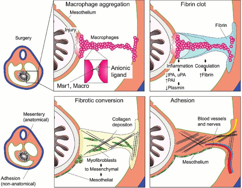
Zwicky et al. Serosal Repair and Adhesion Formation
membrane called mesothelium (2). The somatic mesoderm gives filled cavities from surrounding tissues (Figure 2). Together with
rise to the parietal layer of the mesothelium which lines the body the associated sub-mesothelial connective tissue the serosa is also
wall, and the splanchnic mesoderm gives rise to the visceral layer of called peritoneum, pleura, and pericardium in the peritoneal
the mesothelium which lines the surfaces of organs. The intra- (abdominal), pleural and pericardial cavities, respectively. In
coelomic organs stay connected to the body wall by elongations practice, the terms mesothelium, serosa, and peritoneum (or
referred to as mesenteries which contain blood vessels, lymphatics, pleura or pericardium) are often used interchangeably.
and nerves (1) (Figure 1). The peritoneum is less than 25 µm thick in the mouse (3) and
Later, the coelomic cavity is further subdivided resulting in about 50-100 µm thick in humans (4, 5). Therefore, as we discuss
three embryologically related but anatomically distinct anatomical injury at serosal surfaces, it is important to note that the
compartments: the pericardial cavity, the pleural cavities, and the mesothelium will rarely be injured in an isolated fashion. In
peritoneal (abdominal) cavity. All of these contain vital organs fact, serosal injuries will often compromise the tissues that are
such as heart, lung, and abdominal organs (1). covered by the mesothelium as well. These underlying tissues can
The serous membrane that covers the walls of all coelomic be vastly different such as:
cavities as well as the borders of all organs contained within them is
also called the serosa and is comprised of a flat monolayer of - smooth-muscular wall of the intestines, urinary bladder, uterus,
mesothelial cells. The serosal linings ensure friction-less movement - parenchymal tissue of heart, lung, liver, gallbladder, spleen
of organs and establish a water-tight barrier separating the fluid- (only mouse), ovaries,
A B C
FIGURE 1 | Development of the intra-embryonic coelomic cavity. (A) Schematic cross section human embryo of 3 weeks age. The mesoderm shows a somatic
(dorsal) and splanchnic (ventral) aspect. (B) Cranio-caudal and latero-lateral folding in week 4. (C) After closure of the anterior abdominal wall the intra-embryonic
coelomic cavity is formed. Organs (e.g. gut) are suspended by dorsal and sometimes ventral (not shown) mesenteries carrying blood vessels and nerves.
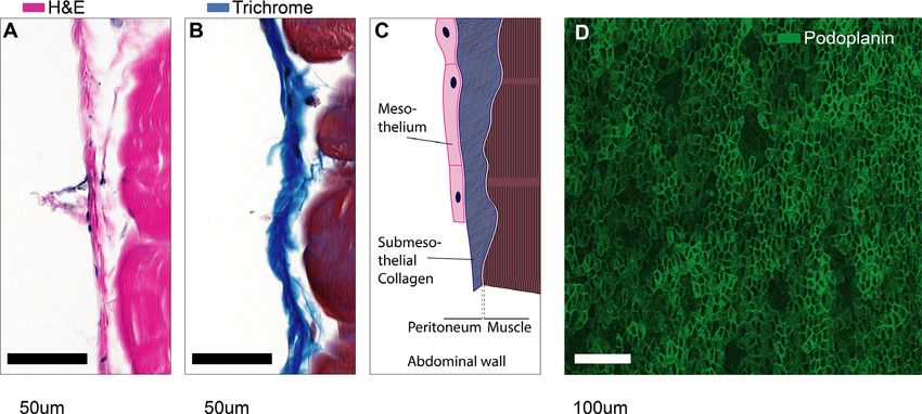
FIGURE 2 | Microanatomy of mesothelial surfaces. (A, B) Cross sections of mouse abdominal wall stained with Hematoxylin & Eosin (A) and Masson’s trichrome
staining (B). Scale bars: 50 µm. (C) Illustration of the structures shown in (A, B). (D) Top view on mesothelial surface stained with anti-podoplanin antibody.
Frontiers in Immunology | www.frontiersin.org 2 May 2021 | Volume 12 | Article 684967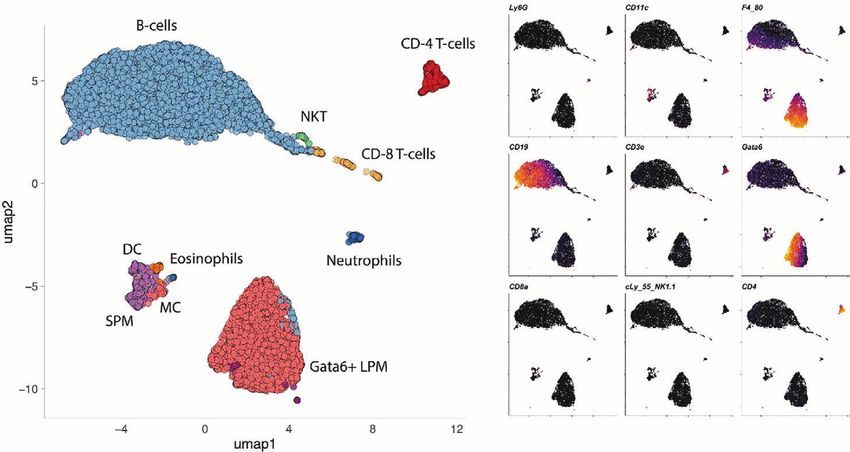
Zwicky et al.                                                                                                               Serosal Repair and Adhesion Formation

- fat tissue of the omentum,                                                        macrophages (SPM) are monocyte-derived, constantly replenished
- striated muscle, fascia, and bone of the thoracoabdominal wall                    and can be recruited within hours in significant amounts (14). At
    and diaphragm,                                                                  baseline, they account for about 5% of all immune cells or about
- and connective tissues such as that of the pericardium.                           10% of all macrophages (14, 18). The majority (90%) of peritoneal
                                                                                    macrophages belong to a distinct tissue-residential macrophage
    Any experimental model system that studies serosal wound                        population. Since these resident cells are slightly larger than their
repair, may invoke some underlying tissue-specific wound repair                      monocyte-derived sisters, they are also referred to as large peritoneal
mechanisms. This review is targeted at serosa specific mechanisms,                   macrophages (14). The large peritoneal macrophages (LPM) are a
but we ask the reader to bear in mind that we use the generalization                self-renewing population characterized by the expression of CD102
“at serosal surfaces” inductively; some of the mechanisms discussed                 (Icam2), high levels of F4/80 and the transcription factor GATA6
here may apply only to specific locations within the coelomic cavity.                (19–22). GATA6+ LPM seem to be well conserved when comparing
                                                                                    the different coelomic cavities of mice and human (23–25).
                                                                                    Canonically, these GATA6+ cavity macrophages are thought to
                                                                                    clear bacteria by phagocytosis (14, 26) and also by inducing intra-
CELLS SUSPENDED IN                                                                  abdominal formation of fibrin clots that immobilize bacteria (21). In
COELOMIC CAVITIES                                                                   primordial species such as the purple sea urchin (Strongylocentrotus
                                                                                    purpuratus), coelomocytes are also crucial for tissue repair, in
The coelomic cavities are filled with fluid that suspend millions
                                                                                    addition to clearing toxins and pathogens (27–30). The
of cells also referred to as coelomocytes. The coelomocyte
                                                                                    importance of GATA6+ cavity macrophages in damage
composition of mice and humans has been reviewed elsewhere
                                                                                    recognition and tissue repair will be discussed in detail.
(6). Briefly, the human peritoneal cavity suspends a total of 107
leukocytes in 5-100ml of peritoneal fluid (6, 7). In mice, the
number of peritoneal leukocytes varies between strains from 3 to
5x10⁶ cells (8). The pleuropericardial cavities contain 0.3-1x10⁶                   DAMAGE RECOGNITION
leukocytes per mouse (6, 9, 10). Most leukocytes in the peritoneal                  AND INFLAMMATION
cavity are lymphocytes (10-60%) and macrophages (40-60%) (8,
11–16). In addition, the peritoneal cavity contains dendritic cells                 Wound repair at large starts with inflammation. Inflammation is
(2 – 6%) (12, 17), mast cells, eosinophils, neutrophils (0-31%),                    induced when a significant deviation from homeostasis is detected.
innate lymphoid cells (ILCs) including natural killer cells and                     According to the current paradigm, such a deviation could be the
mesothelial cells (14, 16) (Figure 3).                                              presence of microbes (infection) or damaged tissue (injury). The
    In terms of wound healing, the role of peritoneal macrophages is                innate immune system has developed an effective arsenal of
best established. Macrophages make up 40-60% of all coelomocytes                    surveillance cells that constantly probe their microenvironment
in both mice and humans. Two major subpopulations of peritoneal                     for deviations from homeostasis. On a molecular level, deviation
macrophages have been described (14). The small peritoneal                          from homeostasis is defined by the occurrence of pre-specified

                              A                                                         B

 FIGURE 3 | Cells in a mouse coelomic cavity. (A, B) Peritoneal cavity lavage of healthy C57Bl/6 mice. Dimensionality reduction dimension 1 (umap1) and 2 (umap2)
 of myeloid lineage markers (mass cytometry) are plotted on x- and y-axis, respectively. Dots (cells) are colored by cluster (A) or marker (B). Data with kindly
 permission from M. Dosch and G. Beldi.

Frontiers in Immunology | www.frontiersin.org                                   3                                          May 2021 | Volume 12 | Article 684967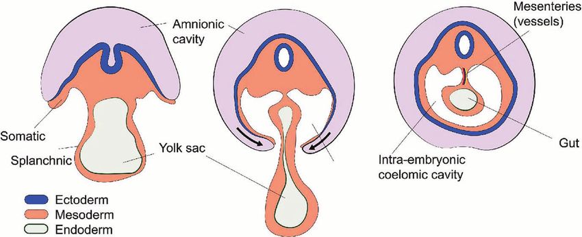
Zwicky et al.                                                                                                   Serosal Repair and Adhesion Formation

molecular patterns. Immunostimulatory molecular patterns that                Mesothelial Damage Is First Recognized
induce inflammation in case of sterile injury, i.e., in the absence of        by Cavity Macrophages
pathogens and their products, have been termed damage associated             Recent advances in intravital microscopy have allowed to
molecular patterns (DAMPs). DAMPs have been extensively                      characterize the sequence of cells recruited to mesothelial
reviewed elsewhere (31). In brief, DAMPs comprise different                  injuries. By using resonant-scanners, multi-photon excitation,
molecules that are not normally present outside of cells such as             and extremely sensitive hybrid detection systems it became
double stranded DNA, nuclear proteins, mitochondrial DNA,                    possible to image the peritoneal cavity through the intact
mitochondrial proteins, and molecules with high cytosolic                    abdominal wall under real-life conditions (21, 44). Second,
concentrations such as ATP or K+ Ions. In addition, damaged                  multi-photon imaging allows the use of near-infrared
cells may induce the production and release of additional DAMPs              microscopy lasers to induce focal thermal injuries during
(iDAMPs) such as heat shock proteins, defensins, galactins and               intravital microscopy with high precision (44–46). By combining
interleukin 1 (IL-1). Furthermore, if proteins that are constitutively       intravital microscopy of the abdominal cavity with peritoneal laser
present in the extracellular space such as hyaluronan, biglycan,             injuries, we were able to image cellular recruitment to mesothelial
heperansulfate and other extracellular matrix (ECM) components               injuries. Surprisingly, the first GATA6+ cavity macrophages
are modified by injuries, they can also become DAMPs. Under                   attached at the injuries within only a few seconds and the
homeostatic conditions, the serosal surfaces are covered with                macrophages completely covered the lesions after 15 minutes of
glycoconjugates such as sialomucins, hyaluronic acid, and                    imaging (44). The recruitment of cavity macrophages to
glycoproteins like fibronectin (32–35). These molecules contain               mesothelial injury was significantly faster than that of
large anionic sites that cover the serosal surfaces with a negatively        neutrophils, which needed much longer (> 40 minutes) (44).
charged coat—also referred to as the glycocalyx—that may help to             Cavity macrophages were present in the peritoneal fluid in vast
repulse invading microbes (32) and ensure friction-less movement             numbers and traversed the peritoneal cavity in a seemingly
of intra-coelomic organs (35). The loss of this negatively charged           random fashion within respiration-dependent movement of
coating due to serosal injury, may serve as mesothelium-specific              peritoneal cavity content (44). The observations that these cells
DAMP or “touch me signal” (36).                                              seemed to rely on passive transportation by peritoneal fluid, and
    Molecules that allow eukaryotic cells to detect the presence of          that they—upon contact with cell already adhering to the injury—
DAMPs have been termed pattern recognition receptors (PRR).                  were forming stable cell-cell aggregates were very reminiscent of
The expression of PRR such as toll-like receptors 1 through 6                the platelet aggregation that took place when a nearby blood vessel
(TLR-1-6), nucleotide-binding oligomerization domain (Nod)-1                 wall was damaged using laser injury. We concluded that cavity
and Nod-2 and advanced glycation end product (AGE)                           macrophages randomly “patrol” the serosal surfaces in a platelet-
receptors, has been demonstrated for murine and human                        like fashion and rapidly form aggregates in response to DAMPs.
mesothelial cells (37). Upon activation, mesothelial cells release           This is consistent with a previous electron microscopy study by
cytokines and inflammatory mediators such as chemokine (C–C                   Haney showing that peritoneal macrophages invariably detected
motif) ligand 2 (CCL2), CCL5, (C–X–C motif) ligand 8                         and migrated to injuries of the peritoneal membrane (47). In
(CXCL8), and nitric oxide (38, 39). Furthermore, mesothelial                 addition, Wang and Kubes showed that cavity macrophages were
cells upregulate adhesion molecules that presumably facilitate               able to detect mesothelial injuries of the liver capsule and migrated
the migration of inflammatory leukocytes across and along                     to the injured liver (36). On a molecular level, this interaction
serosal surfaces. These include intercellular adhesion molecule-             occurred independent of integrins or selectins, instead peritoneal
1 (ICAM-1), vascular cellular adhesion molecule-1 (VCAM-1),                  macrophages relied on different receptor molecules such as
E-cadherin, N-cadherin, CD29 and CD44 (38, 40–42). It is                     macrophage receptor with collagenous structure (MARCO),
important to note that cellular adhesion molecules expressed                 Macrophage scavenger receptor 1 (MSR1), CD44, and
by mesothelial cells play a dual role in serosal wound repair.               purinergic receptor P2X7. The respective DAMPs recognized by
While an initial upregulation may facilitate leukocyte                       CD44 and P2X7 are hyaluronan and ATP respectively (36). The
recruitment, these molecules, especially E-cadherin, are                     ligands that mediate MARCO and Msr1 dependent macrophage
downregulated later during serosal wound repair. The latter is               aggregation are yet to be identified (44).
associated with loss of mesothelial cohesion enabling the                        The function of peritoneal macrophages in sterile injury is multi-
mesothelium to switch to a more mesenchymal program, a                       facetted. Current models indicate that ligation of DAMPs to PRR on
process that we will discuss in detail below. In addition,                   macrophages leads to their inflammatory polarization—also
mesothelial cells modulate inflammation by synthesis and                      referred to as M1 polarization. This activation would result in the
release of hyaluronan (43), which is able to sequester free                  production of pro-inflammatory cytokines such as tumor necrosis
radicals and initiate tissue repair responses (38).                          factor (TNF) and IL-1 (31, 48). However, peritoneal macrophages
    In vivo, the initiation of inflammation at serosal surfaces does          recruited to sterile liver injury were shown to skew their phenotype
not rely on mesothelial cells alone but on a series of events. These         towards alternative or repair polarization—also referred to as M2
comprise specialized cellular and humoral immune mechanisms                  macrophages—increasing their expression of CD273, CD206 and
such as leukocyte recruitment, complement activation and                     Arginase 1 (36). Interestingly, Uderhardt et al. recently investigated
production of natural antibodies. In the rest of this chapter, we            the resident tissue macrophages of the muscular abdominal wall.
will discuss these elements one by one.                                      The abdominal wall macrophages are distinct from the peritoneal

Frontiers in Immunology | www.frontiersin.org                            4                                     May 2021 | Volume 12 | Article 684967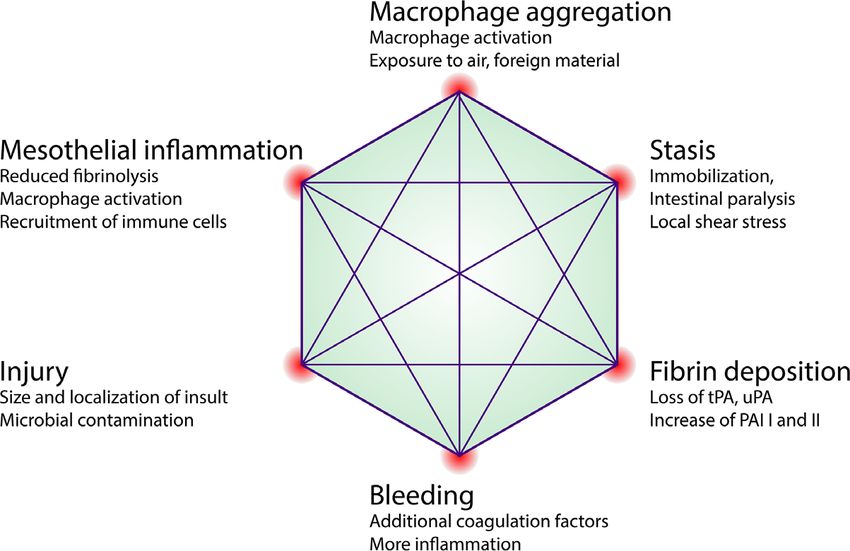
Zwicky et al.                                                                                                   Serosal Repair and Adhesion Formation

cavity macrophages suspended in the peritoneal cavity. They                  indicate that the aggregation of cavity macrophages in response to a
proposed that abdominal wall macrophages can extend their                    strong stimulus, such as peritonitis, causes inflammation. However,
pseudopods toward local injury sites within a radius of 100-                 in the case of smaller insults such as focal injuries or localized
150µm. In their study, resident tissue macrophages were able to              microbial challenges, MDR may compartmentalize the insult, in
completely enclose lesions if their size was below a certain threshold       analogy to the cloaking mechanism described for macrophages of
(microlesions). This—as the authors termed it—cloaking                       the muscular abdominal wall (45). Along those lines, complete
mechanism, was able to block scouting neutrophils from                       MDR could be interpreted as a threshold above which all
interacting with DAMPs and thus prevented subsequent                         macrophages have been “used up” indicating that the attempt at
neutrophil driven inflammation and tissue destruction (45). The               cloaking the insult has failed, which in turn results in inflammation.
cloaking mechanism was described for tissue resident macrophages             Either way, it would be important to study the largely unknown
in the muscular abdominal wall, i.e., on the far side of the                 (intracellular) changes in macrophages undergoing a disappearance
mesothelium with respect to the coelomic cavity. It needs to be              reaction in sterile and microbial models.
determined whether scavenger receptor mediated macrophage
aggregation on the coelomic site of the mesothelium causes                   Dendritic Cells and Mast Cells
inflammation or whether aggregation of cavity macrophages                     The peritoneal cavity harbors CD11c+ dendritic cells as well as
serves to contain injuries and is therefore—in essence—anti-                 cKit+ mast cells both of which are canonical initiators of
inflammatory. Ultimately, aggregation of peritoneal cavity                    inflammation. Their role as antigen presenting cells and
macrophages in response to mesothelial injuries was shown to                 inducers of inflammation in response to bacterial infection is
improve tissue repair (36, 44, 47).                                          well documented. In fact, CD11c+ dendritic cells are required for
                                                                             survival in murine polymicrobial peritoneal sepsis (66). In
Cavity Macrophage                                                            addition to pathogen-derived ligands for PRR, several DAMPs
Disappearance Reaction                                                       have been shown to interact with dendritic cells and dramatically
Aggregation of peritoneal macrophages causes their number in                 affect their function (67, 68). Interestingly, the response of
the peritoneal lavage to drop. The decrease in their number was              dendritic cells to DAMPs is not always clear-cut, with different
correlated with the injury size (44). With larger injuries of the            responses depending on dendritic cell subtypes and location (67).
mesothelium, such as a surgical laparotomy, the number of                    For example, activation of dendritic cells in sterile liver injury
GATA6+ cavity macrophages in the peritoneal lavage was                       leads to the secretion of anti-inflammatory cytokines such as IL-
reduced to zero (44). In other words, these cells disappeared                10 and TGF-b (67) while similar injury models of kidney and gut
from the peritoneal fluid (lavage). However, this was not the first            may lead to a pro-inflammatory response and secretion of IL-6,
time, the sudden absence of macrophages was observed. In fact,               IL-12 and TNF-a (67, 69). So far, the response of peritoneal
over half a century ago, Nelson and Boyden described a sharp                 dendritic cells to serosal injury is not well understood and
decline of macrophage count in peritoneal exudates in response               requires further studies. Mast cells have traditionally been
to a hypersensitivity reaction to tuberculin in Bacille Calmette-            studied in the context off helminthic infections and Ig-E
Gué rin (BCG)-vaccinated guinea pigs. They termed this the                  mediated reactions. It becomes clear, that mast cell
“macrophage disappearance reaction” (MDR) (49). Since then,                  degranulation is also an important modulator of wound
various insults (sterile and microbial) to the peritoneal                    healing of skin wounds (70) and lesions in the gastrointestinal
compartment have been found to induce the MDR (Table 1).                     tract (71–73). Poerwosusanta et al. investigated the role of mast
    These studies indicate that the MDR is not a specific reaction but        cell degranulation in mesothelial injury. Mesothelial injury was
arguably follows any inflammatory challenge to the peritoneal                 carried out by performing laparoscopic surgeries in rats at
compartment. While some reports indicate that peritoneal                     different intra-abdominal inflation pressures (74). They showed
macrophages can leave the peritoneal cavity through the draining             that an increased intraabdominal pressure—and presumably
lymphatics (52, 60, 64), most of the more recent reports suggest that        increased stress to the mesothelium—led to an increased
peritoneal macrophages have the tendency to adhere to each other             number of mast cells that infiltrated the mesothelium. This
(aggregate) as well as to the mesothelium in response to challenge           was correlated with increased mast cell degranulation. This
(Table 1). Therefore, the loss of dispersion and cellular aggregation        increased mast cell count is consistent with findings from skin
are a commonality among the different models of MDR. The MDR                 injury models and is due to chemokine-dependent mast cell
correlates with increased inflammatory cytokine levels in the                 immigration rather than local proliferation. More detailed
peritoneal fluid and the influx of pro-inflammatory leukocytes                  investigation, e.g. based on intravital microscopy, could help to
such as monocytes, eosinophils, and neutrophils into the                     elucidate whether mast cells are recruited to mesothelial injuries
peritoneal compartment (21, 59). Cailhier et al. used CD11b                  by blood or directly from the peritoneal cavity.
driven diphtheria toxin receptor and low dose intraperitoneal
injections of diphtheria toxin to selectively deplete resident               Humoral Pattern Recognition Molecules
peritoneal macrophages. In an experimental peritonitis model,                and Natural Antibodies
this resulted in a significant decrease of inflammation (infiltration           The fluid of the pleural and peritoneal cavity in mice and humans
of neutrophils) that could be restored by the adoptive transfer of           not only contains cells but also large amounts of proteins of the
resident, non-transgenic, peritoneal macrophages (65). These data            coagulation system and complement system as well as large

Frontiers in Immunology | www.frontiersin.org                            5                                     May 2021 | Volume 12 | Article 684967
Zwicky et al. Serosal Repair and Adhesion Formation
TABLE 1 | Macrophage disappearance reaction (MDR). Studies describing MDR from 1963 until now.
MDR Trigger (dose) Time between trigger and Postulated fate of disappeared Molecular mechanism Reference
complete MDR macrophages
Sterile Models
Sterile mesothelial injury 3h Form stable cell-cell aggregates that Scavenger receptors, can be blocked with (44)
(surgery, laser) cover injury and induce post-surgical Heparin and Poly-(I)
adhesions
Sterile Brewer’s Thioglycollate 12-72h Macrophage cell death Not demonstrated (50–52)
Antigen, migration inhibitory 1 to 96h Undergo activation during MDR in MDR Inhibited by Heparin, L-Fucose, (53)
factor, viruses or tumor cells delayed type hypersensitivity or acute Hyaluronidase
inflammatory reaction and then
reappear activated to regulate
responses toward pathogens or tumor
cells.
Egg Antigen (10ug), purified 5h Macrophage activation Desensitization suppress MDR, in (54)
protein derivate (10ug) sensitized animals normal MDR
Tuberculin 2.5 - 6h Not demonstrated MDR completely inhibited by Heparin and (49, 55)
Warfarin
Ova peptide (50ug) into mice 5h Macrophage adhesion Suppressed in fibrinogen- (56)
bearing antigen-primed T cells deficient mice, partially suppressed by
thrombin antagonist
Thrombin (20 Units) 1h -5h Macrophage adhesion MDR suppressed (56)
in fibrinogen-deficient mice.
RGES Peptide 48h Macrophage bind the mesothelium Integrin-mediated mechanisms involving (57)
overlying draining lymphatics VLA-4 and VLA-5 that can be blocked by
RGD (Arg-Gly-Asp peptides) and VLA-4
and VLA-5 blocking antibodies.
Microbes or microbial products
E. coli (5×10⁷ UV-inactivated) 20d Do not undergo fas-mediated apoptosis No difference in fas-deficient mice. (58)
S. aureus 2h (2 × 10⁷) Not demonstrated Not demonstrated (59)
Lipopolysaccharide 3h (10 µg) Accumulation in the omentum Macrophage interaction with mesothelial (19, 40,
5h (1 µg) cells, mainly of the omentum, was 56)
proposed to be a key step in MDR.
Partially inhibited by refludan.
Zymosan 3-4h (1mg) Form large clots to trap MDR reversed completely with Heparin (21, 51,
4h (0.5mg) microorganisms; adherence with tissue and partially with Hirudin/loss Factor V 60–62)
3d (10 µg) and drained to lymph node Expression/loss of Integrin activation
adaptor talin-1 Expression/TF deficiency
INF-g (100 U/mL) + LPS 20h Binding to mesothelial cells Monocyte activated by in vitro exposure to (40)
(100ng/ml) LPS and INF-Y bound with increased
efficiency to mesothelial cells
Synthethic Lipopetid 12h Not demonstrated Not demonstrated (59)
(Pam3CSK4)
Human studies
Bacterial peritonitis 1 day Shedding of surface CD206 Depletion of CD206+ LPM at day 1 of SPB (63)
Peritonitis with normalization to steady
state after resolution of SPB
Liver cirrhosis associated Not demonstrated Severity of liver disease and liver cirrhosis (25)
events (Bacterial peritonitis, are correlated with reduced numbers of
encephalopathy, death) CrIghi macrophages. Human CrIghi
macrophages were transcriptionally similar
to mouse F4/80hi peritoneal macrophages.
amounts of natural antibodies (75, 76). In the peritoneal and correlation of the invasiveness of the procedure with the
pleural cavities, the complement proteins are produced by amount of complement used, indicating that sterile injury
mesothelial cells (75, 77). The complement system is an leads to complement activation in humans (84). Inversely,
ancient enzymatic cascade of proteins with the main function humoral molecules that are canonically associated with innate
of opsonization and lysis of bacteria (78). The alternative immunity, have been shown to mediate tissue repair and regulate
pathway of the complement can be activated by injuries and in fibrosis. For example, pentraxin 3 (PTX3) was shown to reduce
the last decade a role of complement activation in wound healing fibrin deposition and fibrosis in several wound models outside of
(79) and regeneration (80, 81) as well as morphogenetic and the peritoneal cavity. While to our knowledge, no humoral
developmental processes (82, 83) has been suggested. molecule with anti-fibrotic properties has been described in the
Furthermore, some clinical studies have evaluated the role of peritoneal cavity, the discovery of such could have great
blood complement during major surgery and described a therapeutic potential (85–87).
Frontiers in Immunology | www.frontiersin.org 6 May 2021 | Volume 12 | Article 684967Zwicky et al. Serosal Repair and Adhesion Formation
Like complement factors, natural antibodies are abundant in an abdominal adhesion, a pathology we will discuss in more
the coelomic cavity fluids and are primarily thought to combat detail below. See the Perspective Box 1 speculating on parallels
microbes by recognizing a wide variety of different microbial between vascular and mesothelial coagulation control.
antigen patterns (88). The peritoneal natural antibodies are
mainly produced by self-replenishing peritoneal B1 cells in an
antigen-independent manner (89). The repertoire of natural
antibodies also enables recognition of self-antigens such as RECRUITMENT OF NEUTROPHILS
phosphorylcholine, phosphatidylcholine and carbohydrate
AND MONOCYTES
determinants. Furthermore, natural antibodies have been
shown to sense apoptotic cells (88) and electronegative We discovered that the first cells recruited to mesothelial injuries are
DAMPs (90). In addition, natural antibodies were able to the peritoneal cavity macrophages (44). These cells reside suspended
accelerate wound healing by recruiting additional wound in the peritoneal fluid (19) and are recruited directly from their
macrophages (91). Along those lines, Grönwall et al. postulate suspensive state to the mesothelium in case of injury. This
that natural IgM antibodies are part of a synapse between an comprises a special case of leukocyte recruitment that is unique to
apoptotic cell (that binds IgM) and the phagocyte. This synapse coelomic cavities. The canonical route of leukocyte recruitment is
is mediated by complement and complement receptors from the blood stream. The processes of leukocytes leaving the
expressed by phagocytes (92). There is increased interest in blood stream have been referred to as leukocyte adhesion cascade
studying the role of natural IgM, and IgM-dependent, and trans-endothelial migration. The underlying mechanisms have
complement-mediated phagocytosis in several disease models been revisited and reviewed most comprehensively by Nourshargh
(93–95). Although available data is limited, it is conceivable that et al. (97, 98). In perfused organs such as muscle or liver, neutrophils
complement and natural antibodies of body cavities play an are recruited within 30 minutes to the inflammatory site from the
important a role in wound repair at serosal surfaces. bloodstream (46, 99). In the mesothelium, neutrophils are the first
cells that arrive after peritoneal macrophages, after about 40-60
minutes (44). In response to focal sterile injuries, neutrophils show
an extremely high degree of coordination. While intravascular
COAGULATION AND FIBRIN DEPOSITION chemokine gradient seems to be very important for successful
directional migration over the initial distance within the vessel
It is widely accepted that inflammation of the peritoneal, pleural,
(31, 99), different molecules are the most potent chemotactic stimuli
or pericardial compartment, is associated with fibrin exudates.
once the neutrophils are close to the wound (31, 99, 100). This has
After mesothelial injury, the resulting inflammation as well as the
been demonstrated for molecules such as N-formyl peptides, ATP,
injury itself, lead to the activation of tissue factor pathway and
and leukotriene B4 (LTB4) and their respective receptors on
the inhibition of the antithrombin-III (AT-III) pathway (96).
neutrophils called formyl peptide receptor, P2Y2 receptor and
This is followed by the spontaneous cleavage of fibrinogen and
LTB4 receptor (31, 46, 99–102). These so-called gradients and
cross-linking of fibrin. In addition to inflammation, injury causes
autocrine feedback loops (LTB4) have been established for solid
the destruction of and consequent leakage from blood vessels of
organs such as the liver or muscle. Whether neutrophils rely on
the abdominal wall. Extravasation of blood results in the
similar mechanism to reach serosal surfaces has not been
activation of the canonical coagulation cascade which further
demonstrated yet but the technical advances of the last years will
enhances the deposition of fibrin (96). In vivo, the deposition of
now allow us to address these questions using intravital microscopy.
fibrin is limited at serosal surfaces by activated plasmin which
Although the canonical role of neutrophils is to clear
continuously degrades fibrin. Plasmin levels are regulated by
microbes, several reports suggest that they are imperative for
plasminogen activators tPA and uPA and their respective
timely restoration of tissue architecture after sterile injury by
plasminogen activator inhibitors 1 and 2 (PAI 1, PAI 2) (96).
clearing necrotic material (99) and producing growth factors
Homeostatic mesothelial cells produce large amounts of Plasmin,
such as transforming growth factor b (TGF-b) and vascular
uPA and tPA and low amounts of PAI1 and PAI2. Therefore, the
deposition of fibrin at serosal surfaces is tightly controlled.
During inflammation however, the mesothelial production of
PAI1 and PAI2 is significantly increased resulting in a BOX 1 | Perspective.
mesothelial program switch from fibrinolytic towards anti- We find it intriguing that fibrin clots after surgery are not focally limited to sites of
fibrinolytic state, resulting in the visible deposition of fibrinous injury but seem to be formed at distant sites as well while other regions of the
exudates (96). peritoneal cavity appear to be protected. During sepsis, spontaneous
The formation of a stable fibrin clot—also referred to as fibrin disseminated intravascular coagulation (DIC) is a major clinical problem. The
distribution of fibrin clot deposits during DIC is poorly understood. Interestingly,
matrix or cross-linked fibrin—serves as the scaffold for the the response of mesothelium and endothelium to inflammation share certain
subsequent wound repair (granulation) tissue. This includes similarities and both lead to the spontaneous formation of disseminated fibrin
the infiltration of leukocytes and mesenchymal precursors and clots. Studying the analogy between these two pathologies, that both involve an
results in the deposition of ECM, ingrowth of vessels and nerves epithelial-like monolayer of mesodermal origin, may lead to the identification of
common patterns and molecules governing the respective phenomena of DIC and
and finally, the re-mesothelialization of the injured serosa. If
peritoneal adhesions.
these fibrin clots grow too large, they can be the starting point for
Frontiers in Immunology | www.frontiersin.org 7 May 2021 | Volume 12 | Article 684967Zwicky et al. Serosal Repair and Adhesion Formation
endothelial growth factor (VEGF) (103–105). Furthermore, once peritoneal cavity fluid. In fact, within a few hours after
neutrophils have cleared all necrotic tissue, they starve each other of extensive mesothelial injury (surgery), large numbers of Gr1+
DAMPs. This leads to neutrophils becoming apoptotic and cleared cells (neutrophils) and monocyte derived macrophages were
by macrophages, which is another critical step for tissue repair as it recruited into the peritoneal cavity (119). This is consistent
enables macrophages within the wound to switch from an with observations in humans undergoing surgery where
inflammatory to an anti-inflammatory, pro-resolution program billions of neutrophils and monocytes can be isolated from the
(99, 103, 106). In this environment, pro-resolution macrophages peritoneal fluid. This recruitment was proposed to be driven by
and neutrophils start to produce factors that inhibit the recruitment chemokines such as MCP-1 and CXCL1 that were released into
of additional neutrophils and enhance pro-resolution properties of the peritoneal cavity by mesothelial cells in response to injury
macrophages. These factors include lipoxin A4, resolvins, protectins (119, 120). It is not known what molecules and mechanisms
(107), phosphatidylserine containing microvesicles shed by govern the migration of leukocytes from the interstitium, into the
neutrophils, chemokine sequestration through CCR5 modulation peritoneal fluid, where these cells adopt a planktonic form once
by neutrophils (108), Annexin A1 released from neutrophil granules again. Further, it would be interesting to investigate the
or in microvesicles (109) and IL-10 (110). Taken together, migratory patterns of neutrophils and monocytes after they
neutrophils play an important role in clearing debris and setting have reached their suspended state in the peritoneal cavity.
up a pro-resolution environment.
The second major leukocyte population recruited to
mesothelial injuries comprises inflammatory monocytes and SEROSAL (MESOTHELIAL) REPAIR
monocyte-derived macrophages. It is generally accepted that the
recruitment of neutrophils precedes the recruitment of monocytes So far, we have seen how a mesothelial injury leads to
in sterile injury (31, 111) but it remains controversial whether inflammation with the consecutive deposition of a fibrin
neutrophils recruitment is a necessary prerequisite for subsequent matrix and infiltration of immune cells. Normal serosal repair
monocyte recruitment (112, 113). Rather than being pre- is achieved when a) the underlying organ is repaired, b) the sub-
determined, the fate of recruited monocytes appears to be mesothelial connective tissue layer is restored to its original
largely dependent on the environment (62). In the context of composition and thickness and c) the integrity of the
sterile injury repair, monocytes have been shown to differentiate mesothelial membrane is reconstituted (31, 121, 122). The
into mature macrophages at the site of injury. This process has central role of the mesothelium in tissue repair and fibrosis has
been reviewed before (114). In brief, Ly6Chi monocytes are been revisited and comprehensively summarized (38).
recruited to the wound, where they gradually differentiate into The mesothelium is a slowly renewing tissue with less than 1%
Ly6Clow macrophages. One of the molecules necessary for this of cells undergoing mitosis at any time (123). After mesothelial
conversion is nuclear receptor subfamily 4, group a, member 1 injury, activated macrophages induce a pronounced proliferative
(Nr4a1) (115). In the peritoneal cavity, the influx of inflammatory expansion of the mesothelial compartment (124, 125). Genetic
monocytes due to injury or infection, correlates with the increase lineage tracing of mesothelial cells in several injury and disease
of MHCII+ CD102- GATA6- macrophages (26). These are also models indicate that regenerating mesothelium originates from
referred to as small peritoneal macrophages or bone marrow healthy mesothelium rather than submesothelial cells (126–128).
derived macrophages (14, 26). In the peritoneal cavity, PAI-1 Small focal mesothelial injuries that were induced using thermal
and CCL1 were shown to recruit macrophages to the wound probes on the liver capsule and abdominal wall, healed completely
involving the receptor molecules CD11b and CCR8 respectively without any visible defect left after days (36, 44). Because repair of
(116, 117). Depletion of macrophages using clodronate-loaded mesothelial defects is largely independent of the defect size,
liposomes or genetic constructs results in decreased wound investigators have proposed that mesothelial cells not only crawl
healing of sterile injuries of serosal surfaces of the liver and into the wound from the borders, but also detach from opposing
abdominal wall (36, 44). However, novel experimental strategies surfaces and distant sites and migrate in a free-floating state
will be necessary to experimentally isolate the role of recruited through the coelomic cavity until they settle on the wound (125,
macrophages compared to that of the resident GATA6+ 129, 130). This is further supported by the fact that adoptively
macrophages in mesothelial wound healing. Furthermore, transferred mesothelial cells improve mesothelial repair in the
monocyte-derived macrophages have been shown to replenish recipient (130). The response of mesothelial cells to injury can be
the resident GATA6+ macrophages. This process is dependent on summarized as proliferation, loss of epithelial cohesion and
the transcription factor IRF4 (118). The degree of this replacement migration. These phenotypic changes are reminiscent of other
strongly depends on the degree of initial macrophage serosal surfaces that undergo epithelial-to-mesenchymal transition
disappearance (50, 62). Taken together, this suggests a dual role (EMT). In analogy, this reaction has been termed mesothelial-to-
of infiltrating monocytes in wound repair: they act as precursors of mesenchymal transition (MMT) (131, 132). On a molecular level,
bone-marrow derived macrophages that are directly needed in MMT involves the downregulation of epithelial junctional
wound repair, and they can serve to replace the GATA6+ proteins such as E-cadherin and the upregulation of
resident macrophages. mesenchymal marker a-smooth muscle actin (a-SMA) and
Neutrophils and monocytes are not only recruited into the production of ECM (133, 134). These changes correlate with an
abdominal wall (45) but they are also recruited into the upregulation of transcription factors canonically associated with
Frontiers in Immunology | www.frontiersin.org 8 May 2021 | Volume 12 | Article 684967Zwicky et al. Serosal Repair and Adhesion Formation
EMT such as SNAI1, SNAI2, ZEB1, ZEB2, Twist1 (38) and can be deposition led to adhesion formation in many different injury
induced by exposing mesothelial cells to TGF-b1, hepatocyte models and different species (96) and became generally accepted
growth factor, platelet derived growth factor and IL-1b (38). We as the “classical concept of adhesion formation” (96). We have
will next discuss how mesothelial repair can become defective. seen that an exuberant inflammatory response induced by
peritoneal injury or infection promotes an increased
procoagulatory and antifibrinolytic reaction. On a molecular
POST-SURGICAL ADHESIONS level, inflammatory mediators increase the expression of tissue
factor (TF) and PAI-1 and decreases the expression of tPA in
Surgeries within body cavities such as the abdominal cavity are often mesothelial cells resulting in increased fibrin deposition (96). This
lifesaving procedures. Following surgical trauma, mesothelial repair mechanism was confirmed in peritoneal biopsies of inflamed
can lead to a restitution of serosal surfaces at integrum. However, in peritoneum of humans that underwent surgery. The reduction
some patients, the healing process is disrupted, leading to a fibrotic of fibrinolytic activity during inflammation was mediated by PAI-
complication called post-surgical adhesions. Adhesions are fibrous 1 (96). Importantly, a prospective study in humans showed that
bridges of various thickness and length containing blood vessels and PAI-1 concentrations in peritoneal fluid were correlated with the
nerve tissue (135, 136). Adhesions can also be caused by infection occurrence of adhesions after 8 days (96). Strategies to prevent
but today, surgeries comprise by far the most common cause of postoperative adhesion target the dysregulated fibrin clot
mesothelial injury leading to adhesions (96, 135). In the peritoneal deposition by either inhibiting coagulation, increasing
cavity, adhesions result in considerable morbidity as they impair the fibrinolytic activity, or reducing inflammation.
free movement of organs. These problems include potentially life- Administration of fibrinolytic (tPA) or anticoagulant agents
threatening intestinal occlusions, secondary infertility in women, (Heparin) significantly reduced adhesions in different animal
and chronic post-operative abdominal pain (96, 137, 138). models (38, 96, 141–144). However, the only study in humans
Peritoneal adhesions were described for the first time in 1836 in a that enrolled 102 patients, was unable to confirm this effect. In
post-mortem examination of a patient that had died from peritoneal this study 5000 I.U. of heparin were diluted in saline and used to
tuberculosis (139). It was then suggested in 1849 that these wash the peritoneal cavity. The patients in this study then
abnormal structures originate from lymphatic vessels that turn underwent a second operation (laparoscopy) 12 days after the
into fibrinous adhesions (135, 139, 140). Despite tremendous first, to obtain adhesion scores (145). The heparin dose that was
scientific advances since 1849 including the execution of many effectively administered in these patients was not reported but it
clinical and experimental studies on adhesions, understanding of must have been extremely low as most heparin containing lavage
their pathogenesis has not evolved enough to develop effective solution was removed after a few minutes. Based on titration
therapies. To date, few research and development resources are studies in an animal models, the threshold dose for significant
dedicated towards resolving this significant health problem. The anti-adhesion effect was (without occurrence of bleeding after
process of adhesion formation largely depends on the same two days) 7.5x10 U/kg/day, which is equivalent to 5250 I.U/day
mechanisms as “normal” mesothelial repair: Healing is initiated for a person weighting 70kg (142). Therefore, low heparin dose
by damage recognition, inflammation, and coagulation. These steps or low heparin concentration may be the reason no effect on
lead to the recruitment of leukocytes and the deposition of fibrin. adhesion formation was observed in the Jansen study. Further
Then, the stable fibrin matrix (fibrin clot) is infiltrated by studies would be necessary. However, whether the current
myofibroblasts that start to deposit ECM proteins such as collagen. evidence justifies a human trial testing high dose heparin in
The problem with adhesions is that wound healing occurs at major abdominal surgery remains to be discussed.
sites it should not (96). The classical paradigm of adhesion Others have tried to use anti-inflammatory agents to restore
formation states that if serosal surfaces cannot re-establish mesothelial fibrinolytic activity. Cyclo-oxygenase inhibitors and
homeostatic fibrinolysis soon after injury, excessive amounts of steroids were tested in animal models (146–148) with no
fibrin are deposited. We have discussed that macrophage resounding success. Experimental animal models demonstrate
aggregation accompanies fibrin deposition after sterile injury at potent prevention of postoperative adhesion following
serosal surfaces (3). Macrophage-fibrin deposits serve as the intraperitoneal application of HMG-CoA reductase inhibitors
basic scaffold for tissue repair. Clots that span the space (statins) (149). HMG-CoA reductase inhibitors stimulate
between opposing serosal surfaces are dangerous because they fibrinolytic activity in human peritoneal mesothelial cell cultures
can be converted into scars that permanently link these surfaces (150) and exert anti-inflammatory functions (151). In experimental
called adhesions. We will now discuss the events taking place in animal models amelioration of adhesion after administration of
more detail and highlight the respective therapeutic intraperitoneal acylated ghrelin, a 28-amino acid gastric peptide
considerations for each (Figure 4). with anti-fibrotic and anti-inflammatory properties, was
demonstrated. The adhesion prevention by ghrelin application
Macrophage Aggregation and Fibrin was modulated via blockage of the TGF-b signaling pathway (152).
Clot Formation Extending on this classical paradigm we have recently
The idea that mesothelial loss of baseline fibrinolytic activity after proposed yet another factor in adhesion formation: that of
surgical trauma may cause adhesions has been the subject of peritoneal macrophage aggregation. We showed that in
various animal models (96). Interestingly, hypofibrinolytic fibrin macrophage aggregation to focal peritoneal injuries was tightly
Frontiers in Immunology | www.frontiersin.org 9 May 2021 | Volume 12 | Article 684967Zwicky et al. Serosal Repair and Adhesion Formation
A B C
D E F
FIGURE 4 | Post-surgical adhesion formation. (A) Overview of the peritoneal cavity before surgery. (B) Non-focal mesothelial injury such as major abdominal surgery
leads to the uncontrolled aggregation of peritoneal macrophages serving as the nidus for the (C) subsequent Fibrin clot deposition. Inflammation and Coagulation
inter-dependently promote the deposition of fibrin (see text). (D) Overview during or after adhesion formation. The abdominal organs (e.g., intestine) are now attached
to the abdominal wall at anatomic (mesentery) and non-anatomic (adhesion) locations. (E) Mesothelial to mesenchymal transition gives rise to myofibroblasts that
migrate into the wound and into the fibrin clot where they start to deposit extracellular matrix (ECM) such as collagen. (F) Adhesion formation is completed when the
scar tissue is covered with mesothelium. The lesion may become fully perfused and pain-sensitive by ingrowth of blood vessels and nerves.
regulated with just enough cells aggregated to seal the defect. aggregation of macrophages was largely independent of fibrin
However, in response to large peritoneal injuries, such as crosslinking and macrophages showed the ability to aggregate ex
abdominal surgeries, the aggregation of these macrophages was vivo without addition of fibrin (44). Taken together, mesothelial
dysregulated, resulting in the formation of large super aggregates inflammation, macrophage aggregation, and coagulation, can act
that started to join mesothelial surfaces. We found that this cooperatively but do not necessarily depend on each other. The
process was dependent on scavenger receptors MARCO and relative contribution of each of these three processes likely depends
MSR1. Depleting peritoneal cavity macrophages or inhibiting on the type (sterile, microbial, combined) and strength of the insult
their aggregation significantly reduced the amount and severity as well as on local shear (3), which in turn is largely dependent on
of adhesions in a mouse model. We therefore propose an adaptation patient movement and post-surgical intestinal paralysis (Figure 5).
of the classical paradigm to include peritoneal macrophage
aggregation as an additional event (Figure 4). Before we discuss Fibrotic Conversion
the later events in adhesion formation such as fibrotic conversion Alpha smooth muscle actin (a-SMA) positive myofibroblasts are
and remodeling, we would like to note that the early process of considered the main collagen-producing cell in wound healing
adhesion formation such as mesothelial inflammation (chapter 3), and many fibrotic diseases (155–157). The question of the origin
macrophage aggregation (chapter 3) and coagulation (chapter 4) are of a-SMA positive myofibroblasts in adhesions has been a matter
tightly linked. We have discussed how inflammation directly affects of debate. Myofibroblasts in adhesions were believed to be either
coagulation. Inversely, coagulation provides a positive feedback to derived from the mesothelium or alternatively derived from sub-
the mesothelium further increasing inflammation. For example, mesothelial cells (126). Recently, Fischer at al. used a genetic fate
activation of proteinase-activated receptor-2 (PAR2) on mesothelial mapping to permanently and selectively label cells expressing
cells results in increased MIP-2 production and consecutive protein c receptor gene (Procr CreERT x Rosa26 tdTomato ).
neutrophil infiltration (153). Furthermore, peritoneal cavity Tamoxifen administration in these reporter mice resulted in the
macrophages were shown to produce coagulation factors (21, 23, selective and permanent labelling of approximately 50% of all
154) and Factor V produced by peritoneal macrophages was shown mesothelial cells but not submesothelial cells (127). Using this
to be essential for the clotting of peritoneal fluid in response to approach, they were able to show that the majority of platelet
bacteria (21). Inversely, they showed that macrophage aggregation derived growth factor receptor a positive (PDGFRa+)
(disappearance) was partially dependent on coagulation factors. In myofibroblasts in adhesions were of mesothelial origin (127).
other models such as laser-induced sterile mesothelial injury, the This is in line with older studies that relied on non-genetic
Frontiers in Immunology | www.frontiersin.org 10 May 2021 | Volume 12 | Article 684967Zwicky et al. Serosal Repair and Adhesion Formation
lineage tracing methods such as cell tracker dyes or lineage weeks post-surgery the nerve fibers were traversing the whole
markers to infer on source of myofibroblasts in adhesions (122, adhesion from coecum to abdominal wall (161). Human
158, 159). Taken together these data suggest that mesothelial to peritoneal adhesion specimens collected during surgery from
mesenchymal transition (MMT) is the major source of 25 patients contained invariably sensory nerves (162). The
myofibroblasts in adhesion pathogenesis. On a molecular level, sensory innervation could partially explain the chronic pain a
MMT in adhesion formation relies on the same pathways as lot of patients with adhesions experience. Animal studies
mesothelial repair (159). In fact, administration of TGF-b blocking revealed blood vessels in adhesions already 6 hours after injury
peptide P144 resulted in a significant reduction of adhesions in an (163). This process of remodeling from connective tissue to fully
experimental mouse model (159). This was associated with a innervated and vasculated tissue might be modulated.
reduced expression of MMT markers such as Snail, a-SMA and The nerve fibers in adhesions were often associated with blood
Collagen I in P144 treated mice (159). In addition, the exposure of vessels indicating angiogenesis could play a key part in regulating
mesothelial cells to cyclic mechanical forces was shown to increase ingrowth of nerves into adhesions (162, 164). Local production of
MMT in experimental murine and human models. Biomechanical VEGF by mesothelial cells appears to play a central role in the
induction of MMT cooperates with biochemical signals such as process leading to peritoneal angiogenesis (165). In different
TGF-b and seemed to be regulated by caveolin-1, a plasma murine models, postsurgical adhesion formation was reduced by
membrane mechanotransducer (160). Interestingly, MMT-cells inhibition of VEGF suggesting adhesion formation is
in adhesions also express many markers that are found in the angiogenesis-dependent (166, 167). In a human study including
mesothelium during embryonic development but not within the adhesions samples from patients years after first surgery,
adult mesothelium. These markers, including Mesothelin (MSLN), adhesions expressing VEGF A and its receptor showed
Uroplakin-1B and Wilms-tumor 1 (WT1), were upregulated in significantly higher numbers of immature vessels suggesting
adhesions indicating that adult mesothelial cells can repurpose ongoing angiogenesis in mature adhesions (163). In addition to
aspects of fetal development (158). Depletion of mesothelial cells angiogenesis, modulation of the ECM by matrix metallo-
results in a complete reduction of adhesions (127). However, proteinases (MMPs) takes place. MMPs are proteolytic enzymes
totally depleting the peritoneal cavity of potential myofibroblasts involved in degradation of ECM, their activity is opposed by
may compromise wound healing too much for use in the clinic. tissue-derived inhibitors of MMPs (TIMPs) (168). The expression
Strategies that inhibit MMT in adhesions but leave mesothelial of both VEGF and MMPs is upregulated during MMT (131). In a
repair intact need to be developed. human study, peritoneal samples were collected during initial
laparoscopy and during a second-look laparoscopy 48 hours later.
Remodeling Patients with pelvic adhesions exhibited significantly lower
After fibrotic conversion, adhesions are considered irreversible amounts of MMP-9 concentrations and significantly higher
and redundant scar bands. Furthermore, animal, and human MMP-9/TIMP-1 ratios when compared with controls (169). In
studies demonstrated the ingrowth of nerves and vessels into peritoneal fluid of patients with excessive adhesions, higher TIMP-
adhesions. In a murine model, nerve fibers in abdominal 1 levels could be demonstrated compared with those of patients
adhesions were detected already two weeks after surgery and 4 without adhesions (170). Mice treated with instillation of
FIGURE 5 | Factors influencing adhesion formation. Proposed concept of early local determinants that influence the binary outcome of adhesion formation and may
be exploited therapeutically. tPA, tissue plasminogen activator; uPA, urokinase-type plasminogen activator; PAI, Plasminogen activator inhibitor.
Frontiers in Immunology | www.frontiersin.org 11 May 2021 | Volume 12 | Article 684967You can also read