Overcoming immunotherapy resistance in non-small cell lung cancer (NSCLC) - novel approaches and future outlook
←
→
Page content transcription
If your browser does not render page correctly, please read the page content below
Horvath et al. Molecular Cancer    (2020) 19:141
https://doi.org/10.1186/s12943-020-01260-z

 REVIEW                                                                                                                                            Open Access

Overcoming immunotherapy resistance in
non-small cell lung cancer (NSCLC) - novel
approaches and future outlook
Lena Horvath1, Bernard Thienpont2, Liyun Zhao2, Dominik Wolf1,3 and Andreas Pircher1*

  Abstract
  Immunotherapy (IO) has revolutionized the therapy landscape of non-small cell lung cancer (NSCLC), significantly
  prolonging the overall survival (OS) of advanced stage patients. Over the recent years IO therapy has been broadly
  integrated into the first-line setting of non-oncogene driven NSCLC, either in combination with chemotherapy, or
  in selected patients with PD-L1high expression as monotherapy. Still, a significant proportion of patients suffer from
  disease progression. A better understanding of resistance mechanisms depicts a central goal to avoid or overcome
  IO resistance and to improve patient outcome.
  We here review major cellular and molecular pathways within the tumor microenvironment (TME) that may impact
  the evolution of IO resistance. We summarize upcoming treatment options after IO resistance including novel IO
  targets (e.g. RIG-I, STING) as well as interesting combinational approaches such as IO combined with anti-
  angiogenic agents or metabolic targets (e.g. IDO-1, adenosine signaling, arginase). By discussing the fundamental
  mode of action of IO within the TME, we aim to understand and manage IO resistance and to seed new ideas for
  effective therapeutic IO concepts.
  Keywords: NSCLC, Immunotherapy resistance, Tumor microenvironment heterogeneity, Targeted therapy

Background                                                                            Prospective clinical studies to demonstrate treatment strat-
Immunotherapy (IO) and particularly immune check-                                     egies following progression on IO therapy are still lacking.
point inhibitors (ICI), including programmed death re-                                  Various IO resistance mechanisms have been character-
ceptor 1 (PD-1) and PD-ligand 1 (PD-L1) inhibitors                                    ized, involving tumor cell intrinsic as well as environmen-
have revolutionized the treatment landscape of non-                                   tal resistance patterns. The tumor microenvironment
small cell lung cancer (NSCLC). Previously unantici-                                  (TME) plays a critical role by influencing both extrinsic
pated long-term responses in advanced stage disease                                   and intrinsic resistance pathways. A better understanding
have been accomplished, with a 5 year overall survival                                of the heterogenous TME will set stage for further opti-
(OS) of 20% in unselected and up to 40% in PD-L1high                                  mizing strategies and guide new avenues in future IO
expressing patients [1].                                                              treatment stratification.
  Despite the striking clinical improvements, the majority                              This review discusses the multitude of novel preclinical
of patients eventually fails to respond to ICI therapy due to                         and clinical treatment approaches that aim to overcome
the evolution of primary or secondary resistance.                                     IO resistance in NSCLC. The complexity of cellular and
                                                                                      molecular alterations within the immunosuppressive TME
* Correspondence: andreas.pircher@i-med.ac.at
                                                                                      build the fundament for designing rational and synergistic
1
 Internal Medicine V, Department of Hematology and Oncology, Medical                  combination therapies that lower the risk of resistance
University Innsbruck, Anichstraße 35, 6020 Innsbruck, Austria                         and prolong benefit from IO therapy.
Full list of author information is available at the end of the article

                                       © The Author(s). 2020 Open Access This article is licensed under a Creative Commons Attribution 4.0 International License,
                                       which permits use, sharing, adaptation, distribution and reproduction in any medium or format, as long as you give
                                       appropriate credit to the original author(s) and the source, provide a link to the Creative Commons licence, and indicate if
                                       changes were made. The images or other third party material in this article are included in the article's Creative Commons
                                       licence, unless indicated otherwise in a credit line to the material. If material is not included in the article's Creative Commons
                                       licence and your intended use is not permitted by statutory regulation or exceeds the permitted use, you will need to obtain
                                       permission directly from the copyright holder. To view a copy of this licence, visit http://creativecommons.org/licenses/by/4.0/.
                                       The Creative Commons Public Domain Dedication waiver (http://creativecommons.org/publicdomain/zero/1.0/) applies to the
                                       data made available in this article, unless otherwise stated in a credit line to the data.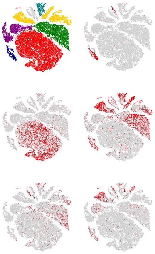
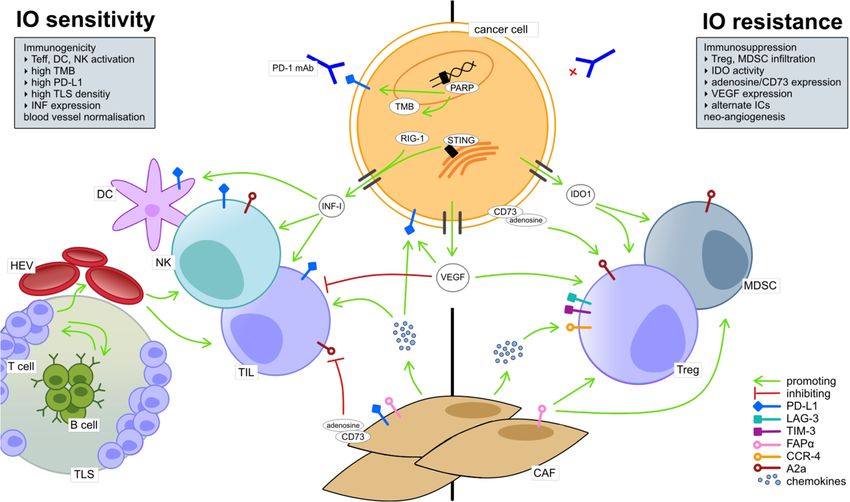
Horvath et al. Molecular Cancer     (2020) 19:141                                                                                      Page 2 of 15

Immunopathology of NSCLC and evolution of IO                                immune responses. The best studied IC are PD-1 and cyto-
resistance                                                                  toxic T lymphocyte antigen 4 (CTLA-4). PD-1 is broadly
IO resistance mechanisms result from the constantly                         expressed on CD8+ T lymphocytes, regulatory T cells (Treg)
evolving interactions between cancer cells and the sur-                     and natural killer (NK) cells and modulates T cell activity via
rounding cell populations within the TME, including im-                     interaction with its ligand (PD-L1) in the TME (Fig. 2).
mune cells, cancer-associated fibroblasts (CAF) and                         CTLA-4 is expressed on CD8+ and CD4+ T lymphocytes
tumor endothelial cells (TEC) (Fig. 1). The following                       and Treg and regulates early naïve T cell activation in sec-
section recapitulates the basic characteristics of the im-                  ondary lymphoid organs [3, 4]. Other IC are constantly being
munogenic TME, particularly focusing on IO response-                        discovered and under investigation for their clinical utility as
or resistance-mediating mechanisms and biomarkers.                          druggable IC (e.g. TIM-3, LAG-3 or TIGIT).

Immune checkpoints                                                          Lymphocytes of the tumor microenvironment
Immune checkpoints (IC) play a central role in negative                     T lymphocytes
regulation of T cell reactivity and their inhibition via mono-              Tumor infiltrating T lymphocytes (TIL) play a major
clonal antibodies can unleash T cell-triggered antitumor                    role in antitumor immune responses within the TME

 Fig. 1 Overview of the cellular TME composition and major molecular pathways associated with IO sensitivity (left) and resistance (right). IO
 sensitivity is depicted by an immunogenic TME, comprising the activation of effector immune cells (e.g. tumor infiltrating lymphocytes (TIL),
 dendritic cells (CD) and natural killer cells (NK)). Naïve T cells undergo activation and priming in close association to B cells within tertiary
 lymphoid structures (TLS). T effector cells transmigrate to the stromal TME compartment via high endothelial venules (HEV), tightly regulated by
 immunomodulatory tumor endothelial cells (TEC; not illustrated) in the HEV endothelium. Cancer cell intrinsic molecular pathways that enhance
 TME immunogenicity involve interferon type I (IFN I) expression, which is, amongst other stressors, induced by cytosolic RIG-I or by an activated
 STING pathway. IO sensitivity is enhanced in a TME with high PD-L1 expression by cancer and immune cells. High neo-antigen expression by
 cancer cells as result of high tumor mutational burden (TMB), e.g. induced by PARP inhibition, enhances TME immunogenicity and IO sensitivity.
 IO resistance is marked by an immunosuppressive TME and includes, on a cellular basis, infiltration of T regulatory cells (Treg) and myeloid
 derived suppressor cells (MDSC) as well as M2 macrophages (not shown). CD73 and, thus, adenosine expression by cancer cells or fibroblasts
 leads to inhibition of TIL and promotion of Treg; CD73 upregulation associates with cancer immune evasion. Also, up-regulation of alternative
 immune checkpoints e.g. LAG-3 and TIM-1 by immune cells enhances IO resistance. Cancer associated fibroblasts (CAF) depict both
 immunosuppressive and immunostimulatory functions, e.g. via chemokine release. Upregulation of the chemokine receptor CCR-4 is associated
 with IO resistance. Vascular endothelial growth factor (VEGF) gets ubiquitously expressed in the TME (not illustrated, see Fig. 2). It has
 immunosuppressive functions by inhibiting effector immune cells (e.g. TIL, NK, DC), upregulating inhibitory immune checkpoints (e.g. PD-L1) and
 by promoting Treg and MDSC. Tumor growth promoting neo-angiogenesis (not illustrated) is driven by hypoxia and, thus, VEGF expression
Horvath et al. Molecular Cancer      (2020) 19:141                                                                                        Page 3 of 15

 Fig. 2 The gene expression heterogeneity of the NSCLC TME, illustrated by gene expression in stromal and cancer cells. 52.698 single cells from 4
 non-malignant and 15 tumor samples of five patients were analyzed. (a-f) tSNE plots of the 52.698 cells, with (a) clusters color-coded according
 to the associated class of cell types, or with (b-f) cells colored according to the expression of the indicated marker gene, illustrating the
 heterogeneity of gene expression by the various cell types within the TME. Gene expression is shown ranging from grey to red (low to high). (e)
 CD274 is the gene alias for PD-L1. (f) NTE5 is the gene alias for CD73. (g) Expression levels of selected genes (gene alias in brackets), involved in
 immunomodulation in tumors shown separately for each cell type based on single cell RNA sequencing. Expression levels in cancer cells are
 shown separately for each patient, while the subtypes of T cells, innate immune cells, endothelial cells and fibroblasts represent pooled patient
 data. Data are derived from reference [2]

[5]. The phenotype of T cell infiltration varies strongly                    effects of therapeutic TLS neogenesis on anti-cancer im-
between different tumors in terms of quantity and distri-                    mune responses [10–12].
bution and is associated with IO efficacy. Classifying tu-
mors based on the cytotoxic T cell infiltration                              B lymphocytes
phenotype might help to rationally guide treatment                           Tumor infiltrating B cells harbor both immunostimula-
stratification [5].                                                          tory [13] and immunosuppressive [14] functions and
                                                                             their effect on IO efficacy is increasingly appreciated. Es-
Tertiary lymphoid structures                                                 pecially those B cells located in mature TLS may exhibit
In chronically inflamed areas such as tumors, B and T                        immunostimulatory functions by closely interacting with
lymphocytes are frequently organized in ectopic lymph-                       local T cells, thereby enhancing anti-cancer immunity.
oid aggregates, so-called tertiary lymphoid structures                       This hypothesis is indirectly supported by the observa-
(TLS), where they convert to effector cells upon antigen                     tion that intra-tumoral B cells are linked to a favorable
presentation. The cellular organization ranges from sim-                     IO response [7, 15, 16].
ple lymphocyte clusters (immature TLS) to highly so-
phisticated structures (mature TLS) [6, 7]. High                             Tumor Mutational Burden (TMB)
endothelial venules (HEVs) are found nearby and pro-                         Somatic mutations in the cancer genome, such as in
mote lymphocyte extravasation [8]. TLS display a surro-                      DNA repair genes including mismatch repair (MMR),
gate marker of prompt immune responses that actively                         homologous recombination (HR) or polymerase epsilon
modulate anticancer immunity [7]. High TLS density as-                       (POLE) increase tumor mutational and neoantigen bur-
sociates with a favorable prognosis in many cancer types,                    den, which has been linked to greater TIL density and
including NSCLC [9] and TLS may also enhance IO effi-                        enhanced ICI efficacy [17–19]. This observation is clinic-
cacy [6]. Preclinical studies demonstrated beneficial                        ally underscored as mutagen-driven cancer types (e.g.
Horvath et al. Molecular Cancer (2020) 19:141 Page 4 of 15
melanoma, NSCLC) typically show high initial ICI re- Clearly, PD-L1 expression is necessary to achieve ad-
sponses [17]. Moreover, components of the major histo- equate responses to PD-1/PD-L1 blockade and numerous
compatibility complex I (MHC I) such as B2M are often studies associated high tumor cell PD-L1 expression with
downregulated (Fig. 2), hence curbing neo-epitope pres- better outcomes to anti-PD-1/PD-L1 monotherapy in
entation to T cells [20]. Antigen presentation pathways NSCLC. Controversially, some patients with very low or
can also be inactivated through mutations (e.g. B2M is even absent PD-L1 expression show durable responses
mutated or deleted in about 5% of lung cancers) [21] [33], an observation currently lacking a mechanistic ex-
and also other pathway members are inactivated [22]. planation see 2.4.1. Besides cancer cells, also PD-L1 posi-
Importantly, IO may increase the frequency of such mu- tive immune cells may exert a predictive value. In the
tations [19, 23, 24] suggesting an active immune-editing Impower110 trial, presence of PD-L1 positive TIL signifi-
of cells failing to present neo-epitopes. cantly associated with enhanced OS in patients treated
Concerning TMB as predictive biomarker of ICI re- with atezolizumab [34]. These results are in line with
sponse, clinical trials report divergent results, possibly other tumor entities (e.g. bladder and breast cancer).
due to technical issues with TMB assessment (e.g. use of
inhomogenous cut-off values) [25]. On the one hand, PD-L1 is not yet a robust biomarker
high TMB was the strongest variable linked to benefit of So far, clinical trials considered tumor PD-L1 expression
combined PD-1 plus CTLA-4 blockade in NSCLC and as the most robust and reproducible biomarker, and
TMB was independent of PD-L1 expression [26]. Ac- clinical NSCLC guidelines are based on this. However,
cordingly, pembrolizumab was recently FDA-approved PD-L1 immunohistochemistry (IHC) has several limita-
in TMBhigh advanced solid cancers (≥10 mutations/ tions (e.g. biopsies from primary versus metastatic le-
megabase) in response to results from KEYNOTE-158. sions, different detection antibodies and cut-offs,
In contrast, in the complex multi-arm CheckMate227 staining procedures) and this may contribute to the
trial testing ipilimumab plus nivolumab versus chemo- above-mentioned controversial observations. Moreover,
therapy or nivolumab plus chemotherapy in NSCLC, the TME is highly heterogenous and a single core biopsy
neither TMB nor PD-L1 expression could segregate only depicts one spatial tumor component, hence some
therapy responsiveness [27]. Concerning CTLA4-specific patients may be PD-L1 negative in one biopsy and PD-
biomarkers, different genomic signatures were correlated L1 positive in other tumor areas. This also explains
with enhanced clinical outcome [28, 29], however none quantification errors in tissue-based biomarkers. One
have been translated into clinical practice yet. approach to resolve the limitation of spatial resolution
involves PET-based PD-L1 imaging with zirconium-89-
labeled atezolizumab. Interestingly, pre-treatment tumor
PD-L1 expression in the TME PET signal was shown to better correlate with clinical
Cancer cells can overexpress PD-L1 upon type I inter- treatment responses than IHC or RNA-sequencing based
feron (IFN I) stimulation [30] to evade cytotoxic im- predictive biomarker-detection [35].
mune responses. Immune cells, including Treg, myeloid-
derived suppressor cells (MDSC), dendritic cells (DC) Tumor-associated macrophages
and TEC can similarly upregulate PD-L1 upon inflam- Tumor-associated macrophages (TAM) are an abundant
matory signals (especially by IFNs) fostering an im- cell type within the TME and despite growing research,
munosuppressive TME [31]. Interestingly, myeloid cells their role in cancer progression remains ambiguous.
show markedly higher PD-L1 expression than cancer Along a functional scale, TAM polarize to either M1 or
cells or lymphocytes (Fig. 2) and especially extra- M2 phenotypes in response to environmental cues, in-
tumoral PD-L1 expressing myeloid cells, e.g. in tumor cluding metabolic changes (e.g. cyclic hypoxia, nitric
draining lymph nodes, might be essential for ICI re- oxide) [36, 37]. The classically activated M1 phenotype is
sponse [31]. A preclinical study demonstrated that mye- stimulated upon type 1 T helper cell (Th1)-produced IFN-
loid progenitors that accumulate during cancer-driven γ or Toll-like receptor (TLR) ligands such as microbiota-
emergency myelopoiesis (in bone marrow, spleen and derived lipopolysaccharide (LPS) and is characterized by
tumor site) show both PD-L1 and particularly prominent phagocytic, cytotoxic and antigen-presenting functions
PD-1 expression. Selective deletion of myeloid-specific and secretion of pro-inflammatory cytokines (e.g. TNFα,
PD-1 by targeting the Pdcd1 gene effectively suppressed IL-1β, IL-6) [36, 38]. Alternatively, the M2 phenotype ex-
tumor growth in several tumor models by mediating an- pands in response to Th2-derived IL-4 and IL-13 [39], but
titumor immunity (enhanced T effector memory cells) cancer cell-derived macrophage-colony stimulating factor
despite preserved T cell-specific PD-1 expression. These (M-CSF) also promotes M2 polarization by binding CSF1
data underline the important role of myeloid-intrinsic receptor (CSF1-R). M2 macrophages express anti-
effects in regulating anti-tumor immunity [32]. inflammatory cytokines (e.g. IL-10, CCL22, CCL18) andHorvath et al. Molecular Cancer (2020) 19:141 Page 5 of 15
low levels of IL-12, thereby exerting anti-inflammatory, Intrinsic cancer cell resistance: immunogenicity
angiogenic and pro-tumoral effects [36]. Impeding M2 Neo-antigen burden of cancer cells markedly determines
polarization to promote anti-tumor immune responses tumor immunogenicity, which enhances ICI efficacy.
has gained clinical interest (e.g. CSF1 inhibition) and also Hence, low tumor immunogenicity may cause primary
preclinical studies of genetic TAM reprogramming are IO resistance. Immune-cancer cell interactions can pro-
promising [40, 41]. mote the evolution of low-immunogenic and low-
antigenic tumor subclones, a process named immuno-
editing [48]. Genetic instability due to impaired DNA re-
Cancer-associated fibroblasts pair can enhance tumor immunogenicity, which is the
Cancer associated fibroblasts (CAF) constitute one of target of later discussed PARP inhibitors [47].
the most prominent, yet highly heterogenous compo-
nents of the TME. They express a variety of molecular Intrinsic T cell resistance: Immuno-adaption
markers, e.g. α-SMA, S100A4, FAP, PDGFRα/β, none of In response to PD-1/PD-L1/CTLA-4 inhibition, T cells
which, however, is unique for the fibroblast lineage. Next can upregulate alternative ICs, including T cell immuno-
to immune cells CAFs have emerged as important medi- globin mucin-3 (TIM-3) or lymphocyte activation gene 3
ators of the complex stroma-tumor interactions, pro- (LAG-3), as adaptive resistance mechanism [49, 50]. Co-
moting local immunosuppression and orchestrating expression of multiple ICs associates with severe T cell
immune cell trafficking [42]. CAFs may express PD-L1 exhaustion, consequently leading to IO resistance [51].
(e.g. upon IFN-γ) (Fig. 2) but may also promote PD-L1
expression on tumor cells via cytokine secretion (e.g. Extrinsic resistance: Treg and MDSCs
CXCL5, CXCL2) [43]. Further knowledge on CAF func- An immunosuppressive TME facilitates tumor cell growth
tionality might unveil insights in IO sensitivity. and tumor infiltrating Treg and MDSC are key players in
sustaining this immunosuppression [52]. IO efficacy has
Tumor endothelial cells been linked to lower Treg and MDSC infiltration in pre-
Tumor endothelial cells (TEC) have immunomodulatory clinical studies [53–55]. Moreover, Indoleamine 2,3-dioxy-
functions by controlling immune cell transmigration, genase (IDO) represents an important promotor of Treg
lymphocyte activation and function. They hold a “senti- and MDSC proliferation/activation [56].
nel” role in detecting foreign antigens as antigen (cross)-
presenting cells, though this has been studied extensively Extrinsic resistance: the chemokine milieu
in non-malignant inflammation and less in TEC [44, 45]. Chemokines mediate immune cell recruitment into the
TEC are strategically positioned at the blood–TME TME and directly impact cancer and endothelial cells to
interface, serving as “immune gatekeepers” by control- regulate tumor cell proliferation, neo-angiogenesis and
ling immune cell trafficking. In NSCLC, TEC may ex- hence cancer progression. Multiple chemokines (four
press PD-L1 (Fig. 2) and downregulate inflammatory major subgroups CC, CXC, CX3C, C) have been identi-
pathways (e.g. antigen presentation, chemotaxis, immune fied with multi-faceted roles, acting both pro- or anti-
cell homing) [2]. On the single cell level, Goveia et al. cancerogenic in different tumor entities. Their impact
identified distinct lung TEC subpopulations carrying the on IO resistance and efficacy remains unclear [57].
transcriptome signature of HEVs and semi-professional
APCs, suggesting a role in tumor immune surveillance. Extrinsic resistance: VEGF
Specific TEC subtypes were associated with prognosis Vascular endothelial growth factor (VEGF) expression
and response to anti-angiogenic therapy [46]. within the TME is heterogenous (Fig. 2) and mainly
hypoxia-driven. VEGF is the key driver of tumor neo-
angiogenesis but also exerts immunosuppressive effects
Resistance mechanisms [58]. Accordingly, anti-PD-1 non-responders showed
It remains to be answered why some patients attain sus- higher VEGF levels compared to responders, suggesting a
tained durable IO therapy response while others evolve potential role of VEGF in IO resistance [59]. This at least
primary or secondary resistance. The mode of action is partly explains potential additive and even synergistic ef-
definitely multifactorial and includes intrinsic (e.g. cell fects of anti-VEGF and IO strategies, as described later.
signaling, immune recognition, gene expression, DNA
damage response) and extrinsic (e.g. T cell activation, Future IO treatment strategies
neo-angiogenesis) mechanisms [47]. The following sec- The treatment landscape of non-oncogene driven NSCLC
tions briefly address relevant resistance mechanisms, has changed dramatically in recent years and IO is an im-
many of which are already used as targets of novel thera- portant cornerstone of front- and later-line therapies (we
peutic strategies as to overcome resistance (see Fig. 1). refer to the latest ESMO and ASCO guidelines [60, 61]).Horvath et al. Molecular Cancer (2020) 19:141 Page 6 of 15
Yet, IO resistance occurs frequently, thus stressing the Retrospective studies have investigated IO re-challenge
need for better therapy allocation based on predictive bio- in a small number of NSCLC patients with clinical bene-
markers. The cellular and molecular heterogeneity of the fit in only a minority of them [66–69]. Recently, a retro-
TME sets the stage for innovative prediction models in spective study including 10.452 NSCLC patients
diagnostics and depicts a pivotal target of many tailored demonstrated the effectiveness of nivolumab retreatment
therapy approaches that aim to overcome IO resistance. after either treatment interruption or interim chemo-
Multiple clinical trials in different cancer types are therapy. OS in the retreatment situation significantly
based on an exploding number of preclinical studies correlated with duration of initial IO exposure, which
using novel IO combinations or targeted therapies. The may be due to a time-dependent consolidation of an im-
following section will discuss the background, mode of mune memory. The median OS for IO retreatment was
action and clinical update of the most relevant up- above 12 months, which compares favorably with OS
coming treatment options in IO-refractory NSCLC. during initial nivolumab treatment or with standard
third-line chemotherapy in advanced NSCLC [70].
IO combination or re-challenge Moreover, the phase III KEYNOTE-024 trial demon-
IC co-inhibition, by expanding the anti-PD-1 or PD-L1 strated the feasibility of a second course pembrolizumab
backbone with a second ICI has been one of the first in 10 NSCLC patients who had progressed after comple-
strategies to overcome IO resistance and most clinical tion of 2 year pembrolizumab monotherapy, with an ob-
experience has been gathered with combinational jective response rate (ORR) in 7/10 patients [71].
CTLA-4 inhibitor. The observed synergistic effect of The question of dual ICI following IO progression has
PD-1/CTLA-4 inhibitors likely depends on the distinct currently been investigated in two RCC studies. A small
patterns of PD-1 and CTLA4 in immune activation, as retrospective study (n = 17) could not show a substantial
PD1 blockade inhibits peripheral and CTLA4-blockade benefit of nivolumab plus ipilimumab after progression
central tolerance see 2.1, 3. on first-line nivolumab [72]. Contrarily, the phase II
TITAN trial (n = 207) showed a significant ORR benefit
Clinical experience of IO combination for the “immunotherapeutic boost” with 2–4 cycles of
The combination of CTLA-4 and PD-1 inhibitors is ef- nivolumab plus ipilimumab in the first-line as compared
fective in melanoma [62] and renal cell carcinoma to nivolumab monotherapy [73].
(RCC) [63] patients, having led to to FDA approval. In
NSCLC, CheckMate227 demonstrated a prolonged OS IO beyond progression
benefit for first-line ipilimumab plus nivolumab in The discussion of continuing IO therapy beyond pro-
advanced-stage disease (median OS 17.1 vs. 13.9 months gression originates from the observation of initial pseu-
with chemotherapy, 2-year OS of 40% vs. 32.8% (HR doprogression preceding objective response. However,
0.79, 97.72% CI 0.65–0.96; P = 0.007)), independent of pseudoprogression is rare (less than 10% of NSCLC pa-
TMB or PD-L1 expression. Intriguingly, the OS effect tients) and hence IO continuation should only be con-
was most prominent in PD-L1low patients. Treatment- sidered in patients with clinical benefit and lack of
related serious adverse events (AE) of any grade were severe AE [74]. Some NSCLC patients treated with ICI
more frequent with ipilimumab plus nivolumab than might present with dissociated response, where some
with chemotherapy (24.5% vs. 13.9%) [27]. tumor areas progress while others regress. Similarly to
Recent results from the phase II CITYSCAPE trial oligometastatic disease, a concomitant local treatment
showed a significant PFS and ORR benefit for the first- approach (radiotherapy, surgery) of the resistant clones
line combination of the TIGIT-inhibitor see 3.1.4 tirago- could be discussed as possible option [75].
lumab plus atezolizumab compared to atezolizumab
monotherapy in PD-L1 positive metastatic NSCLC pa- Alternative immune checkpoints: LAG-3, TIM-3 and TIGIT
tients. Particularly, a meaningful ORR improvement was Apart from PD-1/PD-L1/CTLA-4, other inhibitory IC
seen in PD-L1high (TPS > 50%) expressing patients regulate T cell response and might influence IO resist-
(55.2% vs 17.2%) [64], while toxicity was not aggravated. ance mechanism. Blocking these additional IC has
These data emphasize the potency of IO combination, proven highly efficient in preclinical and clinical studies
but optimal patient selection criteria are still lacking. as monotherapy or in combination with PD-1/PD-L1 in-
hibitors. The following IC have been investigated:
IO re-challenge Lymphocyte activation gene 3 (LAG-3 or CD223) is
In recent years, the dogma of disease progression being expressed on various immune cells (Fig. 2). LAG-3 posi-
synonymous for drug resistance has been questioned tive T cells bind to ligands such as FGL1 expressed by
[65], therefore re-challenging IO after progression dis- cancer cells [76], which inhibits activation and cytokine
plays a possible strategy. secretion via indirectly blocking of TCR signaling [77].Horvath et al. Molecular Cancer (2020) 19:141 Page 7 of 15 Studies showed significant co-expression of LAG-3 and inhibition has the ability to normalize tumor vasculature PD-1 on TILs [78, 79], with PD-1 marking a range of ex- and restore chaotic blood flow, thus reducing tumor haustion phenotypes in T cells, from mild to anergic, hypoxia and facilitating immune cell infiltration [84]. while LAG-3 predominantly marks severely exhausted These mechanisms depict the functional basis of syner- PD-1 positive CD8+ T cells. Hence, LAG-3 synergizes gistic AAD and IO effects. Positive preclinical investiga- with other IC, particularly PD-1, and dual IC blockage tions in different cancer entities build a strong rationale with an anti-LAG3 antibody (e.g. IMP321, relatlimab) for further clinical studies. plus a PD-1/PD-L1 inhibitor has revealed promising pre- clinical results in different tumor entities and numerous Clinical translation clinical phase I/II trials are currently ongoing [77]. A Therapeutic combinations of AAD and IO have already melanoma study (NCT01968109) presented preliminary been approved for RCC and endometrial cancer. In non- efficacy of relatlimab plus nivolumab in LAG-3 positive squamous NSCLC, the IMpower150 trial showed an OS tumors after progression on PD-1/PD-L1 inhibitors. benefit for the first-line quadruple (atezolizumab/bevaci- Further phase I/II studies in NSCLC are ongoing as up- cumab/carboplatin/paclitaxel) therapy versus AAD/doub- front IO combination or in the resistance situation let-chemotherapy with a particular benefit in patients with (NCT02750514, NCT02817633). EGFR-mutant/ALK-positive tumors or baseline hepatic Similar to LAG-3, the T cell immunoglobulin mucin-3 metastases [85]. The observed benefit in patients with liver (TIM-3) negatively regulates T cell activation (Fig. 2). metastasis adds on to previous investigations by Sandler Even though TIM-3 biology is context-dependent, TIM- et al. [86] that showed benefit of the AAD/chemotherapy 3 acts as an IC in severely exhausted CD8+ T cells. Here, combination, suggesting an organotypic vascular pheno- TIM-3 ligands such as galectin-9, HMGB1 or type predisposing to AAD sensitivity. To clinically validate CEACAM-1, expressed by cancer cells, activate TIM-3 these combinational approaches, deeper investigation of and promote T cell anergy [80, 81]. Based on positive synergistic anti-tumor functions and related toxicity is re- preclinical results for anti-TIM-3 antibodies, several quired. Regarding currently ongoing studies and the basic clinical trials are ongoing, testing anti-TIM-3 monother- concepts we refer to other comprehensive reviews [87, 88]. apy or in combination with PD-1/PD-L1 inhibitors [82]: Preliminary results from the phase I Amber trial IO and radiotherapy (NCT02817633) testing anti-TIM3 antibody TSR-022 in Background and rationale combination with a PD-1 inhibitor showed increased Radiation acts cytotoxic by inducing caspase-driven gen- clinical activity in anti-PD-1 refractory NSCLC and mel- omic and mitochondrial DNA fragmentation in tumor anoma. A phase I trial (NCT03099109) investigating cells, promoting the release of cytochrome c from mito- anti-TIM3 antibody LY3321367 monotherapy showed chondria to activate caspase 9 (CASP9) to ultimately preliminary anti-tumor activity and a phase I trial initiate intrinsic apoptosis. Also, radiation alters the in- (NCT03708328) investigates a bi-specific antibody targeting flammatory TME by activating cytosolic DNA sensing TIM-3 and PD-1 in advanced or metastatic solid tumors. pathways (particularly c-GAS-cGAMP-STING cascade, Lastly, T cell immunoglobulin (Ig) and immunorecep- discussed below) in DC [89], possibly also endothelial tor tyrosine-based inhibitory motif (ITIM) domains cells (EC) [90], resulting in IFN I production and activa- (TIGIT) is a lymphocyte-specific transmembrane glyco- tion of anti-cancer immune responses [89]. Irradiated protein receptor (Fig. 2). As a co-inhibitory receptor, it tumor cells often fail to activate DNA sensing pathways exerts direct immunosuppressive effects on these cells to produce IFN I and this barrier most likely depends on through binding to CD155 (and with less affinity CASP9, as blocking radiation-induced CASP9 with a CD112) on APC or target cells. TIGIT is weakly pan-caspase inhibitor emricasan activates tumor- expressed in naïve cells but can be rapidly induced in re- intrinsic type I IFN production, thereby promoting anti- sponse to inflammatory stimuli [83]. It has been shown tumor immune responses. However, in this study CASP9 to impact many steps of the cancer immunity cycle inhibition resulted in PD-L1 upregulation by tumor cells (reviewed in [83]) and TIGIT inhibition can enhance as adaptive resistance strategy. Thus, combinational anti-tumor T cell responses (CITYSCAPE trial), as dis- blockage by emricasan plus PD-L1 inhibitor enhanced cussed in later. radiation effects [91]. IO combined with Anti-Angiogenic Drugs (AAD) Clinical translation Background and rationale for the combination The additive effect of radiotherapy and IO was investi- VEGF is the key promoter of hypoxia-driven neo- gated in the phase III PACIFIC trial. A long-term sur- angiogenesis in the TME and also serves as important vival benefit was seen with PD-L1 inhibitor durvalumab immunosuppressive molecule. Furthermore, VEGF versus placebo when used as consolidation therapy in
Horvath et al. Molecular Cancer   (2020) 19:141                                                               Page 8 of 15

patients with stage III unresectable NSCLC, who did not      STING agonists
have disease progression after concurrent chemoradio-        Background and rationale
therapy [92].                                                The cGAS-STING pathway has been identified as key
                                                             intracellular pathway bridging anti-cancer innate and
DNA damage inhibitors (PARP inhibitors)                      adaptive immunity [103]. Stimulator of Interferon Genes
Background and rationale                                     (STING) is a cytosolic protein of phagocytic immune,
DNA damage occurs frequently during cell replication         endothelial and cancer cells (Fig. 2) that gets activated
and cells have evolved various DNA Damage Response           by the enzyme cyclic-GMP-AMP synthase (cGAS) via
(DDR) pathways to repair damaged DNA, which when ac-         the cyclic dinucleotide (CDN) second messenger
cumulating would lead to cell cycle arrest or apoptosis      cGAMP. The STING pathway senses cytosolic DNA
[93]. One DDR mechanisms involves the poly ADP-ribose        (self or foreign e.g. cancer-derived DNA) and, via activa-
polymerase (PARP), a key protein repairing single-strand     tion of numerous downstream signals, induces IFN I
DNA breakages. Therapeutic PARP inhibition triggers ef-      IFN-ß. IFN-ß plays a major role in priming adaptive im-
fective anti-cancer immune responses [94]. Double-strand     munity, including activation and recruitment of CD8+T
DNA breaks are repaired by homologous recombination          cells and promoting DC migration and maturation, thus
(HR). The germline BRCA1/2 genes are involved in HR          enhancing anti-tumor immune responses [103, 104].
mechanism and their mutation may lead to HR deficiency       Cancer cells can downregulate STING activity to evade
(HRD) [95]. HRD alone does not always induce apoptosis       immune-mediated apoptosis [105].
as other repair mechanisms can prohibit accumulation of
damaged DNA. However, impairing two DDR mecha-               Clinical translation
nisms by adding PARPi to HR-deficient cells can lead to      Based on this understanding, STING agonists, including
cell death (synthetic lethality) [95].                       STING-binding molecules and CDN derivatives, are be-
                                                             ing developed as novel cancer therapeutics. Preclinical
Clinical translation                                         studies showed dramatic anti-cancer effects of intratu-
PARP inhibitors (PARPi) are well established in the          morally (i.t.) applied STING agonist [90, 106–108]. Im-
treatment of BRCA-mutated breast (Olaparib, Talazo-          portantly, the STING induced increase in CD8+ T cells
parib) and ovarian cancer independent of HRD status          at the tumor site can enhance concomitant anti-PD-1 ther-
(Olaparib, Niraparib, Rucaparib), being highly associated    apy effect [109, 110]. The synthetic STING agonist ADU-
with sensitivity to platinum-based chemotherapy [96].        S100 is currently under investigation in clinical phase I/II
   The BRCA-proficient NSCLC is not clinically respon-       trials (NCT02675439, NCT03937141) as i.t. monotherapy
sive to PARPi monotherapy. However, numerous clinical        or in combination with ICI in advanced solid tumors or
studies showed synergistic effects of PARPi and IO in        lymphoma. A first-in-human study (NCT03010176) of
several solid BRCA-proficient malignancies [97]. As ob-      STING agonist MK1454 as i.t. monotherapy or together
served preclinically, PARPi induces genetic instability,     with pembrolizumab in advanced solid tumors or lymph-
increases TMB and neoantigen burden via DDR defi-            omas showed encouraging results with PR in 24% of pa-
ciency and may be involved in PD-L1 upregulation by          tients and substantial tumor size reduction (83% of both
cancer cells [97, 98]. This enhanced tumor immunogen-        injected and non-injected target lesions).
icity explaining potential synergy with IO [97, 99, 100].       In conclusion, i.t. STING agonists may evolve as po-
   Following these encouraging investigations, combin-       tent combination to ICI treatment by “boosting” cancer-
ational IO/PARPi NSCLC studies are ongoing: The phase        directed immune responses and sensitizing tumor cells
II Hudson umbrella trial (NCT03334617) investigates dur-     to ICI.
valumab plus olaparib in PD-1/PDL-1 refractory patients.
The phase II Jasper trial (NCT03308942) studies first-line   IDO inhibitors
Niraparib plus a PD-1 inhibitor in PD-L1 positive patients   Background and rationale
progressive on chemotherapy. Results have not been re-       Tryptophan catabolism, involving the key enzymes indo-
leased, however preliminary data from other tumor en-        leamine 2,3-dioxygenase 1 and 2 (IDO1 and 2) and
tities are promising [101, 102]. Lastly, an ongoing phase    tryptophan-2,3-dioxygenase (TDO2) is a critical meta-
III trial (NCT02106546) investigates first-line veliparib    bolic pathway in cancer progression. IDO is IFN-
plus chemotherapy versus placebo plus chemotherapy in        induced in cancer, stromal non-immune and immune
advanced or metastatic NSCLC patients.                       cells that metabolizes tryptophan to kynurenine. Its
   Altogether, combining PD-1/PD-L1 inhibitors with          overexpression has immunosuppressive functions by de-
PARPi is preclinically active in BRCA-proficient tumors      pleting tryptophan and increasing kynurenine in the
and numerous clinical investigations in NSCLC are            TME. Indeed, kynurenine accumulation and tryptophan
ongoing.                                                     depletion promotes the generation of Tregs and MDSCs,
Horvath et al. Molecular Cancer (2020) 19:141 Page 9 of 15
and inhibits Teff proliferation and activation [111]. IDO1 arginine deprivation by using recombinant human ARG
upregulation has been demonstrated in numerous cancer can induce apoptosis in some tumors, including NSCLC.
types, including NSCLC, and associates with poor prog-
nosis and IO resistance [56]. Various preclinical studies Clinical translation
demonstrated increased T cell proliferation and tumor ARG inhibitors have entered clinical trials and most sub-
infiltration as well as IL-2 upregulation upon IDO1 in- stances competitively target ARG1 and ARG2. In ad-
hibition (reviewed in [112]). Although investigated to a vanced or metastatic solid cancers including NSCLC a
lesser extent, TDO2 exerts similar immunosuppressive phase I/II study (NCT02903914) investigates the small
functions and enhanced expression has been shown in molecule INCB001158 alone or in combination with
NSCLC [56]. pembrolizumab. First results from CRC show manage-
able AEs and clinical responses. The substance OATD-
02 is a selective ARG1/2 inhibitor and has shown signifi-
Clinical translation
cant anti-tumor immunity in preclinical tumor models
IDO1 inhibitors (IDO1i) have been tested in multiple
alone or in combination with PD-1 or IDO1i.
phase I/II trials in combination with PD-1/PD-L1/
CTLA-4 inhibitors with promising results (reviewed in
Epigenetic modulators + IO
[113]). However, the first large phase III ECHO-301 trial
Background and rationale
evaluating the selective IDO1i epacadostat in combin-
Epigenetic-modulating drugs like 5-azacitidin (DNA
ation with pembrolizumab in advanced melanoma was
hypomethylating agent) and entinostat (class I HDAC
terminated early as the primary endpoint (improved PFS
inhibitor) are well established in hematology. In addition
compared to pembrolizumab) was not reached [114].
to reactivating expression of epigenetically silenced
Many flaws, such as insufficient dosing, lack of pharma-
tumor suppressor genes in cancer cells, these drugs may
codynamic surrogates for drug efficacy and testing in an
also selectively inhibit MDSC by induction of viral mim-
unselected patient population (without prior IDO test-
icry via inducing retrotransposon-derived dsRNA. This
ing) limit the value of the trial. Moreover, the inclusion
increases tumor foreignness through enhanced neoepi-
of patients pre-treated with CTLA4- or BRAF inhibitors
tope expression, as well as it upregulates genes related to
might explain the beneficial lack of selective IDO1i, as
immune-evasion, such as B2M. In preclinical models,
these therapies enhance TME levels of IDO1 and the
the combination of epigenetic modulators and PD-1 in-
compensatory molecules TDO2 and IDO2, which may
hibitors has shown major therapeutic effects [54, 118].
have increased cytotoxic TIL and IFN-γ, hence impeding
the effect of concomitant PD-1 blockade [56]. Still, the
Clinical translation
scientific rationale of IDO1i is solidly grounded and
Based on these investigations, numerous phase I/II clin-
further clinical investigation is ongoing. Other drug
ical trials in various solid tumor entities have been initi-
combinations might evolve as efficient partners for
ated, including NSCLC. Though interim analysis (e.g.
IDO1i, e.g. CTLA-4 inhibitors, STING agonists or radio-
ENCORE 601 trial) showed promising results, most of
chemotherapy [115].
these studies are currently still ongoing [119].
Arginase inhibitors Adenosin-signaling pathway (CD73)
Background and rationale Background and rationale
Arginine is a semi-essential amino acid critical for Adenosine is an effective endogenous immunosuppres-
lymphocyte proliferation and function. The enzymes ar- sive mediator in normal and cancerous tissues. It gets ei-
ginase 1 and 2 (ARG1/2) regulate extracellular arginine ther excreted by stressed or injured cells or generated
availability by converting arginine to ornithine and urea. via a multi-staged pathway from extracellular adenosine-
High ARG1/2 expression and activity has been shown in triphosphate (ATP) through dephosphorylation of
various cancer types including NSCLC [116] and associ- adenosine-monophosphate (AMP) by the enzyme CD73
ates with poor prognosis. Within the TME, ARG is [120]. In the TME both CD73 and adenosine are widely
mainly produced by myeloid cells (i.e. MDSC, macro- expressed on a variety of cells (Fig. 2). Adenosine acts
phages) in response to local stimuli (e.g. immunosup- via binding the A2a receptor (A2aR) (expressed on lym-
pressive cytokines, hypoxia, acidosis). ARG impedes T phocytes, myeloid and NK cells, CAF, EC), provoking i.e.
cell function e.g. by downregulation of TCR CD3ζ chain, Treg and MDSC accumulation, Teff and NK cell inhib-
lowers Th1 cytokine production (IFN-γ, TNF-β) and in- ition or CAF proliferation, thereby fostering a tumori-
hibits T cell proliferation and differentiation [117]. Thus, genic TME. CD73 expression and consequently
therapeutic ARG inhibition may enhance anti-tumor im- adenosine generation is regulated via complex molecular
munity. Contrarily, preclinical studies implicated that pathways, including HIF-1alpha, MAPK, mTOR, TGF-Horvath et al. Molecular Cancer (2020) 19:141 Page 10 of 15
beta [120]. Some tumors overexpress CD73 as a possible directed immunotherapy. A phase II trial is currently
immune-evading strategy while others do not. CD73 up- testing the selective CXCR2 antagonist navarixin (MK-
regulation has been associated with an inferior outcome 7123) together with pembrolizumab in advanced solid
in NSCLC [121], and in preclinical cancer models, high tumors including NSCLC (NCT03473925). Although
CD73 expression correlated with a better response to only at the beginning of an understanding, these data
CD73 blockade [122]. In NSCLC, high A2aR expression pinpoint to possible future chemokine-targeted therapies
correlated with lower CD4+ and CD8+ T cell activation in cancer.
and lower PD-L1 expression [123].
CSF1R antagonists
Clinical translation Background and rationale
Therapeutic attempts have focused on inhibiting adeno- Polarization of TAM to the pro-tumorigenic M2 pheno-
sine production by targeting CD73 or interfering with ad- type is promoted by binding of tumor cell-derived M-
enosine signaling by targeting A2aR. Different anti-CD73 CSF to CSF1R on TAM. Anti-CSF1R antibodies can de-
antibodies have entered clinical trials as monotherapy or plete TAM, however clinical studies failed to show po-
in combination with ICI: The anti-CD73 antibody oleclu- tent anti-tumor effects of the monotherapy (e.g.
mab plus durvalumab is being tested in phase II studies in NCT01494688). A study by Kumar et al. showed that
locally advanced or metastatic ICI-refractory (COAST, CSF downregulates granulocytic chemokine (e.g.
NCT03822351; HUDSON, NCT03334617, respectively) CXCL1/2) production by CAF and that anti-CSF1 anti-
or as neo-adjuvant therapy in resectable (NeoCOAST, bodies hence promote TME infiltration by immunosup-
NCT03794544) NSCLC. Concerning A2aR antagonists pressive MDSC. Inhibition of both CSF1R and CXCR2
the two oral small molecules cifroadenant (CPI-444) decreased TME infiltration by TAM and MDSC, signifi-
and AZD4635 are currently under investigation in cantly reduced tumor growth and enhanced the effect of
phase I studies (NCT03337698 and NCT02740985, re- PD-1 inhibitor [129].
spectively) alone or in combination with PD-L1 inhib-
itors. NSCLC-regarding results of both studies have Clinical translation
not been released yet. Numerous ongoing preclinical studies are testing CSF1R
antagonists with different IO partners. In advanced NSCLC,
Chemokine receptor antagonists: CCR4 and CXCR2 two phase I trials (NCT03502330, NCT02526017) are
inhibitors currently investigating the CSF1R antagonist cabiralizumab
Background and rationale in combination with an anti-CD40 mAb or nivolumab,
The CC chemokine receptor type 4 (CCR4) is expressed respectively. Unfortunately, a recent phase II trial
on Treg and other circulating/tumor-infiltrating T cells (NCT03336216) testing cabiralizumab plus nivolumab in
and binding of TME-derived ligands (CCL17, CCL22) to advanced pancreatic cancer failed its primary endpoint.
CCR4 promotes recruitment of immunosuppressive
Treg. Therapeutic Treg depletion may alleviate the sup- RIG-I
pression of anti-tumor immunity and hence synergize Background and rationale
with PD-1 inhibition, as also suggested by a preclinical Retinoic acid Inducible Gene 1 (RIG-I) is a cytosolic
study [55]. Furthermore, the CXCL5/CXCR2-axis medi- RNA receptor ubiquitously expressed in most human
ates myeloid cell recruitment and CXCR2 blockade sig- body cells and is known for its major role in antiviral
nificantly reduced presence of MDSC in murine tumors immune defense by inducing pyroptosis. RIG-I is also
[124]. CCR4 and CXCL5 expression has been associated expressed in cancer cells, acting pro-inflammatory by ex-
with poor prognosis in various cancer types including pressing INF I and other cytokines [130]. In preclinical
NSCLC [125, 126]. models, systemically applied RIG-I agonists were able to
inhibit tumor growth via induction of immunogenic can-
Clinical translation cer cell death [131–133].
The monoclonal anti-CCR4 antibody mogamulizumab
exerts Treg-depleting effects and is FDA-approved for Clinical translation
refractory T cell lymphoma. First results from phase I Intratumoral application of the selective RIG-I agonist
solid tumor trials in combination with PD-1/PD-L1/ RGT100 was investigated in a small phase I/II first-in-
CTLA-4 inhibitors suggest an acceptable safety profile human study (NCT03065023) in advanced or recurrent
[127, 128] and antitumor effects of mogamulizumab/ cancer (n = 15). There were no dose-limiting toxicities,
nivolumab in a small NSCLC subgroup [127]. Different especially as only minimal systemic exposure was found
CXCR2 antagonists are getting investigated preclinically after i.t. application. Interestingly, systemic chemokine
and clinically (reviewed in [124]), acting as neutrophil- elevation and INF-associated gene expression wereHorvath et al. Molecular Cancer (2020) 19:141 Page 11 of 15
detected. RIG-I agonists are only at the starting point of also by the different stromal cell populations [139]. The
clinical applicability. Therapeutic challenges include the heterogeneity and complexity of the stromal TME and
development of highly selective agonists due to ubiqui- associated pathway activities and resistance patterns
tous RIG-I expression and to avoid uncontrolled cyto- were particularly highlighted in lung cancer by recent
kine release. high-resolution profiling [2]. However, it is likely that
many of the here described TME alterations are univer-
Fibroblast Activation Protein (FAPα) sally apparent across different tumor entities and most
Background and rationale preclinical studies and early-phase IO trials include sev-
The immunosuppressive activity of CAF can be ham- eral, mostly solid cancer types. At the current state of
pered by blocking cell surface markers and most experi- knowledge, no NSCLC-specific molecular target has
ence has been gathered with fibroblast-activation protein been identified yet. Nevertheless, differences in the rela-
α (FAPα), a common but non-selective CAF marker in tive abundances of tumor infiltrating immune and stro-
many cancer types [134]. In a mouse model, FAPα- mal cells as well as the mutational burden do exist
blockade resulted in tumor growth inhibition and stro- across different tumor entities [140].
mal reduction of myofibroblasts and vasculature in lung Many of the discussed novel treatment approaches ei-
and colon tumors [135]. Other preclinical strategies in- ther aim to inhibit intrinsic immunosuppressive (IDO,
clude FAPα-targeted oncolytic adenovirus-vaccination CD73/adenosine, VEGF, CCR4, CXCR2, arginase) or
[136] or FAPα-targeted chimeric antigen receptor T cell promote proinflammatory/immunogenic (STING, RIG-I,
(CAR-T) [137]. PARP) pathways. Combinations of these targeted ap-
proaches with different ICI are often synergistic and may
Clinical translation evolve as promising strategies to overcome IO resist-
A recent pioneer study investigated the use of a bispeci- ance. Moreover, dual ICI therapy with PD-1/CTLA-4
fic antibody (RO6874281) consisting of an interleukin-2 antibodies may boost intrinsic anticancer immunity and
variant (IL-2v) domain that binds the IL-2 receptor on has previously been translated into clinical OS benefit
immune cells and a FAPα-specific domain, which tracks (see CheckMate227). Combinations of PD-L1 and alter-
the antibody-drug conjugate inside the tumor and re- native IC (e.g. LAG-3, TIM-3, TIGIT) have shown
duces efflux. RO6874281 showed an acceptable safety promising results in phase I trials.
profile and displayed monotherapy activity in tumor Concerning biomarkers, PD-L1 is still considered the
types not previously reported to respond to IL-2 [138] A most robust biomarker in NSCLC, even though in many
phase II trial (NCT02627274) of RO6874281 together cases its predictive power is insufficient. Thus, the need
with atezolizumab is currently ongoing. CAFs and their for further, more complex biomarker-signatures that
immunosuppressive network present an interesting help to optimize patient selection for the different IO
therapeutic target, however non-specificity of molecular strategies is immense. A priori identification of resist-
markers incorporates a major hurdle and needs further ance mechanisms in order to initiate targeted therapies
exploration. upfront will depict a major challenge. In-depth tumor
analysis including whole-genome sequencing, single cell
Discussion RNA-sequencing, multidimensional flow cytometry or
In this article, we discussed relevant immunomodulatory epigenetics might be implemented in the future as to
pathways imprinted within the TME that fundamentally find individualized treatment strategies.
impact the evolution of IO resistance in NSCLC and
summarized novel therapy approaches targeting many of Conclusion
these alterations. Considering that the majority of IO therapy induces a wide range of cellular and molecu-
NSCLC patients eventually progress on IO therapy, lar alterations in the TME and resistance mechanisms
combinational or multimodal treatment approaches are are only partially understood. However, as research is
an unmet medical need. rapidly growing, numerous targets have been identified
The mechanisms underlying IO efficacy are still in- that may inhibit or override IO resistance. With positive
completely understood. Factors such as the dynamic cel- results from many clinical trials, these novel IO combin-
lular composition and heterogeneity of immunogenic ational approaches pose a promising outlook for future
and metabolic pathways within the TME, as well as the therapies that improve clinical outcome and patient
mutational load driving tumor immunogenicity, all con- survival.
tribute to IO effectiveness and evolution of resistance
mechanisms. Abbreviations
AAD: Anti-angiogenic drugs; AE: Adverse events; AMP: Adenosine-
The hallmarks of carcinogenesis are significantly influ- monophosphate; ARG: Arginase; ATP: Adenosine-triphosphate; B2M: β2
enced not only by cancer cell-intrinsic mechanisms but microglobulin; CAF: Cancer-associated fibroblasts; CCR4: CC chemokineHorvath et al. Molecular Cancer (2020) 19:141 Page 12 of 15
receptor type 4; CDN: Cyclic dinucleotide; cGAS: Cyclic-GMP-AMP synthase; Received: 31 May 2020 Accepted: 4 September 2020
CRC: Colorectal cancer; CSF: Colony stimulating factor; CTLA-4: Cytotoxic T
lymphocyte antigen 4; DDR: DNA Damage Response; FAPα: Fibroblast-
activation protein; αHEV: High endothelial venules; HR: Homologous
recombination; HRD: HR deficiency; IC: Immune checkpoints; ICI: Immune References
checkpoint inhibitors; IDO1: Indoleamine 2,3-dioxygenase; IFN: Interferon; 1. Garon EB, Hellmann MD, Rizvi NA, Carcereny E, Leighl NB, Ahn MJ, et al.
IHC: Immunohistochemistry; IL-2v: Interleukin-2 variant; IO: Immunotherapy; Five-year overall survival for patients with advanced non–small-cell lung
LAG-3: Lymphocyte activation gene 3; LPS: Lipopolysaccharide; M- Cancer treated with Pembrolizumab: results from the phase I KEYNOTE-001
CSF: Macrophage-colony stimulating factor; MDSC: Myeloid-derived study. J Clin Oncol. 2019;37(28):2518–27.
suppressor cells; MHC: Major histocompatibility complex; MMR: Mismatch 2. Lambrechts D, Wauters E, Boeckx B, Aibar S, Nittner D, Burton O, et al.
repair; NSCLC: Non-small cell lung cancer; ORR: Objective response rate; Phenotype molding of stromal cells in the lung tumor microenvironment.
OS: Overall survival; PARP: Poly ADP-ribose polymerase; PARPi: PARP Nat Med. 2018;24(8):1277–89.
inhibitors; PD-1: Programmed death receptor 1; PD-L1: PD-ligand 1; 3. van der Leun AM, Thommen DS, Schumacher TN. CD8. Nat Rev Cancer.
RCC: Renal cell carcinoma; RIG1: Retinoic acid Inducible Gene 1; 2020;20(4):218–32.
STING: Stimulator of Interferon Genes; TAM: Tumor associated macrophage; 4. Buchbinder EI, Desai A. CTLA-4 and PD-1 pathways: similarities, differences,
TCR: T cell receptor; TDO2: Tryptophan-2,3-dioxygenase; TEC: Tumor and implications of their inhibition. Am J Clin Oncol. 2016;39(1):98–106.
endothelial cells; TIGIT: T cell immunoglobulin and immunoreceptor tyrosine- 5. Galon J, Bruni D. Approaches to treat immune hot, altered and cold
based inhibitory motif domains; TIL: Tumor infiltrating T lymphocytes; TIM- tumours with combination immunotherapies. Nat Rev Drug Discov. 2019;
3: T cell immunoglobin mucin-3; TLR: Toll-like receptor; TLS: Tertiary 18(3):197–218.
lymphoid structures; TMB: Tumor mutational burden; TME: Tumor 6. Sautès-Fridman C, Petitprez F, Calderaro J, Fridman WH. Tertiary lymphoid
microenvironment; VEGF: Vascular endothelial growth structures in the era of cancer immunotherapy. Nat Rev Cancer. 2019;19(6):
307–25.
Acknowledgements 7. Helmink BA, Reddy SM, Gao J, Zhang S, Basar R, Thakur R, et al. B cells and
Not applicable. tertiary lymphoid structures promote immunotherapy response. Nature.
2020;577(7791):549–55.
Submission declaration and verification 8. Martinet L, Garrido I, Filleron T, Le Guellec S, Bellard E, Fournie JJ, et al.
This manuscript has not been published previously. It is not under Human solid tumors contain high endothelial venules: association with T-
consideration for publication elsewhere and its publication is approved by all and B-lymphocyte infiltration and favorable prognosis in breast cancer.
authors. If accepted, it will not be published elsewhere in the same form, in Cancer Res. 2011;71(17):5678–87.
English or in any other language, including electronically without the written 9. Fridman WH, Zitvogel L, Sautès-Fridman C, Kroemer G. The immune
consent of the copyright- holder. contexture in cancer prognosis and treatment. Nat Rev Clin Oncol. 2017;
14(12):717–34.
10. Johansson-Percival A, He B, Li ZJ, Kjellén A, Russell K, Li J, et al. De novo
Authors’ contributions
induction of intratumoral lymphoid structures and vessel normalization
LH and AP developed the concept of the manuscript; LH and AP drafted the
enhances immunotherapy in resistant tumors. Nat Immunol. 2017;18(11):
manuscript; AP, DW and BT critically revised the manuscript, LH, BT and LZ
1207–17.
developed the visualization and figures. All authors read and approved the
11. Maldonado L, Teague JE, Morrow MP, Jotova I, Wu TC, Wang C, et al.
final manuscript.
Intramuscular therapeutic vaccination targeting HPV16 induces T cell
responses that localize in mucosal lesions. Sci Transl Med. 2014;6(221):
Funding 221ra13.
This research was solely supported by the "In Memoriam Dr. Gabriel Salzner 12. Lutz ER, Wu AA, Bigelow E, Sharma R, Mo G, Soares K, et al. Immunotherapy
Privatstiftung" and did not receive any other specific grant from funding converts nonimmunogenic pancreatic tumors into immunogenic foci of
agencies in the public, commercial, or not-for-profit sectors. The authors re- immune regulation. Cancer Immunol Res. 2014;2(7):616–31.
ceived no financial support for the research, authorship, and publication of 13. DeFalco J, Harbell M, Manning-Bog A, Baia G, Scholz A, Millare B, et al. Non-
this article. progressing cancer patients have persistent B cell responses expressing
shared antibody paratopes that target public tumor antigens. Clin Immunol.
Availability of data and materials 2018;187:37–45.
The datasets generated and/or analysed during the current study are 14. Shalapour S, Font-Burgada J, Di Caro G, Zhong Z, Sanchez-Lopez E, Dhar D,
available in the Scope repository, http://scope.aertslab.org/#/Bernard_ et al. Immunosuppressive plasma cells impede T-cell-dependent
Thienpont/*/welcome. immunogenic chemotherapy. Nature. 2015;521(7550):94–8.
15. Petitprez F, de Reyniès A, Keung EZ, Chen TW, Sun CM, Calderaro J, et al. B
cells are associated with survival and immunotherapy response in sarcoma.
Ethics approval and consent to participate
Nature. 2020;577(7791):556–60.
Not applicable.
16. Cabrita R, Lauss M, Sanna A, Donia M, Skaarup Larsen M, Mitra S, et al.
Tertiary lymphoid structures improve immunotherapy and survival in
Consent for publication melanoma. Nature. 2020;577(7791):561–5.
Not applicable. 17. Rizvi NA, Hellmann MD, Snyder A, Kvistborg P, Makarov V, Havel JJ,
et al. Cancer immunology. Mutational landscape determines sensitivity
Competing interests to PD-1 blockade in non-small cell lung cancer. Science. 2015;348(6230):
AP has received speaker’s fees and honoraria for advisory boards from Astra 124–8.
Zeneca, BMS, Roche, Pfizer, Takeda and MSD. The other authors have no 18. Chae YK, Anker JF, Oh MS, Bais P, Namburi S, Agte S, et al. Mutations in
potential conflicts of interest to declare. No medical writer or other DNA repair genes are associated with increased neoantigen burden and a
nonauthor was involved in the preparation of the manuscript. distinct immunophenotype in lung squamous cell carcinoma. Sci Rep. 2019;
9(1):3235.
Author details 19. Le DT, Durham JN, Smith KN, Wang H, Bartlett BR, Aulakh LK, et al.
1
Internal Medicine V, Department of Hematology and Oncology, Medical Mismatch repair deficiency predicts response of solid tumors to PD-1
University Innsbruck, Anichstraße 35, 6020 Innsbruck, Austria. 2Laboratory for blockade. Science. 2017;357(6349):409–13.
Functional Epigenetics, Department of Human Genetics, KU Leuven, 20. del Campo AB, Kyte JA, Carretero J, Zinchencko S, Méndez R, González-
Herestraat 49, 3000 Leuven, Belgium. 3Medical Clinic III, Department of Aseguinolaza G, et al. Immune escape of cancer cells with beta2-
Oncology, Hematology, Immunoncology and Rheumatology, University microglobulin loss over the course of metastatic melanoma. Int J Cancer.
Hospital Bonn (UKB), Sigmund-Freud-Street 25, 53127 Bonn, Germany. 2014;134(1):102–13.You can also read