Analysis of NOTCH1 mutations as predictive biomarker for transformation in patients with follicular lymphoma
←
→
Page content transcription
If your browser does not render page correctly, please read the page content below
Analysis of NOTCH1 mutations as
predictive biomarker for transformation in
    patients with follicular lymphoma
A 10-year observational retrospective cohort study

            Author: Anna Hernández Castillo
           Tutor: Dr. Josep Maria Roncero Vidal

                                            Final Year Project
                                              November 2019
Analysis of NOTCH1 mutation as predictive biomarker for transformation in follicular
lymphoma patients
It has been a pleasure to learn from the professionals of the Hematology Department of Hospital
Universitari Dr. Josep Trueta and be a testimony to the affection and commitment they profess to
their patients.
I would like to express my sincere gratitude to Dr. Josep Mª Roncero Vidal, for his invaluable
lessons on Medicine and life.
Last but not least, I would like to thank my loved ones, for their tireless support and inspiration,
that always lead me where I need to be.Analysis of NOTCH1 mutation as predictive biomarker for transformation in follicular
lymphoma patients
TABLE OF CONTENTS
1. INTRODUCTION ................................................................................................................... 3
1.1 Epidemiology ................................................................................................................. 3
1.2 Etiology .......................................................................................................................... 4
1.3 Clinical findings of follicular lymphoma ......................................................................... 4
1.4 Diagnosis, staging and prognosis factors ..................................................................... 5
1.5 Therapeutic strategies in follicular lymphoma ............................................................... 7
1.6 Biology and Pathology ................................................................................................. 12
1.6.1 Can we predict transformation? .............................................................................. 16
1.6.2 The paper of the NOTCH family in B cell malignancies .......................................... 19
1.6.3 Relationship between NOTCH1 and transformation ............................................... 20
1.6.4 Can we prevent transformation? ............................................................................. 21
2. JUSTIFICATION .................................................................................................................. 23
3. HYPOTHESIS AND OBJECTIVES ..................................................................................... 25
4. PATIENTS AND METHODS ............................................................................................... 26
4.1 Study Design ............................................................................................................... 26
4.2 Study Population ......................................................................................................... 26
4.3 Sample Selection......................................................................................................... 27
4.4 Sample Size ................................................................................................................ 27
4.5 Variables and measurement methods......................................................................... 28
4.5 Data Collection ............................................................................................................ 30
5. STATISTICAL ANALYSIS .................................................................................................. 31
6. WORK PLAN AND CHRONOGRAM .................................................................................. 32
7. ETHICAL ASPECTS ........................................................................................................... 34
8. STRENGHTS AND LIMITATIONS OF THE STUDY .......................................................... 35
9. BUDGET .............................................................................................................................. 36
10. FEASIBILITY ...................................................................................................................... 38
11. IMPACT ON THE HEALTHCARE SYSTEM ...................................................................... 39
12. REFERENCES .................................................................................................................... 40
Annexes ...................................................................................................................................... 45
Annex I. Staging and Prognostic Indexes ............................................................................... 45
Annex II. GELF criteria ............................................................................................................ 46
Annex III. Gene-Specific Primer Sequences ........................................................................... 46
Annexes IV and V. Information Sheet and Informed Consent Form of Biobanc HUB-ICO-
IDIBELL ................................................................................................................................... 47Analysis of NOTCH1 mutation as predictive biomarker for transformation in follicular lymphoma patients INDEX OF FIGURES & TABLES Figures Figure 1. Treatment of advanced stage, low-grade FL ................................................................. 9 Figure 2. Treatment options for relapsed FL ............................................................................... 10 Figure 3. Treatment strategies on tFL ......................................................................................... 12 Figure 4. B cell development.. ..................................................................................................... 12 Figure 5. Model of FL pathogenesis.. .......................................................................................... 13 Figure 6. Alterations in epigenetic modifiers. .............................................................................. 15 Figure 7. Reccurrently Mutated Genes in tFL. ............................................................................ 16 Figure 8. NOTCH signalling pathway. ......................................................................................... 20 Figure 9. Chronogram of the study ............................................................................................. 33 Tables Table 1. Transformation rates and clinical risk factors reported in literature .............................. 18 Table 2. Effect of immunotherapy in transformation risk ............................................................. 21 Table 3. PCR Thermal Protocol Example ................................................................................... 28 Table 4. Budget Breakdown........................................................................................................ 37
Analysis of NOTCH1 mutation as predictive biomarker for transformation in follicular
lymphoma patients

ABSTRACT

 BACKGROUND
 Follicular lymphoma is the most common indolent NHL in the western world. As it is an
 indolent lymphoma, the typical clinical practice is to only offer treatment when patients
 develop symptoms or when a high tumour burden is present.

 The prognosis of FL is partially determined by the risk of histological transformation to an
 agressive lymphoma (30% transformation rate at 15-y of follow-up). Recent studies have
 suggested that early treatment with rituximab could reduce the risk of transformation.

 The question that arises is which patients present a higher risk of transformation and
 consequently, which patients will benefit from early treatment of their lymphoma. At this time
 there are no predictive biomarkers to identify this subset of patients. Some studies have
 identified mutations in the NOTCH1 gene as a risk factor for transformation of other indolent
 lymphomas and therefore we presume it could be a predictive biomarker of transformation of
 FL at diagnosis.

 OBJECTIVE
 To analize the relationship between the mutational status of the NOTCH1 gene and the risk
 of transformation in patients with non-transformed follicular lymphoma at the time of
 diagnosis.

 METHODS
 The study is designed as a 10-year observational retrospective cohort of patients from ICO
 Girona-Hospital Dr. Josep Trueta in which 224 cryopreserved samples of non-transformed
 FL will be analyzed for the presence of NOTCH1 mutations. Posteriorly, we will assess what
 percentage of mutated and wild-type NOTCH1 cases suffered transformation.

 KEY WORDS
 Non-Hodgkin’s lymphoma, follicular lymphoma, histological transformation, NOTCH1
 mutation, rituximab

                                               1
Analysis of NOTCH1 mutation as predictive biomarker for transformation in follicular
lymphoma patients
ABBREVIATIONS
ABC-DLBCL Activated B Cell like Diffuse Large B Cell Lymphoma
AlloSCT Allogenous Stem Cell Transplant
AutoSCT Autologous Stem Cell Transplant
BCR B-cell Receptor
CMG Chromatin Modifying Genes
CPC Common Precursor Cell
FDC Follicular Dendritic Cells
FDG Fluorodeoxyglucose
FISH Fluorescence in situ hybridization
FL Follicular lymphoma
FLIPI Follicular Lymphoma International Prognostic Index
GC Germinal Centre
GCB-DLBCL Germinal Centre B Cell like Diffuse Large B Cell Lymphoma
HDT High Dose Therapy
IgVH Immunoglobulin heavy chain variable region
ICO Institut Català d’Oncologia
IPI International Prognostic Index
LDH Lactate Dehidrogenase
NHL Non-Hodgkin’s lymphoma
OS Overall survival
PCR Polimerase Chain Reaction
PET scan Positron Emission Tomography Scan
PFS Progression-free Survival
R-CHOP Rituximab, Cyclophosphamide, Doxorrubicin, Vincristine, Prednisone
RIT Radioimmunotherapy
SHM Somatic Hypermutation
SUVmax Maximum Standarized Uptake Value
TFH T-Follicular Helper
tFL Transformed follicular lymphoma
2Analysis of NOTCH1 mutation as predictive biomarker for transformation in follicular
lymphoma patients
1. INTRODUCTION
Follicular lymphoma (FL) is the second most common type of non-Hodgkin lymphoma (NHL).
With the exception of early stage localised disease, FL is generally considered to be an indolent
though incurable malignancy, characterised by a pattern of multiple relapses, decreased duration
of response and progressive acquisition of resistance to various drugs throughout its natural
history (1–3).
With the emergence of new therapies, especially with the introduction of immunotherapy as part
of standard treatment, patients can experience long-term survival of almost 19 years (4), with
slow progression of the disease. One of the main causes of morbidity and mortality is histological
transformation (HT) into a clinically more aggressive lymphoma, typically diffuse large B-cell
lymphoma (DLBCL).
In the setting of the rituximab-era, HT has decreased in comparison to historical cohorts, and it is
estimated to appear at a frequeny of 2-3% of patients annually during the first 15 years (5,6), after
which the data becomes less reliable (5).
Outcomes following transformation are worse in comparison to those patients that do not
experience transformation, mainly due to refractoriness to treatment and rapid clinical
progression. Median overall survival post-transformation is improving with the use of rituximab,
achieving a median overall survival (OS) at 5 years of 48% (6).
Previous studies have shown that patients with worse prognostic factors (high risk FLIPI and M7-
FLIPI) at diagnosis present an increased risk of HT (6–9) but these factors can’t predict
transformation for the vast majority of patients. For instance, in a recent retrospective study, a
high FLIPI score failed to predict transformation in 53% of cases (10).
At this time, it is unclear what biological and molecular factors are implicated in the pathogenesis
of HT and thus there are no predictive biomarkers that can accurately identify which patients will
transform. Understanding the risk of transformation will be useful to monitor patients and evaluate
novel treatment strategies that could potentially prevent transformation (11).
1.1 Epidemiology
Follicular Lymphoma is the most common indolent non-Hodgkin lymphoma (NHL) in Western
Europe, accounting for 20% of all NHLs according to the World Health Organisation (WHO)
classification of the lymphoid neoplasms (1). The highest incidence rates are reported in USA and
western Europe.
3Analysis of NOTCH1 mutation as predictive biomarker for transformation in follicular
lymphoma patients
The prevalence is of 40/100.000 with similar incidence in men and women. Most cases develop
in adults over 50 years and elderly patients. It is rare disease amongst young adults and
exceptional in adolescents and children. In Spain, 3,000-5,000 new cases of FL are diagnosed
each year.
The incidence of tFL is not well stablished, mainly due to the different criteria for defining and
classifying transformation (histological and/or clinical), the follow-up lenght of the studies and the
fact that not all studies distinguish between actual transformation and de novo DLBCL (12).
Despite the discrepancies mentioned above, the risk of transformation has been estimated to be
2-3% per year from the time of diagnosis for at least 15 years of follow-up. However, overall rates
of transformation might be decreasing since the inclusion of rituximab in most chemotherapy
regimens (6,13,14).
1.2 Etiology
The exact cause of FL is unknown. Agricultural exposure to pesticides and herbicides has been
associated with an increased risk of FL (1).
1.3 Clinical findings of follicular lymphoma
Most cases of disease present with asymptomatic superficial adenopathies (laterocervical,
axillary and/or inguinal) in a patient otherwise asymptomatic. The adenopathies are painless and
might fluctuate in size. The general status of the patient is typically preserved and only 20% of
patients present B symptoms1 or altered performance status (PS).
It is not uncommon for patients to neglect the growing adenopathies for years so at the time of
diagnosis, generalized adenopathies, hepatoesplenomegaly and involvement of the bone marrow
are typically detected. The involvement of other extranodal sites is less frequent than in high-
grade lymphomas.
The course of the disease is extremely heterogenous. Some patients experience long periods
during which the adenopathies grow and shrink and don’t require any specific treatment besides
1 B symptoms include night sweating, >10% weight-loss over 6 months and fever >38º
4Analysis of NOTCH1 mutation as predictive biomarker for transformation in follicular
lymphoma patients
watchful observation. Other patients will present a disseminated, rapidly growing disease that will
require intensive treatment due to large adenopathies or extranodal involvement (15).
Patients will typically present a high response rate to treatment but eventually will relapse. The
pattern of multiple remissions and relapses is characteristic of this disease, with each relapse
occurring at a shorter interval and progressively losing sensitivity to chemotherapy.
Transformation of FL is considered a common event in the natural history of the disease. This
event should be clinically suspected when acute deterioration of the patient is observed. Common
symptoms include rapid lymphadenopathy growth, involvement of novel extranodal sites, new B
symptoms, a rapid increase in LDH 2 to 3 times normal values and hypercalcemia (6,12,16).
It should be taken into account that these symptoms can also be related to progression without
the existence of transformation and that not all cases of transformation will necessarily present
with them. For this reason, performing a biopsy is recommended in all cases where transformation
is suspected.
1.4 Diagnosis, staging and prognosis factors
General clinical history and physical examination, particularly to check for superficial
adenopathies, liver and spleen enlargement should be performed. A blood analysis with complete
blood count, routine blood chemistry, including liver and renal function, LDH, uric acid,
immunoglobulin and β2-microglobulin levels are recommended, as well as a thoracoabdominal
CT scan and a blood marrow biopsy (17).
Diagnosis should be based preferably on an excisional biopsy of a lymph node although a core-
needle biopsy can be considered when an excisional biopsy is not possible (e.g retroperitoneal
adenopathies). A fine-needle aspirate is insufficient for initial diagnosis.
Pathology (18)
The accumulation of malignant cells enlarges the lymph nodes forming a nodular (follicular)
growth pattern recalling the normal follicles. The morphology of FL is described as an effacement
of normal lymph node architecture by neoplastic follicles that show attenuated mantle zones and
abscence of tingible body macrophages that are usually present in non-neoplastic follicles (18).
Although the growth pattern is typically nodular, a diffuse pattern can also be detected.
Outside of lymph nodes, involvement of spleen, peripheral blood and extranodal sites
(gastrointestinal tract, liver, testicle) can be seen. The involvement of the bone marrow is very
common (70-80% of cases), with the cells forming paratrabecular lymphoid aggregates.
5Analysis of NOTCH1 mutation as predictive biomarker for transformation in follicular
lymphoma patients
FL cells typically demostrate coexpression of B cell markers such as CD19, CD20 and CD79a,
germinal centre markers like CD10 and Bcl-6 and are usually positive for Bcl-2. They are negative
for CD43, CD5 and CD23. The characteristic t(14;18) can be detected by fluorescence in situ
hybridization (FISH).
The neoplastic cells consist in small cleaved-nucleus cells (centrocytes) and large cells with an
oval nucleus and several nuclear membrane-bound nucleoli (centroblasts). The grade of FL is
defined by the proportion of centroblasts per high power field (HPF):
- Grade 1: 0-5 centroblasts per HPF
- Grade 2: 6-15 centroblasts per HPF
- Grade 3: >15 centroblasts per HPF
o Grade 3A: Centrocytes still recognisable
o Grade 3B: Only sheets of centroblasts present
The ki-67 index in FL generally correlates with histological grade; most grade 1-2 cases have a
proliferation index 20%.
Staging
Standard staging examinations include positron emission tomography (PET)-computed
tomography (CT) and bone marrow examination. PET-CT is specially useful to confirm localised
disease when considering radiation therapy and to choose the best place to biopsy if HT is
suspected.
The Ann Arbor classification (See Annex I Table 1) is used for the staging of NHL. It is based on
the number of lymph node stations involved, their position with respect to the diaphragm,
involvement of extra-lymphatic tissues and presence of B symptoms.
Prognosis
The OS of patients with FL has drastically improved in the last decades (16). A retrospective study
by the University of Stanford (4) analyzing the different outcomes of patients with FL during 4
decades showed a median OS of 11 years in the pre-anthracycline era (1960-1975) in comparison
with the 18,4 year median OS reported from the more recent data.
To predict FL prognosis, the standard FL International Prognosis Index (FLIPI) and its modified
version, FLIPI22, are widely used (See Annex I Tables 2 and 3).
2 FLIPI-2 has been designed in a prospective study of patients treated with rituximab
6Analysis of NOTCH1 mutation as predictive biomarker for transformation in follicular
lymphoma patients
Each risk factor conferes 1 point. A FLIPI score of 0-1 is considered “low risk” and has a 10 year
OS of 70,7%. A score of 2 is considered “intermediate risk” with a 10 year OS of 50,9%. Finally,
a score of 3 or more is considered “high risk” with a 10 year OS of 35,5% (17).
Recently, a clinicogenetic risk model named m7-FLIPI integrates the mutational status of seven
genes (EZH2, ARID1A, MEF2B, EP300, FOXO1, CREBBP and CARD11), the FL Prognostic
Index and the Eastern Cooperative Ongology Group (ECOG) performance status (See Annex I
Table 4).
This model improves risk stratification for failure-free survival and overall survival in patients with
FL recieving first line of immunochemotherapy (R-CHOP or R-CVP). In the validation cohort this
index outperformed FLIPI alone and FLIPI+ECOG PS. It has a positive predictive value of 72%
and a negative predictive value of 68% at 5 years for failure-free survival. (19)
However, in a recent study, the m7-FLIPI appeared to be dependent on the chosen therapeutic
regimen (20). It is currrently not used in clinical practice
1.5 Therapeutic strategies in follicular lymphoma (16,17,21)
First-line treatment of FL will depend on its histological grade, extension of the disease, tumor
burden and patients symptoms.
There is no evidence that supports different treatment strategies between grade 1 and 2 FL but
different genetic characteristics and clinical behavior suggest that FL grade 3B is a more
aggressive disease and should be treated like a DLBCL (22).
Stage I-II disease
Only a 15-25% of patients present with localised stage at diagnosis (1). These patients can benefit
of localised treatment based on Involved Field Radiotherapy (24-36 Gy) which results in
prolonged remission rates and even curation in some cases. The role of systemic treatment,
including immunochemotherapy or chemotherapy, isn’t well established in this context.
First-line treatment in stage III-IV disease
The majority of patients are diagnosed with an advanced stage. In this setting, the first question
that arises is if asymptomatic patients without a high tumor burden should be administered
treatment immediately or otherwise wait until the eventual progression.
7Analysis of NOTCH1 mutation as predictive biomarker for transformation in follicular
lymphoma patients
Randomised trials demonstrate that the “watch and wait” strategy allows for chemotherapy to be
delayed for years without any survival disadvantage (23). Consequently, in the abscence of risk
factors, postponing treatment until progression is a valid option.
The most used criteria to define patients of low tumour burden are those of the GELF (Groupe
d’Etudie des Lymphomes Folliculaires) (See Annex II Table 5) (24).
In asymptomatic patients for whom W+W is not acceptable, monotherapy with rituximab results
in a high response rate and duration, and does not impair quality life.
Symptomatic patients and those with high tumour burden should be treated with systemic therapy.
The choice of treatment varies from single agent to intensive chemoimmunotherapy.
Many single agents are active against FL: low-dose RT, radioimmunotherapy (RIT), chlorambucil,
rituximab, fludarabine, bendamustine and others. All these agents present similar activity but vary
in their toxicity profile. More intensive treatment with combination schemes obtain higher
response rates and longer progression-free survival (PFS) but cause more side-effects and don’t
improve survival (25).
The addition of rituximab, a monoclonal anti-CD20 agent, to different chemotherapy regimens
was shown to improve PFS (26–28) and, in a meta-analysis, also overall survival (OS), becoming
therefore the standard treatment (29).
The best chemotherapy to partner with rituximab remains unclear. R-CHOP (Rituximab,
Cyclophosphamide, Doxorrubicin, Vincristine and Prednisone) is a very active regimen in FL but
it doesn’t improve OS and it has a relatively toxic profile. Other combinations such as R-CVP
(Cyclophosphamide, Vincristine, Prednisone) or R-fludarabine were not superior to R-CHOP and
a randomised comparison between the three showed that R-CHOP had the best risk-benefit
profile (30).
Bendamustine is an increasingly used drug due to its favorable therapeutic index (high response
rate and little toxicity). In grade 1-2 FL, R-Bendamustine is as active as R-CHOP but induces less
side effects (31).
In patients responding to R-chemo, maintenance treatment with rituximab given every 2 months
during 2 years showed an increase of PFS, but not in OS, in the PRIMA trial (32).
In conclusion, as first-line in patients that need treatment, a combination of chemotherapy with
rituximab followed by 2 years of rituximab maintenance therapy is the best option. For low risk or
unfit patients, the use of single agents, R-monotherapy, RIT or “lighter” chemo regimens such as
R-chlorambucil or R-cyclophosphamide are good choices to consider.
8Analysis of NOTCH1 mutation as predictive biomarker for transformation in follicular
lymphoma patients
Watch and wait
Asymptomatic with low
tumor burden
Rituximab monotherapy
in selected patients
R-chemo
Asymptomatic with high
Advanced stage, tumor burden
grades 1, 2 and
3A Watch and wait in
selected patients
Low dose RT, RIT,
single agent CT or
rituximab
Symptomatic with low Consider patient's age
tumor burden and comorbidities
R-chemo
R-chemo + 2 years
Symptomatic with high
maintenance with
tumor burden
rituximab
Figure 1. Treatment of advanced stage, low-grade FL
Options at relapse/progression
Before any treatment decision a new biopsy should be performed to rule out HT into aggressive
lymphoma, specially if there are indirect signs such as elevated LDH or rapidly growing tumours.
Specimens should be tested for MYC translocations with FISH to rule out transformation into high-
grade double hit lymphoma.
There isn’t a universal standard treatment at relapse, it depends om the efficacy of the previous
treatment, duration of time to relapse, fitness of the patient and goals of therapy.
The different options of treatment vary between observation, chemoimmunotherapy,
radioimmunotherapy or local RT palliation.
It is possible to re-treat with the same chemotherapy regimen that was effective in the first-line,
specially after a long remission. However, a different regimen without cross-resistance (e.g
Bendamustine after CHOP) is preferable. The addition of rituximab should be considered if
patients were able to achieve a durable remission.
Alternatively a single agent, being radioimmunotherapy, rituximab or chemotherapy, can be
sufficient. The choice depends on response duration and prior treatment.
For instance, radioimmunotherapy with 90Y-ibritumomab tiuxetan is an option for patients with
nonbulky, indolent B-cell NHL if the bone marrow is minimally involved. Single agent rituximab is
9Analysis of NOTCH1 mutation as predictive biomarker for transformation in follicular
lymphoma patients
a good option for elderly or fragile patients who will not tolerate cytotoxic agents and are still
sensitive to the drug.
New anti-CD20 monoclonal antibodies such as obinutuzumab and ofatumumab could be used if
refractoriness to rituximab in combination with bendamustine.
A new option for relapsed FL is the phosphatidylinositol 3-kinase (PI3K) inhibitor idelalisib, that
has shown a 57% response rate (RR) in heavily pre-treated FL patients.
If remission lenght is brief (2 chemo
years)
Consider watch &
Low tumor burden Single agent RIT,
wait
R or CT
Relapsed Prior R-chemo
Consider autoSCT
FL
Consider alloSCT if
High tumor burden Short remission fit & motivated and
(Analysis of NOTCH1 mutation as predictive biomarker for transformation in follicular
lymphoma patients
Ideally, a PET-CT scan should be performed prior to biopsy for assessment of higher standarized
uptake value (SUVmax) areas, as research shows the majority of transformed lymphomas present
high SUVmax, similar to DLBCL. Biopsies should be directed to the sites with most FDG avidity
(34).
HT to a high-grade lymphoma is most frequently to DLBCL (80%) and less commonly Burkitt
lymphoma or with features intermediate of DLBCL and Burkitt lymphoma. Transformation typically
involves additional genetic abnormalities, specially MYC translocations. The combination of BCL2
and MYC rearrangement is associated with a particularly agressive course and should be
diagnosed as high-grade B-cell lymphoma with double hit (1).
Treatment at transformation (21)
There are still great differences between the outcome of patients with non-transformed follicular
lymphoma and tFL with the new therapy regimens but the median post-transformation survival
has increased up to 5 years (6,13).
The treatment of HT has to be individualized, as there are no randomized studies to guide
practice. The decision depends essentially on the clinical scenario and the treatment of the prior
indolent lymphoma.
There are several clinical scenarios:
a) Patients presenting with indolent and transformed lymphoma simultaneously
b) Patients with known FL that are treatment naïve before transformation. It would be the
case of “watch and wait” strategy
c) Patients with FL that have been treated previously and have undergone histological
transformation
Patients who are treatment naïve have a better chance of survival, as well as those who have
recieved anthracycline-free CIT for the indolent disease. With the increasing use of
bendamustine-based regimens for initial treatment of FL, more patients will be anthracycline
naïve at the time of transformation but will have recieved another form of CIT. A question that
arises is if R-CHOP will still be a good option at this point and if stem cell transplant would improve
progression and/or OS.
Another aspect that has changed since the introduction of rituximab is the necessity of
consolidative high-dose chemotherapy followed by autoSCT. In the groups where patients have
a simultaneous diagnosis of indolent and agressive disease, as well as those who are treatment
naïve, studies showed no difference in OS between being only treated with R-CHOP or R-CHOP
followed by autoSCT (6). Thus the recommendation is to not offer autoSCT for patients who
haven’t been treated for the indolent lymphoma.
11Analysis of NOTCH1 mutation as predictive biomarker for transformation in follicular
lymphoma patients
There is a lack of evidence regarding the need of consolidation with autoSCT following R-CHOP
in patients who have been previously treated with anthracycline-free regimens. For this reason a
discussion regarding its pros and cons at each individual case is encouraged.
In patients who have transformed after treatment for indolent disease, salvage chemotherapy plus
autoSCT, similar strategy to relapsed DLBCL, is a good option.
Treatment naïve patients R-CHOP
or non anthracycline- *In the group of non
based treatment (e.g R- Transformed anthracycline-based
CVP, R-Bendamustine, lymphoma treatment HDT/ASCT
monotherapy with after R-CHOP should be
rituximab) discussed
Prior anthracycline- Transformed Salvage CIT followed
based treatment** lymphoma by HDT/ASCT
Figure 3. Treatment strategies on tFL depending of previous therapy regimens & lymphoma biology
1.6 Biology and Pathology
FL is composed of the malignant counterparts of normal germinal centre B cells (GCB) that reside
within the follicles. These cells typically express B-cell surface antigens such as CD20, follicle
centre B-cell markers like CD10 and Bcl-6 and, in contrast to normal GCB cells, they also express
cytoplasmic Bcl-2 protein. Overexpression of the Bcl-2 protein is the result of the translocation
t(14;18)(q32;q21), which brings the BCL2 gene on chromosome 18 near the Ig heavy chain gene
on chromosome 14, which acts as a promoter.
Lymphomagenesis
Figure 4. B cell development. Naive B cells in the bone marrow (BM) acquire the t(14;18)
translocation due an error in V(D)J recombination.
12Analysis of NOTCH1 mutation as predictive biomarker for transformation in follicular
lymphoma patients
During early B-cell devolpment in the bone marrow, progenitor B cells undergo V(D)J
recombination processes to assemble the immunoglobulin heavy and light chain genes that
encode the variable parts of antibody molecules.This process of recombination involves DNA
double-strand breaks at specific sequences which are located at the ends of rearranging V, D and
J genes. This proces continues in the dark zone of the germinal centre of the lymph node, where
antigen-activated cells undergo a process of clonal expansion and somatic hypermutation to
modify their variable (IgVH) genes. This allows for even more variability of antigen affinity.
Most cells acquire disadvantageous mutations and are destined to die by apoptosis whereas the
fewer affinity-increasing mutations are positively selected by the T follicular helper (TFH) cells to
become either plasma or memory cells.
Apoptosis of
selected B cells
Mantle
Zone
Rescue by TFH
based on antigen
affinity of BCR
Mature SOMATIC
naïve cell HYPERMUTATION
BCL2 rescue from
CD19+20+ apoptosis
IgM+/IgD+ independent of
T(14;18) Ag presentation affinity Migration to
by dendritic cell secondary lymphoid
organs
Acquisition of “FL like” cell
secondary events and
progression of
Malignant cell disease
forming FL
Figure 5. Model of FL pathogenesis. Naïve cells migrate from the bone marrow to lymph nodes where
they undergo the GC reaction. In the dark zone of GC, B cells proliferate as centroblasts and undergo
somatic hypermutation (SHM) and class switching of their BCRs. Centroblasts then become centrocytes and
migrate to the light zone of the GC where they interact with follicular dendritic cells (FDCs) and are selected
to either undergo apoptosis or be rescued by follicular helper T cells (TFH) based on Ag affinity of their
BCRs. Overexpression of BCL2 provides cells with t(14;18) a way to avoid apoptosis, independent of BCR
affinity. These FL-like B cells then enter the bloodstream where they traffic between follicles and/or the BM
and acquire additional genetic alterations necessary for the development of FL.
Adapted from Brad S. Kahl, David T. Yang, Follicular Lymphoma: evolving therapeutic strategies. Blood
(2016) 127 (17): 2055-2063.
13Analysis of NOTCH1 mutation as predictive biomarker for transformation in follicular
lymphoma patients
In some cases, accidents happen during this attempt of diversification of the immunoglobin (Ig)
genes and when the ends of a rearranging gene in one of the Ig loci are mistakenly joined to DNA
breaks in another chromosome, a reciprocal translocation occurs. Such mechanism is likely to be
the cause of the t(14;18) translocation (35).
Afterwards, naive B cells carrying this translocation exit the bone marrow and colonize secondary
lymphoid tissue, where they are driven into a T-cell-dependent immune response and become a
GCB cell.
As the overexpressed Bcl-2 protein exerts anti-apoptotic functions, the mutated cells lose their
programmed death capacities, start to proliferate due to their survival advantage and acquire new
genetic alterations that lead to the development of the lymphoma.
Although the t(14;18) aberration is considered to be the molecular hallmark of the disease, it is
neither necessary or sufficient for its development, and other genetic alterations are required for
the acquisition of FL.
The t(14;18) translocation has also been identified at low frequencies in the periperal blood of
healthy people who will never develop a FL and furthermore, it is absent in 15% of FLs and
present in about 20-30% of GCB-type DLBLCs (41,42,43). All of this evidence strongly argues to
the fact that this genetic abnormality itself is not sufficient for malignant transformation, and that
other genetic alterations are required for the development of FL.
Follicular lymphoma has become a prototype of cancer that depends not only on genomic
alterations but also on the epigenetic control of gene expression. Mutations in chromatin-
modifying genes (CMGs) such as histone methyltransferases or histone linker proteins occur in
high frequency. Evidence suggests that these mutations may act synergically rather than
independently to promote cancerogenesis (37).
For instance, histone lysine methyltransferase KMT2D (also known as MLL2) inactivating
mutations are observed in 60-70% of FL cases. Its loss results in decreased H3K4 methylation
levels and down-regulation of key genes involved in B cell differentiation and immune signaling
(38,39).
Similarly, histone acetyltransferases CREBBP/EP300 act as transcriptional co-activators in
multiple pathways. About 40% of FLs present mutations that inactivate the histone
acetyltransferase (HAD) domain of these genes. Their loss of function causes acetylation-
mediated activation of the Bcl-6 oncogen and inactivation of TP53 tumor-supressor (40,41).
14Analysis of NOTCH1 mutation as predictive biomarker for transformation in follicular
lymphoma patients
KMT2D CREBBP
Transcriptional
repression
CD40 BCL6
JAK-STAT TP53
TLR, BCR FOXO1
SOCS3 MHC class II
TNFRSF14 genes
Figure 6. Alterations in epigenetic modifiers. Loss of function of histone methyltransferase
KMT2D acts through decreased methylation of the enhancer H3K4 which in turn causes
transcriptional supression of various genes. Inactivation of histone acetyltransferase
CREBBP/EP300 causes altered acetylation of Bcl-6 and TP53, promoting lymphomagenesis.
Adapted from Lackraj, T. Pathogensis of follicular lymphoma. Best Practice and Research. 2018. p.
2–14
All in all, the described genetic and epigenetic lesions contribute to a “freezing” of the FL cells at
a germinal centre stage and prevent their further differentiation into plasma or memory cells (35).
Pathways of transformation (33,42–44)
Although the term “transformation” entails a switch from indolent to aggressive disease, the reality
is that there is not a single moment in time where the so-called transformation occurs, but rather
multiple changes in the biology of the disease accumulate during the life of any patient with this
lymphoma.
Because of the evolutionary dynamics of transformation, there is not a single aberration sufficient
to drive transformation on its own. Be that as it may, the recurrent alteration of genes related with
the control of cell cycle progression and DNA damage responses suggest that a loss of genetic
stability and deregulated proliferation are essential steps in the transformation of FL.
Studies of clonal evolution of FL and tFL have pointed at the existence of a common population
of progenitors that give rise to all FL subclones, including those involved in transformation. This
theory postulates that tFL derives from the divergent evolution of a primary common precursor
cell (CPC) that acquires distinct mutations to become a FL or a transformed FL (42).
The most affected genes in both FL and tFL are those encoding for histone modification enzymes.
Different epigenetic regulatory genes such as CREBBP, KMT2D (or MLL2) and EZH2 are
affected, leading to the loss of normal B-cell phenotype and increased proliferation (42).
Whereas mutations in epigenetic modifiers and anti-apoptotic genes are introduced early in the
common precursor population, transformation is specifically associated with alterations in classic
tumor supression pathways such as TP53 and cell-cycle regulator CDKN2A/B, as well as
15Analysis of NOTCH1 mutation as predictive biomarker for transformation in follicular
lymphoma patients
translocations and amplifications of oncogenes like MYC. One possible mechanism of acquisition
of such alterations could be an aberrant somatic hypermutation (43).
It has to be taken into consideration that despite the great advances in ellucidating the pathways
involved in transformation, these events alone can’t explain the majority of transformed cases,
leaving room for new discoveries of the genetics of transformation.
Non-
malignant TFL
B cell
BCL2 MYC
Apoptosis CDKN2A Cell cycle
FAS DNA damage
KMT2D (MLL2) TP53
Proliferation
CREBBP PIM1 Immune evasion
Chromatin
Remodeling EZH2 B2M
Shared in FL and tFL tFL acquired/selected
Figure 7. Reccurrently Mutated Genes in tFL. Adapted from Pasqualucci, L. Genetics of Follicular
Lymphoma Transformation. Cell Reports. 2014;6(1):130–40.
1.6.1 Can we predict transformation?
As mentioned previously, the importance of histological transformation into a behaviourly more
agressive lymphoma relies on the fact that it’s the event that causes a greater downfall in patient’s
prognosis, including a decrease in life expectancy and survival rates.
To date, predicting the risk of subsequent transformation at the time of diagnosis remains a
challenge. There are different clinical and biological features that have been studied in an attempt
to find predictive markers of transformation but so far none of the reported factors can predict
transformation with enough sensitivity and specificity to imply a change in patient’s management.
Here, we review the reported factors associated to HT to reinforce the necessity of finding more
discriminative biomarkers.
Clinical factors
Various clinical factors have been identified to be associated with a higher risk of HT over the
years (See Table 1). Evidence regarding which of them could help predict transformation is not
consistent, nonetheless, due to their simplicity, they remain to be the only routinely available
method for identifying at risk patients.
16Analysis of NOTCH1 mutation as predictive biomarker for transformation in follicular lymphoma patients For instance, in the follow-up of patients of the PRIMA trial, an ECOG performance status of ≥2 and low hemoglobin levels were found to be independent predictors of transformation in a multivariate analysis. The hazard ratio (HR) was of 5.6 (95% CI, 1.7 to 17.7) for ECOG PS and HR of 3.7 (95% CI, 1.4 to 9.7) for anemia (7). Still, ECOG PS of ≥2 and anemia
Analysis of NOTCH1 mutation as predictive biomarker for transformation in follicular
lymphoma patients

                        Table 1. Transformation rates and clinical risk factors reported in literature
                                  Rituximab      Criteria for       Median       Median time to                                   5-year   10-year   Median OS after
                                                                                                       Clinical risk factors1
                                      era      transformation      follow-up          HT                                           risk      risk          HT
 Bastion et a. (1997) (46)
           N: 220                    No          HT + clinical      9 years             NR           β-2 microglobulin            22%       31%         7 months
         tFL (n): 52

   Giné et al. (2006) (9)                                                                            High risk FLIPI
           N: 276                    No          HT + clinical     6,5 years       3,5 (0,6-20)                                    NR       15%         1,2 years
         tFL (n): 30
                                                                                                     Histological grade 3A

 Montoto et al. (2007) (8)                                                                           Advanced stage
           N: 325                    No                H            15 years       3 (0,1-16,2)                                   17%       28%         1,2 years
         tFL (n): 88
                                                                                                     High risk FLIPI and IPI

Al-Tourah et al. (2007) (5)                       HT (63%) +
           N: 600                    No                             9 years        3,3 (0,2-16)      Advanced stage               15%       30%         1,7 years
        t(FL (n): 170
                                                    clinical

Conconi et al. (2012) (47)                                                                           Age 1
 Wagner-Johnson et al.                                                                               More than 1 extranodal                            4,9 years
     (2014) (13)                                  HT (39%) +
                                     Yes                           6,8 years            NR           site                         12,8%      NR       OS at 5y 75%
          N: 2,652                                  clinical
        tFL (n): 379
                                                                                                     Increased LDH
                                                                                                     B symptoms
 Alonso et al. (2014) (10)                                                                           High FLIPI score
          N: 1,734                   Yes              HT           6,2 years            2,5          Non-response to first-line    5%        8%       OS at 5y 26%
        tFL (n): 106                                                                                 therapy
Federico et al. (2017) (48)                                                                                                                          OS at 5y 43% and
          N: 7,405                   Yes              HT           7,3 years       1,6 (0,2-9,2)     High FLIPI score             5,8%      7,7%
        tFL (n): 439
                                                                                                                                                         10y 32%

tFL, transformed follicular lymphoma; HT, histologically confirmed; NR, not registered; OS, overall survival
1Only statistically significant values measured by multivariate analysis are shown

                                                                                   18
Analysis of NOTCH1 mutation as predictive biomarker for transformation in follicular
lymphoma patients
1.6.2 The paper of the NOTCH family in B cell malignancies (49,50)
The NOTCH signalling pathway activates different cellular processes including cell proliferation,
differentiation and death. Considering its multiple roles in a great variety of organs and tissues,
aberrations resulting in the loss or gain of function of NOTCH signalling components have been
associated with the development of solid cancers and hematological malignancies.
Mutations specifically in NOTCH1 and NOTCH2 have been linked to various lymphoproliferative
disorders of the B cell, namely chronic lymphocitic leukemia (CLL), mantle cell (MCL), splenic
marginal zone (SMZL), diffuse large B cell (DLBCL), Hodgkin’s (HL), Burkitt’s (BL) and follicular
lymphoma (FL).
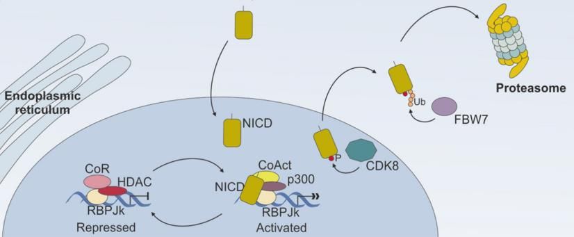
NOTCH pathway components and mechanisms of signaling (50)
There are four NOTCH receptors (NOTCH1-4), each encoded by a different gene, that interact
with five ligands. Out of the four receptors, NOTCH1 and NOTCH2 are the most widely expressed
receptors, being present in many tissues both in the developmental stage and in adults.
NOTCH receptors are single-pass transmembrane proteins composed of extracellular (EC),
transmembrane and intracellular domains (NICD). The IC portion of the receptor consists of a
protein-binding RBPJk-associated molecule (RAM) and a domain that regulates protein stability
and degradation, as it contains the substrate site that is recognized by E3 ubiquitin ligases (PEST
domain).
Ligand binding of a neighboring cell to the EC domain leads to a conformational change of the
receptor that ultimately triggers the proteolytic cleavage of the intracellular domain by a γ-
secretase, releasing it from the membrane and allowing its translocation into the nucleus.
Once in the nucleus, the NICD forms a a transcriptional complex. It mediates the displacement of
co-repressor molecules that are bound to a DNA-binding factor known as RBPJk and directly
interacts with RBJk recruiting co-activators, finally inducing gene transcription (See Figure 8).
One of the genes under its control is MYC, which is an important mediator of the NOTCH effects
in the transformation process.
The activity of NOTCH signaling is then shut down by the phosphorilation and ubiquitinylation of
the PEST domain, driving its degradation by the proteasome.
The most common mutation detected in follicular lymphoma samples (p.P2514fs*4) produces a
truncation in the PEST domain that results in the elimination of the degradation signals of NOTCH
proteins. This leads to a decresed physiological turnover of the protein and consequently a
persistently active NICD and high expression levels of NOTCH1 target genes.
19Analysis of NOTCH1 mutation as predictive biomarker for transformation in follicular
lymphoma patients
Figure 8. NOTCH signalling pathway. NICD translocates to the nucleus where it mediates the displacemet
of co-repressors (CoR) and histone DeAcetylase Complex (HDAC) and interacts with RBJk recruiting co-
activators (CoAct). These modifications shift the conformation of the transcriptional complex from a repressor
to an activator of gene transcription. The signalling pathway is then terminated by the phosphorylation and
ubiquitination of the PEST domain and subsequently degradated via proteosome. Adapted from Arruga F.
The NOTCH pathway and its mutations in mature B cell malignancies. Frontiers in Oncology. 2018; 8:550.
1.6.3 Relationship between NOTCH1 and transformation
A study of a small group of patients (49) analyzed the mutational status of NOTCH1 in follicular
lymphoma samples. Despite the small number of patients analyzed, there were relevant
differences in the risk of transformation between NOTCH1 mutated and NOTCH1 wild-type FL
samples (risk of transformation of 71% vs 23% respectively, p = 0.02). Unfortunately, the number
of cases analyzed was very small and the follow-up time was short.
Although the detected frequency of NOTCH1 mutations in this series was 6,3%, it identified a
subset of cases with distinctive clinipathological characteristics such as lower frequency of
t(14;18), higher incidence of splenic involvement, female predominance and association to
transformation to DLBCL.
Additionally, the mutational analysis of NOTCH1 in paired pre and post-transformation samples
showed no differences, suggesting that the NOTCH1 mutation may already be present in the
initial FL component and may facilitate progression to DLBCL.
Further evidence pointing to the NOTCH1 gene as a factor involved in the biology of
transformation is the fact that mutations of this gene have been found in other indolent lymphomas
acquiring an agressive biology. The paradigm of this matter is CLL’s transformation to DLBCL,
an event known as Richter syndrome.
Genetic lesions of DLBCL-type Richter syndrome recurrently target the TP53, NOTCH1, MYC
and CDKN2A genes, indicating that lesions affecting regulators of apoptosis and proliferation are
20Analysis of NOTCH1 mutation as predictive biomarker for transformation in follicular
lymphoma patients
shared among other transformed lymphomas. For this reason, we presume that NOTCH1
mutations could play a role in driving transformation from indolent FL to DLBCL (51).
Furthermore, in a study characterising the molecular features of DLBCL (52), mutations in the
NOTCH pathway, as well as TP53/CDKN2A genes, conferred a poorer prognosis independently
of IPI score and cell of origin (GCB like DLBCL and ABC like DLBCL) adding up to the evidence
that mutations in the NOTCH pathway are related to tumor agressiveness. Other papers (53,54)
investigating the relation between NOTCH1 or NOTCH2 mutations and other mature B cell
lymphomas (CLL and MCL) have also found that the mutated cases usually have a more
agressive clinical behaviour in comparison to the wild-type cases.
Despite the evidence gathered so far, further analyses are needed to stablish the functional role
of NOTCH mutation in FL lymphomagenesis and fine-tuning its potential role as a therapeutic
target.
1.6.4 Can we prevent transformation?
Given the generally poor outcome of patients with tFL and the lack of response to therapy after
transformation, prevention of the transformation itself appears to be an appealing strategy.
The first question to answer is wether the treatment of FL at diagnosis could prevent
transformation or, at least, reduce its incidence.
There are different studies that have attempted to answer this question (6,13,32,47,48). Evidence
argues strongly to the fact that treatment wih rituximab reduces rates of tFL over time (See Table
2). This is true both for initially untreated patients who recieve rituximab versus observation and
also for patients receiving rituximab maintenance after enduring immunochemotherapy treatment.
Table 2. Effect of immunotherapy in transformation risk
Treatment Effect of treatment
Observation: 33%
Link et al. (2013) (6) Observation vs RTX: Increased risk
RTX mono: 12%
N: 631 RTX-mono: Reduced risk (14.4% vs 3.2% risk of HT
Chemo (+/-RTX):
t(FL (n): 60 at 5y)
42%
Observation vs RTX: Increased risk
Wagner-Johnson et Observation: 21%
Anthracycline vs no athracycline chemo: No
al. (2014) (13) RTX mono: 13%
differences
N: 2,652 R-chemo: 48%
RTX-mono: Reduced risk (HR 0.58)
tFL (n): 379 Other: 19%
RTX (M) vs no RTX (M): Reduced risk (HR 0.67)
Federico et al.
Observation vs RTX: Increased risk
(2017) (48)
4468 pt recieved RTX RTX (I) vs RTX (I+M): Reduced risk (5.9% vs 3.6%
N: 7,405
risk of HT at 10y)
tFL (n): 439
RTX, rituximab; Mono, monotherapy; I, induction; M, maintenance; tFL, transformed follicular lymphoma; HT,
histological transformation
21Analysis of NOTCH1 mutation as predictive biomarker for transformation in follicular
lymphoma patients
The fact that immunotherapy reduces transformation rates and improves its outcomes highlights
the importance of finding predictive biomarkers that allow us to identify which patients present a
higher risk of transformation, as those will be the patients who will benefit the most from early
treatment to prevent transformation.
Evidence suggests that at least in some cases, genetic alterations in the transformation of FL are
early events that already exist at the time of clinical FL diagnosis (42,55), albeit at minuscule
proportions.
This finding opens the door to the possibility of identifying very early pre-clinical transformed
events (perhaps through techniques such as circulating tumour DNA (55) and targeting the
transformed subclone through precision therapy, provided that the biology of such subclone is
understood well enough.
With the current clinical and histological definitions of HT along with the diagnostic methods, it is
clear that we have insufficient tools for adequately identifying at risk of transformation patients.
For this reason, clinical practice at this time is limited to nonspecifically supressing the FL cell
pool to minimize the odds of a cell acquiring the necessary transformed biology, instead of
targeting specific transforming clones.
22Analysis of NOTCH1 mutation as predictive biomarker for transformation in follicular
lymphoma patients
2. JUSTIFICATION
Follicular lymphoma is the second most common NHL in the Western world, accounting for 20%
of all lymphomas (1). It is generally an indolent though incurable disease, with a pattern of
relapses and increasingly long survival rates in the immunotherapy era. However, a minority of
patients suffer histological transformation into an agressive lymphoma, a compelling event that
overshadows clinical course and life expectancy.
In the past decades, extensive research has led to the identification of genetic and epigenetic
alterations involved in the pathogenesis of transformation. Despite the substantial progress
accomplished elucidating the contributing factors leading to transformation, no predictive
biomarkers have been validated for clinical use.
Likewise, adverse clinical factors have been associated with a higher risk of transformation, but
evidence is not consistent and still, these clinical features alone can’t adequately predict the
majority of patients who will transform in the future.
This lack of knowledge is relevant to the prognosis of patients with FL because typically the
majority of patients who are asymptomatic and present a low tumour burden are not treated from
the beginning but rather managed following a “watch and wait” strategy.
Several papers suggest that early treatment with immunotherapy can reduce transformation rates
(15-y risk of transformation of 30% in the pre-rituximab era to 8% in the most recent series) as
well as improve the prognosis of patients with a transformed biology. Accordingly, finding
predictive biomarkers that allow us to detect which patients present a higher risk of HT would be
useful to determine which patients would benefit the most from early treatment to prevent
transformation.
Although NOTCH1 mutations are a rare event in FL patients (6,3% according to literature
research), one paper studying a small group of patients with FL (49) analyzed the mutational
status of this gene and found that this mutation identified a subset of patients with distinctive
clinicopathological characteristics; amongst them being association to transformation to diffuse
large B cell lymphoma. The same study found evidence suggesting that NOTCH1 gene mutations
might be acquired before the transformation occurs, which implies that they could be used as a
predictive biomarker for transformation before its occurrence.
Further evidence pointing to NOTCH1 as a factor involved in the biology of transformation is the
fact that previous research has implicated mutations in this gene as an important driver of other
indolent lymphomas acquiring an agressive biology, as is the case of CLL’s Richter syndrome
(51).
23Analysis of NOTCH1 mutation as predictive biomarker for transformation in follicular
lymphoma patients
Mutations in the NOTCH family have been assessed in previous papers on FL samples. However,
the functional role of this molecule in the FL pathogenesis and its influence on transformation
have not been well determined.
The aim of this study is to characterise the frequency of the NOTCH1 mutations in non-
transformed FL patients and evaluate its potential role as a biomarker for transformation at the
time of diagnosis.
24Analysis of NOTCH1 mutation as predictive biomarker for transformation in follicular
lymphoma patients
3. HYPOTHESIS AND OBJECTIVES
Hypothesis
Aberrant activation of the NOTCH signalling pathway via gain-of-function mutation of the
NOTCH1 gene could predict a higher risk of transformation from indolent follicular lymphoma to
DLBCL at the time of diagnosis.
Objectives
To analize the relationship between the mutational status of the NOTCH1 gene at diagnosis and
the risk of transformation in a cohort of patients with non-transformed follicular lymphoma.
25You can also read