Non coding RNAs (miRNAs and lncRNAs) and their roles in lymphogenesis in all types of lymphomas and lymphoid malignancies (Review) - Spandidos ...
←
→
Page content transcription
If your browser does not render page correctly, please read the page content below
ONCOLOGY LETTERS 21: 393, 2021

              Non‑coding RNAs (miRNAs and lncRNAs) and their
               roles in lymphogenesis in all types of lymphomas
                      and lymphoid malignancies (Review)
                     GEORGIOS DRILLIS1, MARIA GOULIELMAKI2, DEMETRIOS A. SPANDIDOS3,
                               SOFIA AGGELAKI4 and VASSILIOS ZOUMPOURLIS2

              1
             1st Internal Medicine Clinic, Medical School, Laiko University Hospital of Athens, 115 27 Athens;
2
 Biomedical Applications Unit, Institute of Chemical Biology, National Hellenic Research Foundation (NHRF), 116 35 Athens;
    3
     Laboratory of Clinical Virology, and 4Oncology Unit, Medical School, University of Crete, 71003 Heraklion, Greece

                                    Received December 16, 2020; Accepted February 26, 2021

                                                     DOI: 10.3892/ol.2021.12654

Abstract. Contemporary developments in molecular biology              3. Non‑coding RNAs in lymphomas
have been combined with discoveries on the analysis of the role of    4. Anti‑ncRNA therapeutic strategies in lymphoid disorders
all non‑coding RNAs (ncRNAs) in human diseases, particularly          5. Conclusions and future perspectives
in cancer, by examining their roles in cells. Currently, included
among these common types of cancer, are all the lymphomas             1. Introduction
and lymphoid malignancies, which represent a diverse group
of neoplasms and malignant disorders. Initial data suggest that       Lymphomas are a heterogeneous group of cancers; more
non‑coding RNAs, particularly long ncRNAs (lncRNAs), play             specifically, they consist of a group of blood disorders derived
key roles in oncogenesis and that lncRNA‑mediated biology             from lymphocytes and are presented with multiple variations
is an important key pathway to cancer progression. Other              in clinical presentation, long‑term prognosis and pathogenesis.
non‑coding RNAs, termed microRNAs (miRNAs or miRs),                   They represent one of the most common types of cancer world‑
are very promising cancer molecular biomarkers. They can be           wide and affect numerous patients. According to the World
detected in tissues, cell lines, biopsy material and all biological   Health Organization (WHO) classification report, there are
fluids, such as blood. With the number of well‑characterized          approximately 100 different types of lymphoma. The two main
cancer‑related lncRNAs and miRNAs increasing, the study               categories of lymphomas are non‑Hodgkin lymphoma (NHL)
of the roles of non‑coding RNAs in cancer is bringing forth           (consisting 9 out of 10 of all lymphoma cases) and Hodgkin
new hypotheses of the biology of cancerous cells. For the first       lymphoma (HL) (consisting 1 out of 10 of all lymphoma
time, to the best of our knowledge, the present review provides       cases) (1‑3). Furthermore, non‑Hodgkin lymphomas can be
an up‑to‑date summary of the recent literature referring to all       grouped into B‑ and T‑cell NHLs (TNHLs), which account
diagnosed ncRNAs that mediate the pathogenesis of all types           for approximately 90 and 10% of NHLs, respectively (4,5).
of lymphomas and lymphoid malignancies.                               According to the WHO, two other categories are also
                                                                      considered as types of lymphoid tissue tumors: Multiple
Contents                                                              myeloma (MM) and immunoproliferative diseases (3).

1. Introduction                                                       Epidemiology. According to the WHO, there are the following
2. Literature search                                                  lymphoma subtypes (WHO 2016): i) Mature B‑cell neoplasms;
                                                                      ii) mature T‑cell and natural killer (NK) cell neoplasms;
                                                                      iii) precursor lymphoid neoplasms; iv) HL; and v) immunode‑
                                                                      ficiency‑associated lymphoproliferative disorders (4,5).
                                                                           B‑cell NHLs (BCNHLs) are tumors of B‑cells that exhibit a
Correspondence to: Dr Vassilios Zoumpourlis, Biomedical               heterogeneity that is attributed to the fact that these tumors are
Applications Unit, Institute of Chemical Biology, National Hellenic
                                                                      derived from different stages of mature B‑cell differentiation.
Research Foundation (NHRF), 48 Vassileos Constantinou Avenue,
116 35 Athens, Greece
                                                                      The main subtypes of BCNHLs are the following: i) diffuse
E‑mail: vzub@eie.gr                                                   large B‑cell lymphoma (DLBCL); ii) chronic lymphocytic
                                                                      leukemia (CLL); iii) follicular lymphoma (FL); iv) mantle cell
Key words: long non‑coding RNAs, microRNAs, B‑cell lymphoma,          lymphoma (MCL); v) Burkitt's lymphoma (BL); vi) marginal
non‑Hodgkin lymphoma, Hodgkin lymphoma, multiple myeloma,             zone lymphoma (MZL); and vii) mucosa‑associated lymphoid
biomarkers                                                            tissue (MALT) (6). The majority of BCNHLs, such as DLBCL
                                                                      and FL, have passed the germinal center (GC) reaction,
                                                                      indicating that their immunoglobulin (IG) genes have been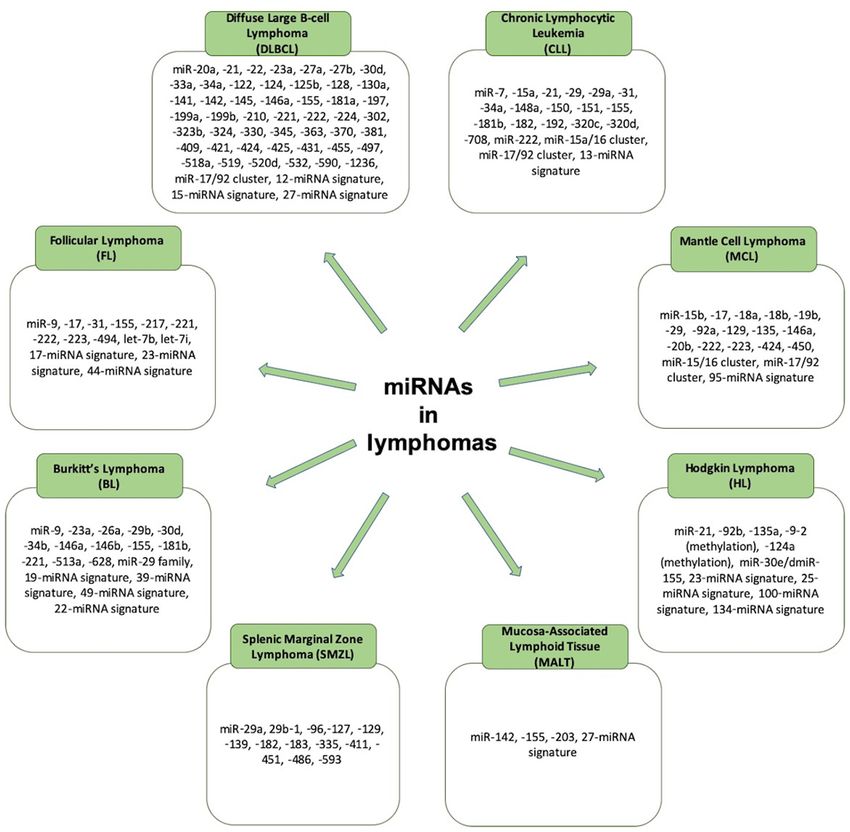
2                                  DRILLIS et al: NON-CODING RNAs IN LYMPHOID MALIGNANCIES

hypermutated. Other subtypes, such as MCL and CLL, are                suspension of the translation of these mRNAs (21). The patterns
derived from GC‑inexperienced B‑cells (7).                            of expression of miRNAs in different cancer types have been
    The most common subtypes of TNHLs and NK‑cell                     well‑observed, and studies have highlighted numerous miRNAs,
NHLS (NK‑NHLs) are the following: i) Cutaneous T‑cell                 such as miR‑10b, let‑7, miR‑101 and miR‑15a‑16 complex‑1,
lymphomas (mycosis fungoides, Sezary syndrome and others);            which have oncogenic or tumor‑suppressive functions (22,23).
ii) adult T‑cell leukemia/lymphoma; iii) angioimmunoblastic
T‑cell lymphoma; iv) extranodal NK/T‑cell lymphoma;                   lncRNAs. Recent observations of new species of lncRNAs
nasal type; vi) enteropathy‑associated T‑cell lymphoma; and           have led to the development of various possible candidates as
vii) anaplastic large cell lymphoma (ALCL) (4).                       lncRNAs. Although a number of RNAs have a length of >200 bp,
                                                                      such as repeat sequence transcripts and pseudogenes (24), the
Risk factors and diagnosis of lymphomas. The most common              term lncRNA (also referred to as lincRNAs, for long transgenic
risk factors for HL are Epstein‑Barr virus (EBV) infection            ncRNAs) is not used in the same manner in all cases.
and a family history of the disease (8). The most common                  A number of common features of lncRNAs have been indi‑
risk factors for several types of NHLs include the following:         cated to confirm their biological identity, such as the following:
i) Autoimmune diseases, such as Sjögren syndrome, celiac              i) Epigenetic regulation as in a transcripted gene; ii) transcrip‑
disease, rheumatoid arthritis and systemic lupus erythema‑            tion performed by RNA polymerase II; iii) poly‑adenylation
tosus; ii) HIV/AIDS infection; iii) human T‑lymphotropic              to the 3'‑untranslated region (3'‑UTR); iv) frequent splicing
virus infection; iv) Helicobacter pylori infection; v) HHV‑8          of multiple exons through specific molecular patterns;
infection; vi) hepatitis C virus infection; vii) medical treat‑       v) regulation by classic transcription factors; and vi) frequent
ments (patients who have been previously treated for Hodgkin          tissue‑specific expression (24) (Fig. 1).
lymphoma, methotrexate and the tumor necrosis factor‑a
inhibitors); viii) genetic diseases; and ix) certain chemical         ncRNAs in normal B‑cell differentiation and T‑cell develop‑
agents (benzene and certain herbicides and insecticides;              ment. B‑cell differentiation in adult humans begins within the
weed‑ and insect‑killing substances) (9‑15). Some environ‑            bone marrow (BM) and is continued thereafter in the lymph
mental agents, such as red meat consumption and tobacco               nodes, tonsils and spleen (25). On the other hand, T‑cells a
smoking may also play a role in increasing the risk of devel‑         derived from bone marrow hematopoietic stem cells (HSCs),
oping NHL (11,12,16).                                                 whose progenitors migrate to and colonize the thymus (26).
    The diagnosis of lymphomas can be achieved due to the                 The most common lncRNAs affecting normal B‑cells are
enlargement of lymph nodes, which can be determined by                the following: i) MYB‑AS1, SMAD1‑AS1 and LEF1‑AS1,
performing a lymph node biopsy (17). A lymph node biopsy              located on 6q23.3, 4q31.21 and 4q25, respectively, are
commonly is followed by performing immunophenotyping,                 involved in early B‑cell development; ii) CRNDE, located
flow cytometry, fluorescence in situ hybridization testing, bone      on 16q12.2‑involved in mitotic cell cycle related processes;
marrow aspiration and bone marrow biopsy (18). Imaging via            and iii) RP11‑132N15.3/lnc‑BCL6‑3, located on 3q27.3, and
computed tomography of the chest and upper‑lower abdomen              involved in the modulation of the GC reaction. However, data
may then be performed to determine the possible expansion of          on the roles of lncRNAs in normal T‑cells are limited (27‑31).
the lymphoma throughout the human body (17).                              miRNAs are also involved in lymphocyte development,
                                                                      as first described in 2004; it was demonstrated that miR‑223,
Non‑coding RNAs (ncRNAs). ncRNAs are RNAs that are not                miR‑181 and miR‑142 were highly expressed in B‑cells (32).
translated to proteins. Over the past ten years, a number of          miR‑181 can also contribute to the regulation of the levels of
ncRNAs have been identified. Any of the three RNA poly‑               CD69, BCL2 and TCR during T‑cell development. In addition,
merases (RNA Pol I, RNA Pol II or RNA Pol III) can perform            miR‑155 and miR‑181 play key roles in the regulation of GC
the transcription of a ncRNA. The ncRNAs are divided into             B‑cell differentiation (33).
the following two main categories: Small ncRNAs, 200 bp in length (19).          role as predictive and prognostic biomarkers in the pathogenesis
    In these two categories, several individual categories of         and progression of lymphomas and lymphoid malignancies in
ncRNAs also exist. These include housekeeping ncRNAs                  general. The main aim of the present review was to provide an
[transfer RNAs (tRNAs) and some ribosomal RNAs (rRNAs)],              up‑to‑date summary of available information on all the known
which are essential for fundamental principles of cellular            miRNAs and lncRNAs that participate in the development of all
biology, small nuclear RNAs (snRNAs), and a number of                 lymphoid disorders, with a main focus on their connection to each
recently observed RNAs which are associated with the                  lymphoma subtype. Furthermore, these molecular biomarkers
transcription of genes into proteins (20).                            may be used, in the near future, in the therapeutic management
                                                                      of the majority of lymphomas. Thus, the present review summa‑
MicroRNAs (miRNAs or miRs). To date, miRNAs are the less              rizes all published data to date on ncRNAs, in order to shed light
extensively studied ncRNAs for their roles in cancer. Over the past   on the future perspectives of lymphoma management.
years, a number of targeted reviews have been published (21‑23),
which have described a complex basic mechanism through                2. Literature search
which miRNAs can lead to the silencing of target gene expres‑
sion; through the formation of a silencing complex induced by         A literature search was performed, including studies published
RISC‑induced RNA, which uses proteins from the Argonaute              up to August, 2020, using the following databases: Medline
family (such as AGO2) for the splicing of target mRNAs or for the     (PubMed), Science Direct, Web of Science and Google Scholar.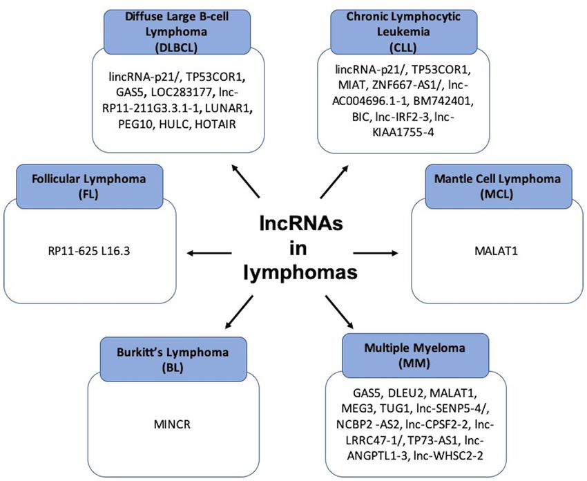
ONCOLOGY LETTERS 21: 393, 2021 3
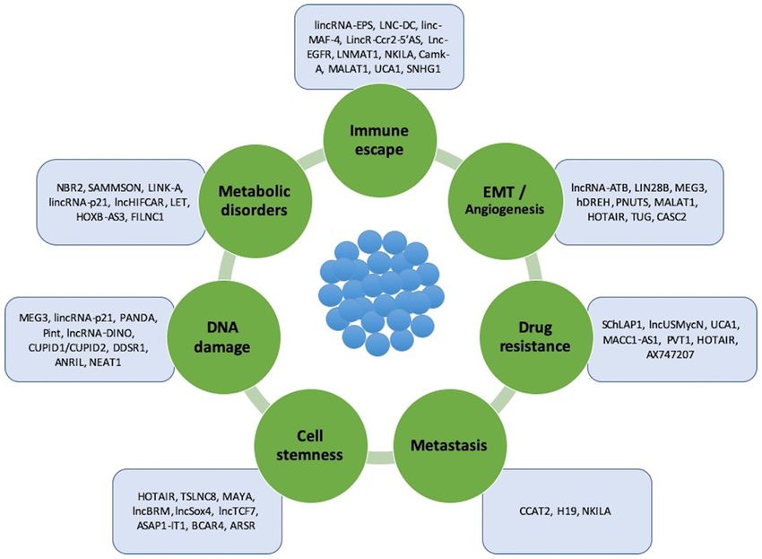
Figure 1. lncRNAs involved in oncogenesis, tumor progression and metastasis. These lncRNAs can be divided into 7 subtypes based on the process/property
in which they are involved: Immune system escape, epithelial‑mesenchymal transition, and angiogenesis, drug resistance, metastasis, cell stemness, DNA
damage and metabolic disorders. lncRNAs, long non‑coding RNAs.
Systematic reviews, uncontrolled prospective, retrospec‑ lymphoma; and v) large B‑cell lymphoma with IRF4
tive and experimental studies were included for each specific rearrangement (38,39).
subject (total no. of studies, n=235). The following inclusion Subtypes of DLBCLs with distinctive clinical issues are
criteria were applied: Studies concerning ncRNAs, lncRNAs, the following: i) Primary mediastinal large B‑cell lymphoma;
miRNAs, cancer and lymphomas: HLs, and BCNHLs and ii) primary cutaneous DLBCL, leg type; iii) primary DLBCL
TNHLs. All studies concerning the association of lncRNAs of the central nervous system; iv) DLBCL associated with
and miRNAs with NHLs and HLs were included. chronic inflammation; v) lymphomatoid granulomatosis; and
vi) primary effusion lymphoma (38,39).
3. Non‑coding RNAs in lymphomas Additionally, there are DLBCLs driven by viruses, such as
the following: i) EBV‑positive DLBCL, not otherwise speci‑
Over the past years, a number of studies have referred to the fied; and ii) HHV8‑positive DLBCL, NOS (Not otherwise
significance of lncRNAs and miRNAs in the pathophysi‑ specified). There are also DLBCLs driven by disorders related
ology of lymphomas, particularly B‑cell NHLs. There are to DLBCL, such as: i) Helicobactor pylori‑associated DLBCL;
different non‑coding RNAs that play a role in each subtype and ii) EBV‑positive mucocutaneous ulcer (40,41).
of lymphoma, and these are referred to the sections and tables In order for a B‑cell to be developed or to progress
below (Figs. 2 and 3). into a DLBCL type, changes in the following genes need
to occur: BCL2 (42), BCL6 (42), MYC (36), EZH2 (43),
a) BCNHLs MYD88 (42), CREBBP (44), CD79A and CD79B (44) and
DLBCL. Diffuse large B‑cell lymphoma is the most common PAX5 (44). Therefore, the neoplastic cells in DLBCL exhibit
form of NHL among adults (34) and it occurs most often a pathologically overactivation of the nuclear factor (NF)‑κ B,
in older‑aged individuals, with a median age of diagnosis phosphoinositide 3‑kinase (PI3K)/AKT/mammalian target of
approaching the seventh decade of a patient's life (35). rapamycin (mTOR), Janus kinase (JAK)/signal transducer and
There are 2 different molecular subtypes of DLBCL: activator of transcription (STAT), mitogen‑activated protein
GC B‑cell like (GC‑DLBCL) and activated B‑cell like kinase (MAPK)/extracellular signal‑regulated kinase (ERK),
(ABC‑DLBCL) (36,37). B‑cell receptor and Toll‑like receptor pathways (42).
Subtypes of DLBCLs with a distinctive morphology or Concerning miRNAs in DLBCLs, it has been shown than
immunophenotype are the following: i) T‑cell/histiocyte‑rich in ABC‑type DLBCL lymphoma, there is a high expression of
large B‑cell lymphoma; ii) ALK+ large B‑cell lymphoma; miR‑21, miR‑146a, miR155, miR‑221 and miR‑363, while in
iii) plasmablastic lymphoma; iv) intravascular large B‑cell GCB‑type DLBCL, there is a high expression of miR‑421 and4 DRILLIS et al: NON-CODING RNAs IN LYMPHOID MALIGNANCIES
Figure 2. Summary of the major lncRNAs that are involved in different types of lymphomas. lncRNAs, long non‑coding RNAs.
Figure 3. Summary of the miRNAs that are involved in different types of lymphomas. miRNAs, microRNAs.ONCOLOGY LETTERS 21: 393, 2021 5
Table I. miRNAs and lncRNAs identified in patients with diffuse large B‑cell lymphoma.
Genome
location Molecular
miRNA(s)/lncRNA (if defined) Role mechanism/sample (Refs.)
miR‑155 Diagnostic biomarker Presence in serum (51)
miR‑34a Diagnostic biomarker Presence in serum (52)
miR‑17/92 cluster Cell survival, prognostic Subtyping (53)
biomarker
miR‑21, miR‑23a, miR‑27b, miR‑34a Poor overall survival, diagnostic Presence in serum (54‑58)
and prognostic biomarkers
miR‑20a, miR‑30d, miR‑22, miR‑146a Prognostic biomarkers Presence in tissues (59)
miR‑21, miR‑210 Diagnostic biomarkers Presence in serum (60)
12‑miRNA signature, 15‑miRNA signature, Diagnostic biomarkers Presence in tissue (61‑64)
27‑miRNA signature
miR‑155, miR‑221, miR‑222, miR‑21, Diagnostic biomarkers Presence in cell lines (65)
miR‑363, miR‑518a, miR‑181a, miR‑590,
miR‑421, miR‑324
miR‑124, miR‑532, miR‑122, miR‑128, Diagnostic biomarkers Presence in plasma (66)
miR‑141, miR‑145, miR‑197, miR‑345, and exosomes
miR‑424, miR‑425
miR‑34a, miR‑323b, miR‑431 Diagnostic biomarkers Presence in serum (67)
miR‑27a, miR‑142, miR‑199b, miR‑222, Predictive biomarkers Presence in tissue (46)
miR‑302, miR‑330, miR‑425, miR‑519
miR‑224, miR‑455, miR‑1236, miR‑33a, Predictive biomarkers Presence in serum (68)
miR‑520d
miR‑125b, miR‑130a, miR‑199a, miR‑497, Predictive biomarkers Presence in tissue, (69‑71)
miR‑370, miR‑381, miR‑409 blood and cell lines
lincRNA‑p21/TP53COR1 17p13.1 Tumor‑suppressor Link to cyclin D1, (72‑76)
(mouse) CDK4 and p21
6p21.2
(human)
GAS5 1q25.1 Tumor‑suppressor Regulation of mTOR (77‑86)
pathway
LOC283177 11q25 Uncharacterized Not described (87)
lnc‑RP11‑211G3.3.1‑1 3q27.3 Uncharacterized Not described (88)
LUNAR1 15q26.3 oncomiR progenitor NOTCH1 regulation. (89,90)
Enhances IGF1R
mRNA expression
PEG10 7q21.3 oncomiR progenitor Activated by c‑MYC (91‑93)
HULC 6p24.3 oncomiR progenitor Not described (94‑97)
HOTAIR 12q13.13 oncomiR progenitor Regulation of the (98)
PI3K/AKT/NF‑κB
pathway
lncRNA, long non‑coding RNA; miRNA/miR, microRNA.
the miR‑17~92 cluster (45‑50) (Table I). In serum samples of seem to be associated with the overall survival of patients
patients with DLBCL, increased levels of miR‑21, miR‑155 and with DLBCL (50). In any case, miR‑155, miR‑34a, miRNA‑21,
miR‑210 have been identified, along with increased levels of miRNA‑23a, miRNA‑27b, miRNA‑34a, 12‑miRNA signa‑
miR‑124, miR‑532‑5p miR‑15a, miR‑16, miR‑29c and miR‑155. ture, 15‑miRNA signature, 27‑miRNA signature, miR‑363,
Decreased levels of miR‑122, miR‑128, miR‑141, miR‑145, miR‑518a, miR‑181a, miR‑590, miR‑421 and miR‑324‑either in
miR‑197, miR‑345, miR‑424 and miR‑425 have been also serum samples, or in tissue or cell line samples of patients with
found in the serum of patients with DLBCL. miR‑27a, miR‑142, DLBCL, can be used as diagnostic biomarkers in patients with
miR‑199b, miR‑222, miR‑302, miR‑330, miR‑425 and miR‑519 DLBCL (50‑71) (Table I and Fig. 3).6 DRILLIS et al: NON-CODING RNAs IN LYMPHOID MALIGNANCIES
Table II. miRNAs and lncRNAs identified in patients with chronic lymphocytic leukemia.
Genome
location Molecular
miRNA(s)/lncRNA (if defined) Role/activation mechanism/sample (Refs.)
miR‑15a/16 cluster, miR‑7, miR‑182, Diagnostic biomarkers Presence in PBMCs and (107,108,115,116)
miR‑320c/d, miR‑29, miR‑192 cell lines
miR‑151, miR‑34a, miR‑31, miR‑155, Diagnostic biomarkers Presence in serum (113‑114)
miR‑150, miR‑15a, miR‑29a
miR‑181b, miR‑21, miR‑155, miR‑708, Prognostic biomarkers Presence in PBMCs, cell (115,133‑135)
miR‑17~92 cluster, 13‑miRNA lines, serum, blood cells
signature, miR‑150, miR‑155
miR‑181b, miR‑155, miR‑21, Predictive biomarkers Presence in PBMCs and (133‑135)
miR‑148a, miR‑222 cell lines
DLEU2 13q14.3 Tumor suppressor NF‑κB activation (117‑120)
NEAT1 11q13.1 Tumor‑suppressor Induction by p53 (121,122)
lincRNA‑p21/TP53COR1 17p13.1 Tumor‑suppressor Induction by p53 (72‑76)
(mouse)
6p21.2
(human)
MIAT 22q12.1 Oncogene Regulatory loop with (123‑126)
OCT4
ZNF667‑AS1/lnc‑AC004696.1‑1 19q13.43 Uncharacterized Not described (127,128)
BM742401 18q11.2 Tumor‑suppressor Not described (129,130)
BIC 21q21 oncomiR progenitor Host of miR‑155‑5p and (131,132)
miR‑155‑3p
lnc‑IRF2‑3 4q35 Uncharacterized Not described (128)
lnc‑KIAA1755‑4 20q11.23 Uncharacterized Not described (128)
lncRNA, long non‑coding RNA; miRNA/miR, microRNA; PBMCs, peripheral blood mononuclear cells.
Concerning lncRNAs in patients with DLBCLs, the monoclonal B‑cells are the following: Low‑count CLL/SLL
following have been observed (Table I): i) lincRNA- with a number of monoclonal B‑cellsONCOLOGY LETTERS 21: 393, 2021 7
Table III. miRNAs and lncRNAs identified in patients with follicular lymphoma.
Genome
location Molecular
miRNA(s)/lncRNA (if defined) Role/activation mechanism/sample (Refs.)
RP11‑625 L16.3 12 Uncharacterized Not described (72)
miR‑9, miR‑155, miR‑31, miR‑17 Diagnostic biomarkers Presence in tissues (136,137)
miR‑217, miR‑221, miR‑222, miR‑223, let‑7i, let‑7b Diagnostic biomarkers Presence in tissues (138,140)
17‑miRNA signature, 44‑miRNA signature, miR‑494 Diagnostic biomarkers Presence in tissues (138,139)
23‑miRNA signature Predictive biomarkers Presence in tissues (139)
lncRNA, long non‑coding RNA; miRNA/miR, microRNA.
Concerning lncRNAs in patients with CLL, the following Low‑grade FLs are grades 1 and 2, as well as grade 3A.
have been observed (Table II): i) DLEU2, located on chromo‑ Grade 3B is regarded as a highly aggressive FL, which can be
some 13q14.3, acting as tumor suppressor by activating the easily transformed into a higher grade (46). The transformation
NF‑κ B pathway (117‑120); ii) NEAT1, located on chromo‑ of FL into a more aggressive state or other type of aggres‑
some 11q13.1, acting as a tumor suppressor via induction sive lymphoma is associated with specific genetic alterations,
by p53 (121,122); iii) lincRNA‑p21/TP53COR1, located on such as in the following genes: CREEBP, KMT2D, STAT6,
chromosome p21.2 (human), acting as a tumor suppressor via CARD11, CD79, TNFAIP3, CD58, CDKN2A or CDKN2B,
induction by p53 (72‑76); iv) MIAT, located on chromosome TNFRSF4 and c‑MYC (45,46,136‑138).
22q12.1, acting as an oncogene by forming a regulatory loop Concerning miRNAs in FLs, a number of studies
with OCT4 (123‑126); v) ZNF667‑AS1/lnc‑AC004696.1‑1, have demonstrated that there is an increase in the levels
located on chromosome 19q13.43, with an uncharacter‑ of 6 particular miRNAs: miR‑223, miR‑217, miR‑222,
ized mode of action (127,128); vi) BM742401, located on miR221, let‑7i and let‑7b in patients with FL, in which
chromosome 18q11.2, acting as a tumor suppressor, with an their lymphoma underwent a transformation. In addition,
uncharacterized mode of action (129,130); vii) BIC, located on the miR‑17~92 cluster can be used as a useful diagnostic
chromosome 21q21, acting as an oncomiR progenitor by being a biomarker found in patients with FL, while miR‑20a/b
host of miR‑155‑5p and miR‑155‑3p (131,132); viii) lnc‑IRF2‑3, and miR‑194 can also be found in patients with FL. Other
located on chromosome 4q35 with an uncharacterized mode useful diagnostic biomarkers in patients with FL may be
of action (128); ix) lnc‑KIAA1755‑4, located on chromosome the following: miR‑9, miR‑155, miR‑31, miR‑17, miR‑217,
20q11.23 with an uncharacterized mode of action (128) (Fig. 2). miR‑221, miR‑222, miR‑223, let‑7i, let‑7b17‑miRNA
Finally, in Table II, references are provided of all the other signature, 44‑miRNA signature, miR‑494 23‑miRNA
miRNAs used as predictive and prognostic biomarkers in signature (136‑140) (Table III and Fig. 3).
clinical trials of patients with CLL (133‑135). Concerning lncRNAs in patients with FL, studies have
demonstrated that there are 3‑fold as many lncRNAs that are
FLs. FL is the second most common type of NHL, and the upregulated than lncRNAs that are downregulated in patients
most common indolent NHL. It derives from the uncontrolled with FL3A stage disease, without their biological functions
division of centrocytes and centroblasts of the follicles in the being cleared yet. The only lncRNA that seems to be upregu‑
GCs of lymph nodes. lated in patients with FL3A grade disease is RP11‑625 L16.3,
The genomic alterations that can be found in FL include located on chromosome 12, with an uncharacterized mode of
the following: i) the t(14:18)(q32:q21.3) translocation (the action (72) (Table III and Fig. 2).
majority of the cases); ii) 1p36 deletions (second most common
genomic alteration in FL) that lead to the loss of TNFAIP3; MCLs. MCL is recognizable as an aggressive and incurable
iii) mutations in PRDM1; and iv) the same mutations observed small B‑cell lymphoma. It predominantly affects older‑aged
in in situ FL (ISFL), including KMT2D, CREEBP, BCL2 and males (>60 years old), and sometimes it may be indolent in
EZH2, as well as other mutations (45). some patients. MCLs arise from the mantle zone of early
According to the WHO criteria, there are differences, B‑cells of the lymph node follicle and they possess the t(11;14)
which can be observed under a microscope, which can be (q13;q32) translocation with an overexpression of cyclin
used to diagnose and categorize FL into the following 3 D1.MCL cells also exhibit CD5+ and CD23 ‑ and surface
grades, with grade 3 comprising A and B subtypes (46): IgM/D expression (141,142).
Grade 1, follicles with 15 centroblasts per hpf; grade 3A, minimally mutated B‑cells and SOX11 overexpression; usually
grade 3 in which the follicles contain predominantly centro‑ presented in lymph nodes and other extranodal sites. Additional
cytes; grade 3B, grade 3 in which the follicles consist almost molecular/cytogenetic abnormalities may be presented in
entirely of centroblasts. blastoid or pleomorphic MCL. Leukemic non‑nodal MCL8 DRILLIS et al: NON-CODING RNAs IN LYMPHOID MALIGNANCIES
Table IV. miRNAs and lncRNAs observed in patients with mantle‑cell lymphoma.
Genome
location
miRNA(s)/lncRNA (if defined) Role/activation Molecular mechanism/sample (Refs.)
MALAT1 11q13 Oncogene Regulation of the bioavailability (146‑152)
of TGF‑β
miR‑15/16, miR‑17/92 Diagnostic biomarker Presence in cell lines (141,142)
95‑miRNA signature Diagnostic biomarker Presence in tissues (143)
miR‑15b, miR‑129, miR‑135, miR‑146a, Prognostic biomarkers Presence in tissues (144,153)
miR‑424, miR‑450, miR‑222, miR‑17,
miR‑18a, miR‑19b, miR‑92a
(miR‑17/92 cluster)
miR‑29, miR‑20b, miR‑18b Prognostic biomarkers Presence in cell lines and tissues (154‑156)
miR‑223 Prognostic biomarkers Presence in PBMCs and cell lines (157)
lncRNA, long non‑coding RNA; miRNA/miR, microRNA; PBMCs, peripheral blood mononuclear cells.
Table V. miRNAs and lncRNAs identified in patients with Burkitt's lymphoma.
Genome
location
miRNA(s)/lncRNA (if defined) Role/activation Molecular mechanism/sample (Refs.)
MINCR 8q24.3 Uncharacterized Induction of MYC (173)
miR‑23a, miR‑26a, miR‑29b, Diagnostic biomarkers Presence in tissues (165)
miR‑30d, miR‑146a, miR‑146b,
miR‑155, miR‑221
22‑miRNA signature, miR‑513a, Diagnostic biomarkers Presence in tissues (166,167)
miR‑628, miR‑9
39‑miRNA signature, 19‑miRNA Diagnostic biomarkers Presence in tissues (168‑171)
signature, 49‑miRNA signature
miR‑34b, miR‑29 family, miR‑181b Diagnostic biomarkers Presence in cell lines and tissues (167,171‑173)
lncRNA, long non‑coding RNA; miRNA/miR, microRNA.
develops from IGHV‑mutated SOX11 B‑cells, and is usually Finally, in Table IV, references of all the other miRNAs that
presented in peripheral blood, BM and spleen (142). have been observed in patients with MCL and used as prog‑
Concerning miRNAs in MCLs, a number of studies nostic biomarkers in clinical trials are presented (153‑157).
have demonstrated the overexpression of miR‑15/16 and
miR‑17~92 in MCL and that this is associated with an aggres‑ BL. BL is a type of aggressive B‑NHL. It may be presented with
sive form of the disease (143,144). In addition, the inhibition any of three main clinical variants: Endemic BL, sporadic BL
of miR‑29 has been demonstrated to lead to the progression and the immunodeficiency‑associated BL (158). In all types of
of MCL (a potential prognostic marker for MCL) (143‑145). BL, the dysregulation of the c‑myc gene is observed (the gene
Additionally, the 95‑miRNA signature can be a diagnostic is found at 8q24), presented with any one of the three known
biomarker for MCL (145) (Table IV and Fig. 3). chromosomal translocations (159). The most common variant
Concerning lncRNAs in patients with MCL, it has been is t(8;14)(q24;q32), which involves c‑myc and IGH (159).
demonstrated that MALAT1 is overexpressed in human The variant at t(2;8)(p12;q24) involves IGK and c‑myc (160).
MCL tissues and cell lines compared to normal B‑cells [a The variant at t(8;22)(q24;q11) involves IGL and c‑myc (160).
high international prognostic index (IPI) is present], and In addition, a last variant of three‑way translocation, t(8;14;18)
is associated with the lower overall survival of patients has been identified (161).
with MCL (146). Thus, MALAT1, located on 11q13 chro‑ Concerning miRNAs in patients with BL, it seems that
mosome, can act as an oncogene in patients with MCL MYC regulates and is regulated by numerous miRNAs
(regulation of the bioavailability of TGF‑ β) (146‑152) (Table V), the most common of which are the following:
(Table IV and Fig. 2). miR‑23a, miR‑26a, miR‑29b, miR‑30d, miR‑146a, miR‑146b,ONCOLOGY LETTERS 21: 393, 2021 9
Table VI. miRNAs identified in patients with splenic marginal zone lymphoma B‑cell lymphoma and mucosa‑associated
lymphoid tissue lymphoma.
Genome Molecular
miRNA(s) Disease type location Role/activation mechanism/sample (Refs.)
miR‑29a, miR‑29b‑1, miR‑96, miR‑129, Splenic marginal Diagnostic Presence in tissues (174)
miR‑182, miR‑183, miR‑335, miR‑593 zone lymphoma biomarkers
miR‑127, miR‑139, miR‑335, miR‑411, Splenic marginal Diagnostic Presence in tissues (175)
miR‑451, miR‑486 zone lymphoma biomarkers
27‑miRNA signature, miR‑142, Mucosa‑associated Diagnostic
miR‑155, miR‑203 lymphoid tissue biomarkers Presence in tissues (176,177)
miR‑142, miR‑155 Mucosa‑associated Prognostic Presence in tissues (177)
lymphoid tissue biomarkers
miRNA/miR, microRNA.
miR‑155, and miR‑221 (162‑165) [widely used as diagnostic to a decrease in cytokine production and a reduced ability in
biomarkers (166‑173)] (Table V and Fig. 3). attracting inflammatory cells (185). In addition, miR‑155, the
Concerning lncRNAs in patients with BL, 13 lncRNAs have 23‑miRNA signature and 134‑ and 100‑miRNA signature,
been identified thus far (173). The most well‑identified lncRNA 25‑miRNA signature and miR‑9‑2 (methylation) can be used as
in patients with BL is MINCR, located on chromosome 8q24.3, diagnostic biomarkers in patients with HL (as they are presented
with an uncharacterized role; but it seems that it causes the induc‑ in HL cell lines and tissues) (186‑193) (Table VII and Fig. 3).
tion of myc and modulates its transcriptional program (173).
c) T‑NHLs and NK‑NHLs. T‑cell lymphomas affect T‑cells
Other indolent BCNHLs. There are also two other types of and they are divided into 4 major types: i) Extranodal T‑cell
BCNHLs which exhibit an indolent course. These are MALT lymphoma; ii) cutaneous T‑cell lymphomas: Sézary syndrome
lymphomas and MZL, particularly the splenic type (SMZL). and Mycosis fungoides; iii) anaplastic large cell lymphoma;
None of the lncRNAs has been thus far identified as playing and iv) angioimmunoblastic T‑cell lymphoma. There is also a
a major role in the the activation or progression of a B‑cell to clinical entity known as aggressive NK‑cell leukemia with an
transform in any of these types of B‑cell lymphomas. aggressive, systemic proliferation of NK cells; it can also be
Concerning miRNAs in SMZL, miR‑96, miR‑129, miR‑29a, termed aggressive NK‑cell lymphoma (194,195).
miR‑29b‑1, miR‑182, miR‑183, miR‑335 and miR‑593 can be As regards miRNAs in T‑cell and NK‑cell lymphomas, very
used as diagnostic biomarkers, although without sufficient little is known so far. MiRNA‑21, miRNA‑155, miRNA‑150,
data to date (174) (Table VI and Fig. 3). miRNA‑142 and miRNA‑494 are present in various forms of
As regards miRNAs in MALT lymphomas, miR‑203 cutaneous T‑cell lymphomas, compared to related benign disor‑
primarily, and secondly, miR‑150, miR550, miR‑124a, ders (196,197). miRNA‑146a and miRNA‑155 are also present
miR‑518b and miR‑539, have been widely recognizable in patients with cutaneous T‑cell lymphomas (1). miRNA‑223,
as being present in gastric MALT lymphoma (175). Other miRNA‑BART‑20, miRNA‑BART‑8, miRNA‑BART‑16 and
miRNAs identified in MALT lymphomas are the following: miRNA‑BART‑9 are EBV‑encoded and are associated with the
The 27‑miRNA signature, miR‑142, miR‑155, miR‑203 activation of the EBV oncoprotein, LMP‑1 (197‑204) (Table VIII).
miR‑142 and miR‑155 (176,177) (Table VI and Fig. 3). Concerning lncRNAs in various types of T‑cell and
NK‑cell lymphomas, MALAT1, located on chromosome
b) HLs. There are two main types of HL: Classical Hodgkin 11q13.1, has been identified as being overexpressed and leads
lymphoma (9 out of 10 cases) and nodular lymphocyte predomi‑ to the induction of BMI1 activation (197); that is the reason
nant Hodgkin lymphoma (1 out of 10 cases) (178,179). There is a why MALAT1 can be used as prognostic marker and thera‑
differentiation in morphology, phenotype and molecular features peutic target in T‑ and NK‑cell lymphomas (195) (Table VIII).
between both these types. Furthermore, classical HL alone can
be subclassified into 4 more pathologic subtypes: i) Nodular scle‑ d) Other common B‑cell malignancies. MM, also known
rosing HL; ii) mixed‑cellularity subtype; iii) lymphocyte‑rich; as plasma cell myeloma, is a fatal malignant hematological
and iv) lymphocyte‑depleted HL (180‑182). disorder which lead to the proliferation of monoclonal anti‑
Compared to B‑NHLs, only limited data are available on body‑secreting plasma cells; the main criterion is the presence
the expression of lncRNAs in HLs. As regards miRNAs in of clonal plasma cells >10% in bone marrow biopsy or in a
patients with HL Hodgkin, there are studies which show that biopsy from other tissues (plasmacytoma). MM accounts for
low miR‑135a levels lead to significantly poorer prognostic 10% of all hematological malignancies (205).
outcome in Hodgkin patients (183,184). The inhibition of let‑7 Compared to all types of B‑cell Lymphomas, very little
and miR‑9 leads to the prevention of plasma cell differentia‑ is known about miRNA expression in patients with MM. As
tion (184). In particular, the inhibition of miR‑9 seems to lead regards lncRNAs in patients with MM, the following have10 DRILLIS et al: NON-CODING RNAs IN LYMPHOID MALIGNANCIES
Table VII. miRNAs identified in patients with Hodgkin lymphoma.
Genome
location Molecular
miRNA(s) (if defined) Role/activation mechanism/sample (Refs.)
miR‑155 Diagnostic biomarkers Presence in cell lines (185,186)
23‑miRNA signature, 134‑miRNA signature, Diagnostic biomarkers Presence in cell lines (187,188)
100‑miRNA signature and tissues
25‑miRNA signature and miR‑9‑2 (methylation) Diagnostic biomarkers Presence in tissues (189,190)
miR‑135a Prognostic biomarkers Presence in tissues (191)
and cell lines
miR‑21, miR‑30e/d, miR‑92b, miR‑124a Prognostic biomarkers Presence in tissues (192,193)
(methylation)
miRNA/miR, microRNA.
Table VIII. miRNAs and lncRNAs identified in patients with T‑cell and NK‑cell lymphomas.
Genome
location Molecular
miRNA(s)/lncRNA Disease type (if defined) Role/activation mechanism/sample (Refs.)
miRNA‑21 NK‑cell lymphoma‑ New biomarker Regulation of apoptosis (199)
derived cell lines or target in of NK‑cell lymphoma cell
primary NKTCLs thetreatment of lines via the PTEN/AKT
NKTCL. signaling pathway
miRNA‑155 NK‑cell lymphoma Potential molecular Regulation of inflammation, (200)
cell lines Primary marker of NKTCL immune cells, and the
NKTCL specimens differentiation and
maturation of tumor cells
miRNA‑142 Under‑expression in Two different forms Downregulation of (201,202)
NKTCLs (miRNA‑142‑3p and RICTOR
lymphomas miRNA‑412‑5p)
miRNA‑142‑3p is
a potential target of
therapy
miRNA‑494 NKTCLs Potential target of Downregulation of PTEN (202)
therapy
miRNA‑223 NKTCLs EBV infection Downregulation of PRDM1 (203)
miRNA‑16 NKTCLs Novel target in Downregulation of (197)
NKTCL treatment CDKN1A
miRNA‑BART‑20 NKTCLs EBV‑encoded Maturation of NK‑cells (204)
miRNA‑BART‑8 NKTCLs EBV‑encoded Induction of apoptosis (204)
miRNA‑BART‑16 NKTCLs EBV‑encoded Induction of cell‑cell (204)
adhesion
miRNA‑BART‑9 NKTCLs EBV‑encoded Induction of cell (204)
proliferation
MALAT1 Various types of T and 11q13.1 Overexpression Induction of BMI1 (195)
NK cell lymphomas Prognostic marker activation
and therapeutic
target in T and NK
cell lymphomas.
lncRNA, long non‑coding RNA; miRNA/miR, microRNA; NKTCL, natural‑killer/T cell lymphoma.ONCOLOGY LETTERS 21: 393, 2021 11
Table IX. lncRNAs identified in patients with multiple myeloma.
Genome
lncRNA location Role/activation Molecular mechanism/sample (Refs.)
GAS5 1q25.1 Tumor‑suppressor Regulation of mTOR pathway (77‑86)
DLEU2 13q14.3 Tumor‑suppressor Host of miR‑15a/16‑1 cluster and targeting (117‑120)
BCL2
MALAT1 11q13 Oncogene Regulation of the bioavailability of TGF‑β (146‑152)
MEG3 14q32.2 Tumor‑suppressor Interaction with p53. (206‑209)
Regulation of P53 gene expression
TUG1 22q12.2 Oncogene Induction by p53 (150,152)
lnc‑SENP5‑4/NCBP2‑AS2 3q29 Uncharacterized Not described (85)
lnc‑CPSF2‑2 14q32 Uncharacterized Not described (85)
lnc‑LRRC47‑1/TP73‑AS1 1p36 Uncharacterized Not described (85)
lnc‑ANGPTL1‑3 1q25 Uncharacterized Not described (85)
lnc‑WHSC2‑2 4p16.3 Uncharacterized Not described (85)
lncRNA, long non‑coding RNA.
been observed (Table IX): i) GAS5, located on chromosome disorders. For example, a viral vector carrying miR‑28 has
1q25.1, acting as a tumor‑suppressor by regulating the mTOR been delivered in DLBCL and BL xenografts and in murine
pathway (77‑86); ii) DLEU2, located on chromosome 13q14.3, models with B‑lymphoma, with acceptable prophylactic and
acting as a tumor‑suppressor by being a host of the miR‑15a/16‑1 therapeutic effects (222).
cluster and targeting BCL2 (117‑120); iii) 3) MALAT1, located Furthermore, the ASO strategy, such as LNA‑anti‑miR‑155,
on chromosome 11q13, acting as an oncogene by regulating the has been used in a B‑cell lymphoma murine model, exhibiting
bioavailability of TGF‑β (146‑152); iv) MEG3, located on chro‑ a significant effect in murine models (223). An anti‑miR‑155
mosome 14q32.2, acting as a tumor‑suppressor by interacting oligonucleotide with the trademark Cobomarsen is currently
with p53 and regulating p53 gene expression (206‑209); v) TUG1, being clinically nowadays in patients with cutaneous T‑cell
located on chromosome 22q12.2, acting as an oncogene by lymphoma (224).
being induced by p53 (150,152); vi) lnc‑SENP5‑4/NCBP2‑AS2, Double‑stranded RNAi and ASOs are the most commonly
located on chromosome 3q29, with an uncharacterized mode used lncRNA‑targeted therapies. When the target lncRNA is local‑
of action (85); vii) 7) lnc‑CPSF2‑2, located on chromo‑ ized in the nucleus, ASOs are the better therapeutic option (225).
some 14q32, with an uncharacterized mode of action (85); The most important finding, by reviewing the literature, is that
viii) lnc‑LRRC47‑1/TP73‑AS1, located on chromosome 1p36, either the lncRNA expression signature or miRNA expression
with an uncharacterized mode of action (85); ix) lnc‑ANGPTL1‑3, may help distinguish between the different lymphoma entities. In
located on chromosome 1q25, with an uncharacterized mode of addition, as certain ncRNAs may be associated with the progres‑
action (85); x) lnc‑WHSC2‑2, located on chromosome 4p16.3, sion of lymphoma or drug resistance, these ncRNAs can be used
with an uncharacterized mode of action (85). as predictive and prognostic markers (225). However, the ncRNA
regulatory network is complex and is not yet fully understood,
4. Anti‑ncRNA therapeutic strategies in lymphoid disorders as the majority of ncRNAs have not yet been thoroughly investi‑
gated. Nevertheless, ncRNA‑based therapeutics can be combined,
There are specific strategies that can be used in order to target in the near future, with other techniques, such as chimeric antigen
ncRNAs in tumor management. These are the following: receptor (CAR) T‑cell immunotherapy, the targeting of tumor
i) Antisense oligonucleotides (ASOs), which can trigger cells, thus improving their therapeutic efficacy (226,227).
RNaseH‑mediated RNA degradation (210); ii) CRISPR/Cas9
genome editing technique which can effectively silence the 5. Conclusions and future perspectives
transcription of the lncRNA‑expressing loci (211,212); iii) viral
vectors (adenovirus, lentivirus and retrovirus) which can be Contemporary developments in biology have been combined
used as a RNA interference (RNAi) method and can lead to the with insightful discoveries analyzing the role of ncRNAs,
knockdown of gene expression by neutralizing the targeted RNA either miRNAs or lncRNAs in human tumors, particularly
through exogenous double‑stranded RNA insertion (213‑215); lymphomas, such as: BCNHLs, HLs, T‑cell/NK cell NHLs
and iv) nanomedicine, including lipid‑based nanoparticles (lipo‑ (T‑/NK‑cell NHLs) and other B‑cell malignancies, such as MM.
somes) (216), polymer‑based nanoparticles and micelles (217), The present review aimed to provide a thorough summary of
dendrimers (218), carbon‑based nanoparticles (219), and metallic the current understanding of ncRNAs in lymphoid malignancies
and magnetic nanoparticles, such as gold nanoparticles (220,221). by summarizing, for the first time, to the best of our knowledge, the
All these novel therapeutic strategies targeting ncRNAs, whole existing ncRNA (and not into different categories), miRNAs
have been tested to date in preclinical models with lymphoid and lncRNAs, which are associated with lymphoid disorders.12 DRILLIS et al: NON-CODING RNAs IN LYMPHOID MALIGNANCIES
The initial data suggest that mostly lncRNAs, play key References
roles in lymphangiogenesis, as a great number of them are
1. Bardia A and Seifter E: Johns Hopkins Patients' Guide to
deregulated in B‑cell malignancies. However, this particular Lymphoma. 1st edition, Jones and Bartlett Publishers, Inc.,
field is still in its infancy, with insufficient data; thus, further pp138, 2010.
studies need to be performed. 2. The Lymphoma Guide: Information for Patients and Caregivers.
Leukemia and Lymphoma Society, New York, USA, 2013.
Concerning the role of miRNAs as biomarkers in all lymphoid 3. World Health Organization: World Cancer Report 2014. IARC
malignancies, ample data are available, although without imme‑ Publications, Lyon, France, pp348-528, 2014.
diate use in clinical practice. A number of miRNA biomarker 4. Swerdlow SH, Campo E, Pileri SA, Harris NL, Stein H, Siebert R,
Advani R, Ghielmini M, Salles GA, Zelenetz AD and Jaffe ES:
studies to date on B‑NHLs, HLs, T‑/NK‑NHLs and MM are not The 2016 revision of the World Health Organization classifica‑
based on multi‑center cooperations, and thus, in most cases, a tion of lymphoid neoplasms. Blood 127: 2375‑2390, 2016.
number of reviews are non‑overlapping and even contradictory. 5. Coiffier B: Monoclonal antibody as therapy for malignant
lymphomas. C R Biol 329: 241‑254, 2006.
For all the above reasons, further multi‑center studies are 6. Siegel RL, Miller KD and Jemal A: Cancer statistics. CA Cancer
warranted with the establishment of a standardized approach J Clin 65: 5‑29, 2015.
and the use of the same techniques: RT‑qPCR, microarrays 7. Küppers R, Klein U, Hansmann ML and Rajewsky K: Cellular
origin of human B‑cell lymphomas. N Engl J Med 341: 1520‑1529,
or next‑generation sequencing (NGS). This is mandatory step 1999.
in order to explore more thoroughly the role and functions of 8. National Cancer Institute: General information about adult
lncRNAs in normal B‑cells and malignant B‑cells; as well as to Hodgkin Lymphoma, 2014.
9. National Cancer Institute: General Information about adult
perform a more in‑depth miRNA biomarker analysis in order Non‑Hodgkin Lymphoma, 2014.
to ensure that these molecules can be effectively used in daily 10. Hu L, Luo D, Zhou T, Tao Y, Feng J and Mei S: The association
practice. These tasks are both compelling and challenging in the between non‑Hodgkin lymphoma and organophosphate pesticides
exposure: A meta‑analysis. Environ Pollut 231: 319‑328, 2017.
next future for the prognosis and potential therapeutic targeting 11. Yang L, Dong J, Jiang S, Shi W, Xu X, Huang H, You X and
of all lymphoid malignancies; leading to a better treatment plan. Liu H: Red and processed meat consumption increases risk for
Non‑Hodgkin lymphoma: A PRISMA‑compliant meta‑analysis
of observational studies. Medicine 94: e1729, 2015.
Acknowledgements 12. Solimini AG, Lombardi AM, Palazzo C and De Giusti M: Meat
intake and non‑Hodgkin lymphoma: A meta‑analysis of observa‑
Not applicable. tional studies. Cancer Causes Control 27: 595‑606, 2016.
13. National Cancer Institute: Cancer Stat Facts: Non‑Hodgkin
Lymphoma, 2014.
Funding 14. Marcus R, Sweetenham J and Williams L (eds): Lymphoma:
Pathology, diagnosis and treatment (2nd edition). Cambridge
Medicine, p326, 2014.
No funding was received. 15. Tepper JE, Niederhuber JO, Armitage JH, Doroshow MB and
Kastan JE: Childhood Lymphoma (5th edition). Abeloff's
Availability of data and materials Clinical Oncology. Chapter 97, Elsevier Inc., 2014.
16. Kamper‑Jørgensen M, Rostgaard KG, Zahm SH, Cozen W,
Smedby KE, Sanjosé S, Chang ET, Zheng T, La Vecchia C,
Data sharing is not applicable to this article, as no datasets Serraino D, et al: Cigarette smoking and risk of Hodgkin
were generated or analyzed during the current study. lymphoma and its subtypes: A pooled analysis from the
I nt er nat iona l Lymphoma Epidem iolog y Consor t iu m
(InterLymph). Ann Oncol 24: 2245‑2255, 2013.
Authors' contributions 17. Manli JN, Bennani N and Feldman AL: Lymphoma classification
update: T‑cell lymphomas, Hodgkin lymphoma, and histiocytic/
dendritic cell neoplasms. Expert Rev Hematol 10: 239‑249, 2017.
GD developed, planned, supervised the review and wrote 18. Sheikhpour R, Pourhosseini F, Neamatzadeh H and Karimi R:
the manuscript. MG created the figure and contributed to the Immunophenotype evaluation of Non‑Hodgkin's lymphomas.
writing of the manuscript. VZ supervised the review, contrib‑ Med J Islam Repub Iran 31: 121, 2017
19. Gibb EA, Brown CJ and Lam WL: The functional role of long
uted to the writing and revisions of the manuscript. DAS non‑coding RNA in human carcinomas. Mol Cancer 10: 38, 2011.
and SA collected relevant literature. VZ and GD confirm the 20. Taft RJ, Pang KC, Mercer TR, Dinger M and Mattick JS:
authenticity of all the raw data All authors read and approved Non‑coding RNAs: Regulators of disease. J Pathol 220: 126‑139,
2010.
the final manuscript. 21. Prensner JR and Chinnaiyan AM: The emergence of lncRNAs in
cancer biology. Cancer Discov 1: 391‑407, 2011.
Ethics approval and consent to participate 22. Cao Q, Mani RS, Ateeq B, Dhanasekaran SM, Asangani IA
and Prensner JR: Coordinated regulation of Polycomb Group
Complexes through microRNAs in Cancer. Cancer Cell 20:
Not applicable. 187‑199, 2011.
23. He Y, Vogelstein B, Velculescu VE, Papadopoulos N and
Kinzler KW: The antisense transcriptomes of human cells.
Patient consent for publication Science 322: 1855‑1857, 2008.
24. Faulkner GJ, Kimura Y, Daub CO, Wani S, Plessy C and
Not applicable. Irvine KM: The regulated retrotransposon transcriptome of
mammalian cells. Nat Genet 41: 563‑571, 2009.
25. Pérez‑Vera P, Reyes‑León A and Fuentes‑Pananá EM: Signaling
Competing interests proteins and transcription factors in normal and malignant early
B cell development. Bone Marrow Res 2011: 502751, 2011.
26. Alberts B, Johnson A, Lewis J, Raff M, Roberts K and Walter P:
DAS is the Editor‑in‑Chief for the journal, but had no personal Molecular Biology of the Cell. T cells and B cells derive their
involvement in the reviewing process, or any influence in names from the organs in which they develop. T cells develop
in the thymus, and B cells, in mammals, develop in the bone
terms of adjudicating on the final decision, for this article. The marrow in adults or the liver in fetuses. Garland Science: New
other authors declare that they have no competing interests. York, NY, pg1367, 2002.ONCOLOGY LETTERS 21: 393, 2021 13
27. Petri A, Dybkaer K, Bogsted M, Thrue CA, Hagedorn PH, 50. Chapuy B, Stewart C, Dunford AJ, Kim J, Kamburov A, Redd RA,
Schmitz A, Bodker JS, Johnsen HE and Kauppinen S: Long Lawrence MS, Roemer MGM, Li AJ, Ziepert M, et al: Molecular
noncoding RNA expression during Human B‑cell development. subtypes of diffuse large B cell lymphoma are associated with
PLoS One 10: e0138236, 2015. distinct pathogenic mechanisms and outcomes. Nat Med 24:
28. Herzog S, Reth M and Jumaa H: Regulation of B‑cell prolifera‑ 679‑690, 2018.
tion and differentiation by pre‑B‑cell receptor signalling. Nat Rev 51. Zhou K, Feng X, Wang Y, Liu Y, Tian L, Zuo Z, Yi S, Wei X,
Immunol 9: 195‑205, 2009. Song Y and Qiu L: miR‑223 is repressed and correlates with infe‑
29. Graham LD, Pedersen SK, Brown GS, Ho T, Kassir Z, rior clinical features in mantle cell lymphoma through targeting
Moynihan AT, Vizgoft EK, Dunne R, Pimlott L, Young GP, et al: SOX11. Exp Hematol 58: 27‑34, 2018.
Colorectal Neoplasia differentially expressed (CRNDE), a novel 52. Bouteloup M, Verney A, Rachinel N, Callet‑Bauchu E,
gene with elevated expression in colorectal adenomas and Ffrench M, Coiffier B, Magaud JP, Berger F, Salles GA and
adenocarcinomas. Genes Cancer 2: 829‑840, 2011. Traverse‑Glehen A: MicroRNA expression profile in splenic
30. Ellis BC, Molloy PL and Graham LD: CRNDE: A long marginal zone lymphoma. Br J Haematol 156: 279‑281, 2012.
NonCoding RNA involved in CanceR, neurobiology, and 53. Fabbri M, Bottoni A, Shimizu M, Spizzo R, Nicoloso MS,
development. Front Genet 3: 270, 2012. Rossi S, Barbarotto E, Cimmino A, Adair B, Wojcik SE, et al:
31. Ellis BC, Graham LD and Molloy PL: CRNDE, a long noncoding Association of a microRNA/TP53 feedback circuitry with patho‑
RNA responsive to insulin/IGF signaling, regulates genes involved genesis and outcome of B‑cell chronic lymphocytic leukemia.
in central metabolism. Biochim Biophys Acta 1843: 372‑386, 2014. JAMA 305: 59‑67, 2011.
32. Chen CZ, Li L, Lodish HF and Bartel DP: MicroRNAs modulate 54. Hanke M, Hoefig K, Merz H, Feller AC, Kausch I, Jocham D,
hematopoietic lineage differentiation. Science 303: 83‑86, 2004. Warnecke JM and Sczakiel G: A robust methodology to
33. de Yebenes VG, Belver L, Pisano DG, Gonzalez S, Villasante A, study urine microRNA as tumor marker: microRNA‑126 and
Croce C, He L and Ramiro AR: miR‑181b negatively regulates microRNA‑182 are related to urinary bladder cancer. Urol
activation‑induced cytidine deaminase in B cells. J Exp Med 205: Oncol 28: 655‑661, 2010.
2199‑206, 2008. 55. He M, Gao L, Zhang S, Tao L, Wang J, Yang J and Zhu M:
34. Teng G, Hakimpour P, Landgraf P, Rice A, Tuschl T, Casellas R and Prognostic significance of miR‑34a and its target proteins
Papavasiliou FN: MicroRNA‑155 is a negative regulator of activa‑ of FOXP1, p53, and BCL2 in gastric MALT lymphoma and
tion‑induced cytidine deaminase. Immunity 28: 621‑629, 2008. DLBCL. Gastric Cancer 17: 431‑441, 2014.
35. Smith A, Howell D, Patmore R, Jack A and Roman E: Incidence 56. Jia YJ, Liu ZB, Wang WG, Sun CB, Wei P, Yang YL, You MJ,
of haematological malignancy by sub‑type: A report from the Yu BH, Li XQ and Zhou XY: HDAC6 regulates microRNA‑27b
Haematological malignancy research network. Br J Cancer 105: that suppresses proliferation, promotes apoptosis and target MET
1684‑1692, 2011. in diffuse large B‑cell lymphoma. Leukemia 32: 703‑711, 2017.
36. Li S, Young KH and Medeiros LJ: Diffuse large B‑cell lymphoma. 57. Gu L, Song G, Chen L, Nie Z, He B, Pan Y, Xu Y, Li R, Gao T,
Pathology 50: 74‑87, 2018. Cho WC and Wang S: Inhibition of miR‑21 induces biological
37. Lenz G, Wright GW, Emre NC, Kohlhammer H, Dave SS, and behavioral alterations in diffuse large B‑cell lymphoma.
Davis RE, Carty S, Lam LT, Shaffer AL, Xiao W, et al: Molecular Acta Haematol 130: 87‑94, 2013.
subtypes of diffuse large B‑cell lymphoma arise by distinct genetic 58. Zheng Z, Li X, Zhu Y, Gu W, Xie X and Jiang J: Prognostic signifi‑
pathways. Proc Natl Acad Sci USA 105: 13520‑13525, 2008. cance of miRNA in patients with diffuse large B‑cell lymphoma:
38. Sukswai N, Lyapichev K, Khoury JD and Medeiros LJ: Diffuse A meta‑analysis. Cell Physiol Biochem 39: 1891‑1904, 2016.
large B‑cell lymphoma variants: An update. Pathology 52: 53‑67, 59. Pillar N, Bairey O, Goldschmidt N, Fellig Y, Rosenblat Y,
2019. Shehtman I, Haguel D, Raanani P, Shomron N and Siegal T:
39. Shimada K, Hayakawa F and Kiyoi H: Biology and management MicroRNAs as predictors for CNS relapse of systemic diffuse
of primary effusion lymphoma. Blood 132: 1879‑1888, 2018. large B‑cell lymphoma. Oncotarget 8: 86020‑86030, 2017.
40. Cheng Y, Xiao Y, Zhou R, Liao Y, Zhou J and Ma X: Prognostic 60. Lawrie CH, Gal S, Dunlop HM, Pushkaran B, Liggins AP,
significance of Helicobacter pylori‑infection in gastric diffuse P ul ford K, Ba n ha m A H, Pezzella F, Boultwood J,
large B‑cell lymphoma. BMC Cancer 19: 842, 2019. Wainscoat JS, et al: Detection of elevated levels of tumourassoci‑
41. Kuo SH, Yeh KH, Chen LT, Lin CW, Hsu PN, Hsu C, ated microRNAs in serum of patients with diffuse large B‑cell
Wu MS, Tzeng YS, Tsai HJ, Wang HP and Cheng AL: lymphoma. Br J Haematol 141: 672‑675, 2008.
Helicobacter pylori‑related diffuse large B‑cell lymphoma of the 61. Bomben R, Gobessi S, Dal Bo M, Volinia M, Marconi D,
stomach: A distinct entity with lower aggressiveness and higher Tissino E, Benedetti D, Zucchetto A, Rossi D, Gaidano G, et al:
chemosensitivity. Blood Cancer J 4: e220, 2014. The miR‑17~92 family regulates the response to Toll‑like
42. Abramson JS: Hitting back at lymphoma: How do modern diag‑ receptor 9 triggering of CLL cells with unmutated IGHV genes.
nostics identify high‑risk diffuse large B‑cell lymphoma subsets Leukemia 26: 1584‑1593, 2012.
and alter treatment? Cancer 125: 3111‑3120, 2019. 62. Calin GA, Ferracin M, Cimmino A, Di Leva GD, Shimizu M,
43. Chavez JC and Locke FL: CAR T cell therapy for B‑cell Wojcik SE, Iorio MV, Visone R, Sever NI, Fabbri M, et al: A
lymphomas. Best Pract Res Clin Haematol 31: 135‑146, 2018. microRNA signature associated with prognosis and progression in
44. Liu Y and Barta SK: Diffuse large B‑cell lymphoma: 2019 update chronic lymphocytic leukemia. N Engl J Med 353: 1793‑1801, 2005.
on diagnosis, risk stratification, and treatment. Am J Hematol 94: 63. Ferrajoli A, Shanafelt TD, Ivan C, Ivan C, Shimizu M, Rabe KG,
604‑616, 2019. Nouraee N, Ikuo M, Ghosh AK, Lerner S, et al: Prognostic value
45. Roehle A, Hoefig KP, Repsilber D, Thorns C, Ziepert M, Wesche KO, of miR‑155 in individuals with monoclonal B‑cell lymphocytosis
Thiere M, Loeffler M, Klapper W, Pfreundschuh M, et al: and patients with B chronic lymphocytic leukemia. Blood 122:
MicroRNA signatures characterize diffuse large B‑cell lymphomas 1891‑1899, 2013.
and follicular lymphomas. Br J Haematol 142: 732‑744, 2008. 64. Iqbal J, Shen Y, Huang X, Liu Y, Wake L, Liu C, Deffenbacher K,
46. Lawrie CH, Chi J, Taylor S, Tramonti D, Ballabio E, Palazzo S, Lachel CM, Wang C, Rohr J, et al: Global microRNA expression
Saunders NJ, Pezzella F, Boultwood J, Wainscoat JS and Hatton CS: profiling uncovers molecular markers for classification and prog‑
Expression of microRNAs in diffuse large B cell lymphoma is nosis in aggressive B‑cell lymphoma. Blood 125: 1137‑1145, 2015.
associated with immunophenotype, survival and transformation 65. Ferracin M, Zagatti B, Rizzotto L, Cavazzini F, Veronese A,
from follicular lymphoma. J Cell Mol Med 13: 1248‑1260, 2009. Ciccone M, Saccenti E, Lupini L, Grilli A, De Angeli C, et al:
47. Caramuta S, Lee L, Ozata DM, Akçakaya P, Georgii‑Hemming P, MicroRNAs involvement in fludarabine refractory chronic
Xie H, Amini RM, Lawrie CH, Enblad G, Larsson C, et al: Role lymphocytic leukemia. Mol Cancer 9: 123, 2010.
of microRNAs and microRNA machinery in the pathogenesis 66. Khare D, Goldschmidt N, Bardugo A, Gur‑Wahnon D,
of diffuse large B‑cell lymphoma. Blood Cancer J 3: e152, 2013. Ben‑Dov IZ and Avni B: Plasma microRNA profiling: Exploring
48. Lawrie CH, Saunders NJ, Soneji S, Palazzo S, Dunlop HM, better biomarkers for lymphoma surveillance. PLoS One 12:
Cooper CD, Brown PJ, Troussard X, Mossafa H, Enver T, et al: e0187722, 2017.
MicroRNA expression in lymphocyte development and 67. Meng Y, Quan L and Liu A: Identification of key microRNAs
malignancy. Leukemia 22: 1440‑1446, 2008. associated with diffuse large B‑cell lymphoma by analyzing
49. Zhong H, Xu L, Zhong JH, Xiao F, Liu Q, Huang HH and serum microRNA expressions. Gene 642: 205‑211, 2018.
Chen FY: Clinical and prognostic significance of miR‑155 and 68. Song G, Gu L, Li J, Tang Z, Liu H, Chen B, Sun X, He B, Pan Y,
miR‑146a expression levels in formalin‑fixed/paraffin‑embedded Wang S and Cho WC: Serum microRNA expression profiling
tissue of patients with diffuse large B‑cell lymphoma. Exp Ther predict response to R‑CHOP treatment in diffuse large B cell
Med 3: 763‑770, 2012. lymphoma patients. Ann Hematol 93: 1735‑1743, 2014.14 DRILLIS et al: NON-CODING RNAs IN LYMPHOID MALIGNANCIES
69. Leivonen SK, Icay K, Jäntti K, Siren I, Liu C, Alkodsi A, 88. Lu Z, Pannunzio NR, Greisman HA, Casero D, Parekh C and
Cervera A, Ludvigsen M, Hamilton‑Dutoit SJ, d'Amore F, et al: Lieber MR: Convergent BCL6 and lncRNA promoters demar‑
MicroRNAs regulate key cell survival pathways and mediate cate the major breakpoint region for BCL6 translocations.
chemosensitivity during progression of diffuse large B‑cell Blood 126: 1730‑1731, 2015.
lymphoma. Blood Cancer J 7: 654, 2017. 89. Peng W and Feng J: Long noncoding RNA LUNAR1 associ‑
70. Thompson MA, Edmonds MD, Liang S, McClintock‑Treep S, ates with cell proliferation and predicts a poor prognosis in
Wang X, Li S and Eischen CM: miR‑31 and miR‑17‑5p levels diffuse large B‑cell lymphoma. Biomed Pharmacother 77:
change during transformation of follicular lymphoma. Human 65‑71, 2016.
Pathol 50: 118‑126, 2016. 90. Trimarchi T, Bilal E, Ntziachristos P, Fabbri G, DallaFavera R,
71. Leich E, Zamo A, Horn H, Haralambieva E, Puppe B, Gascoyne RD, Tsirigos A and Aifantis I: Genome‑wide mapping and charac‑
Chan WC, Braziel RM, Rimsza LM, Weisenburger DD, et al: terization of Notch‑regulated long noncoding RNAs in acute
MicroRNA profiles of t(14;18)‑negative follicular lymphoma leukemia. Cell 158: 593‑606, 2014.
support a late germinal center B‑cell phenotype. Blood 118: 91. Peng W, Fan H, Wu G, Wu J and Feng J: Upregulation of long
5550‑5558, 2011. noncoding RNA PEG10 associates with poor prognosis in
72. Peng W, Wu J and Feng J: LincRNA‑p21 predicts favorable diffuse large B cell lymphoma with facilitating tumorigenicity.
clinical outcome and impairs tumorigenesis in diffuse large B Clin Exp Med 16: 177‑182, 2016.
cell lymphoma patients treated with R‑CHOP chemotherapy. 92. Ono R, Kobayashi S, Wagatsuma H, Aisaka K, Kohda T,
Clin Exp Med 17: 1‑8, 2017. Kaneko‑Ishino T and Ishino F: A retrotransposon‑derived gene,
73. Blume CJ, Hotz‑Wagenblatt A, Hullein J, Sellner L, Jethwa A, PEG10, is a novel imprinted gene located on human chromo‑
Stolz T, Slabicki M, Lee K, Sharathchandra A, Benner A, et al: some 7q21. Genomics 73: 232‑237, 2001.
p53‑dependent non‑coding RNA networks in chronic lympho‑ 93. Li CM, Margolin AA, Salas M, Memeo L, Mansukhani M,
cytic leukemia. Leukemia 29: 2015‑2023, 2015. Hibshoosh H, Szabolcs M, Klinakis A and Tycko B: PEG10 is
74. Huarte M, Guttman M, Feldser D, Garber M, Koziol MJ, a c‑MYC target gene in cancer cells. Cancer Res 66: 665‑672,
Kenzelmann‑Broz D, Khalil AM, Zuk O, Amit I, Rabani M, et al: 2006.
A large intergenic noncoding RNA induced by p53 mediates global 94. Peng W, Wu J and Feng J: Long noncoding RNA HULC predicts
gene repression in the p53 response. Cell 142: 409‑419, 2010. poor clinical outcome and represents pro‑oncogenic activity
75. Yoon JH, Abdelmohsen K, Srikantan S, Yang X, Martindale JL, in diffuse large B‑cell lymphoma. Biomed Pharmacother 79:
De S, Huarte M, Zhan M, Becker KG and Gorospe M: 188‑193, 2016.
LincRNA‑p21 suppresses target mRNA translation. Mol Cell 47: 95. Hammerle M, Gutschner T, Uckelmann H, Ozgur S, Fiskin E,
648‑655, 2012. Gross M, Skawran B, Geffers R, Longerich T, Breuhahn K, et al:
76. Peponi E, Drakos E, Reyes G, Leventaki V, Rassidakis GZ and Posttranscriptional destabilization of the liver‑specific long
Medeiros LJ: Activation of mammalian target of rapamycin noncoding RNA HULC by the IGF2 mRNA‑binding protein 1
signaling promotes cell cycle progression and protects cells (IGF2BP1). Hepatology 58: 1703‑1712, 2013.
from apoptosis in mantle cell lymphoma. The Am J Pathol 169: 96. Xie H, Ma H and Zhou D: Plasma HULC as a promising
2171‑2180, 2006. novel biomarker for the detection of hepatocellular carcinoma.
77. Mourtada‑Maarabouni M and Williams GT: Role of GAS5 Biomed Res Int 2013: 136106, 2013.
noncoding RNA in mediating the effects of rapamycin and 97. Peng W, Gao W and Feng J: Long noncoding RNA HULC is a
its analogues on mantle cell lymphoma cells. Clin Lymphoma novel biomarker of poor prognosis in patients with pancreatic
Myeloma Leuk 14: 468‑473, 2014. cancer. Med Oncol 31: 346, 2014.
78. Coccia EM, Cicala C, Charlesworth A, Ciccarelli C, Rossi GB, 98. Yan Y, Han J, Li Z, Yang H, Sui Y and Wang M: Elevated RNA
Philipson L and Sorrentino V: Regulation and expression of a expression of long noncoding HOTAIR promotes cell prolifera‑
growth arrest‑specific gene (gas5) during growth, differentiation, tion and predicts a poor prognosis in patients with diffuse large
and development. Mol Cell Biol 12: 3514‑3521, 1992. B cell lymphoma. Mol Med Rep 13: 5125‑5131, 2016.
79. Mourtada‑Maarabouni M, Pickard MR, Hedge VL, Farzaneh F 99. Hallek M, Shanafelt TD and Eichhorst B: Chronic lymphocytic
and Williams GT: GAS5, a non‑protein‑coding RNA, controls leukaemia. Lancet 391: 1524‑1537, 2018.
apoptosis and is downregulated in breast cancer. Oncogene 28: 100. Tresckow JV, Eichhorst B, Bahlo J and Hallek M: The treatment
195‑208, 2009. of chronic lymphatic leukemia. Dtsch Arztebl Int 116: 41‑46, 2019.
80. Nakamura Y, Takahashi N, Kakegawa E, Yoshida K, Ito Y, 101. Choi SM and O'Malley DP: Diagnostically relevant updates
Kayano H, Niitsu N, Jinnai I and Bessho M: The GAS5 (growth to the WHO classification of lymphoid neoplasms. Ann Diagn
arrest‑specific transcript 5) gene fuses to BCL6 as a result of Pathol 37: 67‑74, 2018.
t(1;3)(q25;q27) in a patient with B‑cell lymphoma. Cancer Genet 102. Matutes E, Owusu‑Ankomah K, Morilla R, Garcia Marco J,
Cytogenet 182: 144‑149, 2008. Houlihan A, Que TH and Catovsky D: The immunological
81. Qiao HP, Gao WS, Huo JX and Yang ZS: Long non‑coding RNA profile of B‑cell disorders and proposal of a scoring system for
GAS5 functions as a tumor suppressor in renal cell carcinoma. the diagnosis of CLL. Leukemia 8: 1640‑1645, 1994.
Asian Pac J Cancer Prev 14: 1077‑1082, 2013. 103. Hallek M: Chronic lymphocytic leukemia: Update on diagnosis,
82. Shi X, Sun M, Liu H, Yao Y, Kong R, Chen F and Song Y: A risk stratification, and treatment. Am J Hematol 92: 946‑965, 2017.
critical role for the long non‑coding RNA GAS5 in proliferation 104. Deans JP and Polyak MJ: FMC7 is an epitope of CD20.
and apoptosis in non‑small‑cell lung cancer. Mol Carcinog 54 Blood 111: 2492, 2008.
(Suppl 1): E1‑E12, 2015. 105. Palumbo GA, Parrinello N, Fargione G, Cardillo K, Chiarenza A,
83. Williams GT, Mourtada‑Maarabouni M and Farzaneh F: A Berretta S, Conticello C, Villari L and Di Raimondo F: CD200
critical role for non‑coding RNA GAS5 in growth arrest and expression may help in differential diagnosis between mantle
rapamycin inhibition in human T‑lymphocytes. Biochem Soc cell lymphoma and B‑cell chronic lymphocytic leukemia. Leuk
Trans 39: 482‑486, 2011. Res 33: 1212‑1216, 2009.
84. Mourtada‑Maarabouni M, Hasan AM, Farzaneh F and 106. Calin GA, Dumitru CD, Shimizu M, Bichi R, Zupo S, Noch E,
Williams GT: Inhibition of human T‑cell proliferation by Aldler H, Rattan S, Keating M, Rai K, et al: Frequent deletions
mammalian target of rapamycin (mTOR) antagonists requires and down‑regulation of micro‑RNA genes miR15 and miR16
noncoding RNA growth‑arrest‑specific transcript 5 (GAS5). at 13q14 in chronic lymphocytic leukemia. Proc Natl Acad Sci
Mol Pharmacol 78: 19‑28, 2010. USA 99: 15524‑15529, 2002.
85. Ronchetti D, Agnelli L, Taiana E, Galletti S, Manzoni M, 107. Sampath D, Liu C, Vasan K, Sulda M, Puduvalli VK, Wierda WG
Todoerti K, Musto P, Strozzi F and Neri A: Distinct lncRNA and Keating MJ: Histone deacetylases mediate the silencing
transcriptional fingerprints characterize progressive stages of of miR‑15a, miR‑16, and miR‑29b in chronic lymphocytic
multiple myeloma. Oncotarget 7: 14814‑14830, 2016. leukemia. Blood 119: 1162‑1172, 2012.
86. Kino T, Hurt DE, Ichijo T, Nader N and Chrousos GP: Noncoding 108. Rossi S, Shimizu M, Barbarotto E, Nicoloso MS, Dimitri F,
RNA gas5 is a growth arrest‑ and starvationassociated repressor Sampath D, Fabbri M, Lerner S, Barron LL, Rassenti LZ, et al:
of the glucocorticoid receptor. Sci Signal 3: ra8, 2010. MicroRNA fingerprinting of CLL patients with chromosome
87. Conde L, Riby J, Zhang J, Bracci PM and Skibola CF: Copy 17p deletion identify a miR‑21 score that stratifies early survival.
number variation analysis on a non‑Hodgkin lymphoma Blood 116: 945‑952, 2010.
case‑control study identifies an 11q25 duplication associated 109. Visone R, Veronese A, Balatti V and Croce CM: MiR‑181b: New
with diffuse large B‑cell lymphoma. PLoS One 9: e105382, perspective to evaluate disease progression in chronic lympho‑
2014. cytic leukemia. Oncotarget 3: 195‑202, 2012.You can also read