Decellularized Scaffolds for Skin Repair and Regeneration - MDPI
←
→
Page content transcription
If your browser does not render page correctly, please read the page content below
applied
           sciences
Review
Decellularized Scaffolds for Skin Repair
and Regeneration
Mélissa Dussoyer 1 , Anna Michopoulou 2              and Patricia Rousselle 1, *
 1    Laboratoire de Biologie Tissulaire et Ingénierie Thérapeutique, UMR 5305, CNRS—Université Lyon 1,
      Institut de Biologie et Chimie des Protéines, SFR BioSciences Gerland-Lyon Sud, 7 Passage du Vercors,
      69367 Lyon, France; melissa.dussoyer@ibcp.fr
 2    BIOHELLENIKA SA, Company of Biotechnology, 65 Georgikis Scholis Av., 57001 Thessaloniki, Greece;
      amichopoulou@biohellenika.gr
 *    Correspondence: patricia.rousselle@ibcp.fr

 Received: 23 April 2020; Accepted: 12 May 2020; Published: 15 May 2020

 Abstract: The skin is the largest organ in the body, fulfilling a variety of functions and acting
 as a barrier for internal organs against external insults. As for extensive or irreversible damage,
 skin autografts are often considered the gold standard, however inherent limitations highlight the
 need for alternative strategies. Engineering of human-compatible tissues is an interdisciplinary
 and active field of research, leading to the production of scaffolds and skin substitutes to guide
 repair and regeneration. However, faithful reproduction of extracellular matrix (ECM) architecture
 and bioactive content capable of cell-instructive and cell-responsive properties remains challenging.
 ECM is a heterogeneous, connective network composed of collagens, glycoproteins, proteoglycans,
 and small molecules. It is highly coordinated to provide the physical scaffolding, mechanical
 stability, and biochemical cues necessary for tissue morphogenesis and homeostasis. Decellularization
 processes have made it possible to isolate the ECM in its native and three-dimensional form from a
 cell-populated tissue for use in skin regeneration. In this review, we present recent knowledge about
 these decellularized biomaterials with the potential to be used as dermal or skin substitutes in clinical
 applications. We detail tissue sources and clinical indications with success rates and report the most
 effective decellularization methods compatible with clinical use.

 Keywords: skin repair; wound healing; decellularization; scaffolds; skin tissue engineering; skin
 alternate; biomaterial; skin regeneration

1. Introduction
     Skin is the largest organ in vertebrates and fulfills a variety of functions. Acting primarily as
a barrier protecting the internal organs from external insults, it also fulfills other functions such as
controlling fluid homeostasis, sensory detection, vitamin D synthesis, immune surveillance, and
self-healing. Vertebrate skin consists of the epidermis anchored firmly to an underlying dermis,
conferring elasticity and mechanical resistance [1]. The epidermis, which consists primarily of
keratinocytes, is continuously renewed by the proliferation of stem cells, which progressively undergo
terminal differentiation as they move upward from the basal layer toward the surface, where they die
and slough off [2,3]. The dermis consists of the predominating extracellular matrix (ECM), composed
of collagens, elastin, and glycosaminoglycans (GAGs) with embedded fibroblasts, the major cellular
constituents. The dermis mainly provides the physical strength and flexibility of skin and supports its
extensive vasculature, lymphatic system, and nerve bundles. The upper sublayer of the dermis, called
the papillary dermis, is a loose ECM consisting of a fine network of collagen fibers. The papillary
dermis includes a large amount of nerve fibers, capillaries, water, and fibroblasts [4,5]. The reticular

Appl. Sci. 2020, 10, 3435; doi:10.3390/app10103435                                 www.mdpi.com/journal/applsci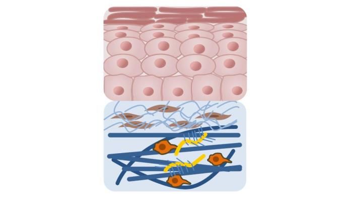
Appl. Sci. 2020, 10, 3435 2 of 24
layer, constituting the lower part of the dermis, has a denser and thicker ECM network with fewer
nerve fibers and capillaries. In this sublayer, collagen fibers are aggregated into thick bundles that
are largely aligned parallel to the skin surface [6,7]. Beneath the reticular dermis is the adipose tissue,
which provides insulation from heat and cold and performs endocrine functions involved in food
intake, glucose homeostasis, lipid metabolism, inflammation, and angiogenesis. Recent findings
henceforth discriminate the dermal white adipose tissue, which consists of intradermal pre-adipocyte
and adipocyte populations, from the subcutaneous white adipose tissue [8].
Damage or loss of integrity of the skin caused by wounds may impair the functions of the skin to
varying extents. Wounds can be caused by mechanical trauma, surgical procedures, reduced blood
circulation, burns, or aging. Most skin wounds can heal naturally through dynamic and interactive
processes, which involve soluble factors, blood elements, ECM components, and cells [9]. However,
in cases of extensive or irreversible damage, surgical strategies are often needed to provide immediate
skin coverage using skin substitutes or scaffolds to guide repair and regeneration. This scenario is
typical for chronic wounds that do not proceed through the normal phases of wound healing for
multifactorial reasons [10]. A chronic wound may enter a persistent inflammatory state and perpetual
non-healing state characterized by chronicity and frequent relapses. A number of pathophysiological
factors can cause a failure of normal wound healing, including inflammation, infection, malnutrition,
age, diabetes, tissue maceration, pressure necrosis, and renal impairment [11–13]. The restoration of
dermis requires three-dimensional (3D) scaffolds to provide elasticity and strength to the epidermal
graft and to feed the keratinocytes in the epidermal layer. Thus, one crucial factor in skin tissue
engineering is the construction of a tissue scaffold that serves as a template to guide restructuring of
cells and subsequent host infiltration of the graft.
In the case of important tissue injury or disease, tissue autografts are often considered the gold
standard, but inherent limitations including donor site morbidity, low availability, and troubling failure
rates highlight the need for alternative strategies [14,15]. To date, engineering human tissues is an
interdisciplinary and active field of research [16–18], but fully reproducing the properties of the ECM is
a great challenge [19–21]. The involvement of ECM proteins in tissue repair is well documented [22–24],
and the development of ECM-biomimetic and -bioinspired materials is undergoing considerable growth.
Moreover, despite technological progress and advances in polymer science, the vast majority of artificial
scaffolds do not perfectly mimic ECM scaffolding and bioactive cell-instructive and cell-responsive
properties. These aspects are particularly challenging for skin repair because regeneration of the
different skin layers requires different cell types and matrix elements.
Among the essential noncellular components, the ECM is a heterogeneous, connective network
composed of fibrous glycoproteins, proteoglycans, and small molecules that coordinate in vivo to
provide the physical scaffolding, mechanical stability, and biochemical cues necessary for tissue
morphogenesis and homeostasis. Decellularization allows acquisition of cell-free, natural ECMs.
The pronounced effect of decellularized ECM scaffolds in supporting tissue regeneration is based on
two major characteristics: first, the maintenance of the 3D structure, providing support, tensile strength,
and attachment sites for cell surface receptors; and second, the availability of bioactive components that
modulate angiogenesis, cell migration, and cell proliferation and orientation in wound healing [25].
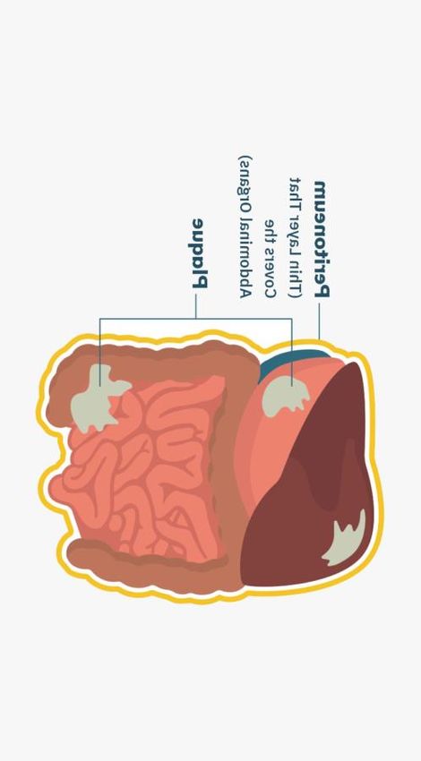
2. Tissue Sources for Preparation of ECM-Based Biomaterials for Skin Repair
In the last decades, decellularized scaffolds for use as skin substitutes have been intensively
developed from various tissue sources such as dermis, skin flaps, peritoneum, intestinal tissues,
and amnion/chorion tissues (Figure 1).
Appl. Sci. 2020, 10, 3435 3 of 24
Appl. Sci. 2020, 10, 3435 3 of 25
A Skin B Peritoneum
C ollagen IV,
Epidermis lam inin,
p roteoglycans
Dermis
C ollagens I-III-
V -V I C ollagen I-III-IV , lam inin,
vitronectin,fibronectin
C Small intestine submucosa D Amniotic membrane
Pla
ce
nta
l cord
Umbilica
C ollagen IV,
lam inin,
p roteoglycans
C ollagen III-IV -V,fibronectin,
lam inin,p roteoglycans
FigureFigure 1. Schematic
1. Schematic representation
representation ofoftissue
tissuesources
sources for
forthe
thepreparation
preparationof decellularized matrix
of decellularized to
matrix to
support skin repair and regeneration. Basic structure, composition and location of each tissue
support skin repair and regeneration. Basic structure, composition and location of each tissue source source
are indicated
are indicated for Skin,
for (A) (A) Skin,
(B) (B) Peritoneum,
Peritoneum, (C)Small
(C) Smallintestine
intestine submucosa,
submucosa,(D) Amniotic
(D) Amniotic membrane.
membrane.
Allogeneic
Allogeneic sources,such
sources, such asastissues from
tissues cadaveric
from donors,donors,
cadaveric and xenogeneic materials, such
and xenogeneic as
materials,
porcine, bovine, and goat tissues, have been used, with different long-term results [26]. The
such as porcine, bovine, and goat tissues, have been used, with different long-term results [26].
decellularization protocols typically combine physical and chemical treatments to remove cellular
The decellularization protocols typically combine physical and chemical treatments to remove cellular
antigens, involving freeze-thawing or detergents and/or enzymes that can alter the 3D structure of
antigens, involving
the ECM freeze-thawing
[27]. Additionally, the US or detergents
Food and Drug and/or enzymes
Administration thatrequirements
(FDA) can alter the 3D structure
impose that
of theacellular
ECM [27]. mammalian-derived products undergo “viral inactivation,” which implies the use impose
Additionally, the US Food and Drug Administration (FDA) requirements of
that acellular
detergentsmammalian-derived
resulting in the removal products
of many undergo
important“viral
biologicinactivation,”
components from whichthe implies theas
tissue, such use of
detergents
lipids, resulting
glycans, and in the removal
elastins of many
[28]. As important
an alternative, skinbiologic components
grafts from fish such asfrom the tissue,
Atlantic such as
cod have
lipids,been proposed
glycans, because[28].
and elastins viralAsandan prion transmission
alternative, risk from
skin grafts is nonexistent.
fish such as These tissues
Atlantic codcan
havebe been
subjected to gentle processing that does not disrupt the structure or bioactive
proposed because viral and prion transmission risk is nonexistent. These tissues can be subjected to composition of the
gentleoriginal ECM that
processing [29]. does not disrupt the structure or bioactive composition of the original ECM [29].
Thus, decellularized tissues can be obtained from a variety of sources and implanted either
Thus, decellularized tissues can be obtained from a variety of sources and implanted either
orthotopically, i.e., the reconstructed tissue exhibits similar properties to those of the source, or
orthotopically, i.e., the reconstructed tissue exhibits similar properties to those of the source,
ectopically, i.e., to a site that is completely different from origin site of the decellularized tissue [30].
or ectopically, i.e., to
In this review, weafocus
site that is completely
on decellularized different from
biomaterials with origin site oftothe
the potential decellularized
be used as dermal or tissue
skin [30].
In thissubstitutes
review, we forfocus onapplications.
clinical decellularized biomaterials with the potential to be used as dermal or skin
substitutes for clinical applications.
2.1. Acellular Dermal Matrix (ADM)
2.1. Acellular Dermal Matrix (ADM)
A decellularized dermal matrix is an acellular tissue made by taking a full-thickness section from
Aa donor source, which
decellularized dermal in most cases
matrix is is
ana acellular
human cadaver
tissue or of porcine
made or bovine
by taking origin. In thesection
a full-thickness case offrom
human
a donor donors,
source, the in
which tissue
mostis cases
screened
is afor infectious
human agents,
cadaver or such as HIV,orhepatitis,
of porcine and syphilis
bovine origin. [31].
In the case of
In the last two decades, ADM has become increasingly popular for the coverage of soft-tissue
human donors, the tissue is screened for infectious agents, such as HIV, hepatitis, and syphilis [31]. open
wounds. Artificial dermal substitutes are routinely used in the treatment of chronic and acute
In the last two decades, ADM has become increasingly popular for the coverage of soft-tissue open
wounds. Artificial dermal substitutes are routinely used in the treatment of chronic and acute injuries,
particularly when autologous intact cutaneous tissue is not available [32]. Although skin grafting is
significantly less expensive, the use of ADM can be a successful alternative to a painful and aestheticallyAppl. Sci. 2020, 10, 3435 4 of 24
undesirable donor site [31]. In addition, as has long been recognized, the presence of dermis enhances
the success of subsequent skin transplantation, inhibits granulation tissue formation, and prevents
scar formation, thereby reducing vascular contraction [26]. Thus, ADM is often used in conjunction
with split-thickness grafts for the treatment of full-thickness wounds [31]. Aside from burn wounds
and diabetic foot ulcers (DFUs), more recently, ADMs have been suggested as a successful alternative
to vascularized flaps for extremity wounds with exposed tendons, bones, and joints [32]. ADMs are
currently produced and developed as various commercial products, and here we focus on commercially
available skin substitutes with substantial relevant evidence of efficacy.
The gold standard for temporary coverage is a skin allograft, i.e., the skin is transplanted from
one person to another. As with autografts involving graft transfer from the same individual, allografts
undergo vascularization within 2–3 days and can provide a variable barrier after a severe burn. Unlike
autografts, however, the skin is highly immunogenic, and when a viable skin allograft is transplanted
onto a healthy recipient, rejection occurs within 6–14 days [33]. Allograft ADM products derived
from donated human skin are supplied by tissue banks compliant with standards of the American
Association of Tissue Banks and FDA guidelines. A long list of products derived from minimally
processed human donor tissue is available, with different durations of covering depending on their
design and composition [34] including the following: AlloDermTM regenerative tissue matrix (RTM;
LifeCell Corp., Branchburg, New Jersey, USA), AlloPatch HD® Pliable (Musculoskeletal Transplant
Foundation), Cymetra™ Micronized AlloDermTM (injectable form of AlloDermTM , LifeCellKCl),
Dermacell® (LifeNet Health), Flex HD® (Ethicon and Musculoskeletal Transplant Foundation),
GammaGraft® (Promethean Lifesciences, Inc.), GraftJacket® RTM (LifeCell, licensed to Wright Medical
Technology and KCl), Glyaderm® (Euro Skin Bank, Beverwijk, The Netherlands), Matrix HD™
(RTI Biologics), Memoderm™ (Memometal, Inc.), Puros Dermis® (Zimmer Dental), and Repliform®
(LifeCell and Boston Scientific) [35]. At least five different manufacturing processes and products
(AlloDermTM , DermaMatrix, Glyaderm® , GraftJacket® , and SureDerm® ) are currently registered for
wound care (Table 1) [33]. We discuss here in detail the most cited versions considering their efficacy
in treating burns and non-healing/difficult-to-heal wounds.
2.1.1. AlloDermTM RTM (LifeCell Corp., Branchburg, NJ, USA)
AlloDermTM (LifeCell Corp., Branchburg, New Jersey, USA) is one of the first FDA-approved
acellular matrix materials and among the most extensively investigated [25]. It has been used
since 1992 to treat burns with successful engraftment of both the AlloDermTM and the subsequent
overlying skin graft in many individuals [36]. This product was FDA approved for the replacement
of inadequate integument tissue, but the most common cited application has been to cover the
skin flaps of donor sites as a sheet graft without an overlying split-thickness graft (Table 1) [37].
AlloDermTM is processed directly from fresh cadaver skin treated with high salt to remove the cellular
components. The treated skin is then freeze-dried, leaving an ADM with an intact basement membrane
complex (Figure 2A) [25]. AlloDermTM is hydrated before use, typically in an antibiotic saline solution
according to the manufacturer’s recommendations. The meshing of the ADM at a 1:1 ratio optimizes
revascularization and nutrition of the overlying skin graft [25]. Finally, in vitro recellularization of
AlloDermTM with allogeneic keratinocytes, adipose-derived stem cells, fibroblasts, and umbilical vein
endothelial cells allows for production of biological skin substitutes [25].Appl. Sci. 2020, 10, 3435 5 of 24
Appl. Sci. 2020, 10, 3435 5 of 25
A Skin B Peritoneal membrane
E p id erm is
M esotheliu m
B asem ent m em brane
0.1 to 0.5 m m
B asem ent m em brane
average of 2 m m
Pap illary Su bm esothelial com p act
D erm is
zone
R eticu lar
A d ip ose connective
A d ip ose connective tissu e
tissu e
C Small intestinal submucosa D Amniotic membrane
E p itheliu m
E p itheliu m
0.02 to 0.05 m m
B asem ent m em brane
Mucosa
B asem ent m em brane
A m nion
C om p act strom al
C onnective tissu e layer
0.1 to 0.15 m m
T hin m u scle layer Fibroblast layer
Sp ongy layer
Su bm u cosa
C horion
T hick m u scle layers
Figure 2. Detailed schematic histological representation of tissues used for decellularization for skin
Figure 2. Detailed schematic histological representation of tissues used for decellularization for skin
repair and regeneration. The various tissue compartments of skin (A), peritoneal membrane (B), small
repair and regeneration.
intestinal mucosa (C) The
andvarious
amniotictissue compartments
membrane of skin by
(D) are represented (A),separate
peritoneal membrane
graphics and (B),
small intestinal mucosa (C) and amniotic membrane (D) are represented by separate
annotated on the side. When a tissue compartment is cellularized, cells are drawn in black and when graphics and
annotatediton is vascularized, vessels
the side. When are represented
a tissue by red circles.
compartment The tissue layers
is cellularized, useddrawn
cells are for decellularization
in black and when it
and clinical applications are shown by arrows on the side indicating their average thickness.
is vascularized, vessels are represented by red circles. The tissue layers used for decellularization and
clinical applications
2.1.2. AlloPatch® are shown
Pliable by arrows on
(Musculoskeletal the side indicating
Transplant Foundation,their average
Edison, thickness.
NJ, USA)
2.1.2. AlloPatch ® Pliable
AlloPatch (Musculoskeletal Transplant Foundation, Edison, NJ, USA)
® Pliable is an aseptically processed human reticular (HR)-ADM, intended for use in
chronic or acute wound coverage [34]. It requires no rehydration or refrigeration prior to use and can
AlloPatch ®
be stored Pliable is an
at ambient aseptically
temperature processed
according to the human reticular
manufacturer’s (HR)-ADM, intended
recommendations. This dermisfor use in
chronic or acute wound coverage [34]. It requires no rehydration or refrigeration prior to use
differs from many other ADMs in that it does not contain a papillary portion, resulting in and
a can be
homogeneous matrix that retains its original architecture and its key ECM
stored at ambient temperature according to the manufacturer’s recommendations. This dermis differscomponents (Figure 2).
The absence of asymmetry or orientation is considered beneficial in the clinical setting, having a
from many other ADMs in that it does not contain a papillary portion, resulting in a homogeneous
positive impact on cell infiltration and native tissue remodeling [38]. In a prospective, randomized,
matrix that retainsmulticenter
controlled, its original architecture
study, this HR-ADM and wasitsshown
key ECM components
to be superior (Figure
in promoting DFU2). The absence
healing as of
asymmetry or orientation
compared is considered
to standard beneficial
of care involving in the changes
daily dressing clinicalwith
setting, having
a collagen a positive
alginate matriximpact
[39]. on cell
infiltration and native tissue remodeling [38]. In a prospective, randomized, controlled, multicenter
2.1.3. DermACELL™ (LifeNet Health)
study, this HR-ADM was shown to be superior in promoting DFU healing as compared to standard of
care involving DermACELL™ is a decellularized
daily dressing changes with regenerative
a collagen human tissuematrix
alginate matrix allograft
[39]. that contains both
the reticular and papillary compartments with a basement membrane (Figure 2). Upon application,
the reticular site is placed against the surgical wound. The patented preparation process for
2.1.3. DermACELL™ (LifeNet Health)
DermACELL™ includes the use of anionic detergents and endonucleases to eliminate more than 97%
of the nucleicisacids.
DermACELL™ It is preservedregenerative
a decellularized and stored at human
room temperature and thus
tissue matrix can bethat
allograft delivered
contains both
the reticular and papillary compartments with a basement membrane (Figure 2). Upon 1).
hydrated [40]. DermACELL™ is indicated for chronic non-healing wounds [34] (Table In a
application, the
prospective, multicenter, single-arm clinical trial for the treatment of large complex DFUs with
reticular site is placed against the surgical wound. The patented preparation process for DermACELL™
exposed tendon or bone, DermACELL™ rapidly reduced the size of the wound. However, that study
includes the use of anionic detergents and endonucleases to eliminate more than 97% of the nucleic acids.
It is preserved and stored at room temperature and thus can be delivered hydrated [40]. DermACELL™
is indicated for chronic non-healing wounds [34] (Table 1). In a prospective, multicenter, single-arm
clinical trial for the treatment of large complex DFUs with exposed tendon or bone, DermACELL™
rapidly reduced the size of the wound. However, that study favored cases that required a single
application of the ADM [41]. Additionally, in a case report [40], DermACELL™ was successfully
used to treat a patient with scarring from a severe burn. More recently, a case report publicationAppl. Sci. 2020, 10, 3435 6 of 24
provided evidence that DermACELL™ can eventually be used to treat fingertip injuries, e.g., after
amputation [42]. An ongoing open-label trial will assess the effectiveness of this ADM in patients with
chronic venous ulcers.
2.1.4. GraftJacket® RTM (Wright Medical Technology, Inc., Licensed to KCl)
GraftJacket® RTM is an intact human ADM containing a basement membrane (Figure 2), processed
with a patented method to minimize damage to the scaffold and freeze-dried to remove moisture.
GraftJacket® RTM is intended for replacement of damaged or inadequate integumental tissue, such
as DFUs, venous leg ulcers, pressure ulcers, or other homologous uses on human integument [43].
A quantitative analysis of comparative literature including randomized, prospective controlled clinical
studies with full-thickness DFU evaluated the efficacy of this ADM compared to standard moist
wound care. The results demonstrated improved healing of DFUs in a significantly reduced time with
this ADM. The authors highlighted the fact that a single application achieved this outcome, whereas
multiple applications are necessary with other commercialized ADMs [44].
Despite their effectiveness, particularly toward wound bed vascularization, skin allografts present
some drawbacks. These include limited availability, potential for pathogen transmission, and cost [33].
2.1.5. Animal-Derived ADMs
Grafts from xenogeneic dermal substitutes are often applied to extensive skin defects caused by
severe burns. Although human ADMs and engineered human skin matrices have shown satisfactory
effects in the treatment of extended second- and/or third-degree burns when xenogeneic ADMs lack
histocompatibility, human ADMs are so expensive that their use is often impossible in these indications.
The necessity of novel cost-effective dermal substitutes with equivalent efficiency has led to the search
for alternative solutions [45]. The critical barrier to xenogeneic scaffolds in translational application
is the recipient immune response to the antigenic components of the xenogeneic tissue [46]. Despite
initial speculation, the decellularization process is insufficient to eliminate antigens from xenogeneic
tissues because both cells and the ECM components themselves can elicit the host immune response.
In addition, the remaining cell debris can be immunogenic [47]. Some strategies to achieve removal
of specific antigens from xenogeneic scaffolds are being developed and are presented later in this
review. These include enzymatic removal of known xenoantigens, such as α-gal epitopes, stepwise
solubilization-based antigen removal, or a combination of these approaches [47].
Although the animal skin can be extremely adherent and occasionally incorporated into the
wound, xenografts are not yet considered true grafts or transplants. Xenografts do not vascularize,
but they can still be beneficial to wound management by reducing the pain in partial-thickness injuries
and improving healing rates. Such ADMs are being used for exfoliative skin disorders [48].
An extensive list of animal-derived ADM products is currently available, most of which achieve
success in clinical use. Since the 1960s, pigs have served as the primary donors for xenografts in the
United States, by virtue of their relative affordability and histological structural similarities to the human
skin [48]. Several reports have attested to the benefits of using porcine skin in the treatment of extensive
wounds. These advantages include reduced healing rates for partial-thickness wounds and granulating
wounds and reduced pain with placement over burns [48]. Currently available acellular porcine dermal
matrices include Permacol™ (Tissue Sciences Laboratories), Strattice™ (LifeCell Corp.), CollamendTM
(Bard), Xenoderm (mbp), and XenMatrixTM (Davol, Inc.) [31]. According to the UnitedHealthCare®
Medicare Advantage Policy Guideline for 2020, porcine skin dressings have been suggested as possibly
necessary as an occlusive dressing for burns, donor sites of a homograft, and decubiti and other
ulcers [49]. Permacol™ is composed of porcine dermal collagen that is subjected to crosslinking, which
is suggested to improve tensile strength. Strattice™ is a non-crosslinked porcine-derived ADM in
pliable and firm versions, which can be stored at room temperature and is available fully hydrated.
Materials from different sources and processing exhibit different biochemical and mechanical
properties and host responses upon implantation [50]. Therefore, suitable dressing material must be
used based on the wound type.Appl. Sci. 2020, 10, 3435 7 of 24
PriMatrix™ (TEI Biosciences) is an ADM processed from fetal bovine skin. It was cleared for
marketing by the FDA for partial- and full-thickness wounds, for diabetic, pressure, and venous stasis
ulcers, and for surgical wounds [51]. This fetal bovine collagen scaffold is processed to remove all
lipids, carbohydrates, and non-collagenous proteins, leaving primarily intact fetal dermal collagen
fibers with a great proportion of fibrillar collagen III [52]. This PriMatrixTM collagen III rich substrate
is associated with healing in developing tissues, whereas the unique structural matrix of fetal tissues
has been identified as a contributing factor in scarless wound healing [50]. SurgiMend™ PRS (TEI
Biosciences) is another ADM processed from fetal bovine dermis.
An acellular fish dermis has also been FDA approved. Kerecis™ Omega3 Wound (Kerecis) is
an ADM rich in omega-3 fatty acids, intended for use in burn wounds, chronic wounds, and other
applications. Human skin differs from fish skin mainly because of the adaptation of the latter to the
aqueous environment through the development of scales, secretion of mucus from epithelial cells,
lack of superficial keratinized layers, and presence of two basement membranes [53]. Additionally,
fish skin heals faster than human skin without leaving scar tissue [54]. The histological analysis of
human skin after a Kerecis™ graft demonstrated that the ADM is incorporated into the damaged area
and infiltrated by autologous cells, is resistant to bacteria, and exhibits anti-inflammatory properties
because of the omega-3 fatty acids [29,54].
2.2. Decellularized Mesothelium
The mesothelium is a simple squamous epithelium lining the walls of large cavities (peritoneum,
pericardium, and pleura) (Figure 1B) [55]. The mesothelial tissues have a thin and strong layer
of ECM that supports epithelial cells, which are capable of rapid wound healing (Figure 2B) [56].
Decellularized mesothelium is obtained using detergent agents in a process designed to maximize
the preservation of the ECM architecture and composition [57]. A list of decellularized mesothelial
matrices of porcine, bovine, equine, and human origins is currently available on the market for use in
regeneration of different tissues and organs, mainly soft tissues including skin, vascular tissues, and
valve replacements [26,58].
2.2.1. Decellularized Intestine and Urinary Bladder
One of the most studied decellularized matrices for a variety of biomedical and tissue-engineering
applications derives from the intestine. The ileum, jejunum, and small intestinal submucosa (SIS) are
the sources of decellularized intestine scaffolds (Figure 1C) [26]. Porcine SIS is derived from the thin,
translucent tunica submucosa layer of the small intestine, which remains after removing the mucosal
and muscular layers (Figure 2C). Once processed, SIS biomaterials constitute cross-linked 3D matrices
consisting of constitutive proteins that are mostly collagen, with smaller amounts of carbohydrates and
lipids [59]. This material supports the formation of a skin barrier 14 days after seeding with human
keratinocytes, fibroblasts, and endothelial cells.
The role of SIS in promoting wound closure has been extensively investigated [25]. Incubation
with SIS inhibits matrix metalloproteinases, which are important contributors to wound chronicity and
are abundantly expressed in chronic wounds [60,61]. Moreover, SIS scaffolds contain transforming
growth factor (GF)-β, basic fibroblastic GF, vascular endothelial GF, and epidermal GF, thus promoting
angiogenesis, cell growth, and differentiation, and consequently tissue regeneration [26]. Advantageous
properties of SIS scaffolds in healing wounds include their effective barrier function of the wound bed
dehydration because of their low porosity, and their strength and flexibility even after a long-term
storage in a lyophilized form [59].
Oasis® Wound Matrix (Cook Biotech, Inc., West Lafayette, IN, USA) is a collagen scaffold derived
from porcine intestine submucosa cleared for marketing by the FDA for the management of partial
and full-thickness wounds, including pressure, venous, diabetic, and chronic vascular ulcers, surgical
wounds, and draining wounds. A clinical study revealed that in a cohort of patients carrying with
chronic leg ulcer treated with SIS, complete wound closure was achieved in 55% cases. The use of SIS
seemed to accelerate healing rates and to reduce the incidence of ulcer recurrence [62]. DecellularizedAppl. Sci. 2020, 10, 3435 8 of 24
porcine urinary bladder matrix (UBM) also has been reported to be an alternative to complex surgical
procedures involving flap-associated donor site morbidity to treat complex extremity wounds. Use of
UBM seemed to be rather beneficial for the management of patients suffering from comorbidities such
as obesity, diabetes mellitus, and cardiopulmonary compromise [27]. After UBM decellularization,
the remaining components include collagen, fibronectin, laminin, GAGs, and GFs. The UBM is likely
to act through induction of bioactive GFs that support angiogenesis and facilitate proliferation of
connective tissues, and the intact basement membrane is conductive to epithelial and endothelial cell
proliferation [63].
Commercially available porcine UBM products include MicroMatrix® (Acell, Inc., Columbia,
MD, USA) and Cytal® Wound Matrix (Acell, Inc., Columbia, MD, USA). Regarding the mechanism
of action, a preclinical study showed rapid cell infiltration into the UBM after application, followed
by a robust angiogenic response and new host tissue deposition [64]. Another study demonstrated
induction of a different immune response as compared to classical wound healing, one that involves
M2-phenotype macrophages related to tissue remodeling rather than the M1 macrophages that are
associated with scar tissue formation [65].
2.2.2. Decellularized Amniotic Membrane (AM)
The AM surrounds the fetus in the uterus and consists mainly from top to bottom of an epithelium
layer, basement membrane, compact ECM layer, fibroblast layer, and spongy layer (Figures 1D and
2D). Fresh human AM contains collagen, fibronectin, and hyaluronic acid along with a combination of
GFs, cytokines, and anti-inflammatory proteins [66]. Several studies have shown that specific AM
products may be useful for treating several conditions in adults, including non-healing lower-extremity
DFUs. AMs are harvested immediately after birth, cleaned, sterilized, and either cryopreserved or
dehydrated, maintaining their properties [67]. Fully decellularized AM can be prepared by incubation
of fresh tissue with hypotonic Tris buffer containing protease inhibitors, sodium dodecyl sulfate
(SDS), DNAase, RNAase, and 1% trypsin-EDTA (ethylene-diamine-tetraacetic acid) for complete
elimination of epithelial and mesenchymal cells [68]. Human AM is characterized by anti-bacterial,
anti-inflammatory, and non-immunogenic properties, promotes reduced pain and dehydration, and
favors the re-epithelialization process [69].
Some commercially available acellular AM products suggested for use in DFUs include
AmnioBand® Membrane (MTF Biologics), Biovance® (Celularity, Inc.), and EpiFix® (MiMedx).
Despite these many advantages, disadvantages of AM include poor mechanical properties and a high
biodegradability rate, which complicate their extensive use in clinic [69]. Finally, the in vitro use of
acellular human AM allows the construction of living skin equivalents through seeding of human
keratinocytes on the epithelial side and fibroblasts on the chorionic side (Figure 2) [70,71].
2.2.3. Decellularized Skin Flaps
A skin flap consists of healthy skin and adipose tissue (Figure 2A) or skin, fat, and muscle.
Clinically, skin flaps are used to cover full-thickness, serious wounds [26] but are not always an option
and can result in donor site morbidity and poor aesthetics. Additionally, split-thickness, autologous
skin grafts often used to cover large wounds transplant only a small portion of the dermis and are thus
subject to scarring and contracture. Full-thickness skin grafts, including epidermis and full dermis,
are difficult to obtain but are subjected to less contracture and result in a more functional graft and a
better cosmetic final outcome. However, they depend on vascular ingrowth. Therefore, generating
a decellularized full-thickness skin flap with a perfusable vascular pedicle could eventually yield
substantial regenerative potential. In two recent studies, decellularized skin flaps from animal sources
were developed and recellularized and/or integrated into host tissue. Zhang et al. [72], using a perfusion
protocol combined with agitation, produced a skin/fat decellularized matrix that maintained the native
ECM architecture and composition of mainly collagen, GAGs, and vascular endothelial GF and absence
of the major class I-histocompatibility complex. Immunohistochemical analysis of the matrix showed
peripheral nerves and vessel structure maintenance, and angiography confirmed the existence of theAppl. Sci. 2020, 10, 3435 9 of 24
vascular pedicle and microcirculatory networks. Those scaffolds could subsequently be re-populated
with human adipose-derived stem cells and endothelial cells and promote neovascularization after
implantation in a rat model [72]. Jank et al. [73], using a porcine model, developed a full-thickness
decellularized skin flap by applying a perfusion technique. The resulting scaffold comprised a vascular
pedicle, maintained the ECM architecture and composition, and could be successfully perfused and
anastomosed to the recipient vascular system. The scaffold demonstrated good biocompatibility and
ability to support human keratinocyte engraftment and formation of an epidermis with intact barrier
function [73]. These results are rather promising and may represent an adequate solution for the
treatment of wounds, especially those characterized by loss of tissue volume.
Table 1. Commercially available decellularized scaffolds with potential use in skin repair.
Source Tissue Product Application Focus
Acellular Dermal Matrix
AlloDerm™ Regenerative Tissue Matrix (RTM, LifeCell Corp.) Burns, Soft tissue ridge augmentation
AlloPatch® Pliable (Musculoskeletal Transplant Foundation,
Chronic or acute wound coverage
Edison, NJ, USA)
Cymetra™ Micronized AlloDerm (injectable form of Damaged tissue, facial reconstructive
AlloDerm, LifeCellKCl) procedures
Reconstructive procedures, chronic
DermaCell™ (LifeNetHeatlh)
wounds
Flex HD® , Structural, Acellular Hydrated Dermis (Ethicon
Abdominal wall repair
and Musculoskeletal Transplant Foundation)
Temporary grafts on burns and chronic
Human skin GammaGraft® (Promethean LifeSciences, Inc.)
wounds
GraftJacket® Regenerative Tissue Matrix (LifeCell, licensed to
Soft tissue repair
Wright Medical Technology and KCl)
Replacement in deep burns, soft tissue
Glyaderm® (EuroSkin Bank, Beverwijk, The Netherlands)
defects, unstable scars
Reconstructive surgery, chronic skin
Matrix HD™(RTI Biologics)
wounds
Memoderm™ (Memometal Inc.) Diabetic foot ulcers, soft tissue repairs
Puros Dermis® (Zimmer Dental) Soft tissue augmentation
Repliform® (LifeCell and Boston Scientific) Repair/replacement of soft tissue
Permacol™ (Tissue Sciences Laboratories) Burns, ulcers, abdominal wall repair
Strattice™ (LifeCell Corp.) Soft tissue repair and body wall defects
Bard CollaMend™ Implant (Davol, Inc.) Soft tissue defects
Xenoderm (mbp) Burns and chronic wounds
Porcine skin Surgical repair of damaged soft tissues,
XenMatrix™ (Davol, Inc.)
plastic and reconstructive surgery
Partial-/full-thickness wounds, burns,
PriMatrix™ (TEI Biosciences)
diabetic foot ulcers
Fetal bovine skin
Hernia repair, plastic and reconstructive
SurgiMend™ PRS (TEI Biosciences)
surgery
Partial-/full-thickness wounds, trauma,
Fish skin Kerecis™ Omega3 Wound (Kerecis)
chronic wounds and diabetic ulcers
Decellularized Small Intestine Submucosa (SIS) and Urinary Bladder Matrix (UBM)
Oasis® Wound Matrix (Cook Biotech, Inc., West Partial-/full-thickness wounds chronic
Porcine SIS
Lafayette, IN, USA) wounds and diabetic ulcers
MicroMatrix® (Acell, Inc., Columbia, MD, USA) Acute and chronic wounds
Porcine UBM
Cytal™ Wound Matrix (Acell, Inc., Columbia, MD, USA) Acute and chronic wounds
Decellularized Amniotic Membrane Matrix
AmnioBand® (MTF Biologics) Acute and chronic wounds
Surgical covering, part-/full-thickness
Human allograft Biovance® (Celularity, Inc.)
acute and chronic wounds
EpiFix® (MiMedx) Acute and chronic wounds
Decellularized Mesothelium Matrix
Porcine
Meso BioMatrix® (MTF Biologics) Soft tissue
mesothelium
Veritas® , Dura-Guard® , Peri-Guard® , Vascu-Guard®
(Baxter
Soft tissue
Healthcare)
Bovine
pericardium CopiOs® (Zimmer) Dentistry
Lyoplant® (B. Braun Melsungen) Dura mater
Perimount (Edwards Lifesciences) Valve replacement
Equine Unite™ Biomatrix (Synovis Orthopedic and Woundcare) Soft tissues and chronic wounds
pericardium DurADAPT™ (Pegasus Biologics) Dura mater
Human
IOPatch™ (IOP) Ophthalmology
pericardiumAppl. Sci. 2020, 10, 3435 10 of 24
3. Decellularization Methods
Both academic and industrial researchers have developed protocols to decellularize various native
tissues, including those mentioned above. They are classically sorted into three categories of chemical,
enzymatic, and physical methods. Almost all protocols, however, consist of combinations of these three
approaches. To investigate these candidates in as many trials as possible, decellularization studies
furthermore apply and compare very different protocols combining various agents instead of varying
only one substance to assess its effects.
We here review decellularization methods specifically used for the production of acellular matrices
for skin repair and evaluate the properties and effects of each substance commonly used in literature.
The results of this analysis led us to propose another classification of these substances into main
decellularizing agents that can disrupt cells and eliminate all DNA content when used alone (detergents,
acids and bases, physical-based methods) and complementary agents that are added to improve
these effects. Particularly, enzymes such as proteases are reviewed as common complementary
agents because their exclusive use, which has been tested several times, never demonstrated total
decellularization or satisfying in vitro cell repopulation of the matrices obtained.
3.1. Complementary Agents
3.1.1. Enzymes
Nucleases are the most common complementary agents included in decellularization protocols
independently of the source of native tissue. Nucleic acids, especially DNA, are well known for their
high immunogenic properties. To limit nucleic acid levels as much as possible, treatments with DNAse
I, DNAse II, or benzonase are performed after exposure of the tissue to the main decellularizing
agent(s) [68,74,75]. Applied concentrations, ranging from 50 U to 10 kU/mL, depend on the properties
of the tissue (type, thickness, cell density) and the exposure to main decellularizing agent(s) (nature,
duration, concentration, temperature). Regarding the choice between benzonase or DNAse, no clear
rationale is available in the literature. Finally, for highly cellularized tissues, processing can also
include RNAse treatments.
As mentioned above, strict protease-based approaches are not sufficient to remove all cells from
the tissues used for the production of skin-repair matrices. Accutase, a commercial mix of proteolytic
and collagenolytic enzymes classically used for soft cell-detachment from two-dimensional (2D) culture
supports, failed to decellularize human AM: the epithelial layer was totally removed after 1 h of
incubation, but many mesenchymal cells were still detected [76]. Trypsin has also been tested on
human AM combined with EDTA, allowing for complete elimination of cells in both epidermal and
mesenchymal compartments and retention of basement membrane components. However, the ability
of the matrix to drive sufficient primary fibroblast infiltration and proliferation after a 7-day in vitro
culture has not been demonstrated. The same group investigated the effects of dispase, a bacterial
protease targeting collagen IV, laminin, and fibronectin [77]. They achieved complete decellularization
but observed ECM alterations and important losses in basement membrane structure, as had been
already reported [78,79]. Cell seeding was not assayed because of the low probability of epithelialization
success using a basement membrane with such damage.
Although still undocumented as an efficient decellularized agent, trypsin can be combined with
physical, acid-, base-, or detergent-based protocols [80–82]. The action of trypsin is achieved by
immersion of the tissue in an isotonic solution containing from 0.25% to 0.1% w/v of enzyme, separately
from the other treatments of the protocol. This exposure is often combined with EDTA (0.1%) and
preferentially applied between delipidation and principal decellularization steps.
Tissue exposure to trypsin needs to be controlled; its ability to cleave a broad range of substrates
means that deleterious effects on ECM structure can be exerted under overly long or too concentrated
trypsin treatments. In porcine skin, different kinds of “fracturations” and density loss were observed
in the ECM of the dermis compartment after an 18-h 0.25% trypsin exposure [80], whereas a denserAppl. Sci. 2020, 10, 3435 11 of 24
and less porous ECM network was obtained for the same decellularization protocol excluding this
preliminary step. Similar observations have been reported for human amnion with shorter exposure at
the same concentration [76]. Surprisingly, none of the studied patents report strict trypsin incubation
during the processing of human skin, human AM, and porcine SIS for acellular matrix production for
skin repair [83–85]. Only one recent patent presenting a decellularization method for fish skin includes
a trypsin step, but at a concentration lower than 0.001% [86].
Dispase, mentioned above as deleterious to human AM in isotonic solution, has surprisingly
been described in two publications as a good complement for skin decellularization. On porcine
skin, application of a soft dispase treatment (0.56 U/mL, 4 ◦ C in isotonic solution), followed by SDS
and soft trypsin exposures, resulted in good cell removal without any adverse effect on ECM post- 4-
and 12-h treatments. These results suggest a softening effect of both temperature and concentration
decreases on dispase treatment [80]. On mouse skin, solubilization of 2 U/mL of dispase in Dulbecco’s
Modified Eagle Medium (DMEM, 37 ◦ C, 2 h) prior to decellularization led to the production of an
acellular matrix compatible with skin re-epithelialization in a mouse model [87]. The use of DMEM
cell culture medium containing various components instead of simple isotonic solution could affect
enzyme activity and explain the absence of deleterious effects. Furthermore, skin is an ECM-rich
tissue containing more layers of epithelial cells than the amnion, so different accessibility to basement
membrane between the two tissues also could explain the differential results with skin and human AM.
3.1.2. Chemicals
EDTA is a well-known chelator of di- and trivalent metal ions, commonly used in biochemistry to
inhibit proteases. Its addition during exposures to trypsin or main decellularizing agents is a common
tactic to prevent ECM degradation by cation-dependent proteases, which are abundant in both intra-
and extracellular compartments. It also allows disruption of the protein–protein interactions based on
metal cation bounding. Concentrations from 0.05% to 0.1% w/v are commonly applied, but levels up to
0.75% w/v have also been reported [80], without any specific association with tissue or decellularizing
agent types. EDTA has been tested as a main decellularizing agent in human AM under various
conditions (0.02–0.1%, 0.5–16 h, 4 or 37 ◦ C), but does not completely decellularize the tissue, with a
maximum DNA reduction limited to 50% of the initial level [88]. Other commercial protease inhibitors
consisting of mixes of chemicals and small peptides can also be added to prevent ECM damage.
Hypo- and hypertonic shocks are applied from 1 to 12 h before the main decellularization steps or
after to wash tissues and ensure the total lysis of remaining cells. To create hypertonic conditions, NaCl
is commonly dissolved at 1 M to weaken cell membranes, as reported in the literature and in patented
protocols to process fibroblast sheets, fish skin, and human skin [83,86,89]. Hypertonic NaCl also has
been used to dissociate epidermis and dermis [90,91]. A hypertonic solution composed of 1 M KI has been
used to process mouse skin [87]. Regarding hypotonic buffers, Tris (tris(hydroxymethyl)aminomethane)
is often used at 10 mM, and immersion baths before decellularization and for solubilization of SDS
have been reported for porcine bladder wall or fibroblast sheets processing [89,92].
3.1.3. Organic Solvents
Organic solvents are used to remove lipid content, an essential step for the preparation of acellular
matrices using SIS and mesothelium, two fat-rich tissues.
A protocol combining methanol and chloroform (1/1, v/v, 12-h immersion) has been reported
with porcine SIS, following manual elimination of the tunica serosa and tunica muscularis and prior
intensive washings and SDS decellularization [81,82]. A degreasing step did not elicit notable changes
in ECM architecture, and no remaining fat cells were detected in the final acellular matrix. The decrease
in GFs reported in the final scaffold has been attributed to the manual removal of some compartments
of the native tissue, not to organic solvent treatment.
Isopropanol (IPL) exposure is another method to remove lipids [76,93]. Two protocols have
been compared with porcine skin: a single final exposure of the tissue to 100% IPL, and two 70%Appl. Sci. 2020, 10, 3435 12 of 24
IPL exposures distributed during the whole process, followed by a final immersion in 100% IPL [93].
The single exposure was associated with the removal of half of the total lipid content, whereas a
decrease of 90% was estimated for repeated treatments. Regarding nucleic acid removal, no significant
variation in remaining DNA levels was detected between the two methods, confirming that IPL is not
a main decellularizing agent. A combination of IPL and acetone is also mentioned in the patent of the
commercial Oasis® matrix derived from porcine SIS [84].
Use of acetone/hexane (40/60, v/v) and pure acetone with porcine mesothelium was reported [94].
Lipid amounts were not assessed, and only a visual evaluation suggested better elimination of fat
using the acetone/hexane combination.
3.2. Main Decellularizing Agents
3.2.1. Detergents
Detergents are the most common decellularizing agents used to process skin, SIS, mesothelium,
bladder, and AM, regardless of the tissue origin. SDS is the strongest detergent used for tissue
decellularization. It is ionic and disrupts all interactions between biomacromolecules. This agent is the
most reported in decellularization protocols, and numerous teams have used it to process porcine SIS,
porcine bladder wall, human amnion, human skin, rat flap, or fibroblast cell sheets [68,80,81,89,92,95].
Patents often mention its use for the production of commercial acellular matrices used in clinic [83,84,86].
SDS treatments are commonly performed by immersion in a 0.5% w/v solution (range of concentrations
reported in literature: 0.1–1%) and last for hours. Thick and complex tissues containing a dense
conjunctive layer or a multilayered epithelium, such as mesothelium, skin, and bladder wall are
processed from 12 to 24 h. In contrast, thinner or less complex structure treatments last from 0.5 h
for fibroblast sheets to 4–12 h for SIS. All decellularization studies that include SDS in concentrations
>0.1% in their protocols, have reported a total removal of cells and cellular debris from the native
tissue and low levels of remaining DNA. Some combinations with nucleases have been reported on
amnion and bladder wall for SDS concentrations lower than 0.1% [68,92].
Although SDS is an excellent agent for decellularization, it has been frequently reported to damage
ECM. Loss of global ECM density as well as GAG content and damage to the collagen network have
particularly been observed in the matrix produced by fibroblast sheets [89] or in complex tissues
such as human skin [96] and porcine SIS [81]. The increase in SDS concentration from 0.05% to 0.5%
on fibroblast sheets or in exposure time from 6 to 12 h on SIS has been directly correlated with the
amplification of ECM alterations. Scanning electron microscopy studies also suggest rearrangements of
ECM fibers and variations in scaffold porosity after SDS treatment, even if no common and systematic
effects can be generalized. The exposure of porcine SIS to the combination of 1% SDS for 12 h and 1%
Triton X-100 for 0.5 h leads to complete cell removal but exerts similar adverse effects on ECM quality,
coupled with a low cell development and metabolic activity after hemangioma stem cell (HEMSC)
seeding [75]. Different SDS/Triton X-100 combinations have also been tested on rat fasciocutaneous
flaps, leading to high DNA content removal after perfusion of 1% SDS for 24 or 72 h, followed by a
0.5-h 1% Triton X-100 treatment [95]. Modifications of the 2D structure of the ECM have been noted
for the 72-h SDS perfusion using histological staining, but no other investigation has characterized
the effects on ECM in detail. The switch of the 72-h SDS treatment from perfusion to immersion was
associated with a decrease in DNA removal.
Basement membrane preservation after exposure to SDS is essential for epithelialization of
the scaffold. The preservation of collagen IV, collagen VI, and laminin has been confirmed by
immunostaining in porcine bladder wall (Tris HCl 10 mM overnight, 0.1% SDS for 24 h) [92] and softly
decellularized human skin (1 M NaCl 24 h, 0.5% SDS 1 h) [97], but not in porcine SIS (0.05% trypsin 12 h,
0.5% SDS 4 h). Trypsin was shown to be compatible with basement membrane preservation at a higher
concentration [81]. In human AM softly decellularized by a 24-h immersion in 0.03% SDS coupled with
DNAse I and RNAse I treatments, maintenance of the basement membrane components but damageAppl. Sci. 2020, 10, 3435 13 of 24
to the mesenchymal matrix architecture have been observed [68]. However, the mechanical properties
were not significantly altered, and the final scaffold exhibited characteristics, e.g., failure strain, failure
stress, an elastin phase slope, and a collagen phase slope similar to native tissue. This study is the
only one to report decellularization of human AM using SDS, and the soft treatment applied could be
an interesting approach to preserving ECM mechanics and integrity. Finally, no other comparison of
tissue biomechanics before and after decellularization has been reported.
Even if ECM damage is often underlined after SDS treatments, biocompatibility of the
resulting scaffolds has been demonstrated for several native tissues. In vitro, matrices derived from
porcine skin [80], porcine SIS [82], human AM [68], and fish skin [98] do not exert cytotoxic or
proliferation-limiting effects. Cytotoxic tests performed by direct or conditioned-medium contact,
metabolic arrays, or visual observations by light microscopy several days after cell seeding have
confirmed the compatibility of the acellular matrices with cell development. These studies used NIH
3T3 fibroblasts, primary fibroblasts, bone marrow mesenchymal stem cells (BMSCs), or human lung
carcinoma cells. Implantation tests have also been reported for some acellular matrices obtained
using SDS. Porcine SIS-derived matrix implanted subcutaneously in rat has shown a very satisfying
colonization and establishment of host fibroblasts 4 weeks post-operatively. A classic immune response
involving giant cells and infiltrated inflammatory cells has been described but started to decrease after
14 days of implantation [81].
Triton X-100 is a non-ionic detergent that can disrupt interactions between biomacromolecules
more gently than SDS. It is very commonly used alone or with other agents to generate matrices
for skin repair derived from porcine SIS [82,99], porcine skin [68], porcine mesothelium [94], mouse
skin [87], and rat flap [72]. Almost all protocols apply this detergent at the same concentration of
0.5% v/v, with a few using up to 1%. The ability of Triton X-100 to fully decellularize a native tissue
has been evaluated with porcine SIS exposure to increasing concentrations of detergent from 0.5% to
2% [99]. Acellular scaffolds were obtained after 48 h of immersion in all conditions, but ECM damage,
especially loss of density, was observed for concentrations higher than 1%. On mouse skin, successful
decellularization using 0.5% Triton X-100 for 24 h also has been reported, associated with matrix
alterations, especially loss of elastin and increased stiffness compared to native skin [87]. Limited
colonization by primary fibroblasts and poor metabolic activity was also described. Surprisingly,
during wound healing experiments in mouse, new blood vessel formation was observed in the grafted
matrix, whereas a low dermis fibroblast infiltration occurred in accordance with in vitro experiments.
Deleterious effects of 0.5% Triton X-100 on mouse skin may be the result of its limited thickness and
cell abundance compared to SIS.
To modulate the effects on ECM and favor cell development, combinations of Triton X-100 with
other agents have been tested at reduced exposure times. As mentioned above, the combination of
0.1% SDS for 12 h with 1% Triton X-100 for 0.5 h failed to produce a satisfying porcine SIS acellular
matrix [75] because the scaffold obtained was not compatible with cell growth in vitro. In contrast,
co-exposure of porcine skin to 0.5% Triton X-100 and hypertonic 1 M NaCl, followed by 2% sodium
deoxycholate and DNAse I treatments, appeared more promising [74]. Density and integrity of dermis
ECM were preserved, especially collagen I and fibronectin networks, and the preservation of basement
membrane was confirmed by the detection of collagen VII and laminin. Other observations revealed
that, after a 6-month abdominal implantation in monkey, new blood vessels and dense colonization by
host cells could be observed (global immune response not estimated). In rat, a dermis/adipose flap
was successfully decellularized by perfusion using a combination of 1 M NaCl (epidermis removal
and tonic shock), trypsin, and 1% Triton X-100 for 48 h [72]. DNA in dermis and fat compartments
was quantified under 50 ng/mg of dry flap (10-fold reduction compared to native tissue). The general
structure (2D and 3D) of the ECM and GAG content were preserved, but no evaluation of the basement
membrane was described. Vascular endothelial GF was easily detectable by immunohistochemistry in
fat tissue, but very low staining appeared in dermis. The growth of human umbilical vein endothelial
cells and adipose stromal cells was shown between days 1 and 7 post-seeding.You can also read