3D Bioprinting at the Frontier of Regenerative Medicine, Pharmaceutical, and Food Industries
←
→
Page content transcription
If your browser does not render page correctly, please read the page content below
REVIEW
                                                                                                                                             published: 28 January 2021
                                                                                                                                       doi: 10.3389/fmedt.2020.607648

                                            3D Bioprinting at the Frontier of
                                            Regenerative Medicine,
                                            Pharmaceutical, and Food Industries
                                            Qasem Ramadan* and Mohammed Zourob

                                            College of Science and General Studies, Alfaisal University, Riyadh, Saudi Arabia

                                            3D printing technology has emerged as a key driver behind an ongoing paradigm shift
                                            in the production process of various industrial domains. The integration of 3D printing
                                            into tissue engineering, by utilizing life cells which are encapsulated in specific natural
                                            or synthetic biomaterials (e.g., hydrogels) as bioinks, is paving the way toward devising
                                            many innovating solutions for key biomedical and healthcare challenges and heralds’ new
                                            frontiers in medicine, pharmaceutical, and food industries. Here, we present a synthesis
                                            of the available 3D bioprinting technology from what is found and what has been achieved
                                            in various applications and discussed the capabilities and limitations encountered in
                                            this technology.
                                            Keywords: 3D bioprinting, tissue engineering, regenerative medicine, drug discovery, in vitro
                          Edited by:
            Khashayar Khoshmanesh,
             RMIT University, Australia
                                            INTRODUCTION
                         Reviewed by:
            Sasan Jalili-Firoozinezhad,
                                            Additive manufacturing (AM), the process of joining materials to make objects from
  Koch Institute for Integrative Cancer
      Research at MIT, United States
                                            computer-aided design (CAD) model data, such as 3D printing, shows a high potential to radically
                        Elena Pirogova,     disrupt the global consumer market and trigger a manufacturing revolution in a broad spectrum
             RMIT University, Australia     of applications in many industry sectors. 3D printing is mostly well-known for custom-fabricating
                  *Correspondence:
                                            of industrial prototypes and parts using standard fabrication materials such as plastics and metals.
                    Qasem Ramadan           This technology has recently infiltrated into many industries such as aviation, automobile, dental,
             qalramadan@alfaisal.edu        electronics, and fashion. The successful implementation of AM in the healthcare industry has
                                            resulted in the development of surgical equipment, prosthetics, medical devices, and implants.
                   Specialty section:       More recently, 3D bioprinting technology has emerged from the existing 3D printing, by utilizing
         This article was submitted to      life cells and gels as printing materials (bioinks) to create ex vivo and in vitro tissue models, which
          Pharmaceutical Innovation,        heralds’ new frontiers in medicine.
               a section of the journal
                                                3D bioprinting is the process of integrating living cells with biomaterials that allows
      Frontiers in Medical Technology
                                            controlled layer-by-layer deposition of cells/bioink, which is characterized by hierarchical structural
      Received: 17 September 2020           properties, with maintained cellular viability in 3D space to create complex, multifaceted tissues.
      Accepted: 08 December 2020
                                            3D bioprinting benefited from several technologies such as tissue engineering, synthetic biology,
        Published: 28 January 2021
                                            micro/nanofabrication, and bioprocessing biomaterial production (1).
                             Citation:          In vitro, cells cannot arrange themselves in 3D structures like that in real tissue in vivo. Various
Ramadan Q and Zourob M (2021) 3D
                                            techniques were utilized and developed, aiming to mimic the living tissue structure and function,
         Bioprinting at the Frontier of
             Regenerative Medicine,
                                            such as scaffold fabrication, tissue culture, bioreactors, ECM self-assembly among others. However,
Pharmaceutical, and Food Industries.        current tissue-engineering strategies lack the capability of fabrication of fully functional tissues and
    Front. Med. Technol. 2:607648.          recapitulate their heterocellular structure (2, 3). 3D printing shares the three basic components
   doi: 10.3389/fmedt.2020.607648           with the conventional Gutenberg paper printing: the 3D model file to be printed (blueprint) is

Frontiers in Medical Technology | www.frontiersin.org                              1                                            January 2021 | Volume 2 | Article 607648
Ramadan and Zourob 3D Bioprinting
analogous to the text file, the bioink (which comprises cells Vessel Bioprinter (Revotek, Sichuan, China), among others.
and other bioactive materials) is analogous to the ink, and The 3D bioprinting market is projected to reach USD 1,647.4
the 3d printer is analogous to the printer that deposit million by 2024, driven by the technological advancements
the ink on a substrate or print platform (Figure 1). The in 3D bioprinters and biomaterials and its application in the
progress in bioprinting technology is going separately through pharmaceutical and cosmetology industries (6). The Food and
these techniques, and the real challenge is to integrate these Drug Administration (FDA) issued a guidance that provides the
technologies in an industrial scalable technology (1). However, Agency’s initial thinking on technical considerations specific to
bioprinting has a great potential to surpass the traditional tissue devices used for 3D printing techniques and products (7).
engineering techniques and can offer solutions to many existed Many technical challenges are still ahead and need to be
technological hurdles such as: solved to enable smoother penetration of this technology into
the market. Several excellent reviews were recently published
• 3d bioprinting allows a high level of control and precise
(8–13) which surveyed the landscape of 3D bioprinting. In this
positioning of several cell types, thanks to the precise control
paper, we briefly presented the current bioprinting techniques
position of the dispenser nozzle in the X–Y–Z space (1,
and other essential elements pertaining to the application of 3D
4), hence enabling accurate recapitulation of tissue/organ
bioprinting for generating 3D tissues/organ. We also discuss the
microstructure with high architectural complexity.
major challenges and exciting opportunities of 3D bioprinting
• 3d bioprinting is amenable to automation and scalable
technologies toward creating realistic tissue/organs in various
technology that would enable the mass production of
market sectors particularly on the potential of creating in vitro
tissue/organ from standard building blocks (4).
models as tools of drug discovery in the pharmaceutical industry.
• 3d bioprinting benefits from the well-established printing
technology. Therefore, the engineering of the modular 3d
bioprinter is advancing with high-speed pace. For example, the 3D BIOPRINTING TECHNOLOGY
3d bioprinter could be evolved as an integrated surgical tool for
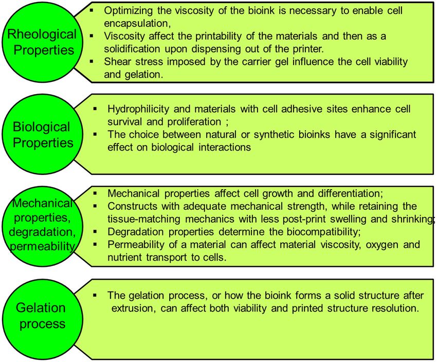
in situ printing (5). Bioprinting starts with obtaining the anatomical structure of the
target tissue by a proper imaging technique such as computerized
The last decade witnessed significant progress in the bioprinting tomography (CT) and magnetic resonance imaging (MRI). A
arsenal with many ground-breaking innovations that makes 3D specialized software is then used to translate the image to a CAD
bioprinting one of the most exciting and promising technologies drawing of cross-sectional layers such that the printing device
that could impact many medical applications. Using bioprinting will be able to add them in a layer-by-layer process. Next, the
technology, scientists may print living de novo organs like heart, bioprinting device constructs the tissue using a specific printing
livers, kidneys, lungs, and skin, which would, therefore, reduce method such as inkjet 3D bioprinting, micro-extrusion 3D
the organ transplant shortage. At the same time, when cells are bioprinting, laser-assisted 3D bioprinting, and stereo-lithography
taken from the patient himself, this would ensure eliminating by employing a combination of printing materials such as
the immune system attack and organ rejection. Another exciting scaffold, bioink, and other additive factors (Figure 2). The
industrial application of 3d bioprinting is in the pharmaceutical accuracy, stability, and tissue viability vary through these
industry. In vivo-like in vitro models can be printed using processes. Finally, the constructed tissue is post-processed in
human cells, and a living organ or a network of organs can a bioreactor to recreate the required in vivo environment to
be created and utilized for preclinical drug screening as an maintain tissue viability during the maturation period.
animal alternative. Another exciting application is using the 3d
printing technology and advanced food formulations to produce
3D Bioprinting Techniques
animal-free meat that mimics the appearance, texture, and taste
3D bioprinting technology was evolved from the traditional
of animal-based meat (Figure 1). These three applications hold
2D printing on paper and later 3d printing of non-biological
great potential in creating new markets and form the major
materials. Therefore, it is not surprising that the engineering
driving forces that accelerate the research and development
aspect is more advanced than bioink material technology.
in academia and industry. The industry sector of this domain
However, since it was initially developed for non-biological
is expanding rapidly with many businesses having been
material printing, each printing technology is still suffering
established that centered around this emerging technology such
from several limitations related to material compatibility when
as Organovo Holdings, Inc. (US), CELLINK (Sweden), Allevi
replacing other building materials with bioink. Several reviews
Inc. (US), Aspect Biosystems Ltd. (Canada), EnvisionTEC GmbH
have been recently published which provide comprehensive
(Germany), Cyfuse Biomedical K.K. (Japan), Poietis (France),
technical information on these techniques (8–14). Therefore,
TeVido BioDevices (US), Nano3D Biosciences, Inc. (US),
here we only briefly discuss these techniques, which are also
ROKIT Healthcare (South Korea), Digilab, Inc. (US), regenHU
summarized in Table 1.
(Switzerland), GeSiM (Germany), Advanced Solutions Life
Sciences (US), and Regenovo Biotechnology Co., Ltd. (China), (a) Microextrusion 3D bioprinting: is a pressure-assisted
Hewlett-Packard (Palo Alto, CA, USA), Novogen MMX Bio- technique commonly used in non-biological material
printer (Organovo, Inc., San Diego, CA, USA), 3D Bioplotter printing. In the bioprinting process, the selected bioinks,
(EnvisionTEC, Gladbeck, Germany), Oxford Performance which are stored in a glass or plastic cartridge, are normally
Materials (South Windsor, CT, USA), and Commercial Blood dispensed through a nozzle by applying pressure using either
Frontiers in Medical Technology | www.frontiersin.org 2 January 2021 | Volume 2 | Article 607648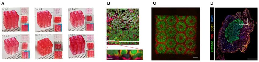
Ramadan and Zourob                                                                                                                                         3D Bioprinting

 FIGURE 1 | 3D bioprinting integrates the conventional 3D printing, imaging, and cell-gel to fabricate functional tissue for regenerative medicine, pharmaceutical
 preclinical drug screening, and animal-free meat.

 FIGURE 2 | Overview schematic of the bioprinting processes.

     a pneumatic or mechanical (piston or screw-driven) method                              (12). In addition, a large variety of bioinks can be used,
     and controlled by a computerized robotic arm. The bioink                               including scaffold-based and scaffold-free bioinks, with high
     is ejected through the nozzle in the form of a thin filament                           printing speed. However, this technique has a low resolution
     and deposited on the substrate based on a CAD design                                   (∼100 µm) (14, 29). The relatively high extrusion pressure
     that determines the position and path of nozzle movement                               through the nozzle imposes high shear stress on the bioink
     to form the tissue in the desired 3d shape. This technique                             components and may lead to loss of cellular viability and
     originated from conventional 3D printing and has been                                  distortion of the tissue structure (16, 30).
     found to be the most suitable method for the creation of                           (b) Inkjet 3D bioprinting: is a non-contact technique that uses
     large-scale constructs, due to its structural integrity, hence                         thermal, piezoelectric, or electromagnetic forces to expel
     becoming more amenable to scale-up for organ fabrication                               bioink droplets onto a substrate replicating the CAD-based

Frontiers in Medical Technology | www.frontiersin.org                               3                                           January 2021 | Volume 2 | Article 607648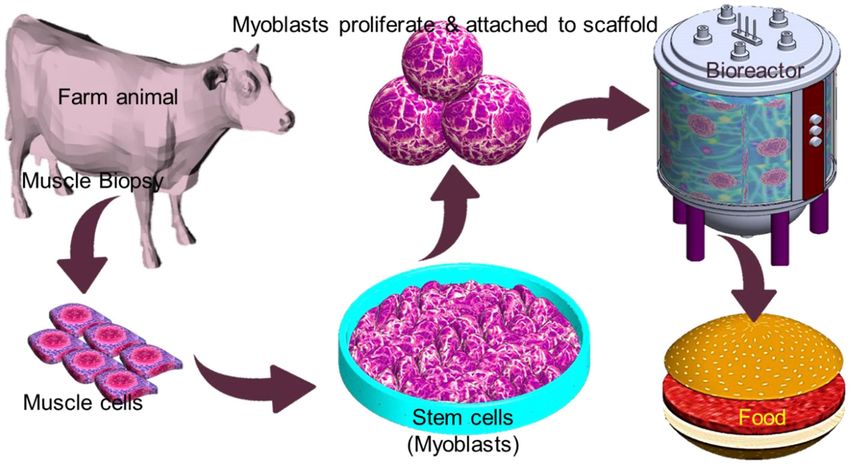
Ramadan and Zourob                                                                                                                                           3D Bioprinting

TABLE 1 | Major 3D bioprinting techniques.

Bioprinting technique               Description                           Advantages                              Disadvantages                             References

Microextrusion 3D bioprinting       Continuous dispensing of the          • The ability to print high-viscosity   • The pressure-driven dispensing             (14–18)
                                    printing materials (bioink) through     bioinks by adjusting the driving        results in high shear stress on the
                                    a nozzle that is driven by a            pressure;                               cells, which dramatically affects the
                                    pneumatic or mechanical (piston       • The ability to print tissues with       cell viability;
                                    or screw-driven) method and             very high cell densities and          • Limited resolution; inability to
                                    controlled by a computerized            scaffold-free bioink;                   construct a microcapillary network
                                    robotic arm                           • Provides good structural integrity
                                                                            due to the continuous deposition
                                                                            of filaments
                                                                          • Amenable to scale-up tissue and
                                                                            organ fabrication process

Inkjet 3D printing                  Droplets of cell-containing bioink    • Non-contact based, which              • Non-uniform droplet size;                  (19–23)
                                    (each contains 10,000–30,000            reduces the chance of                 • Requires bioink with low viscosity
                                    cells) is formed by either heating      contamination;                          (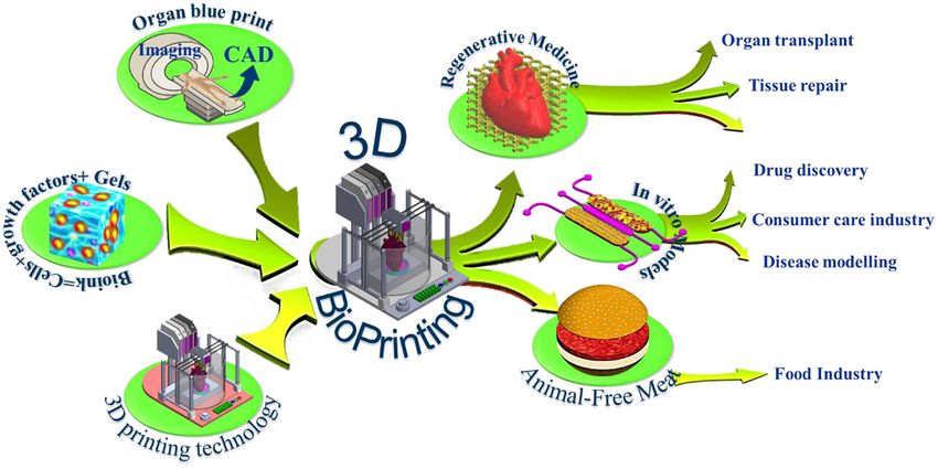
Ramadan and Zourob                                                                                                                    3D Bioprinting

    uses a ribbon, which is coated with an absorbing layer                 cell differentiation (34). Recapitulating the native environment
    such as gold. When a laser pulse is directed and passed                of a specific tissue type promotes stem cell differentiation toward
    through the transparent ribbon, the generated heat induces             that lineage. The ability to create 3D heterogeneous tissue
    a hydrogel droplet and is eventually transferred to the                structures relies on the integration with compatible biomaterials
    receiving substrate. This process is repeated several times,           to support the cellular components. Hydrogels are the most
    through laser pulses, to form a jet, consequently creating             common biomaterials for live-cell printing (5, 35) owing to their
    the final construct in a layer-by-layer fashion. Using this            biocompatibility and their ability to acquire a similar structure
    technique, cells can be deposited with a density of up to 108          the ECM (5, 35–37).
    cells/ml with a single-cell resolution at high speed, enabling             The ultimate aim of bioprinting-based tissue engineering
    high-throughput cell and biomaterial patterning (24–26). In            is to mimic to a certain extent the embryonically developed
    addition, laser-assisted printing offers real-time monitoring          tissue/organs. However, this novel approach is still not close
    of cells, therefore enabling cell selection and transfer (5). On       enough to achieve the same degree of complexity of the in vivo
    the other hand, the excessive heat generated due to the laser          counterparts produced by different specialized cells and dynamic
    energy may damage cells and affect the cell viability in the           extracellular matrix (ECM) composition (9). Bioink is a solution
    printed tissue (32).                                                   of a biomaterial or a mixture of several biomaterials (e.g., in a
(d) Stereolithography-based bioprinting (SLB): utilizes                    hydrogel form), which encapsulates the desired cell types during
    photopolymerizable liquid polymer where a UV light or                  the printing process to create the tissue constructs. Bioinks are
    laser is directed in a predesigned pattern over the polymer,           made of either natural or synthetic biomaterials, or a mixture of
    which leads to cross-linking and hardening of the polymers.            both (38). The biological, mechanical, and rheological properties
    In every polymerization cycle, a thin layer of the structure           of bioinks should be optimized to enable creating the tissue
    is created, and this polymerization cycle is repeated to               that closely mimics the structure and functions of the in vivo
    build the 3D structure in a layer-by-layer fashion. The                counterpart. Different applications and cell types may require
    main advantage of this technology lies in its high resolution          different bioinks. In general, there are essential properties that
    and the absence of harsh shear stress compared to other                need to be considered when choosing a bioink, such as the
    techniques. However, cells are exposed to intense UV                   following (Figure 3):
    radiation for cross-linking, which can cause cell damage.
                                                                           (a) Viscosity: Depending on the bioprinting technique, the
                                                                               bioink matrix should fit in all the bioprinting phases, as fluid
Bioinks                                                                        during cell encapsulation and as a solid after dispensing (39);
The development of printing biomaterials (i.e., bioink) is                 (b) Gelation process and stabilization: the process of how
a cornerstone of 3D bioprinting technology and the most                        the bioink forms a solid structure after extrusion can
challenging task that is still delaying this technology’s progress.            affect the viability and printed structure resolution. This
The ideal properties for bioink must meet both the physical and                process should be fast, and the bioink should retain the
biological material requirements to enable in vivo-like cellular               tissue-matching mechanics after printing and be non-
behavior, such as proliferation, differentiation, migration, and               toxic to cells. Various gelation processes are used, which
maturation. The physical properties are viscosity, structural                  may be determined by bioink material properties and
strength, printing capacity, degradation, and functionality.                   composition, such as ionic (36), thermal (40), stereocomplex
Biological properties include cytocompatibility, biocompatibility,             (29), photocrosslinking (41), enzymatic (42), and click
and bioactivity (16). Bioink viscosity is a crucial parameter of the           chemistry (43);
bioprinting process that always needs optimization to adjust the           (c) Biocompatibility: hydrophilicity and materials with cell-
bioink flow and cell encapsulation efficiency, and tissue structure            adhesive sites enhance cell survival and proliferation (44).
stability (16). Diverse bioink compositions existed to meet the                Also, the choice between natural or synthetic bioinks has
requirements of specific printing technology. Hydrogels are                    a significant effect on biological interactions. Natural-based
promising candidates for developing of bioinks thanks to their                 bioinks may withstand harsh fabrication conditions (e.g.,
biocompatibility, low cytotoxicity, hydrophilicity, and ability to             high temperatures and organic solvents); however, it suffers
form networks of polymers allowing them to acquire ECM with                    from batch-to-batch variability. On the other hand, synthetic
a similar structure.                                                           polymers overcome the batch-to-batch variability, which
    Tissues and organs are self-organizing systems. During the                 offer a high potential for large-scale production but do not
embryonic maturation process, cells undergo biological self-                   offer the natural cell adhesive sites (44).
assembly and self-organization without external influence                  (d) Mechanical properties: Cells are sensitive to their external
or guiding structures (33). However, in vitro, the cell                        mechanical environment, such as matrix elasticity (45),
microenvironment is dramatically simplified or does not                        and can modify their behavior. Therefore, controlling the
exist; therefore, cells fused and slowly aggregate differently.                mechanical parameters of bioinks can be exploited to
In bioprinting, biocompatible scaffolds are usually used as                    control cell behavior, such as their morphology and rate of
structural support for cells to adhere, proliferate, differentiate,            proliferation, within the printed tissue construct, which plays
and eventually form the tissue. Studies showed that the                        an important role in the generation of a functional tissue.
arrangement of integrins within a scaffold highly influenced stem              Another important mechanical property is shear-thinning,

Frontiers in Medical Technology | www.frontiersin.org                  5                                    January 2021 | Volume 2 | Article 607648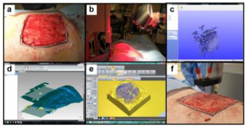
Ramadan and Zourob                                                                                                                                            3D Bioprinting

 FIGURE 3 | The bioink matrix properties play a vital role in the effectiveness of the bioink in the bioprinting process and for creating viable 3D tissues with complex
 geometries.

     which is a non-Newtonian behavior that implies decrease                             formulations are urgently needed to allow utility in different
     of the viscosity as the shear rate increases, which causes                          bioprinting applications.
     reorganization of the polymer chain (46). For example,
     Chen et al. (47) developed a shear-thinning hybrid bioink
     by combining rigid gellan gum, flexible sodium alginate,
                                                                                         Cell Aggregates as Building Blocks
                                                                                         Using a biodegradable solid scaffold is, generally, the dominated
     bioactive thixotropic magnesium phosphate-based gel, and
                                                                                         approach in tissue engineering to construct a living tissue
     thixotropic TMP-BG. The bioink mechanical, rheological,
                                                                                         structure, which provides several supporting functions, including
     and bioactive properties were optimized for printability and
                                                                                         (1) a substrate to cell growth and proliferation; (2) a rigid
     cell viability.
                                                                                         scaffold to provide the desired tissue/organ shape; and (3)
Various natural and synthetic biomaterials have been utilized                            a porous structure of a solid scaffold which allows good
as bioinks. Recently, responsive, dynamic, and supramolecular                            cell viability and enables vascularization (33). However, this
materials are being exploited for bioink development (48).                               classical approach faces some limitations and challenges, which
Morgan et al. (11) provide a comprehensive review on the                                 include (1) vascularization of thick tissue constructs, (2) precise
current trends in bioinks including the mechanical properties                            positioning of multiple cell types inside the 3D scaffolds, and
and dynamic bioinks. Synthetic polymers have good potential                              (3) the effect of scaffold rigidity on cell differentiation (45). The
to be modified to induce bioactivity (38, 49). However,                                  ideal alternative to solid scaffold techniques is to understand
they may generate toxic products and lose their mechanical                               how organs are formed during embryonic development, which
properties during the degradation process (50). In addition,                             would provide a powerful insight into tissue engineering (33,
self-assembling peptides are promising biomaterials for building                         52). Researchers are recently looking at spherical cell aggregates
3D scaffolds that share similar structural and mechanical                                (cellular spheroid) as building blocks of tissue construction.
properties of extracellular matrices. For example, Cofiño et al.                         This development-biology-inspired approach involves utilizing
(51) developed bioink with controlled viscosity by optimizing                            self-assembly of these living microstructures to build tissues
methylcellulose and RAD16-I-based biomaterial to build 3D                                of prescribed shapes (53). During the embryonic maturation
predefined structures. The resultant constructs show high                                process, cells from multiple sources undergo biological self-
shape fidelity and stability. In general, standardized bioink                            assembly and self-organization without any external influence

Frontiers in Medical Technology | www.frontiersin.org                                6                                            January 2021 | Volume 2 | Article 607648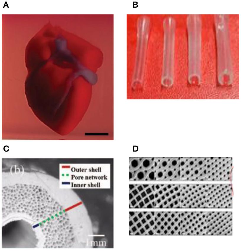
Ramadan and Zourob                                                                                                                       3D Bioprinting

(33). In bioprinting (in vitro), cell aggregates undergo tissue                recently demonstrated (62). An omental tissue biopsy was taken
fusion, where cells organize into multicellular units to create                from patients, and the cells were reprogrammed to become
the final tissue structure. Forgacs et al. (53), showed that the               pluripotent stem cells and then differentiated to cardiomyocytes
fusion of embryonic cushion tissue during heart morphogenesis                  and endothelial cells. The bioink was formed by separately
proceeds similarly in vitro and in vivo and both qualitatively                 combining the two cell types with hydrogels for the cardiac tissue
and quantitatively resembles the coalescence of liquid drops and               and blood vessels. Functional vascularized patches according to
showed that spherical cell aggregates mixed with an appropriate                the patient’s anatomy were demonstrated (Figure 4A). Among
hydrogel behave as self-assembling “bio-ink” particles. The                    the various human tissues, skin was the focus of intensive
authors also demonstrated the print of cellular toroid, tubes,                 research work, aiming to create a replacement of damaged (e.g.,
and “beating” sheets of cardiomyocytes. Cell aggregates can be                 burned) skin and for wound healing and skin ulcer treatment
homogeneous (single-cell type) or heterogeneous (several cell                  purposes. Baltazar et al. (63) described implantable multilayered
types) and can be prepared using different methods (33, 54)                    vascularized 3D-printed skin graft. The skin was constructed
such as hanging drop plates (55), ultra-low attachment (ULA)                   by employing one bioink that contains human foreskin dermal
plates coated with hydrophilic hydrogel (56), and surface coatings             fibroblasts, human endothelial cells, and human placental
that mimic the basement membrane and extracellular matrix                      pericytes suspended in rat-tail type I collagen to form the
(57), among others. Various organoid models are available1 ,                   dermis followed by printing with a second bioink containing
which demonstrates the scalability of this technology and makes                human foreskin keratinocytes to form an epidermis. In this
attractive to be adopted as large-scale industrial bioprinting and             structure, keratinocytes formed a multilayered skin barrier,
tissue/organ engineering industry.                                             while the endothelial cells and pericytes self-assembled into
                                                                               interconnected microvascular networks, which appeared to
                                                                               improve the keratinocyte maturation.
APPLICATIONS OF 3D BIOPRINTING                                                    Table 2 lists some recent achieved 3D-bioprinted organs or
3D Bioprinting for Organ Transplanting                                         functional tissues. Fabrication of fully developed vascularized
Leveraging on the tremendous success of printing industrial                    organs would allow building functional/living human organ
prototypes to prosthetics and surgical instruments, 3D                         constructs suitable for surgical implantation. However, achieving
bioprinting technology shows excellent progress in creating                    this target is still facing many challenges, particularly post-
thick living cellular structures as an intermediate stage toward               processing remodeling associated with tissue fusion, retraction,
organ-level complexity. Despite the limitations with the                       and compaction of the printed soft-tissue construct (1).
associated biology and engineering, bioprinting holds great                    Therefore, blueprint tissue/organs cannot be directly derived
promise in whole-organ printing with an excellent hierarchical                 from clinical scanning images. To get the desirable organ size and
arrangement of cells and building tissue blocks in a 3D                        shape, CAD must include experimentally estimated coefficients
microenvironment. To print living tissues, cells are taken from                of specific tissue compaction, retraction, and remodeling (1).
either patient or adult stem cells and cultivated into a bioink.
These ingredients are held together through a dissolvable gel or
scaffold, which can support the cells and mold them into the
                                                                               3D Bioprinting of Organ Models for Drug
desired shape to obtain the desired function. Current advanced                 Discovery
imaging technology, such as CT, enabled the creation of accurate               The current attempt in the translational medical research
CAD models for 3D printing to ensure a perfect fit into the                    community is to focus more on complex human factors and
desired tissue (58). Building various types of thick tissues with              conditions rather than relying on animal models. While the
different shapes has been reported during the last few years                   simplicity of the traditional in vitro models makes them
with the ultimate target to print the whole organ or body parts                robust and suitable for high-throughput research, unfortunately,
for organ transplantation. Stem cells can be harvested from                    they provide only little biological relevance to the complex
a transplant recipient, and printing them into a replacement                   biological tissues of the human body, which makes the
organ could help bypass complications associated with organ                    technology gap between the lab models and industry/clinic
transplants, such as long waits for a donor or immune rejection                adoptable models dramatically wide. Bioprinting paves the
of the transplanted organ. Several breakthroughs in 3D tissue                  way for creating biomimetic structures and environment that
bioprinting were demonstrated recently to create organ-level                   support in vivo-like cell–cell and cell–matrix interactions with
structures including bone (59), cornea (60), cartilage (61), heart             high-resolution vascularized tissue. Bioprinted tissue would
(62), and skin (63). Zhou et al. (64), constructed a patient-specific          represent powerful tools to provide physiologically relevant
ear-shaped cartilage using expanded microtia chondrocytes and                  in vitro human organ models for drug toxicity assays and
a biodegradable scaffold. The 3D-printed cartilage was used for                disease modeling that faithfully reproduce the complex human’s
the reconstruction of five microtia patients with satisfactory                 key physiological aspects. Typically, organotypic bioprinting
aesthetical outcome. Thick, vascularized cardiac patches and                   requires a large number of cells of different types to achieve
a cellularized human heart with a natural architecture were                    a physiologically relevant heterotypic tissue, which renders it
                                                                               an expensive approach for large-scale and high-throughput
1 Available  online at: https://www.ddw-online.com/therapeutics/p316729-       assays. In addition, without a high-resolution vascularization
spheroids-rapidly-becoming-a-preferred-3d-cell-culture-format.html             that ensures long-term viability, a hypoxic environment may

Frontiers in Medical Technology | www.frontiersin.org                      7                                   January 2021 | Volume 2 | Article 607648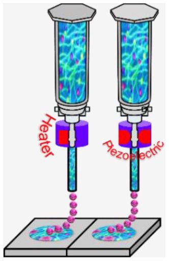
Ramadan and Zourob                                                                                                                                 3D Bioprinting

                                                                                          liver-like tissue constructs. The tissue model was utilized to
                                                                                          monitor the tissue response to amethotrexate and thioacetamide
                                                                                          exposure, such as a liver injury that leads to fibrosis (83). In
                                                                                          another study, Kupffer cells were added to examine their impact
                                                                                          on the injury/fibrogenic response following cytokine and drug
                                                                                          stimuli (84).
                                                                                              The rapid advances in bioprinting technology and the
                                                                                          wide spread of the 3D bioprinter modalities have sparked
                                                                                          unprecedented interest in using this technology to produce
                                                                                          in vitro models in pharmaceutical research. Table 3 lists some
                                                                                          selected examples of using the 3D bioprinting technology
                                                                                          in fabricating in vitro models of tissues/organs for in vitro
                                                                                          drug screening. Bowser and Moore (95), constructed a
                                                                                          neural microphysiological system by employing spheroid
                                                                                          and magnetic-based 3d bioprinting technology. Spinal cord
                                                                                          spheroids, fabricated using magnetic nanoparticles, are
                                                                                          positioned in a three-dimensional hydrogel construct using
                                                                                          magnetically assisted bioprinting method. The constructs
                                                                                          demonstrated localized cell–cell interactions and long-distance
                                                                                          projections that mimic the in vivo structure. Zhuang et al.
                                                                                          (96), combined the extrusion-based bioprinting technique with
                                                                                          an in-built ultraviolet (UV) curing system to enable layer-by-
 FIGURE 4 | (A) A proof of concept of a cellularized human heart with a natural           layer UV curing of bioprinted photo-curable GelMA-based
 architecture is printed using microextrusion 3D bioprinting. Reproduced from             hydrogels. Using this technique, high aspect ratio and stable
 Zhuang et al. (62). Open access (BB-CY). (B) Vessel-like structures printed              cell-laden constructs were achieved without the need of using
 utilizing alginate–gelatin solution. Reproduced from Liu et al. Open access
                                                                                          reinforcement materials such as poly(-caprolactone) (PCL)
 (BB-CY) (65). (C) A porous hydroxyapatite scaffold with unidirectional
 microchannels at the exterior part of the scaffold to facilitate biomineralization       polymer within the 3D-bioprinted constructs (Figure 5A).
 and a central canal that houses the bone marrow. Reproduced from Jang                    A recent study by Grigoryan et al., proved that food dyes
 et al. (66). Open access (BB-CY). (D) An anisotropic glass-ceramic scaffold              could serve as potent photoabsorbers for the production of
 with a mechanical strength comparable to cortical bone to repair large bone              cytocompatible hydrogels with functional vascular topologies
 defects. Reproduced from Roohani-Esfahani et al. (67). Open access (BB-CY).
                                                                                          (85). Using this approach, they demonstrated functional vascular
                                                                                          topologies for various studies (Figure 5B). Another study by
                                                                                          Heinrich et al. demonstrated the construction of mini brains
develop in the fabricated tissue due to the limited diffusion of                          consisting of glioblastoma cells and macrophages as tool for
cell nutrients into the core of the tissue. The integration of                            testing therapeutics that target the interaction between these
bioprinting and microfluidic technology provides an excellent                             two cell types (92). A hybrid 3D cell-printing system was
opportunity to create miniaturized in vitro tissue models                                 developed which utilized both the extrusion-based and inkjet-
“organs-on-a-chip” that overcome these shortcomings. For                                  based dispensing modules to print a 3D human skin model
example, various organotypic tissues can be simultaneously                                within a transwell system (97). A collagen-based construct
printed in a compartmentalized microfluidic chip and then                                 with polycaprolactone was printed using extrusion-based
connected through a vascular network (perfusion channels) to                              printing, and the inkjet-based dispensing module was used
finally create multi organs on a chip “human-on-a-chip.”                                  to uniformly distribute the keratinocytes onto the engineered
    It becomes generally accepted that 3D tissue models are                               dermis. 3D intestinal tissue was also bioprinted using human
superior and physiologically more relevant compared to the 2D                             primary intestinal epithelial cells and myofibroblasts to model
countermodels. Furthermore, these tissue models are not subject                           the architecture and function of the native intestinal tissue.
to the rigorous ethical issues, which makes them an attractive                            The tissue model showed key anatomical and physiological
choice for many relevant industries. However, they are still not                          characteristics such as a polarized epithelium with tight
systematically validated for toxicity prediction. To enable these                         junctions and expression of CYP450 enzymes (98).
powerful models for high-throughput drug discovery, systemic                                  Although various tissues/organ models have been envisioned
validation and standardization are required to certain their                              and manufactured, the level of complexity needed to make
potential value.                                                                          physiologically relevant tissue and organ replacements/models
    Over the past few years, several companies and start-ups                              is still not achieved or clearly defined. In vivo, multiple
have launched 3D tissue in vitro models for toxicity screening                            cell types contribute to tissue development and homeostasis
and disease modeling. For example, Organovo Inc. developed                                in well-connected tissue and organs within the biological
a bioprinting process that can be tailored to produce tissues                             systems. The inherent complexity of interconnected human
in various formats, including microscale tissues in multi-well                            tissues and animal models makes it difficult to mimic their
tissue culture plates. For instance, human primary hepatocytes,                           structure and physiology to enable tracking the physiological
hepatic stellate cells, and endothelial cells were used to bioprint                       events. Until now, it is not clear what level of biomimicry

Frontiers in Medical Technology | www.frontiersin.org                                 8                                  January 2021 | Volume 2 | Article 607648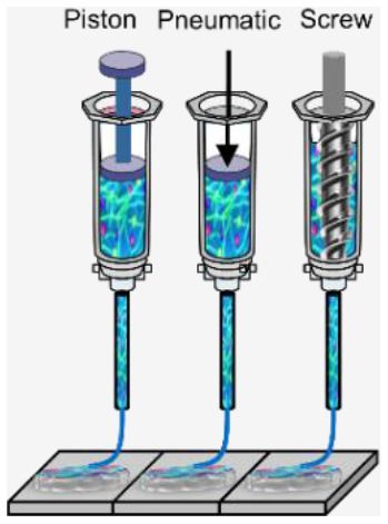
Ramadan and Zourob                                                                                                                                                3D Bioprinting

TABLE 2 | Main bioprinting studies for regenerative medicine.

                    Printing methods                    Description                                                      Specific achievement                      References

Heart & cardiac     Microextrusion 3D bioprinting       Cells from an omental tissue biopsy are reprogrammed to          Bioinks originated from the same              (62)
patches             using 3D printer (regenHU,          become pluripotent stem cells differentiated to                  patient, which would minimize the
                    Villaz-Saint-Pierre, Switzerland)   cardiomyocytes and endothelial cells, while the extracellular    immune response after
                                                        matrix is processed into a hydrogel. The two cell types were     transplantation
                                                        embedded in the hydrogels to form bioinks for the                Whole-organ (heart) bioprinting
                                                        parenchymal cardiac tissue and blood vessel printing. A proof    was demonstrated
                                                        of concept of a cellularized human heart with a natural
                                                        architecture was also demonstrated (Figure 4A)
Blood vessels       Microextrusion 3D bioprinting       Poly(ε-caprolactone) (PCL), low molecular weight chitosan        Endothelial cell line (HUVEC) was             (68)
(vascular                                               (CS), and hydrogels (H) were integrated for building the grafts. used
bypass grafts)                                          PCL has been used for fabricating the scaffolds due to its
                                                        excellent thermal stability and compatibility. Alginate and
                                                        hyaluronic acid were used as a hydrogel matrix, while
                                                        collagen type I was added to the matrix to increase the
                                                        bioactivity properties of the hydrogel matrix
                    In-house built microextrusion 3D An alginate–gelatin solution was used as a bioink material to       A theoretical model was established           (65)
                    printing device                  construct vessel-like structures by employing new rotary            to analyze the vessel thickness under
                                                     forming device (Figure 4B)                                          different conditions.
                                                                                                                         The vessel thickness cannot be
                                                                                                                         adequately predicted by the
                                                                                                                         theoretical model but by controlling
                                                                                                                         the printing parameters (speeds)
Heart valve         Microextrusion 3D bioprinting       Hybrid hydrogels [based on methacrylated hyaluronic acid       Cells in the hydrogel formulations              (69)
                    using Fab@HomeTM                    (Me-HA) and methacrylated gelatin (Me-Gel)] were utilized to maintained a high post-printing
                    open-source, open-architecture      bioprint heart valve conduits containing encapsulated human viability and fibroblastic phenotype
                    RP platform                         aortic valvular interstitial cells (HAVIC). HAVIC encapsulated
                    (www.fab@home.org)                  within bioprinted heart valves maintained high viability and
                                                        remodeled the initial matrix by depositing collagen and
                                                        glyosaminoglycans
Bone                Indirect 3D printing of powder on Biphasic calcium phosphate (BCP) consists of a mixture of     The bioprinted bone constructs were                (70)
                    a Z-printer 310 (Z Corporation, hydroxyapatite (HA), and beta-tricalcium phosphate (β-TCP) implanted subcutaneously in rats
                    Burlington, MA, USA)              matrices were bioprinted as a scaffold to induce ectopic bone
                                                      formation by osteoblast seeding and/or addition of BMP-2
                    An integrated tissue-organ          Cell-laden hydrogel was deposited together with synthetic        Mandible and calvarial bone,                  (42)
                    printer (ITOP)                      biodegradable polymers that impart mechanical strength to        cartilage, and skeletal muscle were
                                                        fabricate mechanically robust tissue constructs (bone,           fabricated with recapitulated native
                                                        cartilage, & skeletal muscle). This was accomplished by          structure
                                                        designing multidispensing modules for delivering various cell
                                                        types and polymers in a single construct. Incorporation of
                                                        microchannels into the tissue constructs facilitates the
                                                        diffusion of nutrients to printed cells
                    Extrusion-based direct writing      Two different GelMA-based hydrogels were synthesized (one Perfusable blood vessel inside a                     (71)
                    bioprinting                         supported vasculogenesis and the other supported                bioprinted bone-like tissue construct
                                                        osteogenesis). GelMA hydrogels containing different
                                                        concentrations of VEGF were bioprinted into well-defined 3D
                                                        architectures to create a gradient of vasculogenic factors. The
                                                        bioprinting and incorporation of a rapidly degradable GelMA
                                                        hydrogel resulted in the formation of a perfusable lumen with
                                                        an endothelial lining at the center of the construct
                    Microextrusion 3D                   A porous hydroxyapatite scaffold was printed to mimic native     Full osteointegration of the scaffold         (66)
                    Bioprinting                         bone through a multipass extraction process with the addition    with the native tissue was observed
                                                        of osteoblast-like cells. The scaffold used is appropriate for   after 4 and 8 weeks of implantation in
                                                        graft without inflammatory reactions and bone formation after    rabbit model
                                                        8 weeks of implantation (Figure 4C)
                    Direct ink writing using 600 µm     A glass-ceramic scaffold, with a dimension of 6 × 6 × 6 mm,      The obtained strength is 150 times            (67)
                    custom-made nozzle                  was bioprinted mimicking cortical bone with scaffold of          more than reported values for
                                                        hexagonal pore shapes (450, 550, 900, and 1,200 µm)              polymeric and composite scaffolds
                                                        (Figure 4D)                                                      and five times more than reported
                                                                                                                         values for ceramic and glass scaffolds

                                                                                                                                                                    (Continued)

Frontiers in Medical Technology | www.frontiersin.org                                 9                                             January 2021 | Volume 2 | Article 607648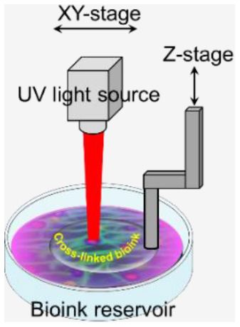
Ramadan and Zourob                                                                                                                                                   3D Bioprinting

TABLE 2 | Continued

                    Printing methods                    Description                                                        Specific achievement                       References

                    Digital laser processing            Haversian bone-mimicking scaffold with integrated                  Effective delivery of osteogenic,              (72)
                    (DLP)-based 3D printing             hierarchical haversian bone structure. The scaffold has the        angiogenic, and neurogenic cells,
                                                        potential to induce osteogenic, angiogenic, and neurogenic         which exhibited favorable
                                                        differentiation in vitro and accelerated the in-growth of blood    osteogenesis and angiogenesis
                                                        vessels and new bone formation in vivo
Cartilage           Simultaneous                    Poly(ethylene glycol) dimethacrylate (PEGDMA) with human               Enhanced proteoglycan deposition               (73)
                    photopolymerization using a     chondrocytes were printed to repair defects in osteochondral           was observed at the interface
                    modified HP Deskjet 500 printer plugs (3D biopaper) in layer-by-layer assembly. Printed human          between printed biomaterial and
                                                    chondrocytes maintained the initially deposited positions due          native cartilage
                                                    to the simultaneous photopolymerization of surrounded
                                                    biomaterial scaffold
                    A hybrid inkjet                     Electrospinning of polycaprolactone fibers was alternated          The chondrocytes maintained 80%                (74)
                    printing/electrospinning system     with inkjet printing of rabbit elastic chondrocytes suspended      viability more than 1 week after
                                                        in a fibrin-collagen hydrogel in order to fabricate a five-layer   printing
                                                        tissue construct of 1 mm thickness cartilage
Skin                Eight electromechanical             Keratinocytes and fibroblasts were used as constituent cells The morphology of the 3D-printed                     (75)
                    dispensers mounted onto a           to represent the epidermis and dermis, and collagen was       skin tissue closely mimics the in vivo
                    3-axis, high-precision robot        used to represent the dermal matrix of the skin. The          human skin tissue
                    stage which enables printing of     3D-printed constructs were cultured in submerged media
                    multiple cell types and scaffold    conditions followed by exposure of the epidermal layer to the
                    materials simultaneously            air–liquid interface to promote maturation and stratification
                    4D bioprinting system (Organ        Extracellular matrix (ECM) which derived from nano-fat             High wound healing rate with                   (76)
                    Regenerator 4D)                     consisting of supportive proteins, growth factors, and             complete closure of wound of 2∼5
                                                        cytokines has been printed with bioinks to apply onto the          weeks after membrane application
                                                        chronic wound site
                    Laser-assisted Bioprinting          Fibroblasts and keratinocytes embedded in collagen were            Successful formation of adhering and           (77)
                                                        printed in 3D as multicellular grafts analogous to native          gap junctions
                                                        archetype and the formation of tissue
                    Pneumatic-based microextrusion An implantable multilayered vascularized skin graft is formed           The human EC-lined structures                  (63)
                    3D                             using 3D bioprinting using a bioink containing human foreskin           inosculate with mouse microvessels
                    bioprinting                    dermal fibroblasts, human endothelial cells derived from cord           arising from the wound bed and
                                                   blood human endothelial colony-forming cells, and human                 become perfused within 4 weeks
                                                   placental pericytes suspended in rat-tail type I collagen to            after implantation
                                                   form a dermis followed by printing with a second bioink
                                                   containing human foreskin keratinocytes to form an epidermis
                    Extrusion-based 3D bioprinting      Full thickness of the human skin model showing undulated           The epidermis–dermis junction was              (78)
                                                        morphology of epidermal rete ridges, architectural,                recapitulated in the 3D bioprinted skin
                                                        mechanical, and biochemical functionalities                        tissue
Ear                 Digital near infrared               Digital near infrared (NIR) photopolymerization (DNP) was          Ear-like tissue constructs with                (79)
                    photopolymerization                 used to spatially induce the polymerization of monomer             chondrification and a muscle tissue
                    (DNP)-based 3D printing             solutions such that the subcutaneously injected bioink can be      repairable cell-laden conformal
                    technology                          noninvasively printed into customized tissue constructs in situ    scaffold
                    The ear scaffold used a PCL         Patient-specific ear-shaped cartilage is fabricated in vitro       Mature cartilage formation during 2.5          (64)
                    mesh as an inner core, which        using expanded microtia chondrocytes, compound, and                years for 5 reconstructed patients
                    was wrapped with PGA unwoven        biodegradable scaffold. Different surgical procedures were         auricles
                    fibers and coated with PLA.         employed to find the optimal approach for handling
                    Expanded microtia cartilages        tissue-engineered grafts
                    were dropped onto the PGA/PLA
                    layer of the ear-shaped scaffold
Liver               Custom-made inkjet 3D               3D liver tissue is constructed using hepatocyte attachment         Controlling cell polarity with                 (80)
                    bioprinter                          and formation of the cell monolayer by interacting with the        galactosylated hydrogels
                                                        galactose chain of galactosylated alginate gel (GA-gel) with
                                                        asialoglycoprotein receptor (ASGPR) of hepatocytes
                    Microextrusion 3D                   Primary hepatocytes with MSCs are used to support                  The 3D hepatic architecture showed             (81)
                    bioprinting                         hepatocyte function and viability time in 3D structures            a higher cell viability compared to the
                                                                                                                           2D system

                                                                                                                                                                       (Continued)

Frontiers in Medical Technology | www.frontiersin.org                                 10                                               January 2021 | Volume 2 | Article 607648
Ramadan and Zourob 3D Bioprinting
TABLE 2 | Continued
Printing methods Description Specific achievement References
Diaphragm Regenova® bio-3D printer with Scaffold-free tissue patches composed of human cells are 3D (82)
cells only (Kenzan method) printed with high elasticity and strength. The resulting tissue is Complete integration of the graft with
cut into a patch for implantation. The patches were the native tissue
transplanted into rats with surgically created diaphragmatic Regeneration of muscle,
defects neovascularization,
and neuronal networks within the
reconstructed diaphragms
Rats survived for 710 days
after implantation
of human physiology is needed and whether we need to cell therapy, where cells can be isolated from a small biopsy
use all the cellular subpopulations to achieve differentiation (103). Zhao and Xu (102) developed a micro-bioprinting system
into the needed phenotypes (9). Recent advances in 3D installed to an endoscope to enable bioprinting inside the human
bioprinting technology show great potential to answer these body and utilized printed circuit micro-electro-mechanical-
critical questions. For example, complex heterogeneous cellular system techniques that allow a high-accuracy tissue printing.
structures can be fabricated with multimaterial depositing Two-layer tissue scaffolds were printed in a stomach model
systems (99, 100), hence enabling the incorporation of vascular using gelatin–alginate hydrogels with human gastric epithelial
and neural networks within the structure of the in vitro cells and human gastric smooth muscle cells as bioinks to
models, thus capturing the complexity of multiple tissue mimic the anatomical structure of the stomach. Kérourédan
and organ systems. However, to achieve this ambitious aim, et al. (104) employed laser-assisted bioprinting (LAB) to pattern
more profound and comprehensive studies are needed. In endothelial cells into a mouse calvaria bone defect, which is filled
addition, integration of the bioprinting techniques with other with collagen-containing mesenchymal stem cells and vascular
technologies such as imaging, bioreactor technology, organ-on- endothelial growth factor. This technique enabled organized
a-chip (OOC), artificial intelligence (AI), and semiconductors microvascular networks into bone defects with promising
would expand tissue engineering capabilities and accelerate vascularization rate for in situ prevascularization that promote
the technology maturation toward organ/tissue production for bone regeneration.
various applications. In situ bioprinting is a contact-based technique that requires
special consideration compared to in vitro printing including
In situ Bioprinting bioink properties, bioprinter setup, and sterilization (105).
One of the promising applications of 3D bioprinting is to For example, in extrusion-based bioprinting, the printing
pattern de novo tissue directly onto the desired location in tip might interfere with the defect surrounding and caused
the body, such as chronic wounds in the skin or bone defect. side-effect damage. Generally, the bioinks used for in situ
With the aid of medical imaging, the topology of printed bioprinting need to be biocompatible with rapid cross-
tissue can be designed to fit into the wound/defects such that linkability to enable shorter surgery time and to retain the
heterotypic cellular structures, hydrogels, and soluble factors can integrity of bioprinted constructs. Several biomaterials show
be precisely deposited inside the defects. This approach, termed high potential for such purposes, such as collage, fibrinogen,
as in situ bioprinting or intraoperative bioprinting (IOB), would gelatin methacrylamide (GelMA), hyaluronic acid methacrylate
minimize the gap between implant–host interfaces and provide (HAMA), and poly (ethylene glycol) (106). Vascularization is a
well-defined structures within zones of irregular topographies major challenging task particularly for in situ bioprinting since
during the healing process, which can effectively recruit desired it takes more than 10 days for angiogenesis to take place in
cells from surrounding tissues where the patient’s body act as living tissue (107). Temporal oxygen supply can be used prior to
a natural bioreactor (5). Compared to the other applications angiogenesis by using oxygen-generating biomaterials or oxygen-
listed above, only a few attempts have been reported. In a filled microparticles which can be bioprinted within the bioink
recent proof-of-concept study, Albana et al. (101) demonstrated (108, 109). Another strategy involves the creation of sacrificial
precise delivery of autologous/allogeneic dermal fibroblasts and porous structure within the bioprinted tissue by using meshed
epidermal keratinocytes directly into an injured area in animals, filaments (110).
replicating the layered skin structure (Figure 6). Excisional
wounds bioprinted with layered autologous dermal fibroblasts
and epidermal keratinocytes in a hydrogel carrier showed Bioprinting Meets Microfluidics and
rapid wound closure, reduced contraction, and accelerated Organ-On-A-Chip
re-epithelialization. These results showed the feasibility of in Recent bioprinting studies leveraged the well-established
situ bioprinting of skin and its potential applications for microfluidic technology to design bioprinting systems that
the regeneration of various body parts. A successful in situ enable precise dispensing of low-viscosity bioink in a well-
bioprinting technique could rapidly accelerate healing using defined template with highly controlled conditions (111–113).
Frontiers in Medical Technology | www.frontiersin.org 11 January 2021 | Volume 2 | Article 607648Ramadan and Zourob 3D Bioprinting
TABLE 3 | Main bioprinting studies for in vitro models for drug discovery.
Printed tissue or Printing methods Description (cell/bioink) Stimuli/effect Ref.
organ
Air-blood barrier Laser-assisted 3d bioprinting Air–blood tissue barrier analogy composed of an endothelial cell Cellular morphology, (40)
with a printing resolution of 5 µ (HUV-EC cell line), basement membrane, and epithelial cell layer (A549 cell–cell contacts, and
m) cell line) (Figure 5B) viability
Multivascular Stereolithography Intravascular and multivascular networks are fabricated with Oxygenation and flow of (85)
networks photopolymerizable hydrogels by using food dye additives as human red blood cells
biocompatible but potent photoabsorbers for projection during tidal ventilation and
stereolithography distension of a proximate
airway
Muscle & tendon Laser-assisted bioprinting Musculoskeletal-tendon-like tissue structures were 3D printed with Electrical stimulation and (86)
tissues (RegenHU, Switzerland) alternating layers of photo-polymerized gelatin-methacryloyl-based calcium signaling
bioink and cell suspension (primary human skeletal-muscle-derived
cells and primary rat-tail tenocytes) in 24-well plates
Liver tissues Microextrusion-based bioprinting A liver tissue-like structure that comprises primary human hepatocytes, Drug (Trovafloxacin)-induced (87)
(NovoGen Bioprinter) hepatic stellates, and HUVEC cells in a defined architecture is 3D liver injury
printed
Custom-built bioprinting system A 3D hydrogel-based triculture model that embeds hiPSC-HPCs with Liver-specific gene (88)
based on digital micro-mirror human umbilical vein endothelial cells and adipose-derived stem cells expression levels, increased
device with motion controller created a microscale hexagonal architecture (Figure 5C) metabolic product secretion
(Newport) that controls a
movable stage
Skin Freeform fabrication technique, Multilayered tissue composites, which consist of human skin fibroblasts Multilayered cell–hydrogel (89)
based on direct cell dispensing and keratinocytes, are printed using a robotic platform that prints composites printing on a
using four pneumatically driven collagen hydrogel precursor, fibroblasts, and keratinocytes. The non-planar surface skin
microvalves as dispensers and a cell-containing collagen was cross-linked by coating the layer with wound repair modeling
three-axis robotic stage nebulized aqueous sodium bicarbonate
Extrusion-based bioprinting Skin is printed with a thickness of 5 mm using a bioink that was Bioink properties (90)
formulated as a mixture of bovine gelatin, very low viscosity alginate,
fibrinogen, and human dermal fibroblasts
Extrusion-based bioprinting Skin tissue equivalents in a multi-well plate format printed using Barrier function (permeability (91)
neonatal human dermal fibroblasts and neonatal normal human tracing with Lucifer yellow
epithelial keratinocytes and biotin tracer)
Mini brain Extrusion-based bioprinting Mini brains consisting of glioblastoma cells and macrophages are Macrophages induce (92)
bioprinted as a tool to study the interactions between the two cell types glioblastoma cell
and to test therapeutics that target this interaction. A two-step progression and
bioprinting process was used in which we first print the larger brain invasiveness in the mini
model encapsulating a mouse macrophages cell line (RAW264.7) with brains
an empty cavity was printed, which in the second step is filled with
mouse glioblastoma cells (GL261) embedded into bioink, followed by
photo-cross-linking of the construct
Tumor breast & Microextrusion-based bioprinting Multiple cell types were incorporated into scaffold-free tumor tissues Cellular proliferation, ECM (93)
pancreatic (NovoGen Bioprinter) with defined architecture. The technique enables modeling deposition, and cellular
patient-specific tumors by using primary patient tissue (Figure 5D) migration are altered in
response to extrinsic signals
or therapies
Laser direct write (LDW) Cell-encapsulating microbeads were generated and further processed The impact of aggregate (94)
bioprinting into core-shelled structures, allowing for the growth and formation of size on the uptake of a
self-contained, self-aggregating cells (e.g., breast cancer cells, commonly employed ligand
embryonic stem cells) for receptor-mediated drug
delivery, transferrin
Microfluidic dispensing technology has been adopted in some aggregates. The ability to create scaffolds with complex 3D
commercial bioprinters. For example, Aspect Biosystems shapes would enable precise control of the microstructures and
developed RX1TM bioprinter, which enables precise motion microarchitecture of tissue constructs and hence the fabrication
and pressure control that allows microscale resolution at high of various tissues and organ as in vitro models for drug discovery.
speed [https://aspectbiosystems.com/technology#bioprinter]. To create miniaturized in vitro models of human organs, also
Abelseth et al. (114) reported using the RX1TM bioprinter to known as organ-on-a-chip and organoids, one option is to follow
create 3D neural tissues derived from hiPSC-derived neural a bottom-up approach by spatially immobilizing various types
Frontiers in Medical Technology | www.frontiersin.org 12 January 2021 | Volume 2 | Article 607648You can also read