Characterization of long G4-rich enhancer-associated genomic regions engaging in a novel loop:loop 'G4 Kissing' interaction
←
→
Page content transcription
If your browser does not render page correctly, please read the page content below
Published online 8 May 2020                                                               Nucleic Acids Research, 2020, Vol. 48, No. 11 5907–5925
                                                                                                                          doi: 10.1093/nar/gkaa357

Characterization of long G4-rich enhancer-associated
genomic regions engaging in a novel loop:loop ‘G4
Kissing’ interaction
Jonathan D. Williams1,2,† , Dominika Houserova3,† , Bradley R. Johnson2 , Brad Dyniewski2 ,
Alexandra Berroyer2 , Hannah French2 , Addison A. Barchie4 , Dakota D. Bilbrey4 , Jeffrey
D. Demeis4 , Kanesha R. Ghee4 , Alexandra G. Hughes4 , Naden W. Kreitz4 , Cameron
H. McInnis4 , Susanna C. Pudner4 , Monica N. Reeves4 , Ashlyn N. Stahly4 , Ana Turcu4 ,

                                                                                                                                                              Downloaded from https://academic.oup.com/nar/article/48/11/5907/5834579 by guest on 22 September 2020
Brianna C. Watters4 , Grant T. Daly3 , Raymond J. Langley3 , Mark N. Gillespie3 ,
Aishwarya Prakash3,5 , Erik D. Larson2,6 , Mohan V. Kasukurthi7 , Jingshan Huang7 ,
Sue Jinks-Robertson1 and Glen M. Borchert 3,4,*
1
 Department of Molecular Genetics and Microbiology, Duke University, Durham, NC 27708, USA, 2 School of
Biological Sciences, Illinois State University, Normal, IL 61790, USA, 3 Department of Pharmacology, University of
South Alabama, Mobile, AL 36688, USA, 4 Department of Biology, University of South Alabama, Mobile, AL 36688,
USA, 5 Department of Biochemistry and Molecular Biology, University of South Alabama, Mitchell Cancer Institute,
Mobile, AL 36688, USA, 6 Department of Biomedical Sciences, Western Michigan University Homer Stryker MD
School of Medicine, Kalamazoo, MI 49007, USA and 7 School of Computing, University of South Alabama, Mobile,
AL 36688, USA

Received February 04, 2020; Revised April 22, 2020; Editorial Decision April 26, 2020; Accepted April 27, 2020

ABSTRACT                                                                                 G4 loops within individual LG4 loci directly basepair
                                                                                         with one another (similar to characterized stem–loop
Mammalian antibody switch regions (∼1500 bp) are
                                                                                         kissing interactions) forming a hitherto undescribed,
composed of a series of closely neighboring G4-
                                                                                         higher-order, G4-based secondary structure we term
capable sequences. Whereas numerous structural
                                                                                         a ‘G4 Kiss or G4K’. In conclusion, LG4s adopt novel,
and genome-wide analyses of roles for minimal G4s
                                                                                         higher-order, composite G4 structures directly con-
in transcriptional regulation have been reported,
                                                                                         tributing to the inherent instability, regulatory capac-
Long G4-capable regions (LG4s)––like those at an-
                                                                                         ity, and maintenance of these conspicuous genomic
tibody switch regions––remain virtually unexplored.
                                                                                         regions.
Using a novel computational approach we have iden-
tified 301 LG4s in the human genome and find LG4s
                                                                                         INTRODUCTION
prone to mutation and significantly associated with
chromosomal rearrangements in malignancy. Strik-                                         Non-coding DNA comprises over 98% of the human
ingly, 217 LG4s overlap annotated enhancers, and                                         genome (1,2) and is predominantly repetitive in nature (3,4).
we find the promoters regulated by these enhancers                                       While traditional concepts hold that such repetitive ele-
                                                                                         ments generally lack biochemical functionality, current es-
markedly enriched in G4-capable sequences sug-
                                                                                         timates are that over 80% of the genome has some func-
gesting G4s facilitate promoter-enhancer interac-                                        tion (5). Some repetitive elements, especially guanine rich
tions. Finally, and much to our surprise, we also find                                   (G-rich) sequences derived from transposable elements, are
single-stranded loops of minimal G4s within individ-                                     capable of forming transient non B-form secondary struc-
ual LG4 loci are frequently highly complementary to                                      tures that have regulatory functions (6).
one another with 178 LG4 loci averaging >35 inter-                                          One prominent secondary structure found in repetitive
nal loop:loop complements of >8 bp. As such, we                                          DNA is G-quadruplex (G4), which can form under phys-
hypothesized (then experimentally confirmed) that                                        iological conditions. G4 is a four-stranded, highly ther-

* To   whom correspondence should be addressed. Tel: +1 251 461 1367; Email: borchert@southalabama.edu
†
    The authors wish it to be known that, in their opinion, the first two authors should be regarded as joint First Authors.


C The Author(s) 2020. Published by Oxford University Press on behalf of Nucleic Acids Research.
This is an Open Access article distributed under the terms of the Creative Commons Attribution License (http://creativecommons.org/licenses/by/4.0/), which
permits unrestricted reuse, distribution, and reproduction in any medium, provided the original work is properly cited.
5908 Nucleic Acids Research, 2020, Vol. 48, No. 11

                                                                            (28). G4 sequences are thought to be tightly controlled dur-
                                                                            ing specific cellular processes and may be particularly ver-
                                                                            satile in regulation due to the variety of different structures
                                                                            they can assume and the potential interplay between struc-
                                                                            ture formation, ligand stabilization, and helicase resolution
                                                                            (12,13).
                                                                               While accurate maintenance of G4-supportive sequences
                                                                            appears crucial for proper cellular function, these loci are
Figure 1. G4 DNA. (A) Illustration of guanine quartet with each guanine     characteristically associated with genomic instability. Both
engaged in four hydrogen bonds and a central potassium cation coordi-       computational analyses and investigations in model sys-
nately bound. (B) Structural illustration depicting unimolecular antipar-
allel G4 DNA.
                                                                            tems have documented the susceptibility of G4 sequences
                                                                            to mutagenesis. Computational analysis of human genome
                                                                            small sequence variation databases has shown an increase

                                                                                                                                              Downloaded from https://academic.oup.com/nar/article/48/11/5907/5834579 by guest on 22 September 2020
mostable, square-planar nucleic acid structure in which                     of small nucleotide variations in sequences that support
guanine repeats are stabilized by Hoogsteen bonds (7–10)                    G4 formation (29). Further, these regions are associated
(Figure 1). DNA replication and transcription require that                  with significant expression variation of downstream genes.
stretches of DNA adopt a single-stranded state, which al-                   Analysis of cancer genomes has also demonstrated that so-
lows for G4 structures to form, and resolution of these                     matic copy-number variations are significantly enriched at
structures involves the action of DNA helicases (11). Se-                   regions that stall DNA polymerase under G4-permissive
quences that support G4 conformations have proven highly                    conditions (18). In addition, analysis of cancer transloca-
variable although the minimum criteria to form intra-                       tions has shown that ∼70% of translocation breakpoints
molecular G4 DNA have classically been described by the                     are capable of G4 formation (30), with structure formation
following motif: GGGnGGGnGGGnGGG. Here, G rep-                              demonstrated in vitro at c-MYC t (8;14), HOX11 t (10;14),
resents guanines that are participating in G4 structure for-                BCL2 (t14;18), and TCF3 t (1;19) breakpoints (22,30–32).
mation, while n denotes DNA spacers of variable length                      In yeast, the TCF3-breakpoint G4 sequence was further
and nucleotide composition (12,13). That said, the defini-                  shown to lead to loss of a chromosomal arm under condi-
tion of G4s has recently been expanded to include struc-                    tions that support G4 formation including high transcrip-
tures containing bulges, guanine vacancies, and or mis-                     tion and increased negative supercoiling (32). Detection of
matches (14). In addition, the spacers separating the gua-                  gross chromosomal rearrangements using other human G4
nine repeats can vary in size (n = 1–24), and the number                    sequences in yeast was magnified by ligand stabilization of
of tandem guanines can go well beyond the minimum of                        G4, increasing G4 repeat number or removal of G4 helicase
three described above (15). Using a stringent loop defini-                  Pif1 (33–35).
tion of n = 1–7 in search algorithms, over 300,000 puta-                       To date, only a small number of G4 loci have been exam-
tive intra-molecular G4-capable sequences have been iden-                   ined, and the number of potential G4 loci (∼300 000–700
tified in the human genome (16,17). More recently, an inno-                 000) constitutes a significant obstacle to identifying biolog-
vative genome-wide mapping of DNA polymerase stalling                       ically relevant loci that are prone to instability and/or reg-
under structure-permissive conditions compared to non-                      ulation. Accordingly, we developed a novel approach and
permissive conditions identified over 700 000 potential G4                  open-source program (LG4ID) that identifies putative G4-
loci (18). Clearly, the sheer number of putative G4 motifs                  forming sequences using search parameters modeled after
represents an obstacle to studying and fully understand-                    the size and composition of human immunoglobulin switch
ing their impact on the human genome, although insights                     region, laboratory-validated G4 sequences (Supplementary
can be gained from studying individual examples and meta-                   Information 1). We further describe the experimental eval-
analyses.                                                                   uation, functional analysis and disease associations of 301
   Recent evidence suggests that G4s participate in mul-                    large-G4 (LG4) sequences thus identified, and document a
tiple genomic events. Computational analysis of the hu-                     novel secondary structure assumed by these sequences. We
man genome indicates that regions capable of G4 forma-                      conclude that LG4ID is highly successful in identifying bio-
tion are not randomly dispersed and are significantly asso-                 logically relevant, large guanine-dense G4 sequences in the
ciated with promoters, 5 untranslated regions, and introns                 human genome.
(12,16,19,20). Recently, over 10 000 unique G4 structures
were physically isolated from HaCaT cells and ∼1000 G4
structures from NHEK cells using a G4-specific antibody
for ChIP-seq analysis (21). Interestingly, G4s from both                    MATERIALS AND METHODS
cell lines were highly enriched in nucleosome-depleted pro-
                                                                            Python 3-based program for identification of LG4s–LG4ID
moter and 5 UTR regions of highly transcribed genes, and
the divergence in number detected per cell line suggests that               In order to identify long G4-capable regions (LG4s) present
G4 formation and resolution can be tightly controlled based                 in the human genome, we wrote a Python 3 program to
on cell type. Notably, G4 sequences have been reported to                   search a FASTA formatted sequence file for long G-triplet
play a role during transcription (22), translation (23), re-                regions likely to form G4. The program identifies LG4 mo-
combination (24), replication initiation (25), aptamer bind-                tifs based on the density of G-triplets within 1.5 kb se-
ing (26), telomere maintenance (27) and mRNA processing                     quence windows sliding one base pair per iteration and does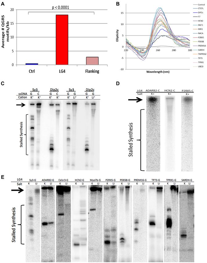
Nucleic Acids Research, 2020, Vol. 48, No. 11 5909

not account for loop length. In order to define the mini-       Database for Annotation, Visualization and Integrated Dis-
mal density of G-triplets needed to call a LG4, we modeled      covery (DAVID) analysis
our search program after the G4-capable sequence density
                                                                All LG4 proteins were analyzed on the Database for An-
within the human immunoglobulin mu (Sμ) switch region
                                                                notation, Visualization and Integrated Discovery (DAVID),
(11–13,24,36). Sliding windows were applied, with a mini-
                                                                a web based program that provides annotation tools for
mal output threshold of (GGG) X 121 for every 1.5 kb of se-
                                                                researchers to understand the biological meaning behind
quence. To identify G-rich sequences on the + and – stands
                                                                large list of genes identified in microarray or bioinformatic
of the genome, CCC density was also determined as above
                                                                studies (46). DAVID can be used to identify protein inter-
then combined with the GGG search output data (for more
                                                                actions, common genetic locations, common pathways, dis-
details see Supplementary Information 1). LG4ID was used
                                                                ease relevance and multiple other analyses. Genes were en-
to identify all LG4s (with parameters stated above) in the
                                                                tered into the web interface by their Ensembl gene ID and
human genome (hg38) after which each LG4’s location and
                                                                then analyzed using the functional annotation tool to inves-
identity were confirmed manually. A Web-hosted LG4ID

                                                                                                                                  Downloaded from https://academic.oup.com/nar/article/48/11/5907/5834579 by guest on 22 September 2020
                                                                tigate any statistically significant similarities and relation-
LG4 search tool and LG4ID source code are available at:
                                                                ships between our genes.
http://omnisearch.socsouthalabama.edu:8080/g4search#

Detailing LG4 genomic locations, putative regulatory abili-     Identification of disease genes
ties, COSMIC and FusionGDB associations and Hi-C inter-         Genes that are potentially regulated by a LG4 (associated
actions                                                         enhancer, gene promoter, or within the transcribed gene se-
Output of the LG4 identification program was used to map        quence) were evaluated for known human disease associa-
each individual LG4 location on Ensembl Release 69 (hg19)       tion. Each corresponding gene was searched on Malacards
(37) and further confirmed for LG4 genes (genes containing      database (47) and Wikigenes literature search (48) to iden-
an LG4 sequence in their transcript or within 10kb of their     tify any potential involvement in disease.
UTR) on Release 77 (hg38) (38). Statistics for genomic lo-
cation with respect to transcription were analyzed using chi-   G4 density calculation
square. The enrichment for chromosome location used one-
way ANOVA followed by individual unpaired two-tailed t-         A program called QGRS mapper (49) was used to de-
tests. Significant enrichment of LG4 at the distal end of the   termine the potential of each sequence to form G4. This
chromosome was calculated using an unpaired two-tailed          was accomplished using the following filters: A max motif
t-test. Potential regulatory functions and ChIP-Seq pull        length of 45 nucleotides, minimum G group of 3, and a loop
down data was obtained using Ensembl77 with the Regu-           size 0–36 nucleotides (selects for intra-molecular G4 only).
latory Build filter turned on (39). Significance for regula-    The output of the analysis was mapped to the location of
tory ability was calculated using chi-square. Full COSMIC       the LG4, and the number of individual non-overlapping G4
(40) and FusionGDB (41) translocation and gene fusion           motifs per kb (G4 density) was calculated for each LG4. Ad-
datasets were downloaded and significant LG4 associations       ditionally, positions directly adjacent of LG4, and control
calculated using an unpaired two-tailed t-test. To enumer-      loci were also calculated in this method. Statistics were cal-
ate reported interactions between LG4s and other regions        culated using one-way ANOVA followed by unpaired two-
of the genome, chromosomal interactions between LG4             tailed t-tests.
loci (or randomly selected, size matched control loci) and
other genomic locations were identified in UCSC Genome
                                                                Identification of human genome variation densities
Browser Hi-C and Micro-C tracks providing chromatin
folding data from Micro-C XL and Hi-C experiments ex-           The location of all individual SNPs, insertions and dele-
amining HFFc6 (foreskin fibroblasts) and H1-hESC (em-           tions were obtained from the dbSNP database (50) and
bryonic stem cell) cell lines (42–44).                          mapped to LG4s found in protein transcript regions as well
                                                                as surrounding introns (exons excluded from analysis) us-
                                                                ing Ensembl release 69 (37). The density of small sequence
Identification of potential loop interactions
                                                                variants was calculated by number of SNPs, insertion, or
Internal LG4 G4 loops were initially identified by extract-     deletion events per 100 base pairs (bp) for LG4 and re-
ing all unique sequences (>4 nt and 9 bp alignments requiring ≥90% comple-          were calculated using one-way ANOVA followed by indi-
mentarity.                                                      vidual unpaired two-tailed t-test.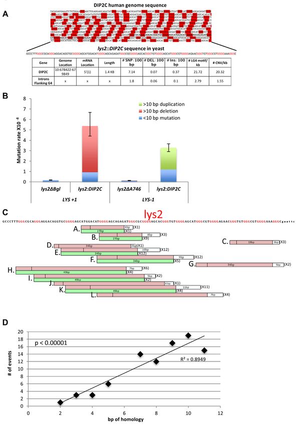
5910 Nucleic Acids Research, 2020, Vol. 48, No. 11
Circular dichroism Mutation rates and spectra
Oligonucleotides for circular dichroism (CD) studies were Cultures inoculated from single colonies were grown to sat-
designed by using representative repeat units found in LG4 uration (3 days) in YEP-GE (1% yeast extract, 2% Bacto-
sequences and synthesized by Operon (Eurofins MWG peptone, 2% glycerol and 2% ethanol). To induce high tran-
operon LLC, Huntsville, AL, USA). CD analysis was per- scription in strains containing the GAL1 promoter, 2%
formed using an Aviv model 215 CD spectrometer at 37◦ C. galactose was used instead of glycerol and ethanol. Lys+
Spectra were taken in 1 cm path quartz cells containing 12 revertants were selected on synthetic complete media lack-
M G4 or GCA oligonucleotide in 10 mM Tris–HCl, pH ing lysine (0.17% yeast nitrogen base, 0.5% ammonium sul-
7.6, 1 mM EDTA and 100 mM KCl. The molar ellipticity fate, 2% agar and 0.13% Hartwell’s complete amino acid
was measured from 220–300 nm and recorded for 3 scans in mix lacking lysine). The total number of cells in each cul-
1 nm increments at a 1 s average time. ture was determined by plating on non-selective YPD (1%
yeast extract, 2% bacto-peptone, 2% dextrose and 2% agar)
Downloaded from https://academic.oup.com/nar/article/48/11/5907/5834579 by guest on 22 September 2020
Primer extension assays medium. Mutation rates were calculated using method of
the median (59), and 95% intervals confidence intervals de-
LG4 containing phagemids for extension assays were ob- termined as previously described (60). The rates of a specific
tained by cloning PCR amplified genomic fragments, mutation type was calculated using its proportion in the cor-
or cloned from amplification products using overlap- responding mutation spectrum; associated confidence in-
ping primers in a standard PCR reaction. PCR prod- tervals were calculated using the right-triangle rule (61).
ucts were gel purified and TOPO cloned (Invitrogen) into The mutation spectrum was generated by isolating genomic
pCR2.1. Fragments were cloned in both orientations and DNA independent mutants, followed by PCR amplification
were verified by Sanger sequencing (University of Illi- and sequencing of the LYS2 reversion window. Mutation
nois Core Sequencing Center). Templates for extension rates were based on 24 independent cultures and spectra on
assays are shown in Supplementary Table S4 and range the analysis of 42 independent revertants.
from 120–1300 bp. The size variation is due to the inabil-
ity for some larger LG4 sequences to be cloned. Closed-
circular single-stranded DNA was obtained using M13K07 In vitro G4-DNA formation and native polyacrylamide gel
helper phage (NEB) according to the manufacturer’s electrophoresis
instructions. Commercially synthesized G4-DNA oligonucleotides (In-
Klenow Polymerase extension assays were performed as tegrated DNA Technologies) were rehydrated using ultra-
described in (52) and developed from previous G4 assays pure DNA grade H2 O to a final concentration of 0.25 mM.
(53,54). A 32 P 5 end labeled forward primer (standard M13 To form tetramolecular G4-DNA, aliquots of each sample
F) was annealed to the single-stranded phagemid templates. were boiled in a thermalcycler at 98◦ C for 10 min, and sub-
In addition to the manufacture’s buffer (NEB), KCl or LiCl sequently held at 80◦ C as previously described (62). Pre-
was added to a final concentration of 25 mM. After anneal- heated KCl solution (folded G4) or an equal volume of
ing, Klenow extension reactions were performed at 37◦ C pre-heated ultrapure H2 O (unfolded controls) was added to
for 8 min, then stopped by the addition of an equal vol- each aliquot. The final concentration of KCl varied from
ume of 90% formamide and 1 mM EDTA followed by heat- 50 to 250 mM. The samples were then allowed to slowly
ing to 90◦ C for 20 min. Products of polymerase extension cool to room temperature and stored at –4◦ C until needed
were resolved by 8% denaturing PAGE (19:1) with 7 M urea (max 3 days). The formation of tetramolecular G4-DNA
and 0.5× TBE, at 700 V at room temperature. Gels were structures was confirmed via gel electrophoresis using 10
then dried and images were captured by phosphorimag- and 20% non-denaturing polyacrylamide gel and Tris/boric
ing using a Molecular Dynamics Storm 840 phosphorim- acid buffer. To visualize the oligonucleotides and differen-
ager (Amersham/GE). Each template was assayed at least tiate between G4/ssDNA and dsDNA, resulting gels were
twice. stained first with ethidium bromide, imaged on UV Tran-
silluminator FBTIV-88 (Fisher Scientific), then re-stained
Yeast strain construction with Thioflavin T and imaged again.
Yeast strains for the LYS2 reversion assay were derived from
W303 (MATa, leu2–3,112 ura3 his3–11,15 ade2–1, trp-1) RESULTS
(55). A130 bp DIP2C LG4 fragment or GCA repeats was
Identification of large G-triplet dense G4 regions in the hu-
used to replace the CORE fragment (56) in the LYS2 re-
man genome
version window to create a +1 or –1 frameshift allele (pre-
viously described in (57)). The orientation and integrity Most G4-predicting programs utilize an algorithm based on
of the inserted sequences for all strains was verified by a minimal definition that solely identifies individual G4 mo-
Sanger sequencing (Eton bio) before further analysis. PIF1, tifs (14). In contrast, next generation G4 search tools em-
RRM3, LIG4 and RAD51 were removed by one-step allele ploy more complex pattern-based rules (recently reviewed
replacement with a PCR-generated cassette containing hy- in (63)). As an example, G4Hunter considers the relative
gromycin or kanamycin selectable markers. High transcrip- likelihood of both canonical and non-canonical structures
tion was driven by the LYS2 promoter with a galactose- (64). That said, both traditional and next generation strate-
regulated (GAL1) promoter linked to a selectable marker gies predict hundreds of thousands of minimal G4 ca-
(58). pable sequences across the human genome (14). Because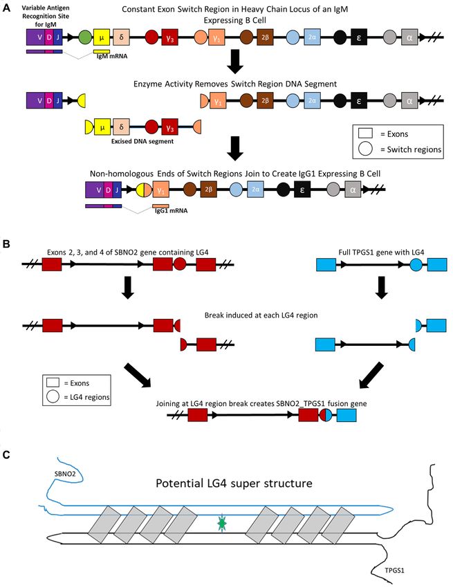
Nucleic Acids Research, 2020, Vol. 48, No. 11 5911

long guanine-rich minisatellites (33) and guanine-rich im-        somes 16 and 19, and depletion on Chromosome 6 (Figure
munoglobulin switch regions (13,24) both adopt G4 struc-          2D). In addition, LG4s within and directly flanking gene
tures and are associated with DNA breaks, we reasoned             transcripts, referred to hereafter as ‘LG4 genes’, were eval-
that a high density of guanine repeats within an ∼1 kb win-       uated in depth using the Database for Annotation, Visual-
dow could likely have similar impacts on the genome. These        ization and Integrated Discovery (DAVID) interface (46).
criteria would identify sequences that are not overly abun-       Similar to the occurrence of LG4 loci, we found Chromo-
dant, thus making an in-depth analysis possible. To iden-         some 16 and 19 both significantly enriched for LG4 genes
tify a panel of loci containing extensive G4 sequence mo-         (Supplementary Table S3.1). Cytogenetic bands are spe-
tifs, we searched for large genomic stretches significantly       cific genetic regions that can be detected using stains on
enriched for guanine-triplets (G-triplets) instead of focus-      metaphase chromosome spreads (69,70). DAVID analysis
ing on shorter, more rigidly defined G4 motifs. G-triplets        also identified significant enrichments of LG4 genes in 18
were counted because they are the basic sequence neces-           cytogenetic bands on 14 chromosomes (Supplementary Ta-
sary for G4 structure formation. Further search parame-           ble S3.2). Finally, we also found LG4 loci significantly en-

                                                                                                                                  Downloaded from https://academic.oup.com/nar/article/48/11/5907/5834579 by guest on 22 September 2020
ters (e.g. window size) were based on the immunoglobulin          riched at the ends of chromosomes, with 46% located less
switch region Sμ (Supplementary Information 1), which is          than two Megabases (Mb) from telomeres, and 67% within
a G4-forming recombination site recognized by mismatch            6 Mb (Figure 2E). To fully ensure the bias towards chromo-
repair factors (24,52). The Sμ guanine density of 120 G-          somal ends is not simply due to the occurrence of expanded
triplets/1.5 kb window, which is a much lower density of G-       telomeric repeats, human telomeric repeat (TTAGGG) se-
triplets compared to other well-known G4s such as telom-          quences (71) were identified in each LG4. While eight
eres (65), was used to train our analyses. Modeling our           LG4s found close to chromosomal ends do average 43.5
LG4ID search program on these parameters, we identified           TTAGGGs/1000 bp suggesting a potential relationship be-
301 loci containing a density of at least 80 GGG repeats/kb       tween (or origin from) these eight LG4s and their neighbor-
(Figure 2A). The 301 long G4-capable regions (LG4s) we            ing telomeres, over 97% (293/301) of the LG4s described in
identified in the human genome ranged from 199 to 4973            this work average only 0.6 TTAGGGs/1000 bp. Taken to-
bp in length (subset shown in Figure 2A). Although the ini-       gether, the significant enrichment of LG4s with defined ge-
tial search window was 1.5 kb, several smaller length re-         netic features indicates that their distribution in the human
gions contained a high density of G-triplets surrounding          genome is non-random and of likely functional relevance.
the larger repetitive unit and, therefore, met our minimal
G-triplet requirements.
   LG4 and control loci (301 sequences of identical lengths
                                                                  LG4 repeats are capable of G4 formation
randomly selected from the human genome) were catego-
rized based on their relative locations to known genes in-        Quadruplex forming G-Rich Sequences (QGRS) mapper
cluding: overlap with or within 5 kb of a known gene              is a web-based program for identifying individual G4 mo-
(Known Gene-associated), overlap with or within 5 kb              tifs in a given DNA sequence (49). We used QGRS map-
of a GENSCAN-predicted gene (66) (Predicted Gene-                 per to corroborate how successful our program (LG4ID)
associated), or unassociated with gene transcripts (Unasso-       was at identifying loci containing a dense concentration of
ciated). Similar to previous reports (19,67), LG4s were sig-      G4 motifs. All individual LG4s, 1.5 kb on the 5 and 3
nificantly (P < 0.00001) enriched within and around known         sides of the LG4 sequence, and control loci were queried
genes with 77.7% of LG4s versus 33.9% of controls oc-             in both orientations with QGRS, and the average number
curring in annotated loci (Figure 2B, Supplementary Table         of non-overlapping G4 motifs/kb (G4 motif density) was
S1). The LG4s associated with known genes were primar-            calculated. On average, LG4s contained 18 individual G4
ily located in introns (∼77%) or upstream of the transcrip-       motifs/kb, a 45-fold increase compared to control loci, and
tion start site (∼12%). One exonic LG4 was identified and         a 6.4-fold increase compared to sequences directly flank-
located in homeobox gene TPRX1, and comprised a ma-               ing LG4 (P < 0.0001) (Figure 3A). Notably, the increase
jority of the coding region. Intronic LG4s were found to          in the density of G4 motifs in the sequences directly flank-
occur in fairly similar percentages on transcribed (56.5%         ing LG4 compared to control loci indicates LG4 are found
CCC mRNA) and non-transcribed strands (43.5% GGG                  in G-rich areas compared to control loci. To confirm these
mRNA), and although beyond the scope of the current               findings, we elected to employ another G4 prediction algo-
work, a more thorough evaluation of these loci will ulti-         rithm (G4IPDB G4 predictor tool) (72) as a second means
mately be necessary to determine if this difference is sta-       of evaluating G4s density. We find the programs largely in
tistically significant (68). In contrast, LG4s located within 5   agreement with QGRS and G4IPDB respectively predicting
kb upstream of an annotated TSS are more than 3.5 times           an average of 18.3 and 21.7 putative G4s/1000 bp within
more likely to occur on the transcribed strand (Supplemen-        LG4 loci versus 0.4 and 1.3 in control sequences. Also of
tary Table S2).                                                   note, we find three additional in-silico G4 prediction tools,
   All LG4 regions identified by LG4ID were visually con-         ImGQFinder (14), Quadparser (73) and AllQuads (74) each
firmed in human genomic sequence, and the endpoints of            independently predict the formation of at least one G4 sec-
G-triplet containing repetitive units used to refine locus        ondary structure by sequences within each of the LG4s de-
length. All but three loci identified were over 500 bp in         scribed in this work, and furthermore, that 100% of the
length; the largest was 4973 bp and the average size was 1843     LG4s we find embedded within known protein coding loci
bp (Figure 2C). The chromosomal distribution of LG4 loci          were previously identified as likely forming G4 in vitro by
was not random with significant enrichment on Chromo-             Chambers et al. (75) via a high-resolution sequencing-based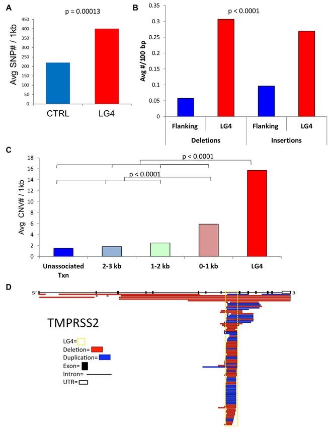
5912 Nucleic Acids Research, 2020, Vol. 48, No. 11

                                                                                                                                                             Downloaded from https://academic.oup.com/nar/article/48/11/5907/5834579 by guest on 22 September 2020

Figure 2. LG4s in the Human Genome. (A) Genome-side distribution of LG4 (red bars) on the left and examples of hits on the right with >3 bp G-repeats
that are highlighted red. (B) Distribution of locations with respect to annotated genes for LG4 and control loci. (C) The length distribution of LG4 loci.
(D) The number of LG4 and control loci on each chromosome, with asterisks indicating a significant difference. (E) Distribution of LG4 loci with respect
to the distance from the ends of each chromosome.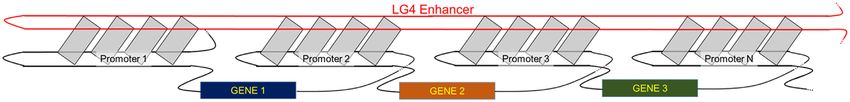
Nucleic Acids Research, 2020, Vol. 48, No. 11 5913

                                                                                                                                                              Downloaded from https://academic.oup.com/nar/article/48/11/5907/5834579 by guest on 22 September 2020

Figure 3. LG4s are capable of G4 formation. (A) The average number of non-overlapping G4 motifs predicted by QGRS mapper per kb (G4 motif density)
in LG4s, regions directly flanking LG4s (flanking) and control loci (Ctrl). (B) Circular dichroism ellipticities of oligonucleotides representing LG4s. (C)
Klenow DNA polymerase primer-extension reactions of G- or C-rich single stranded Sγ 3 or DIP2C DNA templates in buffer containing K+ or Li+ . (D)
Primer extension reactions of the C-rich LG4 strand of loci shown to stall polymerase in G4 supportive and non-supportive conditions. Reactions were
in G4-supportive conditions (K+ ) (E) Klenow primer-extension reactions on LG4 G-rich templates in different G4-permissive conditions. LG4 sequences
are denoted at the top of lanes; areas of stalled DNA synthesis is denoted by the brackets and full-length replication products are denoted by arrows. Sγ 3
and Sμ are G4 model sequences previously shown to form G4 in vitro (24,52).
5914 Nucleic Acids Research, 2020, Vol. 48, No. 11
G4-sequencing (G4-seq) approach based on the fact that sayed in K+ , and we found none were capable of stalling
G4s can block polymerases. Klenow extension (Figure 3D). As such, we conclude that
While computer algorithms can predict the G4 folding the LG4 G-rich strands stalled polymerase advancement
potential of LG4 sequences, the actual ability of these se- (Figure 3E), consistent with the formation of G4 structures
quences to form structures must be confirmed in vitro. Al- in the template.
though the LG4s described in this work average over 1800 Finally, as an initial examination of the ability of LG4
bp in length, they are composed of shorter minimal G4- sequences to form G4s in vivo, we asked if the number of
capable sequences much like telomeres whose minimal G4 LG4s immunoprecipitated (IP’d) in existing G4 ChIP-Seq
motif G4 structures have been extensively verified (65). As datasets (obtained using a G4-specific antibody) (86) were
such, we have employed two separate verifications that indi- enriched over matched control loci. Notably, we find a sta-
vidual minimal G4 motifs within LG4s do in fact form G4 tistically significant (P < 0.0001) enrichment for LG4 loci
secondary structures similar to minimal G4 motifs in telom- sequences (29.1%) over control loci sequences (4.9%) in G4
eres. To achieve this, subsets of transcribed LG4s were se- IPs (Supplementary Table S6).
Downloaded from https://academic.oup.com/nar/article/48/11/5907/5834579 by guest on 22 September 2020
lected for experimental validation by two distinct methods.
Firstly, minimal G4 motifs found in 15 LG4s (chosen due
LG4s are enriched for regulatory sequences
to their diverse G-repeat sequence composition and being
located in frequently transcribed regions) (Supplementary As previous genome-wide analyses (19) have found G4
Table S4) were assayed using circular dichroism (CD). CD sequences highly enriched in promoters, we next asked
measures the differential absorption of left and right po- whether LG4 sequences are associated with known regu-
larized light from chiral molecules in solution in order to latory elements. Because of its comprehensive nature, we
identify structural conformations (76). G4 can adopt two initially selected the Ensembl Regulatory Build Database
different conformations: parallel and anti-parallel, which (38,39) (based on publicly available, experimentally derived
describes the directionality of the DNA strands compos- data sets from DNase1-Seq, FAIRE-Seq and ChIP-Seq
ing the structure (15). Parallel G4 DNA results in a CD studies) to examine potential regulatory roles of all gene-
spectrum (ellipticity) with a peak at ∼260 nm and dip at associated LG4s (along with size matched control loci) and
∼240 nm. Anti-parallel G4 structures show a peak at ∼295 found LG4s associated with regulatory elements two-fold
nm and dip at ∼260 nm (77,78). Notably, all LG4 oligonu- more often than control loci (P < 0.001) (Supplementary
cleotides tested produced spectra characteristic of parallel Table S6). We also examined available NCBI SRA datasets
G4, although there was also evidence of anti-parallel G4 (87,88) to determine if LG4 sequences in available transcrip-
formation for F7 LG4 (black line peak 295, Figure 3B). tion factor (TF) ChIP-Seq datasets were significantly en-
These results are not that surprising and corroborate a study riched over controls. We found a significant (P < 0.0001)
demonstrating that parallel G4s are abundant throughout enrichment for LG4-loci sequences (44/198) over control-
the human genome (79). loci sequences (6/144) and identified over 80 interactions
Next, as a second, independent confirmation that LG4 at 44 LG4 loci involving 26 different TFs (Supplementary
sequences can form G4 DNA in vitro, 11 transcribed LG4 Table S6). The most prominent LG4-interacting TF identi-
sequences were selected for testing by a polymerase ex- fied was the DNA damage-associated protein Early Growth
tension assay. Sequences representing LG4 repeats rang- Response 1 (EGR1), with significant enrichments for 32 dis-
ing from 120 to 1300 bp were cloned and closed-circular tinct LG4s observed in EGR1 IPs. The TF with the sec-
single-stranded templates generated (Supplementary Table ond most LG4 enrichments was Specificity Protein 1 (SP1),
S5). In polymerase extension assays, polymerase pausing at which has previously been reported as associating with G4
G4 is monovalent-cation dependent and occurs only when promoter regions (67).
the guanine-rich strand serves as the template (53,80). There To further explore putative roles for LG4s in transcrip-
is a hierarchy of monovalent cations able to stabilize G4 tional regulation, we next examined their potential associa-
that is dependent on the temperature, cation concentration, tion with known, human-specific super-enhancers (SE) and
specific sequence, and therefore structure. K+ ions strongly super-enhancer elements (SEL). SEs are multi-enhancer
promote G4 assembly while other monovalent ions such as clusters that are characterized by higher TF density and
Li+ are poorer stabilizers (81–85). For the Sγ 3 control se- broader regulatory impact than typical enhancers. Using
quence, extension by Klenow polymerase was blocked in a comprehensive SE-SEL database containing 331 601
an orientation and K+ -dependent manner, which indicates unique super-enhancers (89), we found that LG4 loci were
that G4 formation on the template strand blocks DNA 70.4% (±12.9%) more likely to overlap super enhancers
synthesis (52). Similarly, a 130 bp segment of the DIP2C and/or their elements than randomly selected, same-sized,
LG4 sequence, which was used in subsequent yeast genetic control genomic loci (n = 3). Interestingly, only a subset
assays, also stalled Klenow extension in orientation- and (137/301) of LG4s are within close proximity to these regu-
K+ -dependent manners (Figure 3C). Notably, all LG4 se- latory clusters, while others are mostly intronic or in prox-
quences examined exhibited K+ -associated stalling relative imal promoters, which further signifies their importance
to Li+ , although to varying degrees; three of these LG4 se- to transcriptional regulation. To better examine the rela-
quences (P2RX5, HCN2 and ADARB2) were also able to tionship between LG4s and enhancers, we next correlated
stall Klenow in Li+ , reflecting the capacity of this ion to our data with GeneHancer (90), a novel database of 285
weakly support G4 folding. In order to rule out stalling due 000 candidate human enhancers (covering 12.4% of the
to non-G4 conformations, such as hairpin DNA, the reverse genome) integrating a total of 434 000 reported enhancers
complement C-rich strands of these three LG4s were as- from four different genome-wide databases: the Encyclope-Nucleic Acids Research, 2020, Vol. 48, No. 11 5915
dia of DNA Elements (ENCODE), the Ensembl regulatory in oncogene TMPRSS2 is shown in Figure 4D with dele-
build, the functional annotation of the mammalian genome tions and duplications occurring throughout the LG4 se-
(FANTOM) project and the VISTA Enhancer Browser. quence. Although how LG4 deletions and duplications af-
Strikingly, 180 of our LG4 sequences had either fully or fect TMPRSS2 regulation is unknown, we note that TM-
partially overlap with an annotated GeneHancer human en- PRSS2 is the oncogene most frequently involved in gene
hancer (Supplementary Table S7) as compared to an aver- fusions (41,95). That said, we find the majority of LG4 loci
age of only 84 overlaps (n = 5) between matched controls are similarly associated with annotated deletions and dupli-
and enhancers. cations (Supplementary Information S2).
LG4s have increased small and large-scale genome variation Deletions/duplications >10 bp accumulate within the DIP2C
LG4 in yeast
A recent analysis of single G4 motifs demonstrated enrich-
ment for small nucleotide variations that include single nu- The budding yeast Saccharomyces cerevisiae has been a
Downloaded from https://academic.oup.com/nar/article/48/11/5907/5834579 by guest on 22 September 2020
cleotide polymorphisms (SNPs) and insertions or deletions powerful tool to study instability associated with G4 and
(indels) less than 50 bp, although the two were not distin- other repetitive DNA sequences (32–34,91,93–104). Since
guished in this study (29). SNPs have been shown to dis- our computational evidence indicated that indels and CNVs
rupt the regulatory ability of G4, indicating that regions were elevated at LG4 loci in the human genome, we adapted
prone to single base-pair changes have a high potential to a versatile LYS2-based frameshift reversion assay to directly
loose regulatory ability (22,28). Using identified variations assess the instability of a LG4 sequence in yeast. This sys-
from genome-wide sequence studies available in the db- tem capitalizes on a 150 bp, functionally dispensable seg-
SNP database (50), the number of SNPs for each LG4 was ment of the LYS2 gene that is defined by stop codons in
counted and the average per 1000 bp calculated. There was alternative reading frames. A frameshift mutation in this
a significant ∼82% enrichment (P < 0.00001) of SNPs in region can be reverted by any sequence addition/deletion
LG4s compared to randomly selected, size-matched control of net opposite sign that occurs within the ‘reversion win-
regions (Figure 4A, Supplementary Table S2), suggesting dow’ demarcated by the stop codons (105). Prior stud-
that LG4s, much like conventional G4 motifs (91), may be ies demonstrated that most compensatory frameshifts are
more prone to base damage or have a decreased ability to deletions/insertions of single base pairs in short, mononu-
be accurately repaired. To determine if LG4s are similarly cleotide runs (57,105,106). Insertion of an out-of-frame
prone to indels, the average number of indels/100 bp for LG4 sequence into the reversion window allows detection of
each transcribed LG4 and the surrounding non-exonic re- additional mutation types specifically associated with G4-
gion was calculated separately from SNPs present in the db- forming potential.
SNP database. LG4 insertion events/100 bp were increased The DIP2C sequence (Figure 5A) was selected for this
>5-fold and deletions >2.5-fold (P < 0.0001) as compared analysis because of our in vitro data confirmed its potential
to surrounding non-exonic regions (Figure 4B). The ob- to form G4 structures and our computational analysis iden-
served increase in human genome variation (SNPs and in- tified its significant association with indels and CNVs. Al-
dels) indicates that LG4s are prone to mutagenesis com- though over 800 bp of G-rich repetitive sequence have previ-
pared to surrounding loci. ously been inserted into LYS2 (107), we were unable to clone
In the human genome, copy-number variations (CNVs), the full-length DIP2C sequence into bacterial plasmids and,
which are defined as indels >50 bp, are also major con- therefore, used a 130 bp segment representative of the LG4
tributors to genetic diversity and increase susceptibility (Figure 5A). The DIP2C insertions created a +1 or −1 lys2
to a range of genetic disorders (92,93). A CNV break- frameshift mutation (lys2::DIP2C+1 and lys2::DIP2C-1 al-
point is defined as the genomic location where a dupli- leles, respectively) that revert by net −1 or +1 changes in the
cation or deletion occurs, and CNVs detected through extended ∼280 bp reversion window.
genome-wide sequencing studies are available in dbVAR The reversion rates of the lys2::DIP2C frameshift al-
database at NCBI.org (51). We calculated the number of leles were measured and compared to those of lys2 +1
CNV breakpoints/kb for each transcribed LG4, 3 kb 5 and and −1 alleles without the DIP2C insertion. Although ad-
3 of LG4 in 1 kb increments, as well as for the remaining dition of the DIP2C sequence roughly doubled the size
transcript not associated with LG4 (referred to as unasso- of the theoretical reversion window where compensatory
ciated transcripts). LG4 regions contained a significant (P frameshift mutations can be detected, the reversion rates
< 0.0001), ∼10-fold increase in CNV breakpoints as com- of the lys2::DIP2C+1 and lys2::DIP2C-1 alleles were ele-
pared to nearby unassociated transcripts, and an 8-fold in- vated 15–20-fold relative to relative to alleles without the
crease relative to sequence >2 kb away (Figure 4C). Unex- DIP2C insertion (Figure 5B). The region of the reporter
pectedly, regions within 1 kb of LG4 had a significant (P < containing the DIP2C insertion was sequenced to deter-
0.0001) ∼3-fold increase in CNVs over unassociated tran- mine the types of compensatory frameshift mutations that
scripts, suggesting that LG4s can invoke instability at prox- occurred. Approximately 85% of the lys2::DIP2C+1 rever-
imal sequences (Figure 4C). This supports the findings of tants (53/64) contained deletions >10 bp, while ∼65% of
a previous report suggesting that DNA structures can in- lys2::DIP2C-1 revertants (37/58) were duplications >10 bp.
duce mutagenesis in surrounding regions (94). Importantly These deletions/duplications are summarized in Figure 5C
our analysis provides evidence that many other transcribed where the size and number of each are indicated (see also
G4 regions may be capable of repeat expansion and con- Supplementary Table S8). Importantly, all events had at
traction. For example, a schematic representation of CNVs least 2 bp of perfect homology (a direct repeat) at the end-5916 Nucleic Acids Research, 2020, Vol. 48, No. 11
Downloaded from https://academic.oup.com/nar/article/48/11/5907/5834579 by guest on 22 September 2020
Figure 4. LG4s are associated with increased small and large-scale genome variation. (A) Entries from the dbSNP database for LG4s and size matched
control regions. (B) Entries from the dbSNP database for LG4 and intronic regions directly surrounding LG4 (flanking). (C) CNV sizes from dbVAR and
the average number of breakpoints/kb (y-axis) was calculated for each transcribed LG4, in 1kb increments away from LG4, and the rest of the transcript not
directly associated with LG4 (Unassociated TXN). (D) Schematic diagram of the location and size of copy-number variants with respect to the predominant
TMPRSS2 transcript. Introns, exons and UTRs denoted by lines, solid boxes, or open boxes, respectively. The yellow dashed box highlights the location
of the TMPRSS2 LG4 and individual duplications (blue boxes) and deletions (red boxes) are shown.Nucleic Acids Research, 2020, Vol. 48, No. 11 5917
Downloaded from https://academic.oup.com/nar/article/48/11/5907/5834579 by guest on 22 September 2020
Figure 5. A DIP2C intronic LG4 is prone to deletions and duplications over 10 bp. (A) A 130 bp fragment of DIP2C LG4 was cloned into yeast. The
G-repeats are in red. A table of LG4s genetic traits and human genome variation compared to non-exon regions surrounding all LG4s is below. (B) The
LG4 LYS2 reversion window was sequenced for revertants and rates adjusted by proportion of a given mutation type. Error bars are 95% confidence
intervals and adjusted for the total rate. (C) Mutation spectrum for deletions (red bars) and duplications (green bars) with corresponding size and number
detected for both Lys+1 sequences (top) and Lys −1 (bottom). (D) The bp of perfect homology at the end of duplications/deletions.5918 Nucleic Acids Research, 2020, Vol. 48, No. 11
points. The largest flanking repeat was 11 bp and in gen- gene fusions occurred within or in close proximity to an
eral, there was a correlation between the number of times LG4 present in the TMPRSS2 locus (Figure 4D). Gene
a given event was detected and the length of the endpoint fusions involving TMPRSS2 are the most frequently re-
homology (Figure 5D). For deletions, one copy of the re- ported across all malignancies, with approximately half of
peat and the intervening sequence was deleted; for duplica- all prostate cancers containing TMPRSS2-ERG fusions
tions, one copy of the repeat plus the intervening sequencing (110). There were additionally 23 unique gene fusions an-
was duplicated. There were 12 distinct events (A–L) among notated in the FusionGDB that corresponded to genomic
the 53 deletions detected; among the 37 duplications, there rearrangements occurring between distinct LG4 sequences
were six distinct types (Figure 5C). Each of the duplica- resembling rearrangements occurring between long G4-
tions had a corresponding deletion event with the same end- capable-motif dense switch regions during mammalian im-
points, and these were given the same letter designation as munoglobulin class switch recombination (Figure 6A). As
the deletion. Finally, all but one event within this initial an example, a genomic rearrangement occurring between
sample of deletions/duplications was either 19, 34 or 49 bp. LG4s found in the SBNO2 and TPGS1 loci has been repeat-
Downloaded from https://academic.oup.com/nar/article/48/11/5907/5834579 by guest on 22 September 2020
This striking periodicity of 15 bp reflects both the repeti- edly observed in various malignancies (Figure 6B) (41,95).
tive structure of DIP2C as well as the constraint to delete In light of the frequent participation of LG4s in chromo-
a non-multiple of 3 bp in order to restore the LYS2 read- somal translocations, it is tempting to speculate that G4
ing frame. Although similar deletions/duplications can be sequences may directly facilitate trans interactions (Figure
generated during the repair of double-strand breaks (108), 6C).
neither loss of homologous recombination nor nonhomol-
ogous end-joining pathway affected their rates (rad51Δ
Neighboring LG4 loops are frequently complementary and
and dnl4Δ backgrounds, respectively; Supplementary Ta-
base pair in vitro
ble S8). This suggests that slippage between the flanking di-
rect repeats during DNA replication is the most likely cause A detailed inspection of individual LG4 sequences revealed
of the large deletion/duplications in the DIP2C sequence single-strand loops of neighboring G4s within individual
(108,109). That said, although we find sequence repetitive- LG4 regions that were frequently complementary to one
ness rather than the ability to form G4 structures is typ- another (Figure 7A, Supplementary Table S2, Supplemen-
ically the principle driver of deletions and duplications in tary Information 4). Whereas the complement to a 6 nt
this sequence, we have extensively examined the relation- loop would only be expected to occur (at random) once
ship between G4 formation and LG4 mutagenesis forma- every 4,096 bp and the average LG4 length is only 1843
tion (Supplementary Information S3) and find that over bp, 178 of the 301 individual LG4 sequences we identi-
half of all duplication events occurring during periods of fied contained internal G4-loop complementarities. Each
high transcriptional activity are likely directly attributable of these 178 loci had, on average, 34.8 complementary
to G4 structure. loops of 8.2 bp in length. We hypothesized that G4 loops
within individual LG4 loci directly pair with one another
in a manner similar to numerous, well-documented kiss-
LG4s are significantly associated with genomic rearrange-
ing stem–loop interactions characterized in various RNA
ments
(111,112) and (less frequently) DNA structures (113,114)
Having confirmed LG4s are associated with increased small (Figure 7A). To examine whether neighboring G4 loops can
and large-scale genomic variation, we next examined if base pair with one another in vitro via a loop:loop kiss-
LG4s are similarly associated with mutation in malignancy ing interaction (termed a G4 Kiss or G4K), we synthe-
through mining the somatic mutation information avail- sized oligonucleotides containing two minimal G4-capable
able in COSMIC (Catalogue of Somatic Mutations in Can- sequences separated by a polyA linker. Oligonucleotides
cer) (40). The COSMIC database contains genome-wide contained either (i) neighboring G4-capable sequences with
mutational data from over 32 000 cancer genomes derived putative kissing loops from the LG4 shown in A (Figure
from peer-reviewed, large-scale genome screening datasets 7B), (ii) two minimal G4-capable sequences separated by
and other databases such as TCGA and ICGC (40). No- a polyA linker with loops containing characterized viral
tably, the identified LG4s are 31 times closer to chromo- kissing-loop complements (Figure 7C) or (iii) controls for
somal breakpoints contained within the COSMIC dataset each of these that lacked complementarity. These oligonu-
than matched controls (Supplementary Table S2). In ad- cleotides were evaluated by nondenaturing G4 gel elec-
dition to this, we also screened genes containing LG4s trophoresis and sequential staining (as assessed in Supple-
against the FusionGDB (41). FusionGDB is a publicly mentary Information 5). Importantly, the affinity of ethid-
available database consolidating data from three primary ium bromide (EtBr) for dsDNA is 25 times greater than its
fusion gene resources: chimeric transcripts and RNA-seq affinity for ssDNA, while Thioflavin T associates with G4
data (ChiTaRS 3.1), TumorFusions, and fusions identi- DNA but not normal duplex DNA (115,116). Sequential
fied in The Cancer Genome Atlas (TCGA). In all, the Fu- EtBr and Thioflavin staining (pink and blue, respectively)
sionGDB contains over 48 000 unique gene fusions char- confirmed that an intramolecular G4 structure formed by
acterized from an array of malignancies. Strikingly, 144 of a DNA oligonucleotide with complementary loops directly
the 185 LG4s located in annotated protein-coding loci were engages in an observable double-strand interaction (upper
in genes involved in fusions listed in the FusionGDB (P < gels) whereas the G4 structure formed by nearly identical
0.001) (Supplementary Table S2). Thirty-seven of the chro- control oligos lacking complementary loops does not (Fig-
mosomal breaks contributing to annotated FusionGDB ures 7B, C). Furthermore, we found that LG4s containingNucleic Acids Research, 2020, Vol. 48, No. 11 5919
Downloaded from https://academic.oup.com/nar/article/48/11/5907/5834579 by guest on 22 September 2020
Figure 6. Recurrent translocations between LG4s. (A) Mechanism of isotype switching in activated B cells. Resulting mRNA indicated below genomic
sequences. V; variable exon. D; diversity exon. J; joining exon. IgM; immunoglobulin M. IgG1; immunoglobulin G subclass 1. ; immunoglobulin M
heavy chain exon. ␦; immunoglobulin D heavy chain exon. ␥ 3; immunoglobulin G subclass 3 heavy chain exon. ␥ 1;immunoglobulin G subclass 3 heavy
chain exon. ε; immunoglobulin E heavy chain exon. ␣; immunoglobulin A heavy chain exon. 2; immunoglobulin G subclass 2 beta heavy chain. 2␣;
immunoglobulin G subclass 2 alpha heavy chain (32,52,127–130). (B) Gene fusion (40,41) resulting from distinct LG4 breaks independently reported
in breast (TCGA-BRCA) and cervical (TCGA-CESC) malignancies. SBNO2; Strawberry notch homolog 2. TPGS1; Tubulin Polyglutamylase Complex
Subunit 1. (C) Model of potential LG4 super structure.5920 Nucleic Acids Research, 2020, Vol. 48, No. 11
Downloaded from https://academic.oup.com/nar/article/48/11/5907/5834579 by guest on 22 September 2020
Figure 7. LG4 Kissing Loops. (A) Predicted model of neighboring G4 loop interaction in human LG4 locus occurring at hg38:chr17:1052333–1053488 is
illustrated. Complementary loops are highlighted in yellow. Guanine triplets are highlighted in red. (B) Nondenaturing G4 gel electrophoresis of minimal
G4 capable sequence (taken from the LG4 detailed in A). Lane 1 – tandem of two minimal G4 capable sequences with complementary loops. Lanes 2 and 3
– tandem of two minimal G4 capable sequences with complementary loops replaced by adenosines. Upper gel image: EtBr stain (orange) only. The affinity
for EtBr binding of dsDNA is ∼25 times its affinity for ssDNA. Lower gel image: Subsequent Thioflavin (blue) staining of the identical gel shown in the
upper image. (C) Nondenaturing G4 gel electrophoresis of G4 capable sequences with complementary loops from known HPV kissing hairpins. (Left)
Control oligonucleotide lacking complementary loops. (Right) Oligonucleotide containing complementary loops known to participate in HCV hairpin
kissing (111). Upper gel image: EtBr stain (orange) only. Lower gel image: Subsequent Thioflavin (blue) staining of the identical gel shown in the upper
image. (D) Additional example of a LG4 locus with loops described as ‘Self complements’ (like the LG4 in A). (E) Example of a LG4 locus with loops
described as ‘Neighboring complements’.Nucleic Acids Research, 2020, Vol. 48, No. 11 5921
complementary loops typically assume (and can be fairly K+ -independent stalling was also reported in telomeres, and
evenly divided between) one of two general configurations. to date, details of an alternative structure to G4 formation
The first of these consists of a regularly repeating series of remain elusive (119). Further analysis of the identified LG4
self-complementary loops ‘self complementary’ (Figure 7D, regions will be needed to determine what sequence require-
Supplementary Information S4). In contrast, ‘neighboring ments, if any, lead to K+ -independent stalling on only the
complementary’ LG4s are generally much less organized G-rich strand.
but also more complex in that they typically contain sev- We found that LG4s were associated with both sequence
eral different putative loop:loop interactions (Figure 7E, variations and known promoter/enhancer elements, sug-
Supplementary Information S4). Together, these findings gesting a connection between G4 structures, site-specific in-
suggest LG4s adopt a novel, higher order, composite G4K stability, and genetic regulation. As the mechanism(s) of in-
structure potentially driving the formation and/or mainte- creased SNPs and indels at LG4s cannot be deciphered from
nance of these conspicuous genomic regions. computational data, and were not found to be elevated in
our DIP2C yeast studies, more direct experimentation will
Downloaded from https://academic.oup.com/nar/article/48/11/5907/5834579 by guest on 22 September 2020
be needed to delineate the role(s) of G4 structures in LG4
DISCUSSION mutagenesis. The use of only a 130 bp portion of DIP2C
We have developed a novel algorithm for identifying long in the yeast studies may have resulted in a significant un-
stretches of tandem G4s resembling Ig switch regions in derrepresentation of the true mutagenic capacity of the in-
genomic sequences, LG4ID, and used it to identify 301 tact LG4, although duplications and deletions reflecting the
LG4 regions averaging over 1.5 kb in length in the human repetitive nature of the sequence were evident. It should also
genome. Subsequent analyses of these LG4s suggest that be noted that the LG4s identified in this work contain a
these unusually large, G4-dense regions are capable of com- wide variety of repeat sequence compositions (Supplemen-
plex G4 formation and possess a high probability of regu- tary Table S4), and we predict that the mutagenic propensity
latory and mutagenic potential. Notably, shorter LG4 re- and genetic control of instability will likely differ between
gions (e.g. 200–1500 bp), although smaller than the Sμ or distinct LG4s.
Sγ 3 G4-containing immunoglobulin switch regions, might Strikingly, 119 of 185 protein-coding genes that contain
also be biologically relevant, and we suggest it would be regulatory LG4s have known disease associations (as in-
interesting to determine if shorter LG4s are similarly lo- dicated in Supplementary Table S2). Intergenic LG4s may
cated in regions with regulatory potential and subject to in- also be disproportionately associated with disease, as the
creased genomic variation. That said, we find over 6,000 hu- disruption of proper gene regulation due to CNVs, SNPs
man genomic loci between 500 and 1500 bp in length with or indels in regulatory LG4s could contribute to cellular
a G-triplet/sequence length ratio equal to or greater than dysfunction and/or disease. For example, overexpression
that used in the current search. As the number of LG4 calls of Anoctamin 9, ANO9, is associated with the progression
decreases dramatically as search window size increases (as of metastatic colorectal cancer (120). The ANO9 LG4 is a
detailed in Supplementary Table S9), we elected to focus site for EGR1 transcription factor interactions, suggesting
the current study on LG4s 1500 bp and greater in length this locus is highly involved in gene regulation. Further, the
with a density of at least 8 GGGs per 100 bp (as the core ANO9 LG4 is significantly enriched for CNVs, SNPs and
Sμ G4-containing immunoglobulin switch region is 1489 indels making it possible that an increase of site-specific
bp in length with a GGG density of ∼8.0 GGGs per 100 mutagenesis could disrupt proper ANO9 regulation leading
bp (Supplementary Information S5). Importantly, the flex- to the progression of late-stage colorectal cancer. Consid-
ibility of LG4ID allows window size (total length) to be ering most genome-wide association sequencing studies of
reduced while maintaining a constant G-triplet/sequence human disease have historically focused on coding DNA,
length ratio, or alternatively, for the density of G-triplets mutations in regulatory non-coding regions, such as LG4s,
to be adjusted, and as such, have made the LG4ID source could provide previously missed insights into the etiology
code available for download on the LG4ID website to allow of multiple diseases.
users to explore manipulating these parameters. Importantly, in addition to high levels of CNVs, SNPs,
Although the present search focused on identifying re- and indels being associated with LG4s, translocation
gions of G4 formation, we find several LG4s are capable of hotspots have also previously been reported to be enriched
assuming other non-B form DNA structures, forming stable at sites harboring G4-capable sequences (30). In agreement
hairpins, and/or contain long purine repeats (ex AGGGA) with this, we similarly found translocations and gene fu-
capable of triplex structures (data not shown) (117). We ad- sions associated with LG4s (Figure 6, Supplementary Ta-
ditionally found that a subset of cloned LG4 motifs stalled ble S2). As such, it is tempting to speculate that interchro-
Klenow polymerase in a K+ independent manner, suggest- mosomal and long-range intrachromosomal hybrid G4 for-
ing that Li+ is capable of stabilizing G4, or conversely, other mation between LG4 sequences may directly facilitate re-
G-rich structures are formed in addition to G4. Although current translocations (Figure 6B, C). Potentially related
stalling was enhanced by K+ and confined to the G-rich to this, we found that 217 of the 301 LG4 loci identified
strand and CD scans demonstrated parallel G4 formation in this study either fully or partially overlap with an an-
(Figure 3), non-G4 structures may also form from LG4 se- notated human enhancer (Supplementary Tables S2 and
quences. Hairpins, for instance, can also stall replication S7) (versus only 84 average overlaps (n = 5) between size
(118). Since the LG4 sequences that showed stalling during and nucleotide composition matched control loci and en-
Klenow extension in Li+ did not contain any long stretches hancers). Our preliminary analyses of the gene promoters
of purines, it is unlikely that this was due to triplex DNA. likely regulated by these enhancers found them significantlyYou can also read