A structural inventory of native ribosomal ABCE1- 43S pre-initiation complexes
←
→
Page content transcription
If your browser does not render page correctly, please read the page content below
Article

A structural inventory of native ribosomal ABCE1-
43S pre-initiation complexes
Hanna Kratzat1,†, Timur Mackens-Kiani1,†, Michael Ameismeier1 , Mia Potocnjak1,
Jingdong Cheng1 , Estelle Dacheux2, Abdelkader Namane2 , Otto Berninghausen1, Franz Herzog1,
Micheline Fromont-Racine2 , Thomas Becker1,* & Roland Beckmann1,**

Abstract                                                                             regulation is the initiation phase; however, in eukaryotes the indi-
                                                                                     vidual phases of translation were found to be coupled, especially
In eukaryotic translation, termination and ribosome recycling phases                 termination with ribosome recycling and a new round of initiation.
are linked to subsequent initiation of a new round of translation by                 Two prominent examples are the conserved multisubunit complex
persistence of several factors at ribosomal sub-complexes. These                     eIF3, which has been described as a factor functioning across the
comprise/include the large eIF3 complex, eIF3j (Hcr1 in yeast) and                   translation cycle (Valasek et al, 2017), as well as the ATP-binding
the ATP-binding cassette protein ABCE1 (Rli1 in yeast). The ATPase is                cassette (ABC) ATPase ABCE1 (Rli1 in Saccharomyces cerevisiae),
mainly active as a recycling factor, but it can remain bound to the                  which was shown to enhance termination activity of the eRF1
dissociated 40S subunit until formation of the next 43S pre-initiation               release factor and which represents the key enzyme for ATP-depen-
complexes. However, its functional role and native architectural                     dent ribosome recycling (Pisarev et al, 2010; Shoemaker & Green,
context remains largely enigmatic. Here, we present an architectural                 2011). Moreover, ABCE1 was found associated with initiation
inventory of native yeast and human ABCE1-containing pre-initiation                  factors (Chen et al, 2006; Dong et al, 2004) and as a part of eIF3-
complexes by cryo-EM. We found that ABCE1 was mostly associated                      containing 43S or 48S pre-initiation complexes (Andersen & Leevers,
with early 43S, but also with later 48S phases of initiation. It adopted             2007; Preis et al, 2014; Mancera-Martinez et al, 2017).
a novel hybrid conformation of its nucleotide-binding domains, while                     The ABCE1 ATPase consists of two nucleotide-binding domains
interacting with the N-terminus of eIF3j. Further, eIF3j occupied the                (NBDs) that are forming two nucleotide-binding sites (NBSs) at their
mRNA entry channel via its ultimate C-terminus providing a struc-                    interface, as well as an essential iron–sulfur cluster domain (FeSD) at
tural explanation for its antagonistic role with respect to mRNA                     its N-terminus (Barthelme et al, 2007; Hopfner, 2016). ABCE1 binds
binding. Overall, the native human samples provide a near-complete                   the 80S ribosome during canonical stop codon-dependent termination
molecular picture of the architecture and sophisticated interaction                  or during rescue of stalled ribosomes and splits the 80S ribosomes into
network of the 43S-bound eIF3 complex and the eIF2 ternary                           40S and 60S small (SSU) and large (LSU) subunits, respectively. This
complex containing the initiator tRNA.                                               recycling reaction requires an A site factor in the ribosome, either
                                                                                     release factor eRF1 (after termination) or its homologue Pelota
Keywords ABCE1; eIF3; cryo-EM; translation initiation; ribosome recycling            (Dom34 in S.c.; for ribosome rescue), in order to form part of the
Subject Categories Structural Biology; Translation & Protein Quality                 interaction network for ABCE1 (Becker et al, 2012; Brown et al, 2015;
DOI 10.15252/embj.2020105179 | Received 3 April 2020 | Revised 21 September          Preis et al, 2014). ABCE1 binds these pre-splitting complexes in a
2020 | Accepted 29 September 2020 | Published online 8 December 2020                 semi-open state with respect to its NBSs. Splitting requires binding of
The EMBO Journal (2021) 40: e105179                                                  ATP and site-occlusion to both NBS (Barthelme et al, 2011; Gouridis
                                                                                     et al, 2019; Nurenberg-Goloub et al, 2018). According to current
                                                                                     models, the conformational change occurring during site-occlusion
Introduction                                                                         would be transmitted via the FeSD of ABCE1 to the bound A site
                                                                                     factor (eRF1 or Dom34), whereby the FeSD exerts a force on the A site
Translation of an mRNA into a polypeptide sequence is a central                      factor which ultimately leads to ribosome splitting (Becker et al, 2012;
cellular process, which is highly regulated and linked to other cellu-               Heuer et al, 2017; Nu € renberg-Goloub et al, 2020). The splitting reac-
lar processes like ribosome biogenesis, mRNA turnover, and ribo-                     tion can be recapitulated in vitro (Becker et al, 2012; Nurenberg-
some quality control. Most decisive for translational efficiency and                 Goloub & Tampe, 2019; Pisareva et al, 2011; Shao et al, 2015;

1 Gene Center and Center for Integrated Protein Science Munich, Department of Biochemistry, University of Munich, Munich, Germany
2 Genetique des Interactions Macromoleculaires, UMR3525 CNRS, Institut Pasteur, Paris, France
  *Corresponding author. Tel: +49 89 2180 76915; E-mail: becker@genzentrum.lmu.de
  **Corresponding author. Tel: +49 89 2180 76900; E-mail: beckmann@genzentrum.lmu.de
  †
    These authors contributed equally to this work
  [Correction added on 5 February 2021 after first online publication: Publishing license has been changed]

ª 2020 The Authors. Published under the terms of the CC BY NC ND 4.0 license                                       The EMBO Journal   40: e105179 | 2021   1 of 24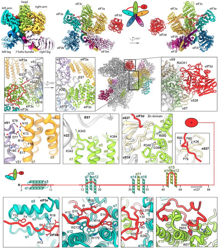
The EMBO Journal                                                                                                                     Kratzat et al

Shoemaker & Green, 2011), where ABCE1 was observed to remain               Llacer et al, 2015; Llacer et al, 2018; Mancera-Martinez et al, 2017).
bound to the 40S small subunit to form a post-splitting complex            During 43S assembly, the 40S subunit gets prepared to thread the
(PSC), in which the two NBDs are present in a closed, nucleotide-          mRNA into the mRNA-binding channel between the 40S body and
occluding state (Heuer et al, 2017; Kiosze-Becker et al, 2016;             the head. The main constriction for mRNA is at the so-called
N€urenberg-Goloub et al, 2020). Therefore, it was assumed that in vivo     “latch”, a structural element formed between ribosomal RNA
as well, ABCE1 may remain bound to the 40S for a defined time span         (rRNA) helix h18 and ribosomal protein (r-protein) uS12 on the 40S
(Gerovac & Tampe, 2019) to prevent re-association of the LSU (Heuer        body, and h34 and uS3 on the head (Schluenzen et al, 2000). Empty
et al, 2017) or to coordinate assembly of initiation factors on the 40S    or only ABCE1-bound 40S usually does not adopt a defined head
subunit. However, a direct physical involvement of ABCE1 in the            conformation, and the latch is rather closed (Heuer et al, 2017; Pass-
translation initiation process has not been shown to date.                 more et al, 2007). Binding of eIF1 and especially eIF1A, which
    In eukaryotes, the start of translation initiation requires the        bridges the body with the head, seems to prime and confine the 40S
assembly of the 43S pre-initiation complex (PIC). It consists of the       by inducing a small rotation of the 40S head (Llacer et al, 2015;
40S subunit, eIF3, eIF1, eIF1A, eIF5, and the ternary complex (TC)         Passmore et al, 2007), but the latch still remains in a closed position
formed by the trimeric eIF2αβγ, initiator methionyl tRNA (tRNAi),          (Llacer et al, 2015). Latch opening was only observed in in vitro
and GTP. After 43S PIC assembly, the mRNA—in collaboration with            reconstituted partial 48S ICs containing mRNA and both eIF3 and
the eIF4F complex (the cap-binding protein eIF4E, the helicase             the eIF2 TC in addition to eIF1 and eIF1A (Llacer et al, 2015; Llacer
eIF4A, and the scaffolding protein eIF4G)—can be recruited to the          et al, 2018). Here, two conformations of the 48S IC can be distin-
43S PIC, forming the 48S initiation complex (IC). This event is coor-      guished: the open POUT and the closed PIN conformation, which dif-
dinated by interactions between eIF3 and eIF4F as well as eIF4B, a         fer in the orientation of the 40S head and the TC. Compared to the
single-stranded RNA-binding protein that attaches to the 40S               empty and eIF1/1A-bound structures, the head is moved upwards
subunit (Walker et al, 2013) and stimulates the helicase activity of       away from the body in the POUT conformation. This leads to widen-
eIF4A. The 48S complex then scans the mRNA for the first cognate           ing of the latch and the P site tRNAi in the TC is only bound via the
AUG codon. After start-codon recognition, inorganic phosphate (Pi)         anticodon loop (AL) to the 40S head but not the body. In the PIN
is released from the eIF2 complex, which is stimulated by eIF5             conformation, the AL moves down and engages in stable codon–
acting as a GTPase-activating protein, likely via an arginine-finger       anticodon interactions with the cognate start codon in the P site,
mechanism (Algire et al, 2005; Das et al, 2001; Paulin et al, 2001).       accompanied by a downward movement of the 40S head.
Subsequently, initiation factors apart from eIF1A and eIF3 dissociate          In all eIF3-containing structures, the PCI-MPN core was located
(Mohammad et al, 2017; Sha et al, 2009) and subunit joining with           on the back of the 40S subunit, from where peripheral subunits
the 60S LSU is then mediated by the GTPase eIF5B (Acker et al,             stretch out. In 43S PICs, the YLC was found close to the mRNA entry
2006; Acker et al, 2009; Lee et al, 2002; Pestova et al, 2000).            site of the 40S (Aylett et al, 2015; des Georges et al, 2015; Eliseev
    An important regulatory and scaffolding role in these processes        et al, 2018; Erzberger et al, 2014), however only at low resolution.
is taken on by the multisubunit complex eIF3 (Cate, 2017; Hinneb-          Moreover, the YLC module has been shown to relocate to the inter-
usch, 2006), which can be structurally divided into the so-called          subunit space (ISS), as observed in in vitro reconstituted partial 48S
PCI-MPN core and the peripheral subunits. In yeast, the PCI-MPN            complexes (Llacer et al, 2015), thereby occupying the position of
core consists of the two subunits eIF3a (Rpg1/Tif32) and eIF3c             ABCE1. The other peripheral subunits eIF3d and the eIF3c N-termi-
(Nip1), whereas in mammals, it is formed by an octamer of eIFs 3a,         nal domain have been localized near the mRNA exit site (eIF3d:
3c, 3e, 3f, 3h, 3i, 3k, and 3l (Valasek et al, 2017). The peripheral       Eliseev et al, 2018) and in the ISS (eIF3c-NTD: Llacer et al, 2015;
subunits consist of the so-called yeast-like core (YLC) module,            Obayashi et al, 2017). Interestingly, two structures of partial native
containing eIF3b (Prt1), eIF3g (Tif35), and eIF3i (Tif34), as well as      43S/48S complexes exist in which ABCE1 could be visualized in
the C-terminus of eIF3a, the N-terminal domain of eIF3c that inter-        substantial quantities (Simonetti et al, 2016, re-interpreted in
acts with eIF1 and eIF5 (Valasek et al, 2003; Valasek et al, 2004;         Mancera-Martinez et al, 2017; Heuer et al, 2017). Notably, both
Yamamoto et al, 2005; Zeman et al, 2019), and in mammals eIF3d.            samples were obtained after adding non-hydrolyzable AMP-PNP
In addition, eIF3j is associated with eIF3 but does not belong to its      and/or GMP-PNP to either yeast (Heuer et al, 2017) or rabbit reticu-
core, and plays a special role (Block et al, 1998; Valasek et al,          locyte (Simonetti et al, 2016) lysates and subsequent isolation of the
1999). It was shown that eIF3j participates during termination by          43S peak from a sucrose gradient. This may have led to non-physio-
recycling eRF3 (Beznoskova et al, 2013) and during ribosome recy-          logical locking of ABCE1 on the 40S subunit, thereby limiting any
cling by assisting ABCE1 in subunit splitting (Young & Guydosh,            conclusions about a putative role of ABCE1 during the phase
2019). Furthermore, it is involved in dissociation of mRNA from the        connecting recycling with initiation. Furthermore, apart from a low-
40S subunit (Pisarev et al, 2007; Pisarev et al, 2010). In the context     resolution cryo-EM map (Aylett et al, 2015) no structural data exist
of initiation, eIF3j is believed to participate in the recruitment of      on eIF3j in the context of the native 43S PIC. Therefore, the native
eIF3 to the 40S (Elantak et al, 2010; Fraser et al, 2004; Nielsen et al,   structural landscape enabling the transition from translation termi-
2006), to antagonize premature mRNA recruitment (Fraser et al,             nation via recycling to initiation is not yet well-understood.
2007), and to regulate start-site selection (Elantak et al, 2010).
    For a better mechanistic understanding of this complicated inter-
play, a number of cryo-EM structures of 43S PICs and partial 48S           Results
ICs gave first insights into the architectural variety of initiation
complexes (Aylett et al, 2015; des Georges et al, 2015; Eliseev et al,     In this work, we set out to provide a structural inventory of ABCE1-
2018; Erzberger et al, 2014; Hashem et al, 2013; Hussain et al, 2014;      containing 43S or 48S initiation complexes from native small

2 of 24     The EMBO Journal 40: e105179 | 2021                                                                                  ª 2020 The Authors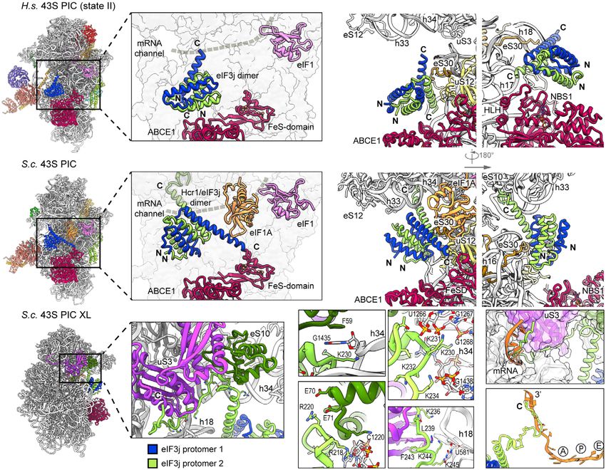
Kratzat et al The EMBO Journal
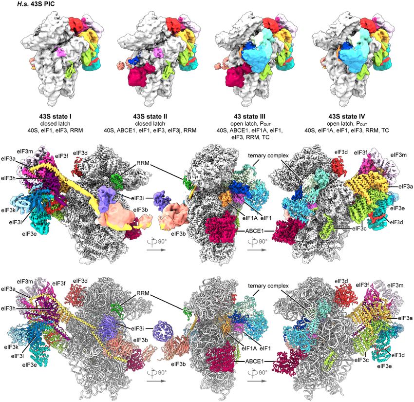
ribosomal subunits (SSU). We first asked if substantial amounts of associated 40S subunits. The majority of these complexes (62%)
ABCE1 are associated with initiation factor-bound 40S under native contained ABCE1, and the most interesting classes consisted of 43S
conditions. To that end, lysates from a yeast strain (S.c.) containing particles containing ABCE1, eIF3, eIF1, eIF1A, and eIF3j on the 40S
TAP-tagged ABCE1 (Rli1) were subjected to density gradient (Aylett et al, 2015; Heuer et al, 2017). The mRNA path (latch) was
centrifugation followed by Western blotting of fractions (Fig EV1A). in the closed conformation (Passmore et al, 2007), and at the mRNA
In agreement with previous studies (Andersen & Leevers, 2007; entry, we found a density for a typical RNA recognition motif
Pisarev et al, 2010; Pisareva et al, 2011), we observed that ABCE1 (RRM) (see below). Importantly, in these classes we observed an
was especially enriched on 40S and 80S ribosomes. We further interaction between the FeSD of ABCE1 and eIF3j (Fig 1D). More-
performed affinity purification from the lysates under varying buffer over, we found one class of particles with mRNA bound, apparently
conditions but without any stabilizing non-hydrolyzable ATP or representing a partial 48S IC complex. It contained eIF3, eIF1, tRNAi
GTP analogs, and analyzed the elution fractions by quantitative in the PIN conformation, as well as the N-terminal domain (NTD) of
mass spectrometry (LC-MS/MS) (Figs 1A and EV1B and C). We eIF5 as observed before (Llacer et al, 2018), and, to our surprise,
found that the expected SSU proteins but also eIF3 core components also ABCE1 (Fig 1E). The classes representing 43S PIC and 48S IC
and especially eIF3j (Hcr1) were enriched by ABCE1 affinity purifi- were refined to a resolution of 5.3 and 6.2 A, respectively, allowing
cation, indicating that both proteins were indeed integral compo- us to fit molecular models of existing structures as rigid bodies (Fig
nents of native pre-initiation complexes. Because of this finding and 1D and E, Appendix Fig S3, Appendix Table S1).
since eIF3j was implicated in ABCE1-dependent ribosome splitting In the human sample, we also found 40S subunits associated
in vivo (Young & Guydosh, 2019), we tested if eIF3j together with with initiation factors, similar to the yeast sample. After classifi-
ABCE1 had a direct impact on ribosome splitting in a reconstituted cation, four major stable eIF3-containing classes could be obtained
system. To this end, we performed in vitro splitting assays in yeast (Fig 2A). The 40S in State I resembled the state of an empty 40S
and tested if eIF3j can play a stimulatory role. Purified 80S ribo- subunit with a closed latch (Heuer et al, 2017; Passmore et al,
somes were incubated with the purified splitting factors Dom34, 2007), and only the core eIF3 subunits and weakly bound eIF1 were
Hbs1, Rli1 (ABCE1), eIF6 to prevent re-association of ribosomal found. State II had a similar conformation, and we found extra
subunits, ATP and GTP as well as different amounts of eIF3j. Split- densities in the ISS for eIF1, eIF3j, and ABCE1. State III additionally
ting efficiency was assessed from sucrose density gradient UV pro- contained eIF1A and the ternary eIF2-GTP-tRNAi complex (TC) in
files by monitoring 80S versus ribosomal subunit amounts (Figs 1B the open POUT conformation (Llacer et al, 2015), whereas State IV
and C, and EV1D). Indeed, we observed that an addition of eIF3j in was similar to State III but lacked ABCE1. Notably, in contrast to the
molar excess increased the ratio of split subunits to 80S when yeast sample, we did not find any 48S classes containing mRNA.
compared to a reaction containing the splitting factors only (Fig 1C). Thus, our human sample mainly represented 43S post-splitting or
Increasing amounts of eIF3j resulted in higher splitting activity. pre-initiation complexes prior to mRNA recruitment.
However, eIF3j alone did not exhibit any activity (Fig EV1E). In Independent focused classification and multi-body refinements
addition, we found that eIF3j and substoichiometric amounts of focusing on individual sub-complexes (Fig EV2 and Appendix Fig
ABCE1 remained bound to the 40S after splitting (Fig EV1F). To S2) enabled us to obtain molecular resolution for large parts of the
further confirm that eIF3j can still be associated with the 40S-ABCE1 human 43S sub-complexes. Therefore, we were able to build models
complex after splitting, we employed the “facilitated splitting” assay for the octameric eIF3 PCI-MPN core at the backside of the 40S,
as described before (Heuer et al, 2017). In this assay, ribosomes are parts of the YLC at the mRNA entry site and most factors located in
allowed to dissociate under splitting-promoting conditions (low the ISS, including ABCE1, eIF3j, eIF1 (including the N-terminal tail),
Mg2+ and high salt) and in the presence of putative subunit-binding eIF1A, the full eIF2 TC, and the eIF3c N-terminal domain, thus
factors (see Materials and Methods). Indeed, in this assay we resulting in a near-complete molecular model of the human 43S
observed that eIF3j remained on the 40S SSU together with ABCE1, particle bound to ABCE1 (Fig 2B and C, Appendix Table S2).
confirming that the two factors remain together on the 40S for
downstream events such as initiation after collaborating during Conformation of ABCE1-bound 40S-initiation complexes
splitting (Fig EV1G and H).
To gain further insights into the composition of native small Strikingly, we observed ABCE1 associated with 40S subunits during
subunits in yeast and human cells, we adopted a shotgun cryo-EM all stages of 43S PIC assembly in humans and even with 48S IC
approach. Yeast SSU complexes were obtained after harvesting the complexes in the yeast sample. In all complexes, the FeSD of ABCE1
crude 43S/48S peak from a preparative sucrose density gradient of was in the extended conformation packed against h44, and the
yeast cell lysate that was not further treated or stabilized with a ATPase body occupied the universal translation factor binding site
non-hydrolyzable nucleotide analog. Similarly, human native 40S on the 40S, which is highly similar to previous observations of non-
was obtained from untreated lysates of HEK Flp-In 293 T-Rex cells native complexes (h8-h14 junction; h5-h15 junction) (Heuer et al,
after serendipitous non-specific enrichment on sepharose material 2017; Mancera-Martinez et al, 2017; N€ urenberg-Goloub et al, 2020)
during unrelated affinity pullouts (see Materials and Methods). Of (Fig 3A). Here, the 40S subunit is engaged in a very similar way as
these samples, large enough cryo-EM data sets were collected in in the archaeal 30S-ABCE1 structure (N€ urenberg-Goloub et al, 2020)
order to analyze their complex composition by extensive 3D classifi- via the ABCE1-specific helix-loop-helix (HLH) domain and the open
cation (Appendix Figs S1 and S2). conformation with respect to the composite hinge regions (h1 and
In the yeast data set, as expected, the selected particles contained h2). Surprisingly, however, in all structures we observed the
pre-initiation complexes, which could be further classified into ATPase in a novel state that has not yet been described for ABC-type
defined states varying in composition and conformation of eIF- ATPases (Figs 3B, C and D, and EV3A): Compared to the closed
ª 2020 The Authors The EMBO Journal 40: e105179 | 2021 3 of 24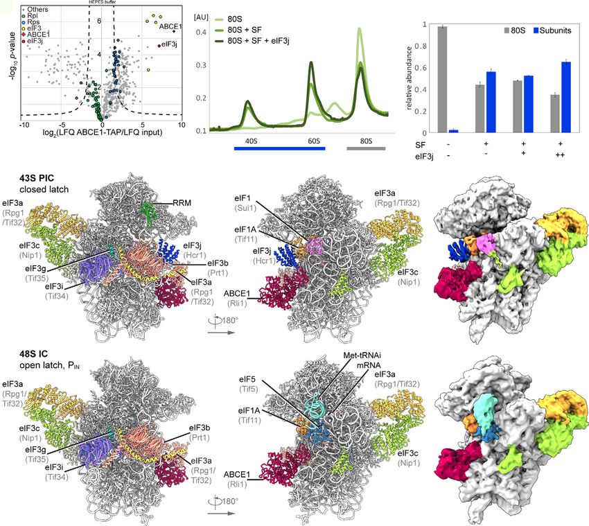
The EMBO Journal                                                                                                                                                  Kratzat et al

 A                                                       B                                                              C

 D

 E

  Figure 1. Biochemical analysis and cryo-EM structures of yeast ABCE1-containing initiation complexes.
  A    Volcano plot representing the statistical analysis of the fold enrichment of proteins after affinity purification in HEPES buffer of ABCE1-TAP followed by label-free
       quantification (LFQ) using liquid chromatography–tandem mass spectrometry (LC-MS/MS). Proteins above the curved lines show a statistically significant
       enrichment according to the t-test value.
  B, C Sucrose density gradient UV profile after in vitro splitting assays (B) and relative abundance of 80S and subunits as calculated from triplicates and displayed as
       mean  SD. (C); SF = splitting factors including Dom34, Hbs1, ABCE1, and eIF6; (+) = 4-fold molar excess of eIF3j; (++) = 20-fold molar excess of eIF3j.
  D, E Cryo-EM maps low-pass filtered at 6  A and models of the yeast subclasses representing an ABCE1- and eIF3j-containing 43S PIC (D) and an ABCE1- and eIF5-
       containing partial 48S IC (E).
  Source data are available online for this figure.

conformation as observed in in vitro reconstituted 30S and 40S PSCs                        was obtained after focused classification on ABCE1, we unambigu-
(Heuer et al, 2017; N€urenberg-Goloub et al, 2020), we found that                          ously identified an Mg2+-ATP (Fig 3E) occluded in NBSII, similar to
only NBSII is closed whereas NBSI adopts a half-open conformation                          the archaeal 30S-ABCE1 structure with Mg2+-AMP-PNP (N€    urenberg-
comparable to the one observed in several 80S pre-splitting                                Goloub et al, 2020). In the human structure, residues of the typical
complexes (Fig 3B) (Becker et al, 2012; Brown et al, 2015; Preis                           conserved motifs of ABC-type ATPases are involved: Lys386 of the
et al, 2014). When analyzing our best-resolved human map, which                            Walker A, Gly220 of the NBD1-Signature loop, and His521 of H-loop

4 of 24       The EMBO Journal 40: e105179 | 2021                                                                                                           ª 2020 The Authors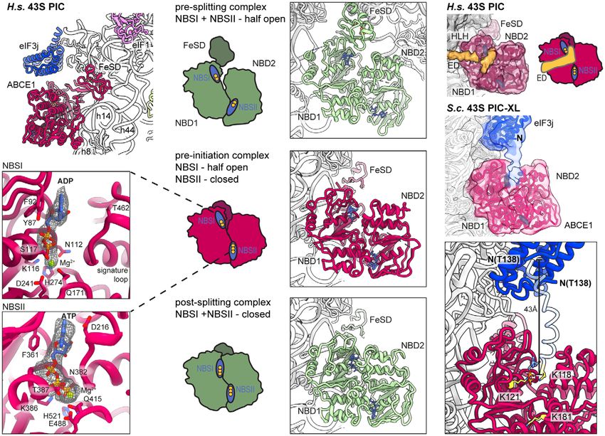
Kratzat et al                                                                                                                   The EMBO Journal

contact the γ-phosphate, and the Mg2+ ion is coordinated by Thr387        location previously described in low-resolution maps (Aylett et al,
(Walker A) and Gln415 (Q-loop). In contrast, for NBSI we observed         2015) (Fig 4). The main difference between the maps was the
Mg2+-ADP bound exactly as observed in the crystal structures of           absence (human) or presence (yeast) of eIF1A. However, apart from
open archaeal ABCE1 (Barthelme et al, 2011; Karcher et al, 2008):         a small rotation around the neck (approx. 3°), we did not observe
Y87 of the A-loop stacks on the adenine base, F92 on the ribose, the      significant conformational changes in the 40S when comparing the
Walker A-loop (Asn112-Ser117) binds the α- and β-phosphates, and          two structures.
the Mg2+ ion is coordinated by the β-phosphate, Ser117, Gln171 (Q-            In the low-pass-filtered human State II, which lacks eIF1A, we
loop) and Asp241, Glu242 (Walker B). Importantly, the signature           identified the eIF3j 6-helix bundle located above the ABCE1
loop of NBD2 (Leu463-Glu467), which occludes ATP in the catalyti-         ATPase body and in close vicinity to NBD1 (Fig 4A and B), but no
cally active closed state, is moved by 3.5     A away from NBD1. In      direct contacts were formed with ABCE1. On the 40S, eIF3j
conclusion, our data suggest that—in contrast to the nucleotide-          contacted the N-terminal tail of eS30 (protomer 1) and the C-termi-
occluded state observed in vitro—in native SSU-ABCE1 complexes,           nus of uS12 (protomer 2). The C-terminal helix of protomer 2
ATP hydrolysis in NBSI has already occurred, whereas NBSII is still       further projects toward the three-way junction formed by h32, h33,
inhibited.                                                                and h34 at the 40S head, whereas in protomer 1 it points toward
    As an additional difference to previous structures, we observed a     h17 and the HLH of ABCE1 (Figs 4B and EV3E). In this position,
rod-like extra density (ED) after low-pass filtering in all native 43S    the N-termini of eIF3j are located above the ABCE1 ATPase body
PIC structures, protruding from h17 of the 40S body via the HLH           close to the NBD1-NBD2 cleft.
motif into the cleft between NBD1 and NBD2 of ABCE1 (Fig 3F).                 In the yeast 43S PIC, in which eIF1A was present, we found eIF3j
However, local resolution in both human and yeast samples was too         in a similar position, but different conformation compared to the
low to identify this factor. To stabilize this assembly, we generated a   human structure (Fig 4C and D). Here, the 6-helix bundle is stably
chemically crosslinked yeast initiation complex sample derived from       anchored between the 40S beak at rRNA h33 on one side and the
a strain harboring TAP-tagged eIF3c (Nip1) and performed a cryo-EM        40S body near the ABCE1 FeSD and eIF1A on the other side. The
analysis focused on the ABCE1 and the adjacent eIF3j (Appendix Figs       two sides of the anchor are formed again by the C-terminal helices
S1B and S4). Indeed, in this reconstruction, we clearly observed an       of eIF3j: protomer 2 contacts eS30 at a similar site as in the human
extra density protruding from eIF3j into the composite NBSs of            structure but now the entire helix bundle was rotated by approxi-
ABCE1. At a resolution of 3.0   A, we built the model for yeast eIF3j    mately 100 degrees (Fig EV3B, C, and D). Consequently, the tip of
(Fig 3G and 4, Appendix Fig S4) based on the human eIF3j dimer (un-       the protomer 2 C-terminal helix now pointed toward the 40S head,
published; PDB 3BPJ; lacking 137 residues at the N-terminus and 28        whereas the C-terminal helix of protomer 1 projected toward the
residues at the C-terminus). In brief, this dimer folds into a stable     ABCE1-FeSD, thereby passing along eIF1A (Figs 4D and EV3F).
entangled 6-helix bundle that is arranged such that the N-termini are     Molecular details of the eIF3j-40S interaction were derived from the
in close vicinity. Yet, the C-termini face into opposite directions,      high-resolution structure of the crosslinked 43S-PIC (Fig 4E). In
whereby the C-terminal tail of one protomer reaches into the mRNA         brief, the 6-helix bundle accommodates between the 40S body and
entry channel (see below). On this basis, we could assign the extra       head via interactions of both protomers. The body is contacted by
density in ABCE1 as a part of the eIF3j N-terminus. This assignment       the first and third helix of protomer 2 (to the h17-h18 junction and
was further confirmed by protein crosslinking coupled with mass           eS30) mainly by basic residues. The third helix projects toward the
spectrometry (XL-MS) using a lysine-specific BS2G crosslinker             beak to contact the phosphate backbone of h33 (G1264). Following
(Appendix Fig S5, Appendix Table S3). Two crosslinks between the          this helix, the ultimate eIF3j C-terminus forms a loop inside a pocket
Lys118 of eIF3j with Lys121 and Lys181 of ABCE1, both located near        formed by h33, h34, and eS10 and from there runs along h18 and
the ATP-binding site of NBD1, were identified (Fig 3G). In this posi-     uS3, parallel to the latch, to position the ultimate C-terminal tail
tion, the eIF3j N-terminus may easily modulate the ATPase activity of     inside the mRNA entry channel (Figs 4F, EV3G, and H; for a detailed
ABCE1 by restricting further movements of the HLH or the two NBDs         description of molecular contacts see Appendix Text 1). In this posi-
with respect to each other. Interestingly, the position of the eIF3j-     tion, eIF3j directly overlaps with the mRNA path and would possi-
NTD on ABCE1 is similar to the one observed in a recent structure of      bly interfere with mRNA loading during 48S-IC formation (Fig 4G).
archaeal ABCE1 co-crystallized with an 18-mer fragment from the C-            Taken together, our structural data explain how eIF3j could exert
terminus of the archaeal 50S stalk protein aP1 (Imai et al, 2018). This   its functions during key steps of translation initiation in conjunction
suggests that ABCE1 possesses a multivalent interaction patch in this     with eIF1A.
region, which would allow for regulation of its ATPase activity. The
observed stabilization of ABCE1 in the half-open conformation with        Molecular architecture of the PCI-MPN core and
one ADP still bound in NBS1 may indicate an inhibition of ADP             its interactions with 40S
release, which would explain its rather stable association with the
40S subunit.                                                              State I of the human sample represented a stable class with mainly
                                                                          eIF3 and weak density for eIF1 bound to the 40S SSU. This appears
Conformation of eIF3j in human and yeast                                  plausible when considering that eIF3 activity during termination
40S-initiation complexes                                                  and ribosome recycling has been proposed (Beznoskova et al, 2013;
                                                                          Pisarev et al, 2007; Valasek et al, 2017), which further indicates that
As described above, we found yeast and human 43S PIC sub-popula-          eIF3 can already bind the 40S before eIF1A comes into play. The
tions concomitantly bound to ABCE1 and eIF3j. The eIF3j subunit           lack of ABCE1 in this complex may be a result of fast dissociation
was positioned on the intersubunit side, roughly resembling the           after splitting or of an alternative splitting mechanism. In any case,

ª 2020 The Authors                                                                                   The EMBO Journal 40: e105179 | 2021   5 of 24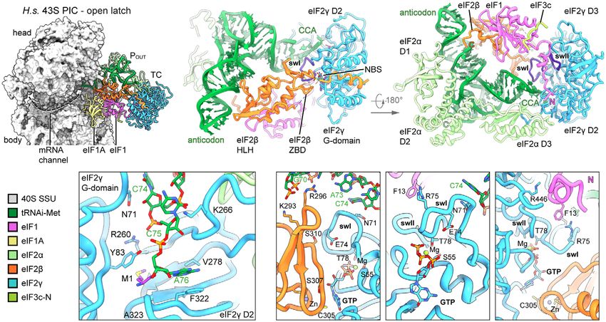
The EMBO Journal                                                                                                                                          Kratzat et al

 A

 B

 C

  Figure 2. Cryo-EM structures of human 43S PICs in different assembly states.
  A Overview of four selected compositional states present in the human 43S PIC data set.
  B Composite map of the complete human 43S PIC after focused and multi-body refinements on individual sub-complexes, filtered at local resolution.
  C Composite model of the complete human 43S PIC, as represented by state III.

after accommodation of eIF1 and eIF1A, the eIF2 TC binds to the                       by multi-body refinement of State I and State II particles. The struc-
43S to induce the POUT conformation (State III-IV). Here, the                         ture assembles into β-sheets with the shape of an arc formed by PCI
improved resolution allowed us to describe the interaction network                    domains of eIF3 subunits a, c, e, l, k, and m. The arc wraps around
of these factors at unprecedented molecular detail.                                   a seven-helix bundle formed by the C-terminal helices of subunits c,
    The PCI-MPN core is located at the backside of the 40S as                         e, f, h, k, and l (Figs 5A and EV4A), resulting in the typical five-
observed before (des Georges et al, 2015; Hashem et al, 2013;                         lobed structure (left and right arm, left and right leg and head),
Srivastava et al, 1992), and high resolution of the core was obtained                 which was visualized at a local resolution of 3.4 A (left arm, head

6 of 24      The EMBO Journal 40: e105179 | 2021                                                                                                      ª 2020 The Authors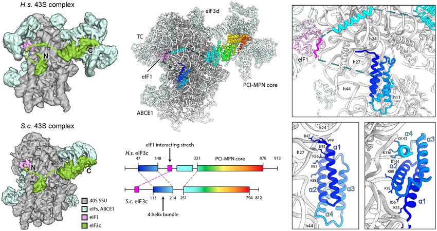
Kratzat et al                                                                                                                                       The EMBO Journal

  A                                              B                                                                       F

                                                                                                                         G

                                                 C
  E

                                                 D

  Figure 3. Conformation of ABCE1 in native 40S initiation complexes.
  A   Overall position of ABCE1 in 40S initiation complexes, here representatively shown for the human State II with eIF3j.
  B–D Schematic representation and structure of semi-open ABCE1 as in 80S-pre-splitting complexes (Brown et al, 2015, PDB 3JAH) (B), hybrid semi-open/closed ABCE1 as
                                                                                                                            €renberg-Goloub et al, 2020, PDB 6TMF) (D).
      in native 40S initiation complexes (C) and fully closed ABCE1 as in in vitro reconstituted post-splitting complexes (Nu
      Nucleotide-binding sites colored in blue and bound nucleotide indicated with yellow circles (one circle per phosphate group).
  E   Zoom into NBSI and NBSII showing bound Mg2+-ADP (in NBSI) and Mg2+-ATP (in NBSII) fit in density as obtained after focused classification on ABCE1 and
      refinement.
  F   View focusing on the NBDs and the unassigned extra density (ED) reaching from the 40S via the HLH into NBSI. The ABCE1 map was low-pass filtered at 6      A.
      Schematic representation highlighting the position of the ED with respect to the NBSs.
  G   Upper panel: Position of eIF3j and ABCE1 in the crosslinked yeast 43S-PIC (43S-PIC-XL) sample. View focusing on the ABCE1-eIF3j interaction (same view as (F)): An
      extra density attributing to the eIF3j N-terminal region is connecting the eIF3j 6-helix bundle with NBSI of ABCE1. The map was low-pass filtered at 8
                                                                                                                                                             A. Lower
      panel: N-terminally extended model of eIF3j (transparent blue) highlighting the position of K118, which was found crosslinked to K121 and K181 of ABCE1 (atoms
      colored in yellow).

and right arm) and 3.8   A (left and right leg) (Fig EV2C). This                      interactions). A second contact site was established between Glu17,
allowed for an almost complete molecular interpretation (Fig EV4A,                     Phe18, and Val21 of eIF3a and the eS1 Pro190 as well as adjacent resi-
Appendix Table S4), thus refining previous low-resolution models                       dues. The loop H1-H2 of eIF3c (residues 340-345) interacts with rRNA
(des Georges et al, 2015; Eliseev et al, 2018; Erzberger et al, 2014),                 h22 (G929, C930) and multiple sites at the Zn-knuckle domain of eS27
for example, by correcting the register of helices and extending                       (Figs 5C and EV4B). Furthermore, the β-sheet insert between PCI
molecular models (Appendix Fig S6).                                                    helices 4 and 5 (residues 417-441) of eIF3c forms interactions with
    The main anchor of the eIF3 PCI-MPN core to the 40S is provided                    uS15, and basic residues in the PCI loops of both eIF3a and eIF3c are
by the eIF3a and eIF3c subunits, which form the “head” and the “right                  positioned to interact with the flexible tip of rRNA ES7 (Fig 5B).
arm” of the PCI-MPN core, respectively. eIF3a contacts eS1 via its N-                     An additional anchor of the eIF3 PCI-MPN to the 40S is provided
terminal PCI helix H1 and the loop between H1 and H2. Here, Arg14                      by the N-terminus of eIF3d (from A2 to D84) (Figs 5C and D, and
forms salt bridges to Glu78 and Asp77 of eS1 (Fig 5B and C, see                        EV4C). Interestingly, we found that it meanders along the PCI helices
Appendix Table S4 for an inventory of observed molecular                               1 to 3, 7, 9, 10, and 12 of eIF3e (left arm) and bridges eIF3e with

ª 2020 The Authors                                                                                                     The EMBO Journal 40: e105179 | 2021       7 of 24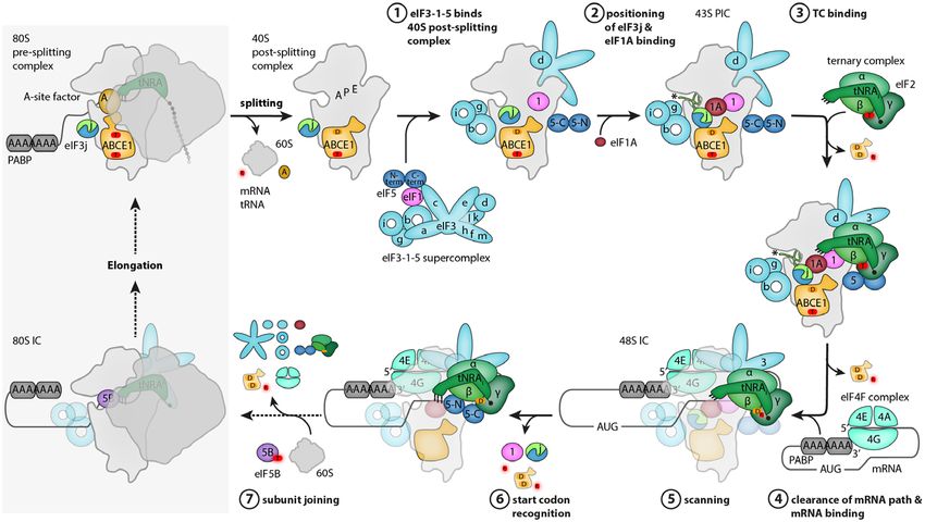
The EMBO Journal                                                                                                                                                 Kratzat et al

  A                                                                                            B

  C                                                                                            D

  E                                                                                F                                                     G

  Figure 4. Two conformations of eIF3j in human and yeast 43S PICs.
  A Overview and zoomed view on the model of human 43S PIC II (lacking eIF1A), focusing on the two protomers of the dimeric eIF3j 6-helix bundle in the ISS. eIF3j is in
    close vicinity to NBD1 of ABCE1 but only forms contacts to the 40S. The mRNA channel is indicated by a dashed gray line.
  B Two different views showing the interaction of the two Homo sapiens (H.s.) eIF3j protomers with the 40S.
  C Same views as in (A) on the model of the yeast 43S PIC. Here, eIF3j (Hcr1) is turned approximately 100 degrees around a pivot formed by the C-terminal helices
    contacting eS30 and uS12. Protomer 1 thereby contacts eIF1A and the FeSD of ABCE1 and protomer 2 contacts h33.
  D Two different views showing the interaction of the two S.c. eIF3j protomers with the 40S and ABCE1.
  E Overview and zoomed view highlighting the position of the eIF3j C-terminus in the yeast 43S-PIC-XL structure.
  F Zoomed views focusing on interactions of the eIF3j C-terminus with the 40S. The loop following the third helix of eIF3j protomer 2 is in a pocket formed by the 40S
    h33, h34, and eS10. Lys230 of eIF3j C-terminus (protomer 2) and Phe59 of eS10 are sandwiching the flipped-out G1435 base of h34 (upper left); Lys231 and Lys234
    interact with h33 (U1266 and G1267) and h34 (G1438) (upper right); salt bridges between Arg220 and Glu70-Glu71 of eS10 further stabilize the loop (lower left).
    Following the loop, the eIF3j C-terminus bridges the 40S body and head in the latch and contacts are formed with h18 (via Lys236) and via hydrophobic interactions
    with uS3 (lower right). See Appendix Text 1 for more molecular details.
  G Position of the ultimate eIF3j C-terminus in the mRNA entry tunnel (upper panel) and steric clash with mRNA as positioned in an 80S ribosome stalled during
    translation (PDB 5MC6); for clarity, in the lower panel only eIF3j and mRNA are shown, A/P/E, respectively indicate the positions of aminoacyl, peptidyl, and exit site in
    the 80S ribosome.

eIF3c (head) by interacting with PCI helices 12, 14, and 16 (eIF3e)                       eS27 is established via its Zn knuckle, where Phe80 of eIF3d is sand-
and PCI helix 11 (eIF3c). Another specific contact between eIF3c and                      wiched between the side chains R80 and K36 of eS27.
eIF3e is formed by stacking of Y286 (eIF3e) to Y583 (eIF3c). More-                           Taken together, the PCI-MPN core of eIF3 establishes a multi-
over, eIF3d also interacts with PCI helices 10, 13, and 14 of eIF3c by                    modal molecular interaction pattern with the 40S involving the
forming a large loop, which is anchored by the conserved Trp45 (in-                       eIF3a, c, and d subunits, which display an unexpected degree of
teractions to Pro603, Ile607, and Glu666 of eIF3c). The interaction to                    inter-connectivity.

8 of 24       The EMBO Journal 40: e105179 | 2021                                                                                                           ª 2020 The Authors
Kratzat et al The EMBO Journal
Structure and location of the peripheral subunits density for the eIF3c-NTD was present at the eIF1 loop between
helix α 1 and helix α 2 (Asp53-Lys58) as well as Ile100 and Gly101
The peripheral subunits, which consist of the YLC, the eIF3c-NTD, of α 2 (Fig EV5D). This observation is highly consistent with the
and in humans the eIF3d cap-binding protein domain, are connected NMR study, in which the same interacting region on eIF1 is identi-
to the PCI-MPN scaffold via flexible linkers. While eIF3a connects fied for the eIF3c-NTD of yeast. Together, these observations lead us
via its CTD to the YLC module located close to the mRNA entry site, to the conclusion that the density observed near eIF1 in the human
the N-terminus of eIF3c protrudes from the mRNA exit toward the structure corresponds to this insertion C-terminal of the helix
ISS, where it interacts directly with eIF1. While the N-terminus of bundle, fulfilling an analogous role to the previously characterized
eIF3d as an integral part of the PCI-MPN core is anchored to the 40S N-terminal stretch of eIF3c in yeast.
body, the cap-binding protein domain of eIF3d is located on the 40S From local classification, we also obtained one class with strong
head close to the mRNA exit site as observed before (Eliseev et al, density for the YLC module including the eIF3a-linker that connects
2018). Here, it contacts the 40S SSU via its highly conserved helix it to the PCI-MPN core (Appendix Fig S7). In brief, the YLC module
α10 (Lee et al, 2016) that packs upon eS28 via Gln416, Thr423, and contains two β-propellers: the 7-bladed WD40 repeat of eIF3i and
Lys426 and reaches into the interface between eS28 and uS7, where the 9-bladed WD40 repeat near the C-terminus of eIF3b. The two
Gln416 stacks on Arg51 (eS28), which in turn stacks on Phe61 propellers are held together by the C-terminal helical domain of
(uS7). The eIF3d helix α12 lies on top of uS7 and forms contacts via eIF3b, which is formed by 3 α-helices: the most C-terminal one
Lys472, Glu475, Ser478, and Gln479. Notably, since eIF3d is bridg- binds to eIF3i, while the two preceding α-helices are bracketing the
ing the 40S head with the eIF3 PCI-MPN core anchored to the 40S eIF3a C-terminus against the eIF3b β-propeller (des Georges et al,
body, it could serve to relay conformational rearrangements of the 2015; Herrmannova et al, 2012). N-terminal of its β-propeller, eIF3b
40S head—as occurring during the assembly of 43S and 48S contains a noncanonical RNA recognition motif (RRM) (ElAntak
complexes—to the PCI-MPN core or, vice versa, allow the eIF3 et al, 2007) that can form further interactions with the eIF3a-CTD
complex to directly control the conformational state of the 40S head (Dong et al, 2013; Khoshnevis et al, 2014; Valasek et al, 2002;
(Figs 5D and EV4C and D). Valasek et al, 2001) as well as the N-terminus of eIF3j (Elantak
For eIF3c, only a part of its NTD could be located on the ISS of et al, 2010; Valasek et al, 2001).
the 40S so far, where it forms a helix bundle (Llacer et al, 2015). We For the CTD of eIF3a, we could build a long α-helix (residues
found a particularly stable arrangement of the eIF3c NTD in classes 602-743) into the elongated rod-like density protruding from the
containing the eIF2 TC and, after multi-body refinement, local reso- PCI-MPN core to contact uS2 and eS21 (Appendix Fig S7A). This
lution of 3 to 4
A (Figs EV2B, EV5A and B) allowed us to determine helix extends further toward the YLC where it forms a hinge-like
the register of the four eIF3c-NTD helices (Val47 to Y149) (Fig 6). A structure and then connects to the stretch of the eIF3a helix that is
stretch preceding the first helix (47-51) contacts h24 and h27 via bound to the eIF3b β-propeller. It thereby contacts the tip of the
R47 to the backbone phosphate of C1039 and the 2’-OH of A1181. otherwise flexible rRNA expansion segment ES6C, which in turn
The peptide bond of Val49 of eIF3c stacks on base C1180, which is contacts the loop between the first two helices of the eIF3b helical
also contacted by the first helix (52-74) of the bundle. Here, the two domain. In this arrangement, the eIF3b WD40 is rigidly confined
charged residues K55 and R56 interact with the backbone of rRNA between rRNA h16 and uS4 on one side, and ES6C on the other side,
G1179 and C1180. Backbone–phosphate interactions were also and is thus well resolved in the proximity of the 40S (Appendix Fig
formed by the second helix (76-92) to rRNA h11 (A364) and h27 S7B, Appendix Table S4). The eIF3i-eIF3g complex and the eIF3b-
(U1178), by the fourth helix (136-143) to rRNA h11 via K136 (to RRM, however, remained rather flexible as observed before
U367), and finally by the peptide bond of Thr140 stacking upon the (Erzberger et al, 2014). Nonetheless, we observed a stabilization of
U367 base, as well as Gln143 hydrogen bonding to U367. Additional the eIF3b-RRM in ABCE1- and eIF3j-containing classes, possibly due
but less rigid contacts were established by the K-rich loop between to an interaction of the eIF3b-RRM with the eIF3j N-terminus (Elan-
helix 3 and helix 4 of eIF3c (Figs 6E and F, and EV5A and B, tak et al, 2010; Valasek et al, 2001).
Appendix Table S4). In yeast, the positioning of the YLC module at the mRNA exit
Notably, when low-pass filtered, a rod-like extra density for the was the same, because here it was also held in place by ES6C
eIF3c N-terminus became apparent, bridging the 4-helix bundle with (Appendix Fig S7C). However, in the majority of particles in the
eIF1 near rRNA h23 and h24. This density was neither present in yeast dataset (approximately 85%), we could observe a conforma-
our nor in other (Llacer et al, 2015) yeast 43S/48S reconstructions, tional change in the eIF3i-eIF3g module relative to the ES6 anchor
where the four-helix bundle was directly connected to the eIF3c core point. Especially in the eIF3j-containing 43S class, the eIF3i-eIF3g
moiety, and a site N-terminal of this region interacted with eIF1 (Fig entity rotates by approximately 120 degrees away from the mRNA
6A and B). Sequence alignments of the yeast and human eIF3c N- entry toward ES6C and ES6B. The loop preceding the eIF3i-contact-
termini revealed an insertion on the C-terminal side of the ing helix of eIF3b (Thr697-Asp701) appears to serve as a hinge for
conserved four-helix bundle in humans (Figs 6C and D, EV5C). This this rotation (Appendix Fig S7D).
insertion from residue 165 to 213 displays 32.0% sequence identity Apart from the YLC, we observed an additional density near the
and 56.0% sequence similarity with a stretch at the N-terminus of mRNA entry at the tip of h16 in all of our 43S structures, which was
yeast (42-92), which was previously shown to be involved in the previously assigned to the RRM of eIF4B (Eliseev et al, 2018)
interaction of eIF3c with eIF1 by NMR studies (Obayashi et al, (Appendix Fig S8). This density is especially prominent in
2017). Here, chemical shift perturbation after eIF1 binding is subclasses of the human dataset lacking the TC, in which we could
observed for Glu51, Ala67, and a stretch between Lys68 and Lys77. unambiguously identify the typical RRM fold at a local resolution
Moreover, in our human complex one stretch of well-resolved around 4 A (Appendix Figs S8C and D). Notably, besides eIF4B, the
ª 2020 The Authors The EMBO Journal 40: e105179 | 2021 9 of 24The EMBO Journal Kratzat et al
A
B
C
D
Figure 5. Molecular interactions of the human PCI-MPN core of eIF3 in the 43S PIC.
A Isolated map and molecular model of the eIF3 PCI-MPN core color coded as in Fig 2. Structural hallmarks are indicated, and a scheme shows the composition of
the lobes.
B, C Interactions of eIF3a, eIF3c, and eIF3d with the ribosome: (B) shows an overview of the structure and zoomed views highlighting the interactions of eIF3a, eIF3c, the
eIF3d N-terminal tail and the eIF3d cap-binding domain with the 40S, (C) shows molecular details of eIF3a interacting with eS1; eIF3c interacting with rRNA h22
and eIF3c and the N-terminal tail of eIF3d with the Zn-knuckle domain of eS27.
D Interactions of the eIF3d N-terminal tail with the PCI-MPN core.
10 of 24 The EMBO Journal 40: e105179 | 2021 ª 2020 The AuthorsKratzat et al The EMBO Journal
largely flexible eIF3g subunit is a potential candidate for this density further missing parts of eIF3g, eIF4B, the C-terminus of eIF3j as
because it also contains an RRM, which shares very high structural observed in yeast maps, the CTD of eIF3a, or the ribosome hiberna-
and sequence similarity (50.0%) to eIF4B (Appendix Fig S8D and tion factor SERBP1 (Stm1 in yeast) (Anger et al, 2013; Ben-Shem
E), and it was crosslinked to the nearby proteins uS10 and uS3 et al, 2011; Brown et al, 2018). In any case, it is apparent that
(Cuchalova et al, 2010). Unfortunately, at the current resolution we accommodation of mRNA in the 48S IC complex would require its
cannot unambiguously distinguish these two RRMs in our maps and relocation, which may allow for allosteric communication between
it is possible that both compete for the same binding site. Next to the different eIFs.
this domain, we observed density reaching from the RRM into the
mRNA channel in all human early 43S PIC structures with a closed Conformation of the ternary complex
latch (Appendix Fig S8A and B). Close to the RRM, this density
forms a loop that shows multiple contacts to uS3 before winding After analyzing the eIF3 complex, we also gained molecular infor-
along uS3 toward the mRNA channel. Within the channel, one side mation on the human eIF2 TC by focused classification. The TC as
chain can clearly be identified as a tryptophan facing toward uS3 well as eIF1 and eIF1A were observed on the intersubunit side in a
(contacting Lys148 and Met150) and further interacting with uS3 similar overall position and conformation as described before for
Leu142 and Val115. The stretch also contacts 18S rRNA G626, other ICs in POUT conformation at low resolution (PDB 6GSM, PDB
A628, and U630 of h18 as well as C1698 of h28, C1331, and A1489 3JAQ (Llacer et al, 2015)) (Appendix Fig S2). Briefly, eIF2 consists
of h34 (all in the A site). Thereby, this peptide stretch blocks the of three subunits, α, β, and γ: The eIF2γ subunit shares structural
entire mRNA channel down to the P site where it contacts the homology to EF-Tu-like translational GTPases (e.g., Schmitt et al,
flipped-out base C1701 at the tip of h44. Unfortunately, local resolu- 2002) and consists of a G-domain (domain I), including the regula-
tion in this region is insufficient to provide further molecular detail tory switch loops (swI and swII), followed by two β-barrel domains.
and clearly identify this entity, yet considerable candidates may be eIF2α consists of an N-terminal OB-fold domain, a central helical
A C E
B F
D
Figure 6. Arrangement of the eIF3c-NTD in human and yeast 43S PICs.
A Cryo-EM map obtained after focused sorting of the human 43S PIC on the TC: when low-pass filtered at 6 A, it shows the density of almost complete eIF3c-NTD in
the ISS.
B Cryo-EM map of the yeast 43S PIC low-pass filtered at 6 A.
C Model for human eIF3c in the TC-containing 43S colored in rainbow (C) and scheme of the alignment between human and yeast eIF3c sequences, colored accordingly
(D). The eIF1-interacting stretch present in the N-terminus of S.c. eIF3c shows 32.0/56.0% sequence identity/similarity with an insert C-terminal of the conserved 4-
helix bundle conserved in mammals.
D Zoomed view highlighting the position of the eIF3c NTD and eIF1 in the 40S ISS.
E Molecular model for the 4-helix bundle interacting with 40S rRNA and r-proteins.
F Molecular model for the 4-helix bundle interacting with 40S rRNA and r-proteins.
ª 2020 The Authors The EMBO Journal 40: e105179 | 2021 11 of 24The EMBO Journal Kratzat et al
domain, and a C-terminal α-β domain. The eIF2β subunit has an Taken together, we found the TC in a stable state within the 43S
unstructured N-terminal domain, followed by a central helix-turn- PIC, in an open conformation in the absence of mRNA. An intricate
helix (HTH) domain and C-terminal zinc binding domain (ZBD). In interaction framework is established by the 40S and eIF1 to accom-
solution, tRNAi was shown to be bound to the TC in a distinct way modate the GTP-bound eIF2-tRNAi in a rigid position. The switch
different to canonical tRNA-bound EF-Tu/eEF1A by employing addi- loops are kept in a rigid conformation stabilized by tRNAi, eIF2β,
tional composite interactions with both eIF2α and eIF2γ (Schmitt and the eIF1 N-terminal tail, and the GTPase pocket of eIF2γ is
et al, 2012). The eIF2β subunit, however, has never been sufficiently closed by eIF2β. This may prevent premature release of the bound
resolved to elucidate its molecular contribution to tRNAi binding nucleotide and, at the same time, may restrict access for eIF5-NTD
and 43S PIC formation. to avoid premature GAP activity.
In our structure, we found the tRNAi embraced by all three eIF2 Following TC assembly on 43S PIC and opening of the latch,
subunits (Fig 7A and B). Similar to the 5 A resolution crystal structure mRNA can be threaded into the mRNA binding site, followed by
(3V11 Schmitt et al, 2012), the methionylated CCA-end is sandwiched scanning for the first AUG codon by the 48S particle. While we do
between the GTPase domain and domain II of eIF2γ. The terminal not find scanning intermediates in either yeast or human datasets,
adenine base A76 is accommodated in a pocket formed by the β-sheets in our yeast native 40S population we find one state containing
of the eIF2γ domain II including Val278, Phe322, Gly340, and Arg260 eIF1A, tRNAi in the PIN state, and the eIF5-NTD instead of eIF1
(Fig 7C, Appendix Fig S9C). The 2’-OH group of the ribose moiety (yeast 43S PIC). Apart from weaker density for eIF2, this state is
interacts with the carbonyl group of Ala323 and the methionyl side similar to one observed before (Llacer et al, 2018), where it was
chain stacks on Tyr83 of eIF2γ G-domain. The CCA-end is further interpreted as a late state after start-codon recognition. However, to
stabilized by contacts including a cation-π stack of Lys266 on tRNAi our surprise we still find ABCE1 in this complex. This suggests that
C75 and Asn71 of the eIF2γ swI loop with tRNAi C74. Moreover, ABCE1 may play further roles even in later stages of initiation, or
Arg296 of the eIF2β ZBD intercalates into the major groove of the that its dissociation is not required at this stage.
acceptor-stem helix (G70; supported by Lys293 contacting the phos-
phate backbone of U69) (Fig 7D, Appendix Fig S9C). eIF2α contacts
the T- and D-loops mainly via its central helical domain whereas the Discussion
N-terminal OB-fold domain intercalates between anticodon stem and
uS7 in the E site on the head of the 40S. The central eIF2β HTH domain While the role of highly conserved ABCE1 during ribosome recy-
contacts the anticodon from the A site and thereby forms multiple cling has been studied in mechanistic details (Becker et al, 2012;
contacts to eIF1, also involving residues of the newly built C-terminus Nurenberg-Goloub et al, 2018; N€ urenberg-Goloub et al, 2020), its
(I314-R329), which stretches below the tRNAi anticodon stem toward role after 60S dissociation remained largely elusive. However, when
the E site and contacts C1057 of rRNA h24 (via N327). first characterized biochemically, ABCE1 was found associated with
Notably, in the GTP binding pocket of eIF2γ we clearly identified 43S/48S pre-initiation complexes in yeast, humans, and Drosophila
a Mg2+-GTP (Fig 7D). Ser55 of the conserved P-loop and Thr78 of (Andersen & Leevers, 2007; Chen et al, 2006; Dong et al, 2004).
swI coordinate the Mg2+-ion, whereas Asp134 and Pro135 of swII Since then, it is a long-standing question what the function of
likely contact the γ-phosphate. Compared to the crystal structure of ABCE1 in these complexes is. Our extensive single particle analysis
the archaeal TC (Schmitt et al, 2012), th, 2012 so that this citation of native small subunits from yeast and human cells captured a vari-
matches the Reference List. Please confirm that this is correct."-->e ety of states throughout the assembly of the 43S PIC prior to mRNA
guanine base is rotated by 90° and accommodated in a pocket loading, in which ABCE1 can stay associated with the 40S. Surpris-
between Asn190 and Ala226 of eIF2γ and Cys305 of the eIF2β ZBD, ingly, in yeast we even find ABCE1-48S complexes beyond the stage
which is tightly packed upon the nucleotide-binding pocket. of mRNA engagement and start-codon recognition as indicated by
Interestingly, both switch loops were embedded in a tight interac- the presence of the eIF5-NTD (Fig 8).
tion network involving interactions with tRNAi, eIF2β, and the eIF1 We further observe that in all ABCE1-containing 43S structures
N-terminal tail, which we built de novo. The N-terminal tail of eIF1 its NBDs are in an unusual hybrid conformation, where NBS2 is
protrudes from the 5-stranded β-sheet and binds to Arg446 of eIF2γ closed and NBS1 is semi-open. This is contrary to previous in vitro
domain III, where it forms a loop and projects toward Arg75 of eIF2γ studies showing SSU-associated ABCE1 in the ATP-occluded fully
swI, forming a cation-π stack with Phe13 (Fig 7D, Appendix Fig S9C closed state. Notably, the two NBSs in ABCE1 were shown to be
and D). Furthermore, the conformation of the swI loop was stabilized highly asymmetric and NBSII has a low ATP-turnover rate
by the tRNAi via Asn71 (see above) and an interaction between compared to NBSI (Gouridis et al, 2019; Nurenberg-Goloub et al,
conserved Ser310 of the ZBD of eIF2β with Glu74. 2018). Consistent with this behavior, we find Mg2+-ATP still bound
In close vicinity to the guanosine binding pocket, we find eIF2β in the closed NBSII, whereas Mg2+-ADP is present in NBSI. This is
Ser307, the equivalent of yeast eIF2β Ser264. In yeast, a Ser264Tyr in agreement with the most recent model for the ABCE1 ATPase
mutation causes the Sui- (suppressor of initiation codon) phenotype, cycle, in which closure of the NBSII was discussed to be the decisive
leading to increased utilization of UUG start codons (Huang et al, step for disassembly of 80S pre-splitting complexes, a process that is
1997). This mutation was shown to increase GTP hydrolysis rates then triggered by subsequent closure and ATP hydrolysis in NBS1.
and stabilize the closed PIN conformation of the 43S PIC (Martin- Subsequently, re-opening of NBSI would be expected on the small
Marcos et al, 2014). In the observed position, the tyrosine mutation subunit. But if ATP hydrolysis is prevented either by usage of a non-
of Ser307 could easily interfere with the bound nucleotide, for hydrolyzable ATP analog or by hydrolysis-deficient Walker B
example, by stacking on the guanine base, and thus alter the geome- mutants, ABCE1 can be trapped in the fully closed state on the small
try of the nucleotide-binding pocket. subunit under facilitated splitting conditions (Heuer et al, 2017;
12 of 24 The EMBO Journal 40: e105179 | 2021 ª 2020 The AuthorsKratzat et al The EMBO Journal
Kiosze-Becker et al, 2016; N€ urenberg-Goloub et al, 2020). In native mRNA channel near the entry site. This position explains, how eIF3j
ABCE1-associated complexes, however, NBSI is already in a more could exert its roles as an antagonist of mRNA binding, for example
open conformation and additionally obstructed by a part of the eIF3j by recycling of mRNA from the 40S subunit (Pisarev et al, 2007;
N-terminal domain, which intercalates between the two NBDs close Pisarev et al, 2010), or during initiation by preventing premature
to NBSI. Thus, eIF3j may keep NBSI from closing (after putative mRNA recruitment (Fraser et al, 2007). Notably, its position close to
binding of another ATP), or alternatively, prevents further opening eIF1A and thus near the A site may also explain its suggested role in
into a state as observed in free ABCE1. This brings up the question regulating start-site selection (Elantak et al, 2010). Moreover, the
of why ATP hydrolysis in NBSII, which would lead to dissociation comparison of yeast with the human structures of early 43S PICs
from the 40S SSU, is inhibited. We find NBSII in a very similar suggests that eIF3j and ABCE1 may be beneficial for binding of eIF1A.
conformation as in the fully closed archaeal structure (N€ urenberg- In the yeast conformation, eIF3j appears like a molecular ruler read-
Goloub et al, 2020), and the structure reveals no clues to explain ing out the exact distance between the post-splitting-specific FeSD
why ATP hydrolysis is slowed down. Thus, we speculate that a conformation of ABCE1 and the 40S head and beak conformation as
further and likely only small-scale allosteric signal into NBSII may adopted after eIF1A binding. Thus, it is tempting to speculate that the
be necessary for its activation. This may occur after dissociation of observed conformational change in eIF3j may play a role in priming
the eIF3j N-terminus upon further opening of NBSI and be accompa- the 40S for eIF1A binding and/or stabilizing the early closed-latch
nied by changes in the ABCE1-specific HLH and hinge regions. conformation of the 43S PIC when eIF1A is bound. Notably, eIF1A is
The observation that ABCE1 dissociation can apparently be the only factor that was not found to be pre-assembled in a 40S-free
actively prevented points toward a direct role in 43S PIC and even multi-factor complex (MFC) consisting of eIF1, eIF2-tRNAi-GTP, eIF3,
48S IC assembly, most likely in concert with eIF3j. We could corrobo- and eIF5 in yeast (Asano et al, 2000; Zeman et al, 2019), plants, and
rate the finding that eIF3j assists in ABCE1-dependent splitting by mammals (Sokabe et al, 2012). While eIF1A is capable of binding 40S
in vitro dissociation assays, and furthermore, we established that SSU independently and adopting a similar conformation as within the
eIF3j remains bound to the 40S together with ABCE1 after the split- context of initiation (Yu et al, 2009), it is possible that after binding
ting cycle. A high-resolution structure of a crosslinked yeast 43S-PIC of the MFC eIF3j binding between the 40S head and body in concert
revealed that dimeric eIF3j is highly stabilized in the presence of with rigidifying the latch structure may be constructive for its produc-
ABCE1, positioning the ultimate C-terminus of one protomer in the tive integration into the 43S complex.
A B
C D
Figure 7. Conformation of the TC in the complete human 43S PIC.
A Overview highlighting the positions of TC, eIF1, and eIF1A in the complete human 43S PIC.
B Interactions of eIF2 subunits and domains and eIF1 with methionylated tRNAi; switch loops (sw) of eIF2γ are labeled and colored in purple; nucleotide-binding site
(NBS) with Mg-GTP bound; the de novo built N-terminal tail of eIF1; and the C-terminus of eIF2α and eIF2β are labeled with N and C, respectively.
C Molecular interactions of the methioninylated CCA-end of tRNAi and eIF2γ.
D Molecular interactions within the nucleotide-binding pocket and conformation of sw loops stabilized by the eIF1 N-terminal tail, the eIF2β ZBD, and tRNAi.
ª 2020 The Authors The EMBO Journal 40: e105179 | 2021 13 of 24You can also read