DNA Damage: From Threat to Treatment - Review - MDPI
←
→
Page content transcription
If your browser does not render page correctly, please read the page content below
cells
Review
DNA Damage: From Threat to Treatment
Antonio Carusillo 1,2 and Claudio Mussolino 1,2, *
 1    Institute for Transfusion Medicine and Gene Therapy, Medical Center—University of Freiburg,
      79106 Freiburg, Germany; antonio.carusillo@uniklinik-freiburg.de
 2    Center for Chronic Immunodeficiency, Faculty of Medicine, University of Freiburg, 79106 Freiburg, Germany
 *    Correspondence: claudio.mussolino@uniklinik-freiburg.de; Tel.: +49-761-70-77738

 Received: 4 June 2020; Accepted: 7 July 2020; Published: 10 July 2020

 Abstract: DNA is the source of genetic information, and preserving its integrity is essential in order
 to sustain life. The genome is continuously threatened by different types of DNA lesions, such as
 abasic sites, mismatches, interstrand crosslinks, or single-stranded and double-stranded breaks.
 As a consequence, cells have evolved specialized DNA damage response (DDR) mechanisms to
 sustain genome integrity. By orchestrating multilayer signaling cascades specific for the type of
 lesion that occurred, the DDR ensures that genetic information is preserved overtime. In the last
 decades, DNA repair mechanisms have been thoroughly investigated to untangle these complex
 networks of pathways and processes. As a result, key factors have been identified that control and
 coordinate DDR circuits in time and space. In the first part of this review, we describe the critical
 processes encompassing DNA damage sensing and resolution. In the second part, we illustrate the
 consequences of partial or complete failure of the DNA repair machinery. Lastly, we will report
 examples in which this knowledge has been instrumental to develop novel therapies based on genome
 editing technologies, such as CRISPR-Cas.

 Keywords: genome integrity; DNA damage; DNA damage response (DDR); cell-cycle; NHEJ; HDR;
 cancer; gene editing; CRISPR-Cas

1. DNA Lesions as a Constant Threat to the Cell
     DNA harbors the genetic information necessary to build an organism, and its maintenance is
pivotal for sustaining life. In order to ensure its faithful replication and transfer to daughter cells,
multiple mechanisms have evolved across the centuries to detect and counteract possible DNA lesions
that may threat genome integrity (Figure 1) [1]. Each human cell experiences more than 10,000 DNA
lesions per day, most of which are typically caused by normal cellular processes [2]. For example,
spontaneous errors during the replication of DNA might result in the incorporation of wrong nucleotides
in the newly synthesized DNA molecule, causing mismatched base pairing [3]. Similarly, Reactive
Oxygen Species (ROS) or Reactive Nitrogen Species (RNS) are generally produced as byproducts
of multiple physiological activities in diverse subcellular sites [4]. These chemical compounds are
responsible for a variety of DNA lesions derived from oxidative stress, including the generation
of apurinic/apyrimidinic sites (AP), single- or double-stranded breaks, and base substitutions [5].
Both ROS and RNS are considered an endogenous source of DNA-damaging agents. However, also
exogenous sources, such as ionizing radiations (i.e., X-rays), cosmic radiation, mutagenic chemicals (like
polycyclic aromatic hydrocarbons, a common byproduct in tobacco smoke) and ultraviolet (UV) light
contribute to the load of DNA lesions that a cell has to counteract daily [6,7]. As all natural compounds,
DNA also undergoes natural decay processes, such as alkylation, oxidation and deamination, which
are mutagenic if not repaired as they lead to incorrect base pairing with consequent base substitutions
during DNA replication [8]. Another common type of DNA lesion is the single-stranded break (SSB),

Cells 2020, 9, 1665; doi:10.3390/cells9071665                                        www.mdpi.com/journal/cells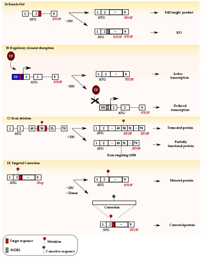
Cells 2020, 9, 1665                                                                                     2 of 20

which may occur spontaneously, for example, during DNA replication, fork stalling or collision of the
transcriptional machinery [9], or as a consequence of UV or gamma irradiation [10]. Accumulation
of SSBs or the occurrence of multiple SSBs in close proximity may lead to complex lesions, where
both strands of the DNA are severed, and so-called double-stranded breaks (DSBs) which are one of
the most dangerous DNA lesions that the cell can experience [10]. Hence, genetic information and
genomic stability are constantly threatened by multiple causes and the cells have evolved complex
mechanisms to sense, monitor and repair this wide variety of DNA lesions.

      Figure 1. Schematic of the major DNA lesions experienced by cellular genomic DNA: The table below
      indicates the type of DNA lesion depicted in the figure above, its leading cause and the DNA repair
      pathway engaged for its resolution.

2. The DNA Damage Response Is Activated upon Sensing of DNA Lesions
     The DNA Damage Response (DDR) can be considered an endogenous alarm system constantly
monitoring the genome integrity and ensuring the faithful transmission of the genetic information
to daughter cells. Each DNA lesion described above leads to specific DNA alterations for which
the cells have evolved lesion-specific repair mechanisms (Figure 1). Here, we provide a simple
overview of these mechanisms. For a comprehensive description of the complex pathways involved,
we refer the reader to extensive review articles previously published [11–13]. Interstrand crosslinks
(ICLs), generated either by natural or synthetic compounds, are mostly resolved via the Fanconi
Anemia (FA) pathway [14]. An increasing body of evidence suggests that ICLs are recognized by
dedicated proteins, such as ubiquitin-like containing plant homeodomain, RING finger domains 1
(UHRF1) and UHRF2 that interact and facilitate the recruitment and retention of Fanconi anemia
group D2 (FANCD2) protein at the ICL site [15]. This event is central to the function of the FA
pathway, and FANCD2 posttranslational modifications are essential for proper regulation of this DNA
repair pathway and subsequent resolution of ICLs [14]. Bulky DNA lesions, as a consequence of UV
irradiation, environmental mutagens and certain chemotherapeutics are sensed by the xeroderma
pigmentosum group C (XPC) protein that activates the Nucleotide Excision Repair (NER) pathway.
On the other hand, alkylated, oxidized and deaminated bases, resulting from natural DNA decay
or endogenous DNA damaging agents, are recognized by specific DNA glycosylases that activate
the Base Excision Repair (BER) pathway. In case of mismatched bases incorporated during DNA
replication, the MutS Homolog (MSH) heterodimer complexes lead to the activation of the Mismatch
Repair (MMR) pathway that secures the resolution of the lesion. If SSBs form, typically because of
oxidative stress or as intermediate products of different DNA repair pathways, they are sensed via
the Poly(ADP-ribose)polymerase 1 (PARP-1) complex and usually resolved via a pathway similar to
the BER pathway [16]. When a DSB occurs, phosphorylation of the H2A histone variant X (termed
γH2AX), mostly at Serine 139 [17], is one of the first events priming the DDR signaling cascade.
Cells 2020, 9, 1665                                                                                 3 of 20

The γH2AX histone decorates the DSB site and acts as a signaling beacon, marking genomic lesion.
Phosphorylation of H2AX is promoted by the phosphatidylinositol 3-OH-kinase-like family including
ATM (ataxia-telangiectasia mutated), DNA-PKcs (DNA-dependent protein kinase catalytic subunit)
and ATR (ataxia telangiectasia and Rad3-related), which are all activated in response to DSB [17] or
replicative fork stalling respectively [18].
     The exact cascade of events leading to sensing of a DSB is still debated. The most accredited model
as of today suggests that changes in chromatin architecture, as a result of a DSBs, are sensed by ATM.
This results in its activation via the autophosphorylation of Serines S367, S1893, S1981 and S2996 and
eventually leads to ATM localization at the DSB site [17]. The subsequent phosphorylation of H2AX
recruits MDC1 (mediator of DNA damage checkpoint protein 1) that functions as docking site for the
MRE11-RAD50-NBS1 (MRN) complex [19]. The signaling is reinforced by further ATM deposition
promoted by MRN, and the recruitment of additional DNA repair factors eventually leads to the
formation of the so-called Ionizing Radiation Induced Foci (IRIF) [20]. To provide the cells enough time
to repair the DNA lesion, ATM is also capable of modulating the function of the checkpoint kinases
1 and 2 (CHK1 and CHK2) and of slowing down the cell cycle [21], as discussed in the next section.
However, when the DSBs cannot be repaired, the unresolved DDR signaling triggers cell senescence
or apoptosis via p53-induced cell-cycle arrest. This fascinating mechanism is extensively reviewed
elsewhere [22].

3. DNA Damage and Cell Cycle Checkpoints
     The cell cycle is divided into four main phases, and internal checkpoints guarantee normal
progression if the necessary condition for cellular growth and division are met [23]. Although the
checkpoints are distinct, they all respond to lesions in DNA and, partially, some of the effector proteins
are shared among the different checkpoints [24]. Cell cycle progression is mostly orchestrated by the
cyclin-dependent kinases (CDKs), a family of protein kinases that phosphorylate key substrates to
promote DNA synthesis and cell cycle progression [25]. In mammals, the CDK family is composed
of more than 20 members acting at different cell cycle phase transitions such as CDK2 and CDK4
that regulate G1–S transition [26,27] or CDK1 and CDK2 that control G2–M transition instead [28].
Cross-talk between DDR and cell cycle progression secures that the latter is modulated to either favor
the repair of dangerous DNA lesions or to induce senescence or cell death when repair fails [29,30].
When DNA damage is sensed during the G1 phase, the activity of the transcription factor p53 is critical
before entry into the S phase. Upon its activation and stabilization, p53 is capable of driving the
expression of a large number of genes, including the cyclin-dependent kinase inhibitor p21 (CDKN1A),
leading to G1 arrest [31]. While the cell cycle is halted, the driving DNA lesion is removed by the
specific DDR mechanism harnessed prior to DNA replication. A contribution to the G1-S checkpoint
comes also from the p38 mitogen-activated protein kinases (MAPK) pathway, which is activated in
response to a wide range of DNA damage events [32]. Particularly, upon DSB formation, p38 MAPK is
activated by ATM through the Tao (thousand and one) kinases [33]. Upon activation, p38 MAPK is able
to directly activate p53, leading to the accumulation of p21. This protein counteracts CDK2 activation
by directly inhibiting its cyclin interactors (cyclin D and CDC25A) [34]. Additionally, p38 MAPK is
also able to directly phosphorylate and stabilize p21, further promoting G1 arrest [35]. Importantly,
p21 levels are crucial to promote G1 arrest, and only when a certain threshold is reached, the block
occurs, thus preventing premature cell-cycle arrest [36]. Once the threat has been resolved, the cell
cycle can proceed to the S phase, during which synthesis and replication of DNA occurs. During
this phase, the sensing of DNA damage typically results in slowing down rather than halting of
DNA synthesis in order to accommodate but not necessarily repair the lesion [37]. The biological
significance of this checkpoint is to provide the repair machinery with sufficient time to resolve
aberrant DNA structures and to prevent cells from dividing before their entire genomes are faithfully
duplicated [38]. This is possible through the activation of multiple origins of replication which have
been established early during cell cycle and can be activated if necessary by the concerted action of
Cells 2020, 9, 1665 4 of 20
ATM and ATR [39]. Once the G2 phase of the cell cycle is initiated, an additional checkpoint secures
that the DNA is completely replicated prior to mitosis. In this case, cell-cycle progression is regulated
by the accumulation of inactive CDK1. This leads to an excess of non-phosphorylated retinoblastoma
(Rb) tumor suppressor, which is able to inhibit cell-cycle progression [40]. Another key player in G2
arrest is p21 that is able to directly block Rb phosphorylation and to sequester inactive CDK1 in the
nucleus, inhibiting entry to the mitotic phase [40]. In conclusion, despite the DNA lesion that triggers
DDR, the cell has developed unique mechanisms to halt or slow down the progression of the cell cycle
to allow repair of the injury. Importantly, besides the type of DNA lesion, also the phase of the cell
cycle during which DNA damage is sensed dictates which DNA repair pathway will be engaged [41].
A detailed explanation of the different pathways that secure seamless replication of the genomic DNA
will be given in the next section.
4. DNA Repair Pathways
As described in the sections above, cells possess surveillance systems able to recognize DNA
damage, allowing a proper response according to the type of lesion experienced. Upon DNA damage
detection, information is transferred to the mechanisms managing the cell cycle progression in order
to pause or slow down cell division. This in turn activates a state during which the cell can adopt
the repair mechanism suitable to repair the lesion. In this section, we will describe the mechanisms
activated by the cell to endure and resolve DNA damage.
4.1. Fanconi Anemia (FA) Pathway
Several natural or artificial compounds might promote crosslinking of two DNA strands. This form
of DNA damage, typically referred to as interstrand crosslink (ICL), is highly dangerous as it prevents
transcription and replication of DNA by inhibiting strand separation, leading to p53-dependent
apoptosis [42]. Studies aimed at better understanding the mechanisms leading to a severe genetic
disorder, known as Fanconi anaemia (FA), have shed light on how cells react and repair this DNA
lesion. Indeed, typically, FA patients suffer from high sensitivity to ICL-forming agents. For a detailed
description of this complex DNA repair pathway, we refer the reader to a comprehensive review
previously published [14]. In the majority of cases, ICL recognition occurs during DNA replication
and involves UHRF1, which in turn recruits FANCD2 at the damaged site. The latter, together with
Fanconi anemia group I protein (FANCI), orchestrates the complex pathway, leading to ICL resolution.
Ubiquitination of the FANCD2/FANCI complex promotes the recruitment of endonucleases, such as
DNA excision repair protein (ERCC1) and Xeroderma Pigmentosum group F (XPF), that cleave the
DNA and unhook the lesion [43]. The resulting DSB is then repaired via the concerted activity of
exonucleases, polymerases and ligases and is detailed elsewhere [42].
4.2. Nucleotide Excision Repair (NER)
In eukaryotes, NER can be divided in two distinct pathways, the global-genome and the
transcription-coupled NER extensively reviewed previously [44]. These two sub-pathways differ only
in the mechanism by which they recognize the DNA lesion. In GG-NER, bulky DNA lesions lead to a
major distortion of the DNA helix and are recognized by the XPC protein, which is in turn stabilized
by RAD23B [45]. On the other hand, TC-NER is independent from XPC, but it occurs when RNA
polymerase II stalls at a DNA lesion serving as a signal for damage recognition. Once the DNA lesion
is recognized, the 10-subunit complex of transcription factor IIH (TFIIH) is recruited. This comprises
the XPD (Xeroderma Pigmentosum group D) helicase, which unwinds the DNA helix, and two
other members of the complex, XPA (xeroderma pigmentosum group A) and replication protein A
(RPA), which stabilize the DNA. Subsequently, the endonucleases within the complex, namely XPG
(Xeroderma Pigmentosum group G) and XPF (Xeroderma Pigmentosum group F), cleave the damaged
DNA strand at both sides of the lesion and remove it. Subsequently, the gap is filled by the concerted
action of delta/kappa and epsilon polymerases, along with DNA ligase IIIα or DNA ligase I [46].Cells 2020, 9, 1665 5 of 20
4.3. Base Excision Repair (BER)
As mentioned previously, natural DNA decay or endogenous DNA-damaging agents can cause
small alterations of the DNA helix. These lesions are recognized by one of the eleven known
damage-specific DNA glycosylases, which in turn activate the BER pathway [47]. In the first phase,
the glycosylase removes the aberrant base by cleaving the N-glycosylic bond between the base and the
deoxyribose and generates an abasic site (AP). Depending on the length of the gap, further processing
occurs via distinct mechanisms, namely short-patch (for single nucleotide processing) or long-patch (for
2–10 nucleotide processing) BER. The AP is sensed and bound by the PARP-1 and its homolog PARP-2,
which recruit the human AP-endonuclease 1 (APE1). This protein nicks 50 of the AP, generating proper
30 -hydroxyl and 50 -deoxyribose phosphate termini in which the DNA polymerase beta can insert the
missing nucleotide(s). In short-patch BER, the gap is finally sealed by the concerted action of the X-ray
repair cross-complimenting protein 1 (XRCC1), DNA ligase I and DNA ligase III. In long-patch BER,
gap sealing is mediated by proliferating cell nuclear antigen (PCNA) [47,48]. When AP sites are not
properly resolved, they might lead to a peculiar type of DNA lesion, the SSB. These lesions are fixed via
a pathway similar to the BER. Indeed, SSB detection is mediated by PARP-1, which recruits the APE2
endonuclease to the SSB site. This enzyme unmasks a longer single-stranded DNA portion. which is
in turn recognized by a complex cascade of different proteins that eventually recruit polymerases and
ligases to seal the gap [9].
4.4. Mismatch Repair (MMR)
DNA mismatch repair (MMR) is a highly conserved DNA repair system that critically contributes
to maintaining genome stability through the correction of mismatched base pairs. Mismatches primarily
occur as errors during DNA replication, although they can arise also from other biological processes [12].
Mismatched bases are recognized by two main MutS Homolog (MSH) heterodimer complexes: MutS
alpha, which comprises the MSH2 and MSH6 subunits, and MutS beta, including the MSH2 and MSH3
subunits [49]. The former mainly identifies single base pair mismatches and small insertion/deletion
(indel) mutations of one or two bases. while the latter mediates the recognition of larger indels [50].
Mismatch recognition by MutS alpha and beta complexes causes a conformational change of the
complex itself into a sliding clamp which primes the recruitment of an exonuclease, probably EXO1,
which leads to the removal of about 150 nucleotides surrounding the mismatched region. The gap is
then filled by the DNA polymerase delta and DNA ligase I [49].
4.5. DNA Double-Stranded Break Repair Pathways
DSB is one of the most critical and dangerous types of DNA lesions leading, if not repaired, to cell
death. Mammalian cells have evolved multiple mechanisms that contribute to DSB repair (Figure 2).
In general, these mechanisms can be divided into conservative, characterized by the accurate repair of
the DSB, and nonconservative that might lead to the alteration of the original DNA sequence. The
cell cycle phase during which the lesion occurs and the extent of resection that the broken DNA ends
undergo contribute to the type of repair mechanism engaged [51]. Multiple mechanisms are involved
in the repair of DSBs, and details are given in the following paragraphs.Cells 2020, 9, 1665 6 of 20
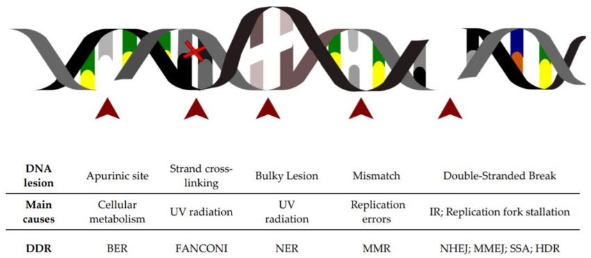
Figure 2. Resolution of a DNA double-stranded break (DSB): Following the recognition and marking of
a DSB by the concerted action of key proteins such as ATM (ataxia-telangiectasia mutated), ATR (ataxia
telangiectasia and Rad3-related) and DNA-PKcs (DNA-dependent protein kinase catalytic subunit),
the repair can follow different pathways. On one hand, if the DNA ends are protected by the
Ku70/Ku80 complex, the DSB is generally repaired via the non-homologous end-joining (NHEJ)
pathway (depicted in A). In this case, an intermediate end-processing step might be necessary
if the protected ends are not compatible. On the other hand, resection of the DNA ends by the
MRN/CtIP (MRE11-RAD50-NBS1/C-terminal-binding protein interacting protein) complex results in
NHEJ inhibition. Based on the length of homology DNA fragments revealed during end-resection,
the DSB can be repaired either via homology-directed repair (HDR) (B) or microhomology-mediated
end joining (MMEJ)/ single-strand annealing (SSA) (C). The key factors involved in the DNA repair
pathways depicted are indicated within the figure.
4.5.1. Non-Homologous End-Joining
Non-homologous end-joining (NHEJ) is the pathway preferentially adopted by the mammalian
cells to resolve DSBs (Figure 2A). As mentioned in the second paragraph of this review, DSBs are
signaled through the phosphorylation of the H2AX histone variant which activates the cellular DDR
machinery. The first component of the NHEJ pathway recruited at the DSB is the Ku70-Ku80 dimer.
This binds to the two ends of the DNA break and protects them from end-resection, thus antagonizing
the HDR pathway [52]. The C-terminal domain of Ku80 promotes the recruitment of the DNA-PKcs
to form the DNA-PK complex [53]. DNA-PK can recruit the X-Ray repair cross-complementing
protein 4 (XRCC4), XRCC4-like factor (XLF) and the DNA ligase IV to seal the break if the ends
are compatible [54–56]. If the ends are not compatible, DNA-PKcs undergoes self-phosphorylation
at the so-called ABCDE cluster and promotes the activation of the Artemis nuclease [57]. This, in
a complex with DNA-PKcs, resects the DNA ends which are then stabilized by XRCC4 and XLF
prior to their ligation operated by the DNA ligase IV [54], with the contribution of the recentlyCells 2020, 9, 1665 7 of 20
discovered paralogue of XRCC4 and XLF (PAXX) [58]. In case of chemistry of the DNA ends not
supporting direct ligation, additional modifications might be required. Editing of aberrant ends, either
lacking a 50 -phosphate or having a 30 -phosphate, is typically processed by the polynucleotide kinase
(PNK), which possesses both kinase and phosphatase activity [59]. The eventual ligation step of the
two DNA ends is further enhanced by the activity of polymerases that support the DNA ligase IV
activity when ends are not compatible. For example, the template-independent Polµ promotes the
rejoining of noncompatible 30 -overhangs by generating microhomology stretches at the DNA ends [60].
On the other end, compatible ends that still require the fill in of missing nucleotides are processed
by Polγ in a template-dependent manner. Even though the DNA-PK complex is mostly involved in
promoting NHEJ-mediated repair, it can also favor HDR. Indeed, when NHEJ is nonfunctional, the
autophosphorylation of the ABCDE cluster releases the DNA ends from the Ku70-Ku80 dimer. This in
turn results in HDR-promoting factors gaining access to the DNA ends, thus initiating the extensive
DNA resection necessary to engage the HDR pathway [61]. In case of defective NHEJ, this alternative
resolution ensures that DSBs are repaired prior to activation of senescence and apoptosis [62,63].
4.5.2. Homology-Directed Repair
The homology-directed-repair (HDR) pathway promotes the scarless resolution of the DNA lesion
by using the sister chromatid as a template (Figure 2B). In this case, the DSB is sensed by two MRN
complexes that localize at both ends of the DSB [64] even though the mechanism by which MRN is
able to get access to DNA ends that are protected by Ku70-Ku80 heterodimers is still elusive [65].
This complex contributes to both antagonizing NHEJ and to amplifying the γH2AX phosphorylation
signal [64,66]. Subsequently, the two MRN complexes are tethered together in a process mediated by
RAD50 [67]. However, to properly engage HDR, an extensive DNA end resection is necessary [52].
This is initiated by MRE11 that, through its endonuclease activity, induces a distal nick up to 300
nucleotides away from the DSB. Subsequently, its 30 –50 exonuclease activity extends the nick back
towards the DSB [68]. This first short resection is intended to displace the Ku complex and inhibits further
processing by NHEJ factors [69]. Resection is extended with the contribution of C-terminal-binding
protein interacting protein (CtIP) exonuclease, which is activated by phosphorylation at Serines S233,
S276 and S347 [70]. Upon reaching the DSB, the CtIP dissociates and the resection is extended by
Exonuclease 1 (EXO1) with the support of endonuclease DNA 2 and the Bloom syndrome helicase
(BLM), which mediates DNA unwinding [70,71]. Although DNA end resection is critical for the
harnessing of HDR, this process leads to cell death if not properly regulated. Indeed, when the HDR
antagonist p53-binding protein 1 (53BP1) is absent, extensive and uncontrolled DNA end resection leads
to deleterious chromosomic rearrangements and eventually to apoptosis [72,73]. To avoid excessive
DNA processing, the exonuclease activity of EXO1 is limited by ATM-mediated phosphorylation or its
proceeding is inhibited by physical barriers, such as the presence of 53BP1 [74]. The single strand DNA
(ssDNA) generated during the end resection process is immediately recognized and bound by the
Replication Protein A (RPA) complex (RPA1, RPA2 and RPA3), which also removes secondary structures
formed during the process [75,76]. Next, the DNA Repair Protein RAD52 mediates the removal of RPA
and allows RAD51 recombinase loading onto the 30 -end of the long ssDNA. In this process BRCA2
(Breast Cancer Type 2 Susceptibility Protein) plays a key role, too, both sustaining the displacement of
RPA and the loading of RAD51 [77]. The formation of the RAD51–ssDNA nucleoprotein filament is
crucial for the subsequent steps of HDR as it mediates the search for homologous sequences on the
sister chromatid and formation of D-loop triple-helix structures, essential for transferring the genetic
information to the new DNA strand [78,79]. Eventually, repair can be completed via mechanisms that
lead to either crossover or non-crossover events as reviewed elsewhere [80].
4.5.3. Alternative DSB Repair Pathways
DSBs in mammalian cells are typically repaired either via NHEJ or HDR. However, cells possess
alternative pathways which are distinguishable when NHEJ is not properly functioning. These areCells 2020, 9, 1665 8 of 20
classically referred to as alternative end-joining (A-EJ) or single-strand annealing (SSA; Figure 2C)
and are detailed elsewhere [53,81]. Both A-EJ and SSA are error-prone as they involve the loss
of nucleotides and require stretches of homologous sequences of different lengths on the same
chromosome, which are used as a repair template: short homology stretches (i.e., 2–20 nucleotides)
activate microhomology-mediated end joining (MMEJ), while longer stretches (i.e., >25 nucleotides)
promote SSA [81]. As explained above, when the DSB is sensed, the first short resection step mediated
by MRN and CtIP occurs. If short homologous DNA sequences are exposed on the two resected
ends, PARP-1, MRN and polymerase theta mediate the alignment of the complementary DNA strands.
The non-homologous 30 -flaps are removed, and the gaps are ligated by the XRCC1/ligase III complex.
If homology regions longer than 25 nucleotides are unmasked, RPA protects the single-stranded DNA
while RAD52 mediates the base pairing of homologous sequences. The non-homologous 30 -flaps are
removed by the ERCC1/XPF endonuclease complex and the gap is filled by the coordinated action of
DNA polymerases and DNA ligases [82].
5. DNA Repair Defects
5.1. Linking DDR Failure to Cell Disorders and Cancer
We have largely described the critical role that the DNA damage repair pathways have in the
maintenance of genome integrity. It is therefore not surprising how defects in these mechanisms might
be detrimental for cell physiology. Mutations might accumulate if specific DNA repair pathways and/or
checkpoints operate aberrantly or if the DDR is overburdened. These events might generate first hits,
leading to genome instability and malignant transformation. The Cancer Genome Project and Cancer
Genome Atlas have analyzed more than nine thousand samples encompassing 33 different cancer types,
providing a general signature of cancer associated aberrations. These mutations are typically divided
into “driver”, that directly promotes cancer initiation and progression, and “passenger”, contributing
to cancer development as a consequence of their accumulation [83]. Somatic mutations affecting
DDR-related genes were found in about a third of the cases. Examples are represented by mutations
in the MLH1 and MSH2 genes that belong to the MMR pathway. When these genes are mutated,
the resulting dysfunctional MMR leads to failure in properly recognizing and resolving errors arising
from physiological processes, such as DNA replication, therefore priming malignant outcomes [84]
or predisposing to cancer [85]. However, alteration in DDR can trigger disorders other than cancer.
For example, loss of protection against UV-mediated DNA damage resulting from inactivation of key
players in NER is one of the causes leading to rare autosomal recessive diseases, such as xeroderma
pigmentosum (XP), cockayne syndrome (CS) and trichothiodystrophy (TTD) [86]. Alterations in NHEJ
have been associated with devastating immunologic and developmental defects [87]. While the majority
of DSBs result from unwanted DNA lesion, immune cells harness this type of DNA damage to create
diversity in crucial physiological processes such as V(D)J recombination, somatic-hyper-mutation (SHM)
and class-switch recombination (CSR) [88]. These “programmed genomic alterations” are critical for the
development of B and T lymphocytes during the generation of immunoglobulins (Ig) and T cell receptor
(TCR) repertoire, respectively. Ig and TCR are made of variable regions which are shuffled and rejoined
in various combinations to generate the variability necessary for recognition of multiple antigens.
The mechanism by which shuffling is achieved comprises the activity of the RAG1/RAG2 complex
that recognizes specific recombination signals flanking the DNA sequence of each V(D)J segment and
introduces a nick at each site. Subsequently, each nick reacts with the opposite strand, generating
the so-called covalently sealed hairpins at the two sites resulting in a DSB. The intervening sequence
containing the recombination signals circularizes and is eventually lost during cell division. The two
hairpins are then opened by the Artemis nuclease, upon its activation through the phosphorylation
mediated by DNA-PKcs, and are sealed via the NHEJ machinery [89]. Therefore, defects in NHEJ
factors critical for V(D)J recombination, such as Artemis, DNA-PKcs or LIG4, might lead to partial
or complete absence of specific immune cells, resulting in a broad spectrum of immunodeficiencies,Cells 2020, 9, 1665 9 of 20
including severe combined immunodeficiency (SCID) [90]. As seen for NHEJ, inherited defects in
HDR are also pathologic. Mutations in the BRCA1 and BRCA2 genes have been associated with
predisposition to various cancers, including malignancies affecting breast tissue or ovaries, and with
lower frequency in the prostate or pancreas [91,92]. Recently, other HDR-related genes have been
associated with carcinogenesis when mutated, such as PALB2 [93,94] and CtIP [95]. These multiple
examples clearly show that failures in DDR can fuel and sustain cancer progression. On a positive
note, many current cancer therapies, including radiotherapy and chemotherapy, exploit the failure of
tumor cells to respond properly to DNA damage by inducing DNA lesions that prompt senescence.
5.2. Exploiting Defects in DNA Repair to Treat Cancer
The main goal of cancer therapy is achieving complete elimination of the tumor either through
surgical procedures or via the more or less selective killing of cancerous cells. Multiple strategies
have been devised that target metabolic processes which are altered in cancer cells. Transformed
cells are typically characterized by an extraordinary high replication rate. The use of antimetabolites,
such as 5-fluorouracil (5-FU) or thiopurines, has been explored to inhibit nucleotides biosynthesis,
thus depleting cells of the essential components to replicate their DNA and to proliferate [96]. Similarly,
cell replication can be hampered by inhibiting the topoisomerase enzyme, which is essential to resolve
DNA torsional stress occurring during replication. As a consequence, accumulation of DSBs and
supercoiled structures before the replication fork limits cancer cell proliferation [97]. Since defects in
DNA repair pathways are a fairly common feature in cancer cells, in principle, these cells are more
vulnerable to DNA-damaging agents. The use of drugs to inhibit the remaining functional DNA
repair pathways, an approach termed “synthetic lethality”, is often exploited to selectively kill the
malignant cells [98]. A similar principle makes use of cancer cell vulnerability to oxidative stress
for eradicating malignant cells. The high replication rate of tumor cells results in high oxidative
stress, and cancer cells are highly dependent on pathways that prevent DNA damage under these
circumstances [99]. Inhibitors of polymerase beta (POLB), a major factor in the BER pathway, have
been exploited to drive cancer cell death [100]. However modest success and concerns related to
inhibition of other polymerases has limited this approach [101]. Another common characteristic of
tumor cells is a dysfunctional HDR repair pathway. Also in these cases, the use of agents that lead to
the formation of DSB is typically explored to create overt DNA damage in cancer cells, leading to their
death [102,103]. For instance, PARP-1 inhibitors have been widely used to inhibit SSB repair and to
promote DSB formation in ovarian or prostate cancer [104,105]. Another mechanism to induce DNA
damage makes use of cisplatin, which reacts with nucleophilic centers on purine bases, causing intra-
and interstrand crosslink. This in turn leads to critical distortions of the DNA double helix and, if not
resolved, to apoptosis [14]. Interestingly, combination of the PARP-1 inhibitor Niraparib with cisplatin
has been used to overcome the acquired platinum-resistances of cancer cells that typically arises in
some cases of metastatic ovarian cancer [106,107]. Although the potential of “synthetic lethality”
strategies to combat some forms of tumors is unquestioned, in some cases. it fails due to the presence
of a few tumor cells capable of counteracting the mode of action of the used compound. For example,
BRCA1-deficient tumors treated with the PARP inhibitor Olaparib developed drug resistance in vivo
in a murine model of BRCA1-associated breast cancer [106]. Further elucidation of the underlying
mechanism revealed an upregulation of the Abcb1A/B gene encoding for the corresponding drug efflux
transporters. This led to drug clearance, and the effect could be reversed by using Tariquidar, a drug
that specifically blocks Abcb1 transporter activity [106]. Different drug resistance strategies have been
reported that hamper the activity of PARP inhibitors. The use of Olaparib for breast cancer patients
has been associated with acquired resistance to this inhibitor. This is achieved for example through the
partial restoration of HDR as a result of reversion mutations in the BRCA1 gene [108]. Although escape
mechanisms exploited by cancer cells might dampen the full efficacy of “synthetic lethality”, they also
offer the opportunity to reveal backup strategies adopted by tumors for survival. Further studies
are necessary to generate novel therapeutics that can be rationally combined to neutralize multipleCells 2020, 9, 1665 10 of 20
pathways at once. Ongoing studies on large patient cohorts will certainly contribute to improving our
knowledge of escape mechanisms and to developing new strategies to counteract tumor progression.
6. Exploiting DSB Repair to Develop Innovative Therapeutic Strategies
In the last decades, understanding the genetic bases of many human disorders has made
unprecedented steps forward. This has fostered the development of novel therapeutics aimed at
curing patients affected by genetic defects by means of gene therapy. This typically includes the
development of viral vectors transferring a correct copy of the mutated gene. Once present in the host
cell, the expression of the correct gene complements the missing gene function in the patient cells,
resulting in a functional cure [109]. With the dawn of targeted genome editing, the chase for new
approaches aimed at precisely correcting the disease-causing mutation was launched [110]. In this
case, the first step relies on the introduction of a DSB in close proximity to the genomic site where the
change is desired. This is made possible by using programmable designer nucleases, such as zinc finger
nucleases (ZFNs), transcription activator-like effector nucleases (TALENs) or the recently introduced
CRISPR-Cas (clustered regularly interspaced palindromic repeat-CRISPR-associated) systems. For a
comprehensive description of the different types of designer nucleases, we refer the reader to detailed
reviews published elsewhere [111–113]. As described in the previous sections, the cells have developed
sophisticated mechanisms to sense and resolve DNA lesions, such as DSBs, in order to maintain
genome integrity. The precise insertion of a DSB by such programmable nucleases triggers either of
the two major repair mechanisms, NHEJ or HDR, which can be harnessed to achieve precise and
permanent changes of the human genome.
6.1. Use of NHEJ-Mediated Repair for Therapy
In mammalian cells, NHEJ is the most commonly used pathway to repair a DSB. This mechanism
is generally fast and promotes the ligation of the broken DNA without loss of genetic information
in less than an hour [114]. However, the activity of the designer nucleases typically lasts for several
hours, or longer. This results in the selection of repair events which lead to insertion or deletions
of few nucleotides at the DSB site, the so-called indel mutations, that prevent further nuclease
cleavage. This outcome has been used with the aim of inactivating endogenous genes or regulatory
elements to provide a beneficial loss of function characteristic to patient cells (Figure 3A). The most
well-documented example is the first clinically approved trial involving the use of designer nucleases
to combat infections with the human immunodeficiency virus (HIV), which leads to a disease known
as acquired immunodeficiency syndrome (AIDS). In this case, ZFNs have been used ex vivo to
introduce indel mutations in the coding region of the CCR5 gene in CD4+ T cells from HIV-positive
patients (NCT00842634). This gene codes for the C-C chemokine receptor type 5, which is used as a
co-receptor by HIV to infect host cells [115]. Its inactivation, as result of the ZFN-induced mutations,
leads to acquired HIV-resistance, as documented by multiple studies [116]. Encouraging results
have fostered further exploitation of this strategy in patient-derived hematopoietic stem cells with
the goal of providing transplanted patients with a new immune system that is resistant to the virus
(NCT02500849) [117]. Similarly, the targeted introduction of indel mutations can be explored to
inactivate regulatory elements (Figure 3B). Beta-hemoglobinopathies include a set of different disorders
caused by mutations in the HBB gene that result in impaired oxygenation of organ tissues leading to
multi-organ failure and death. IVSI-110 G>A is a common intronic mutation in the β-globin encoded
by the HBB gene. The point mutation leads to the formation of an aberrant splice acceptor that drives
abnormal splicing and hence nonfunctional β-globin protein [118]. In the attempt to restore normal
splicing, both CRISPR-Cas and TALENs have been used to introduce indel mutations to disrupt the
aberrant splice site. The high efficiency of this approach and the large number of mutations that
could be treated using such strategies are promising to prompt future investigation [119]. Recently,
genome editing for the treatment of beta-hemoglobinopathies has entered clinics with two ongoing
trials. Reactivation of fetal hemoglobin expression holds promise to complement the adult hemoglobinCells 2020, 9, 1665 11 of 20
function [120]. In both clinical studies, the goal is to reactivate the expression of the fetal hemoglobin
by inhibiting the expression of the BCL11A (B cell lymphoma/leukemia 11A) gene using either ZFN
or CRISPR-Cas (NCT03432364 and NCT03655678, respectively). This gene encodes for a critical
transcription factor that regulates the switch between fetal and adult hemoglobin [121], acting as a
repressor of the former. Preclinical studies have shown that the disruption of the erythroid-specific
enhancer of the BCL11A gene leads to its reduced expression and to the reactivation of fetal hemoglobin
to therapeutic levels [122]. The results of these clinical studies will be certainly instrumental to further
develop new therapeutics for patient affected by hemoglobin disorders. The therapeutic potential of
targeted indels has been explored also to mitigate one of the most devastating human genetic disorders.
Duchenne muscular dystrophy (DMD) is a deadly X-linked disease resulting from mutations in the
dystrophin gene. Dystrophin is a structural protein that connects actin fibers and cytoskeleton to the
muscle fibers via a rod-shaped domain. In its absence, muscle cells progressively degenerate, resulting
in slow but constant inflammation and fibrosis, which eventually leads to death due to respiratory
insufficiency or heart failure in early adulthood [123]. Different mutations have been described that
result in premature stop codons leading to a lack of the dystrophin protein. Since dystrophin is a
structural protein, amino acid substitution or truncations within the rod domain are not necessarily
critical for its function. This has prompted the development of new strategies to create mutations
that eliminate the stop codon, thus restoring the reading frame of the protein [124]. Considering
the stochastic nature of this approach, only a third of the mutations result in dystrophin expression.
Therefore, to increase the likelihood of creating indels that restore protein expression, researchers
have explored alternative strategies. Introducing a DSB in between regions of microhomology leads
to its repair preferentially via the MMEJ pathway that, as explained in Section 4.5.3, results in the
formation of precise microdeletions. MMEJ has been exploited to precisely correct frameshift mutations
resulting from microduplications in patient-derived cells for diverse rare diseases without the need
for additional donor templates [125]. Recently, a bioinformatics tool has been described that allows
the identification of targets which are amenable for MMEJ repair, further promoting the possibility of
achieving accurate repair in an HDR-independent way [126]. This will certainly provide in the future
new exciting opportunities for preclinical exploitation of DMD therapeutics based on genome editing.
In a different approach, using two designer nucleases targeting sites flanking a mutation-bearing exon
led to its deletion with consequent reconstitution of dystrophin expression (Figure 3C) [124]. In both
cases, the expression of a new dystrophin harboring a shorter rod domain resulted in a milder form of
DMD similar to Becker-muscular dystrophy (BMD), with patients having typically a prolonged life
expectancy if the heart muscle is not affected [127]. However, immunological concerns related to the
expression of novel epitopes due to restored dystrophin expression must be considered [128].
In conclusion, the predominant use of NHEJ and the newly explored MMEJ in mammalian cells
enable the highly efficient introduction of loss of function mutations with allelic frequencies as high
as 90% in some instances [129]. This high efficacy supports the development of further strategies
exploiting this mechanism for other therapeutic purposes.Cells 2020, 9, 1665 12 of 20
Figure 3. Genome editing using designer nucleases (DN): (A) Targeting of the DN to the coding region
of a gene promotes the formation of a DSB which is typically repaired via NHEJ. The subsequent
formation of indel mutations (grey box) may generate a premature stop codon, eventually leading to
gene inactivation. (B) DN can be targeted to regulatory elements (RE) in order to disrupt the binding site
of an activating transcription factor (TF). As a result, downstream gene transcription might be reduced
or abolished in a cell-specific fashion as described in Section 6.1. (C) Two DNs can be directed to sites
flanking an exon containing a non-sense mutation for its deletion. The resulting gene might result in
the generation of a truncated, albeit partially functional, protein. (D) Targeting of a DN in proximity
of a genetic mutation and simultaneous delivery of a donor template harboring the correct genetic
sequence might result in precise gene editing via harnessing of the homology directed repair pathway.Cells 2020, 9, 1665 13 of 20
6.2. Use of HDR-Mediated Repair for Therapy
In some instances, the stochastic occurrence of indel mutations upon NHEJ-mediated DSB repair
is not sufficient to secure a therapeutic benefit. In such cases, strategies aimed at harnessing the HDR
mechanism to achieve precise editing at the DSB site are paramount. Two major challenges have to be
considered in this context: (i) a typical human cell rather engages NHEJ than HDR to repair a DSB, and
(ii) the edited gene can be cleaved again by the designer nuclease in certain circumstances. In the 1990s,
pioneer studies from Maria Jasin’s lab have shown that the introduction of a targeted DSB and the
simultaneous delivery of an exogenous DNA fragment that is homologous to the target site improve the
efficiency of HDR by several orders of magnitude [130]. The goal of biasing the DNA repair towards
HDR has been pursued by several laboratories worldwide with different strategies. These include the
use of compounds to either synchronize the cells or to restrict the expression of the designer nuclease
in the cell cycle phases that support HDR [131]. Similarly, the use of small molecule drugs to either
directly inhibit NHEJ or to boost HDR has been explored [132,133]. While promising, such approaches
rely on global alterations of cell physiology, with potential deleterious effects on key mechanism that
rely on NHEJ or HDR, with devastating consequences as discussed in Section 5.1. A localized approach
in which the increase of HDR is restricted to the target site would be preferable. Hence, direct fusion
of the Cas9 endonuclease to proteins that inhibit NHEJ or promote HDR at the target site, such as
dominant negative forms of 53BP1 or CtIP, respectively, have been explored [134,135]. The knowledge
acquired in the last decades has fueled the development of precise genome editing strategies to treat
human genetic disorders. Even though current clinical trial explore the harnessing of NHEJ-based
correction to treat beta-hemoglobinopathies [136], as described in the previous paragraph, this group
of disorders is a particularly fit candidate for the development of precise genome editing therapeutics
(Figure 3D). In this context, it has been indeed estimated that gene correction frequencies as low as
2% in hematopoietic stem cells might result in therapeutic benefit when edited cells are transplanted
back to sick patients [137]. Different gene-correction strategies have been pursued with promising
success to correct the adenine to thymidine mutation that eventually leads to the production of sickling
hemoglobin and to the development of sickle cell disease (SCD) [138]. The development of protocols
capable of isolating edited stem cells which retain engraftment potential suggests the great potential that
precise genome editing strategies have for curing devastating disorders such as SCD [139]. Similarly,
also primary immunodeficiencies (PIDs) are considered a suitable candidate for therapeutic strategies
based on precise genome editing. A successful example is the development of genome editing strategies
to treat X-linked chronic granulomatous disease (X-CGD) caused by mutations in the CYBB gene. In this
case, either the correction of the CYBB causative mutation [140] or the integration of a CYBB expression
cassette in a “safe harbor” locus to complement the missing gene function has been pursued [141].
The results achieved so far underline that precise genome editing is certainly an opportunity for patients,
and further studies will be required to dissect its potential in the clinics.
7. Concluding Remarks
Understanding how genome stability is maintained and investigating the mechanisms adopted by
the cells to withstand DNA lesions has been paramount to explore the therapeutic potential of genome
editing. We have illustrated how failure in DNA repair mechanisms may contribute to the onset of
detrimental conditions, such as cancer. Moreover, we have described how this knowledge is exploited
to develop new therapeutics based on “synthetic lethality” or genome editing using designer nucleases.
While the activity of designer nucleases at sites that share a certain sequence identity with the target
site, the so-called off-targets, poses concerns [142], the increasing number of genome editing trials
approved thus far [143] and the first in human application of CRISPR-Cas to treat a blindness disorder
(NCT03872479) recently described suggest that new therapeutics are on the horizon. Once current
approaches are substantiated in conditions that better resemble those of transplanted patients and
genome-wide analysis to dissect the genotoxic potential of these approaches are in place, it will beCells 2020, 9, 1665 14 of 20
reasonable to believe that new treatment opportunities will be available for more and more human
disorders using a new generation of therapeutics.
Author Contributions: A.C and C.M. conceptualized, wrote and edited the manuscript. Figures were prepared
by A.C. and edited by A.C. and C.M. All authors have read and agreed to the published version of the manuscript.
Funding: This work was financed by the German Federal Ministry of Education and Research (BMBF-01EO0803)
and the European Union’s Horizon 2020 research and innovation programme under the Marie Skłodowska-Curie
grant agreement No. 765269 to C.M.
Conflicts of Interest: The authors declare no conflict of interest.
References
1. Jackson, S.P.; Bartek, J. The DNA-damage response in human biology and disease. Nature 2009, 461, 1071–1078.
[CrossRef] [PubMed]
2. Lindahl, T.; Barnes, D. Repair of endogenous DNA damage. Cold Spring Harb. Symp. Quant. Boil. 2000, 65,
127–134. [CrossRef] [PubMed]
3. Ganai, R.A.; Johansson, E. DNA Replication—A Matter of Fidelity. Mol. Cell 2016, 62, 745–755. [CrossRef]
[PubMed]
4. Cadet, J.; Wagner, J.R. DNA Base Damage by Reactive Oxygen Species, Oxidizing Agents, and UV Radiation.
Cold Spring Harb. Perspect. Boil. 2013, 5, a012559. [CrossRef]
5. Van Houten, B.; Santa-Gonzalez, G.A.; Camargo, M. DNA repair after oxidative stress: Current challenges.
Curr. Opin. Toxicol. 2017, 7, 9–16. [CrossRef] [PubMed]
6. Chatterjee, N.; Walker, G.C. Mechanisms of DNA damage, repair, and mutagenesis. Environ. Mol. Mutagen.
2017, 58, 235–263. [CrossRef]
7. Tubbs, A.; Nussenzweig, A. Endogenous DNA Damage as a Source of Genomic Instability in Cancer. Cell
2017, 168, 644–656. [CrossRef] [PubMed]
8. Lindahl, T. Instability and decay of the primary structure of DNA. Nature 1993, 362, 709–715. [CrossRef]
[PubMed]
9. Hossain, A.; Lin, Y.; Yan, S. Single-Strand Break End Resection in Genome Integrity: Mechanism and
Regulation by APE2. Int. J. Mol. Sci. 2018, 19, 2389. [CrossRef]
10. Abbotts, R.; Wilson, D.M. Coordination of DNA single strand break repair. Free. Radic. Boil. Med. 2017, 107,
228–244. [CrossRef]
11. Callen, E.; Zong, D.; Wu, W.; Wong, N.; Stanlie, A.; Ishikawa, M.; Pavani, R.; Dumitrache, L.C.; Byrum, A.K.;
Mendez-Dorantes, C.; et al. 53BP1 Enforces Distinct Pre- and Post-resection Blocks on Homologous
Recombination. Mol. Cell 2020, 77, 26–38.e7. [CrossRef] [PubMed]
12. Li, Z.; Pearlman, A.H.; Hsieh, P. DNA mismatch repair and the DNA damage response. DNA Repair 2016, 38,
94–101. [CrossRef] [PubMed]
13. Wang, K.; Li, L.; Zhang, Y.; Gao, D. Crosstalk between signaling pathways and DNA damage response.
Genome Instab. Dis. 2019, 1, 81–91. [CrossRef]
14. Martinez, D.L.; Liang, C.-C.; Cohn, M.A. Cellular response to DNA interstrand crosslinks: The Fanconi
anemia pathway. Cell. Mol. Life Sci. 2016, 73, 3097–3114. [CrossRef] [PubMed]
15. Motnenko, A.; Liang, C.-C.; Yang, D.; Martinez, D.L.; Yoshikawa, Y.; Zhan, B.; Ward, K.E.; Tian, J.; Haas, W.;
Spingardi, P.; et al. Identification of UHRF2 as a novel DNA interstrand crosslink sensor protein. PLoS Genet.
2018, 14, e1007643. [CrossRef] [PubMed]
16. Eustermann, S.; Wu, W.-F.; Langelier, M.-F.; Yang, J.-C.; Easton, L.E.; Riccio, A.A.; Pascal, J.M.; Neuhaus, D.
Structural Basis of Detection and Signaling of DNA Single-Strand Breaks by Human PARP-1. Mol. Cell 2015,
60, 742–754. [CrossRef] [PubMed]
17. Blackford, A.N.; Jackson, S.P. ATM, ATR, and DNA-PK: The Trinity at the Heart of the DNA Damage
Response. Mol. Cell 2017, 66, 801–817. [CrossRef]
18. Alexander, J.L.; Orr-Weaver, T.L. Replication fork instability and the consequences of fork collisions from
rereplication. Genes Dev. 2016, 30, 2241–2252. [CrossRef] [PubMed]
19. Reginato, G.; Cejka, P. The MRE11 complex: A versatile toolkit for the repair of broken DNA. DNA Repair
2020, 102869. [CrossRef]Cells 2020, 9, 1665 15 of 20
20. Polo, S.; Jackson, S.P. Dynamics of DNA damage response proteins at DNA breaks: A focus on protein
modifications. Genes Dev. 2011, 25, 409–433. [CrossRef] [PubMed]
21. Awasthi, P.; Foiani, M.; Kumar, A. ATM and ATR signaling at a glance. J. Cell Sci. 2015, 128, 4255–4262.
[CrossRef]
22. Chen, J. The Cell-Cycle Arrest and Apoptotic Functions of p53 in Tumor Initiation and Progression. Cold Spring
Harb. Perspect. Med. 2016, 6, a026104. [CrossRef] [PubMed]
23. Shaltiel, I.A.; Krenning, L.; Bruinsma, W.; Medema, R. The same, only different—DNA damage checkpoints
and their reversal throughout the cell cycle. J. Cell Sci. 2015, 128, 607–620. [CrossRef]
24. Houtgraaf, J.H.; Versmissen, J.; Van Der Giessen, W.J. A concise review of DNA damage checkpoints and
repair in mammalian cells. Cardiovasc. Revascularization Med. 2006, 7, 165–172. [CrossRef] [PubMed]
25. Lim, S.; Kaldis, P. Cdks, cyclins and CKIs: Roles beyond cell cycle regulation. Development 2013, 140,
3079–3093. [CrossRef] [PubMed]
26. Trimarchi, J.M.; Lees, J.A. Sibling rivalry in the E2F family. Nat. Rev. Mol. Cell Boil. 2002, 3, 11–20. [CrossRef]
[PubMed]
27. Harbour, J.W.; Dean, D.C. The Rb/E2F pathway: Expanding roles and emerging paradigms. Genes Dev. 2000,
14, 2393–2409. [CrossRef]
28. Ding, L.; Cao, J.; Lin, W.; Chen, H.; Xiong, X.; Ao, H.; Yu, M.; Lin, J.; Cui, Q. The Roles of Cyclin-Dependent
Kinases in Cell-Cycle Progression and Therapeutic Strategies in Human Breast Cancer. Int. J. Mol. Sci. 2020,
21, 1960. [CrossRef] [PubMed]
29. Petr, M.A.; Tulika, T.; Carmona-Marin, L.M.; Scheibye-Knudsen, M. Protecting the Aging Genome.
Trends Cell Boil. 2020, 30, 117–132. [CrossRef]
30. Wang, J.Y.J. Cell Death Response to DNA Damage. Yalej. Biol. Med. 2019, 92, 771–779.
31. Fischer, M.; Quaas, M.; Nickel, A.; Engeland, K. Indirect p53-dependent transcriptional repression of Survivin,
CDC25C, and PLK1 genes requires the cyclin-dependent kinase inhibitor p21/CDKN1A and CDE/CHR
promoter sites binding the DREAM complex. Oncotarget 2015, 6, 41402–41417. [CrossRef] [PubMed]
32. Thornton, T.M. Non-Classical P38 Map Kinase Functions: Cell Cycle Checkpoints and Survival. Int. J.
Boil. Sci. 2009, 5, 44–52. [CrossRef] [PubMed]
33. Raman, M.; Earnest, S.; Zhang, K.; Zhao, Y.; Cobb, M.H. TAO kinases mediate activation of p38 in response
to DNA damage. Embo J. 2007, 26, 2005–2014. [CrossRef] [PubMed]
34. Reinhardt, H.C.; Aslanian, A.S.; Lees, J.A.; Yaffe, M.B. p53-Deficient Cells Rely on ATM- and ATR-Mediated
Checkpoint Signaling through the p38MAPK/MK2 Pathway for Survival after DNA Damage. Cancer Cell
2007, 11, 175–189. [CrossRef]
35. Kim, G.-Y.; Mercer, S.E.; Ewton, D.Z.; Yan, Z.; Jin, K.; Friedman, E. The Stress-activated Protein Kinases p38α
and JNK1 Stabilize p21Cip1 by Phosphorylation. J. Boil. Chem. 2002, 277, 29792–29802. [CrossRef] [PubMed]
36. Barr, A.R.; Cooper, S.; Heldt, F.S.; Butera, F.; Stoy, H.; Mansfeld, J.; Novak, B.; Bakal, C. DNA damage
during S-phase mediates the proliferation-quiescence decision in the subsequent G1 via p21 expression.
Nat. Commun. 2017, 8, 14728. [CrossRef] [PubMed]
37. Rhind, N.; Russell, P. Checkpoints: It takes more than time to heal some wounds. Curr. Boil. 2001, 10,
R908–R911. [CrossRef]
38. Bartek, J.; Lukas, C.; Lukas, J. Checking on DNA damage in S phase. Nat. Rev. Mol. Cell Boil. 2004, 5, 792–804.
[CrossRef] [PubMed]
39. Yekezare, M.; Gómez-González, B.; Diffley, J.F.X. Controlling DNA replication origins in response to DNA
damage–inhibit globally, activate locally. J. Cell Sci. 2013, 126, 1297–1306. [CrossRef]
40. Gire, V.; Dulić, V. Senescence from G2 arrest, revisited. Cell Cycle 2015, 14, 297–304. [CrossRef] [PubMed]
41. Bennett, G.; Papamichos-Chronakis, E.; Peterson, C.L. DNA repair choice defines a common pathway for
recruitment of chromatin regulators. Nat. Commun. 2013, 4, 2084. [CrossRef] [PubMed]
42. Deans, A.J.; West, S.C. DNA interstrand crosslink repair and cancer. Nat. Rev. Cancer 2011, 11, 467–480.
[CrossRef] [PubMed]
43. Douwel, D.K.; Boonen, R.A.; Long, D.; Szypowska, A.A.; Räschle, M.; Walter, J.C.; Knipscheer, P. XPF-ERCC1
acts in unhooking DNA interstrand crosslinks in cooperation with FANCD2 and FANCP/SLX4. Mol. Cell
2014, 54, 460–471. [CrossRef]You can also read