Recent Advances in Gene Therapy for Cardiac Tissue Regeneration - MDPI
←
→
Page content transcription
If your browser does not render page correctly, please read the page content below
International Journal of
               Molecular Sciences

Review
Recent Advances in Gene Therapy for Cardiac
Tissue Regeneration
Yevgeniy Kim, Zharylkasyn Zharkinbekov                        , Madina Sarsenova, Gaziza Yeltay and Arman Saparov *

                                          Department of Medicine, School of Medicine, Nazarbayev University, Nur-Sultan 010000, Kazakhstan;
                                          yevgeniy.kim@nu.edu.kz (Y.K.); zharylkasyn.zharkinbekov@nu.edu.kz (Z.Z.);
                                          madina.sarsenova@nu.edu.kz (M.S.); gaziza.yeltay@nu.edu.kz (G.Y.)
                                          * Correspondence: asaparov@nu.edu.kz; Tel.: +7-717-270-6140

                                          Abstract: Cardiovascular diseases (CVDs) are responsible for enormous socio-economic impact and
                                          the highest mortality globally. The standard of care for CVDs, which includes medications and
                                          surgical interventions, in most cases, can delay but not prevent the progression of disease. Gene
                                          therapy has been considered as a potential therapy to improve the outcomes of CVDs as it targets the
                                          molecular mechanisms implicated in heart failure. Cardiac reprogramming, therapeutic angiogenesis
                                          using growth factors, antioxidant, and anti-apoptotic therapies are the modalities of cardiac gene
                                          therapy that have led to promising results in preclinical studies. Despite the benefits observed
                                          in animal studies, the attempts to translate them to humans have been inconsistent so far. Low
                                          concentration of the gene product at the target site, incomplete understanding of the molecular
                                          pathways of the disease, selected gene delivery method, difference between animal models and
                                          humans among others are probable causes of the inconsistent results in clinics. In this review, we
                                discuss the most recent applications of the aforementioned gene therapy strategies to improve cardiac

                                          tissue regeneration in preclinical and clinical studies as well as the challenges associated with them.
Citation: Kim, Y.; Zharkinbekov, Z.;      In addition, we consider ongoing gene therapy clinical trials focused on cardiac regeneration in
Sarsenova, M.; Yeltay, G.; Saparov, A.    CVDs.
Recent Advances in Gene Therapy for
Cardiac Tissue Regeneration. Int. J.
                                          Keywords: gene therapy; cardiovascular diseases; cardiac regeneration; cardiac reprogramming;
Mol. Sci. 2021, 22, 9206. https://
                                          therapeutic angiogenesis; growth factors; reactive oxygen species; apoptosis
doi.org/10.3390/ijms22179206

Academic Editors: Nicole Wagner
and Kay-Dietrich Wagner
                                          1. Introduction
Received: 31 July 2021                         Cardiovascular diseases (CVDs) remain the principal cause of mortality and morbidity
Accepted: 16 August 2021                  worldwide. In 2019 alone, it was estimated that there were approximately 18.6 million
Published: 26 August 2021                 deaths due to CVDs, which accounted for almost one third of all global mortalities [1].
                                          At the same time, the prevalence of CVDs exceeded half a billion cases in 2019 [1]. A
Publisher’s Note: MDPI stays neutral      more concerning fact about morbidity and mortality related to CVDs is that they are
with regard to jurisdictional claims in   continuing to grow. According to the Global Burden of Disease 2019 Study, the number
published maps and institutional affil-   of deaths caused by CVDs increased by approximately 6.5 million for the period between
iations.                                  1990–2019, while the amount of prevalent cases of CVDs almost doubled for the same
                                          period [1]. In addition to the huge impact on people’s health and quality of life, CVDs are
                                          blameworthy for huge economic losses. In the countries of the European Union in 2017,
                                          total spending associated with CVDs were estimated to be €210 billion euro [2]. In the
Copyright: © 2021 by the authors.         USA in 2017, CVD-related healthcare and productivity loss costs exceeded $320 billion
Licensee MDPI, Basel, Switzerland.        in 2017 [3]. The estimated healthcare costs are predicted to increase more than 2.5-fold,
This article is an open access article    reaching $818 billion, by 2030 [3].
distributed under the terms and                The standard treatments for CVDs include pharmacologic agents, therapeutic devices
conditions of the Creative Commons        and surgical interventions. Although these therapies improve survival, reduce symptoms,
Attribution (CC BY) license (https://     and improve quality of life, they do not reverse the pathologic processes associated with
creativecommons.org/licenses/by/          CVDs and because of this, the health condition of most patients deteriorate with time [4].
4.0/).

Int. J. Mol. Sci. 2021, 22, 9206. https://doi.org/10.3390/ijms22179206                                        https://www.mdpi.com/journal/ijms
Int. J. Mol. Sci. 2021, 22, 9206 2 of 21
Advances in cellular and molecular biology shed light on the pathogenetic and patho-
physiologic mechanisms of CVDs and give promising opportunities for the use of novel
therapies to combat heart and vascular diseases. Since these therapies are targeted towards
the molecular cause of a specific CVD, they could hold a promising approach to cure the
disease. Such treatments include cell-based therapies, therapies with growth factors and
other bioactive molecules, biomaterials, and others [5–8]. Another therapeutic strategy that
could likely improve the outcome of CVDs is gene therapy.
Gene therapy has been investigated for the treatment of a multitude of CVDs and
CVD-related conditions such as atherosclerosis, coronary artery disease (CAD), myocar-
dial infarction (MI), peripheral arterial disease (PAD), hypertension, various types of
arrhythmias, heart failure (HF), restenosis of coronary stents, failure of vein grafts, and
others [9–13]. Preclinical trials involving gene therapy for the aforementioned conditions
were largely successful, however, there are a number of challenges associated with the
translation of this therapeutic modality to clinical use [9]. Although there were several
clinical trials for gene therapy, which showed positive results in terms of safety and certain
therapeutic efficiency (specifically, trials for CAD, PAD, and HF), many other randomized-
controlled studies failed to show the benefits of gene therapy over standard treatments [9].
This lack of consistent results in clinical trials can be attributed to multiple reasons includ-
ing low concentration of the transferred gene or its product at the target site, incomplete
knowledge of the disease mechanism and hence wrong gene therapy strategy, significant
differences between animal models and human subjects, and others [4,9].
In this review, we discuss the latest preclinical findings and clinical trials related to
the application of gene therapy for the treatment of CVDs. Specifically, we consider how
cardiac gene therapy can be used to enhance angiogenesis, remodel scar tissue, alleviate
production of reactive oxygen species, and prevent apoptosis.
2. Cardiac Tissue Regeneration in Adult Heart
2.1. Mechanisms of Cardiac Tissue Regeneration
The myocardium of adult mammals including humans has an extremely low regen-
erative capacity due to low proliferation rates of cardiomyocytes and scarcity of cardiac
stem cells. In fact, for a long time, it was believed that cardiomyocytes in the adult heart
are completely unable to proliferate. However, several studies using carbon-14 dating
have detected a slow but persistent turnover of cardiomyocytes throughout life [14–16].
According to the American Heart Association’s 2017 Consensus Statement, the annual
turnover rate was reported to be between 0.5% and 2.0% [17]. Moreover, after injury to
the heart muscle, the cardiomyocyte proliferation rate increases several times [18]. In
contrast to adults, the neonatal mammalian heart possesses an outstanding capacity for
regeneration. In the last decade, there have been multiple studies that demonstrated a
robust regenerative response following apical resection and ischemic injury in mice shortly
after birth [19–23]. The ability of neonatal myocardium to completely regenerate was also
established in porcine and rat models of MI [24–26]. Importantly, neonates exhibit this
remarkable regenerative capacity only for a brief period of time after birth, i.e., it is rapidly
lost two to seven days postnatally [19,25,27].
Cardiac tissue regeneration is a very complex process that involves multiple genes and
signaling pathways. Many reports showed that myocardial recovery is mainly mediated by
the proliferation of existing cardiomyocytes with little contribution from cardiac progenitor
cells [19]. Cardiomyocyte proliferation in turn is regulated by multiple cells and signaling
interactions that is demonstrated by recent gene expression and chromatin modification
profiling studies [28,29]. Using single-cell RNA sequencing, Wang and colleagues have
identified 22 non-cardiomyocyte cell types involved in cardiac regeneration, including
endothelial cells, fibroblasts, epicardial cells, pericytes, smooth muscle cells, and a number
of immune cells—namely, macrophages, monocytes, dendritic cell-like cells, granulocytes,
T cells, B cells, and glial cells [29]. In another recent transcriptome profiling study, two novel
regulators of neonatal heart regeneration were found, specifically, C-C motif chemokineInt. J. Mol. Sci. 2021, 22, 9206 3 of 21
ligand 24 (CCL24) and insulin-like growth factor 2 messenger RNA-binding protein 3
(Igf2bp3) [28].
The main players associated with cardiac tissue regeneration are growth factors, cell
cycle regulators, and miRNAs. In addition, several intrinsic signaling pathways were
found to be essential in cardiomyocyte proliferation and heart regeneration. Neuregulin 1
(NRG1) is probably the most studied growth factor involved in heart development and
regeneration [30]. It signals via Erb-B2 receptor tyrosine kinases 2-4 (ERBB2-4) and medi-
ates the formation of a normal heart during embryonic development as well as promotes
proliferation of cardiomyocytes after cardiac injuries [31]. Other growth factors reported
to have a role in cardiac regeneration are follistatin-like 1 and insulin-like growth factor 2,
which can mediate cell cycle re-entry of cardiomyocytes [30]. Another group of molecules
that participates in cardiac regeneration are cell cycle regulators, i.e., cyclins and cyclin-
dependent kinases (CDKs). Earlier studies found that cyclins A2, D1, and D2 can promote
cardiomyocyte proliferation in adult hearts [30]. A recent study by Mohamed and col-
leagues discovered that in addition to the cell cycle regulators mentioned above, cyclins B1
and D1 as well as CDK 1 and CDK 4 are capable of inducing cell division in adult cardiac
tissue [32]. This effect was evidenced by the overexpression studies as well as inhibition of
Wee1 and TGFβ, which directly or indirectly suppress the activity of the aforementioned
cell cycle regulators. Yet another class of molecules involved in heart regeneration are
miRNAs. Recently, a large-scale screen of miRNAs revealed that there are 96 miRNAs that
can mediate cardiomyocyte proliferation [33]. The same study discovered that the majority
of these miRNAs acted via Hippo pathway and depended on the pathway’s effector YAP.
Interestingly, individual silencing of the miRNA did not suppress cell division indicating
that none of these miRNAs on its own is essential for cardiomyocyte proliferation. The
Hippo pathway is likely the most investigated intrinsic pathway that regulates cardiac
tissue regeneration and cardiomyocyte proliferation. This evolutionary conserved path-
way controls heart size by inhibiting cardiomyocyte division [30]. In addition, the Hippo
pathway was shown to suppress cardiac tissue regeneration since the deletion of some
of its components enhanced heart repair. Hippo pathway’s downstream effector YAP, by
contrast, was found to have pro-proliferative effects on cardiomyocytes. Thus, Monroe
and colleagues recently demonstrated that YAP5SA causes expression of fetal genes and
promotes proliferation in mouse cardiomyocytes [34].
The loss of cardiac regenerative capacity shortly after birth is caused by an exit of
cardiomyocytes from the cell cycle and an entering into quiescent state. It is suggested
that a change in extracellular matrix (ECM) and cytoskeleton architecture is a main factor
that is culpable for the transition of cardiomyocytes to a senescent state. In particular, as
cardiac cytoskeleton and ECM become stiffer and more organized and stable, it becomes
more difficult for cell division to occur [35]. This was confirmed in a recent study by
Notari and colleagues [27], in which the transcriptome of mice on the first and second
postnatal days were compared. The study revealed that P2 murine neonates could not
regenerate myocardium after resection of about 15% of the apex and responded with
fibrosis. Importantly, cardiomyocytes of P2 mice did not lose their proliferative capacity,
which indicates that the loss of regenerative ability was not associated with quiescence
of the cells. On the other hand, a transcriptome analysis found a significant difference
in the expression of genes related to ECM and cytoskeleton between P1 and P2 neonates.
Specifically, mice on the second postnatal day overexpressed the aforementioned genes [27].
These findings support the theory that ECM and cytoskeleton growth and development
are responsible for the loss of cardiac regenerative capacity shortly after birth.
2.2. Overview of Strategies to Enhance Cardiac Tissue Regeneration
Given the fact that poor cardiac regeneration lies in the root of the pathogenesis and
pathophysiology of many CVDs, multiple therapeutic strategies have been suggested in
order to enhance the regenerative capacity of the adult heart. A multitude of approaches
that were shown to improve cardiac repair and regeneration can be roughly dividedInt. J. Mol. Sci. 2021, 22, 9206 4 of 21
into three main categories, namely, bioactive molecules and secretory factors, cell-based
strategies, and biomaterials. Paracrine effects of growth factors and cytokines were found
to improve the regeneration of cardiac tissue. For instance, vascular endothelial factor A
(VEGF-A) and fibroblast growth factor 2 (FGF2) enhanced cardiac repair by reducing scar
size and mediating angiogenesis in animal models of MI [36]. In addition to growth factors
and cytokines, microRNAs (miRNAs) have also been considered for regenerative therapy.
MiRNAs are highly conserved, short, single-stranded non-coding RNA molecules, which
control gene expression at the post-transcriptional level [18]. Since miRNAs are important
regulators of cardiomyocyte proliferation, it was proposed that their application could
promote cardiac regeneration in the adult heart. Specifically, this could be done by the
activation of miRNAs that upregulate cardiomyocyte proliferation such as miR-199a and
miR-590, or by suppression of miRNAs that inhibit cardiomyocyte division, namely, miR-
195, miR-15a, miR-15b, miR-16, and miR-497 [18,36,37]. However, the wide application of
growth factors and other secreted molecules for cardiac regeneration is limited by their
short half-life.
Early studies involving cell-based approaches for cardiac regeneration utilized skeletal
myocytes, resident cardiac-derived cells, bone marrow-derived cells, and mesenchymal
stem cells [36,38,39]. Despite certain success achieved by some research groups, the results
of preclinical studies with these cells were either inconsistent or failed to further translate
to clinical trials [36]. The next stage in the development of cellular therapy for cardiac
repair was the application of human embryonic stem cells (hESCs) and human induced
pluripotent stem cells (hiPSCs) [40]. In both cases, multiple animal studies reported positive
results, i.e., hESCs and hiPSCs differentiated into cardiomyocytes in vitro, survived after
transplantation to the injured heart and significantly enhanced cardiac function [38]. In
addition to direct benefits of cardiomyocyte replacement, hESCs and hiPSCs contributed to
the regeneration of cardiac tissue via paracrine mechanisms secreting factors that promoted
cardiomyocyte proliferation and angiogenesis, and inhibited apoptosis and fibrosis [8].
Despite the promising results of cell-based therapy observed in animals, there are a number
of challenges associated with its translation to human studies. Firstly, cell transplantation
could induce tumorigenesis and life-threatening arrhythmias [36,41]. In addition, it was
found that the efficiency of a cell-based approach is significantly limited because of low
survival and poor engraftment of the transplanted cells [38,41]. Rapid cell death following
transplantation was related to preparation techniques, harsh environment of the injured
heart and immune response [36,38]. Current studies involving cell-based strategies are,
therefore, focused on approaches that could address the hurdles mentioned above.
The major approaches utilized to optimize stem cell therapy for cardiac regeneration
are preconditioning, genetic manipulations, and use of biomaterials. Stem cell precondi-
tioning using hypoxia, chemical and pharmacologic agents, and bioactive molecules was
shown to significantly improve survival of the cells after transplantation as well as enhance
their beneficial effects on the heart [8,42,43]. The positive effects of preconditioning on stem
cells are related to induction of pro-survival and inhibition of apoptotic genes [42]. For
instance, hypoxia activates hypoxia-inducible factor-1α (HIF-1α), which in turn promotes
the expression of VEGF and angiotensin, two molecules that stimulate angiogenesis [44,45].
At the same time, it suppresses the expression of apoptosis-related proteins Bcl-2 and Bcl-
xL [8]. Biomaterials such as nanoparticles, hydrogels, cryogels, coacervate, and scaffolds
were also utilized to deliver various cytokines and growth factors for improving tissue
regeneration [46–49] and to enhance the efficiency of cell therapy for cardiac diseases [50].
In multiple preclinical studies on MI, the use of biomaterials combined with stem cells was
associated with enhanced survival and engraftment of the cell [7]. Moreover, stem cells
combined with biomaterials stimulated cardiomyogenesis and angiogenesis as well as the
release of factors that are important for cardiomyocyte survival and proliferation [7,51]. An-
other strategy to improve cellular therapy for cardiac regeneration is genetic manipulation,
which will be further discussed in the upcoming sections of the paper.Int. J. Mol. Sci. 2021, 22, 9206 5 of 21
Overall, adult cardiac tissue in humans has an extremely low ability to regenerate and
because of this, injury to myocardium results in the formation of non-contractile fibrotic
tissue eventually leading to higher morbidity and mortality. Cardiac regeneration strategies
involving bioactive molecules, cells, and biomaterials attempt to improve the regenerative
capacity of adult myocardium.
3. Gene Therapy for Cardiac Tissue Regeneration
3.1. Gene Therapy with Growth Factors for Improving Angiogenesis
Therapeutic angiogenesis can be a potential treatment option for improving cardiac
tissue functioning by stimulating blood vessel growth, increasing tissue perfusion and
recovery [52]. In this regard, gene-based therapy with a qualitative gene delivery system (a
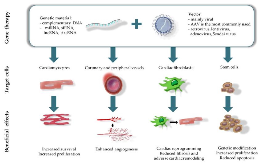
plasmid or viral origin) in combination with adequate pro-angiogenic genes can serve as a
promising tool for successful cardiac tissue regeneration (Figure 1). A well-developed gene-
based system must include the following requirements: sustained long-term therapeutic
effect, ability to target specific cell types and a decreased risk of systemic side-effects
as compared to regular pharmacotherapy. To date, gene transfer systems include two
approaches such as direct tissue injection and intravascular infusion with or without
surgical or catheter-mediated interventions [53]. In addition, for gene therapy in heart
diseases, the goal in most cases is to deliver genetic material directly to the myocardium.
Expression of the targeted gene needs to be sustained over an extended period of time
unless therapy is meant to repair a specific structural defect. Most importantly, the gene
must encode a molecule that plays a critical role in the disease pathogenesis such that by
altering expression of that gene alone, cardiac function will be improved sufficiently or
will favorably alter the disease flow [54]. Thus, gene therapy using appropriate delivery
systems, which meet the aforementioned requirements, and pro-angiogenic factors that
act in processes such as blood flow, metabolic activity, and cardiac functioning are being
applied for cardiac angiogenesis [55]. Among these factors are VEGF, FGF, and hepatocyte
growth factor (HGF). In this section, we will focus on the insights from recent growth factor
gene-based therapy approaches.
Among the pro-angiogenic factors, VEGF has been the most extensively studied. It is
a 45-kD homodimeric glycoprotein that has four main isoforms: VEGF-A (possesses the
ability to bind heparin), -B, -C, and -D. There are additional isoforms in VEGF-A: VEGF121,
VEGF165, which is the most biologically active, VEGF189 and VEGF206 [56]. Many other
known angiogenic factors act at least partly via VEGF-A. VEGF-B has some exceptional fea-
tures compared to other VEGFs since it induces myocardium-specific angiogenesis without
the risk of hyperpermeability and edema. Interestingly, it also seems to alter myocardial
energy metabolism and fatty acid uptake and promote cell survival and compensatory
hypertrophy [57]. The receptors for VEGF are FLT-1 and FLK-1, which activate intracellular
tyrosine kinase. Neuropilin 1 (NP-1) is another receptor for VEGF that binds VEGF165.
NP-1 and FLK-1 are key mediators of the phosphoinositide-3-kinase, and Akt (PI3K/Akt)
and mitogen-activated protein kinase (MAPK) kinase pathways [58].Int. J. Mol. Sci. 2021, 22, 9206 6 of 21
Figure 1. Gene therapy and therapeutic outcomes in CVDs treatment.
VEGF-A isoform has been well studied in a number of preclinical studies and several
clinical trials. However, VEGF-B and VEGF-D isoforms have attracted much attention from
scientists in the past few years [59]. In this regard, Nurro and colleagues demonstrated an
angiogenic capacity of new members of the VEGF family, VEGF-B186 and VEGF-D∆N∆C,
in porcine myocardium [60]. Both adenovirus-mediated VEGF-B186 (AdVEGF-B186)
and AdVEGF-D∆N∆C gene transfers induced efficient angiogenesis in the myocardium,
possibly due to their high solubility in tissues since they do not bind effectively to heparan
sulfates, suggesting that they could be suitable candidates for the induction of therapeutic
angiogenesis for the treatment of refractory angina. In a study by Huusko and colleagues,
it was demonstrated that VEGF-C and VEGF-D are associated with the compensatory
ventricular hypertrophy and that adeno-associated virus subtype 9 (AAV9)-VEGF-B186
gene transfer can rescue the function of the failing heart and postpone the transition
towards HF in mice [61]. Another strategy of gene therapy is based on the concept of a
combined approach using a simultaneous delivery of VEGF165 and HGF genes to alleviate
the symptoms of MI in rats [62]. Combined application of these genes resulted in an increase
of the number of cardiac stem cells in the peri-infarct area and sporadic proliferation of
mature cardiomyocytes. These effects may be explained by the activation of VEGFRs and
c-Met (HGF receptor) that initiate mitogenic signaling cascades. Furthermore, it is reported
that VEGF-D stimulates both angiogenesis and lymphangiogenesis, which has been also
tested in animal models and confirmed by a phase I/IIa clinical study with one-year
follow-up [63]. Another clinical study investigated a novel VEGF-D, a new member of the
VEGF family called VEGF-D∆N∆C, in 30 patients with refractory angina. This phase I/IIa
randomized clinical trial used increasing doses of endocardial adenoviral (ad)-injections
with electroanatomical targeting of injections using a NOGA catheter system. AdVEGF-
D∆N∆C gene therapy is considered to be safe and well tolerated, and confirmed by a
consistent report that lipoprotein (a) level in 50% of patients with refractory angina was
significantly decreased [64].
FGF is another angiogenic factor that has been studied in CVDs. FGF is related to a
family of 22 identified members of pleiotropic proteins in human and mouse and regulatesInt. J. Mol. Sci. 2021, 22, 9206 7 of 21
crucial functions in the heart, ranging from development to homeostasis and disease,
and is considered a cardiomyokine [65,66]. The mechanism of action of FGF proteins
is based on FGF/FGFR signaling, which plays an important role in angiogenesis and
lymphangiogenesis. FGF signaling can influence the whole process of angiogenesis and
cardiomyocyte mitosis. Activation of FGFR1 or FGFR2 has been demonstrated to have a
positive effect on vascular endothelial proliferation. The first step of angiogenesis is ECM
degradation. For instance, FGF1, FGF2, and FGF4 promote the expression of matrix metallo-
proteinases (MMPs) in endothelium cells. The second step in angiogenesis is endothelium
migration. FGF1, FGF2, FGF8, and FGF10 were demonstrated to stimulate endothelium
chemotaxis [67]. In clinical studies, FGF4 showed positive results in patients with stable
angina following a series of AGENT-trials representing a single intracoronary infusion
of adenovirus type 5 vector (Ad5FGF-4) [68]. In an ASPIRE trial, a single intracoronary
infusion of Ad5FGF-4 using catheter-based administration was compared to standard care
without a placebo group. It was planned to recruit 100 participants; however, the study
was terminated apparently due to a low number of patients [69].
Another promising pro-angiogenic factor is HGF. It participates in mediation of
angiogenesis by inducing endothelial cell proliferation, migration and survival. The
HGF receptor c-Met is reported to be expressed on vascular endothelial cells. HGF/c-
Met signaling induces endothelial cell proliferation through the MAPK/ERK and STAT3
pathways. In addition to the direct effect on stimulating endothelial cells to form tubular
structures, HGF can recruit bone marrow endothelial progenitor cells to participate in
angiogenesis in the ischemic area [70]. For cardiac tissue regeneration, HGF either in
plasmid or adenoviral constructs has been tested but only in a small number of studies.
Preclinical studies demonstrated the therapeutic effects of adenovirus carrying the HGF
gene (Ad-HGF) in a minipig model of chronic myocardium ischemia in which an Ameroid
constrictor was placed around the left circumflex branch of the coronary artery. The
data showed that Ad-HGF significantly improves the heart function and blood supply in
chronic myocardium ischemia models [71]. Furthermore, a catheter-based intramyocardial
delivery (NavX) of Ad-HGF was proved to be safe and feasible for Ad-HGF delivery in
pigs [70,72]. In a study by Rong and colleagues, skeletal myoblasts were transduced with
Ad-HGF. Transplantation of HGF-engineered skeletal myoblasts results in reduced infarct
size and collagen deposition, increased vessel density and improved cardiac function
in a rat MI model [73]. The effect might be explained by the overexpression of HGF in
skeletal myoblasts that confers resistance to ischemia in MI. Based on preclinical studies,
the safe application of Ad-HGF was additionally confirmed by clinical trials. One such
application is the use of two delivery approaches: an intramuscular myocardium direct
injection while performing coronary artery bypass surgery (CABG) and a catheter-based
intramyocardial injection guided by the NavX system. An open-label safety trial of Ad-HGF
by a direct multipoint injection into the myocardium of patients, who suffer from coronary
heart disease, showed that no subjective or objective adverse reactions were detected.
Furthermore, there was evidence of revascularization in ischemic regions confirmed by
instrumental examination and physical symptoms [71].
Additionally, some studies have reported the use of the stem cell factor (SCF), which is
a ligand for the c-kit that is a receptor tyrosine kinase. SCF binding to c-kit leads to receptor
dimerization and activation of multiple signaling pathways related to cell recruitment,
differentiation, angiogenesis, and survival [74]. It has been shown that local overexpression
of SCF post-MI induces the recruitment of c-kit+ cells at the infarct border area. Moreover,
gene transfer of membrane-bound human SCF improved cardiac function in the model of
ischemic cardiomyopathy in Yorkshire pigs [75].
The poor angiogenic nature of the adult heart prompted scientists to establish new
therapeutic approaches for cardiac tissue regeneration. Thus, the effectiveness of gene-
based therapy using pro-angiogenic factors may represent a promising therapeutic strategy
to improve cardiac function. However, even though some clinical trials have shownInt. J. Mol. Sci. 2021, 22, 9206 8 of 21
promising results, further large-scale clinical trials need to be performed to clarify their
efficacy and potential clinical application.
3.2. Gene Therapy for Scar Tissue Remodeling
Scar tissue forming after MI has a profound impact on the physiology of cardiac
tissue. Since post-infarction scar is comprised of non-contractile fibrotic tissue, the scarred
region of the heart can no longer contract and relax, leading to systolic and diastolic dys-
functions [76,77]. In addition, the fibrotic area becomes the source of various arrhythmias
because it interferes with the conductive system of the heart [76,77]. Therefore, patients
surviving MI are at a greater risk for morbidity and mortality due to cardiac dysfunction
and arrhythmogenicity caused by the scar tissue.
Several gene therapy strategies have been successfully utilized for scar tissue remod-
eling (Figure 1). One of which is the direct reprogramming of cardiac fibroblasts into
functional cardiomyocytes using a set of specific transcription factors. In vitro studies
found that such a transformation required just three factors—such as Gata4, Mef2c and
Tbx5, which were collectively designated as GMT [78]. Further studies identified that
the use of combinations of 3–4 of the following factors—GATA4, MEF2C, TBX5, HAND2,
MESP1, NKX2.5, and MYOCD—could be as efficient as or even better than the original
GMT combination [79]. Cardiac reprogramming using this gene transfer was further
accomplished in murine MI models using retroviral and lentiviral vectors. The in vivo
reprogramming induced the formation of more mature cardiomyocytes compared to the
same procedure in vitro. In addition, it led to the reduction of fibrosis and improved
cardiac contractility of the injured heart. Despite the fact that early preclinical studies using
cardiac reprogramming have achieved formation of new cardiomyocytes, the efficiency of
this transformation was relatively low, frequently less than 30–40% [79]. Therefore, recent
studies have attempted to combine cardiac reprogramming with other strategies to enhance
cardiac repair [80]. In one of the studies, E-twenty-six Variant transcription factor 2 (ETV2)
was introduced along with GMT genes into the genome of rat MI models using adenoviral
and retroviral vectors, respectively [81]. ETV2 is crucial for the development of new blood
vessels both during embryonic development and repair after injury [82]. Genetic therapy
with a combination of ETV2 and GMT had positive effects on the recovery of the infarcted
hearts. Namely, it induced the formation of new myocardium and blood vessels, reduced
scar size and improved left ventricular function. Notably, the combinational gene transfer
of GMT and ETV2 exhibited synergistic effects, i.e., the beneficial effects of the dual therapy
were significantly more pronounced compared to individual gene transfers.
Direct cardiac reprogramming using conventional retroviral and lentiviral vectors
possesses several limitations, such as low efficiency of fibroblast to cardiomyocyte transd-
ifferentiation as well as potential insertional mutagenesis [83]. In order to address these
obstacles, alternative gene transfer strategies for reprogramming of fibroblasts in the scar
tissue have been considered. Isomi and colleagues have used Sendai virus as a vector to
introduce GMT genes into cardiac fibroblasts of a mouse MI model [84]. The gene transfer
induced the differentiation of fibroblasts into cardiomyocytes which in turn led to reduction
of scar tissue and improved contractile function of the heart for 12 weeks. Importantly,
this is the first study which observed long-term effects of the GMT-Sendai virus vector-
based reprogramming. Another alternative gene carrier for direct cardiac reprogramming
is nanoparticles. In a study by Chang and colleagues [83], cationic gold nanoparticles
loaded with GMT transcription factor genes effectively induced transdifferentiation of
mouse and human fibroblasts into cardiomyocytes in vitro, which was confirmed by their
morphology and expression of cardiac-specific genes. Moreover, the direct injection of
a gold nanoparticles-GMT construct into the infarct area of the murine heart generated
more mature cardiomyocytes compared to in vitro experiments. In addition, the treatment
resulted in a significant decrease in the scar dimensions two weeks after MI in mice. Impor-
tantly, it was shown in this study that the gold nanoparticles-carrier system did not cause
integration of the delivered DNA into the host genome [83].Int. J. Mol. Sci. 2021, 22, 9206 9 of 21
Besides direct cardiac reprogramming, gene therapy to attenuate fibrosis and scar
tissue was also accomplished via ablation of genes regulating fibrosis. In a recent study,
Adapala and colleagues [85] investigated the function of the transient receptor potential
vanilloid 4 (TRPV4) mechanosensitive ion channel in cardiac remodeling after MI using
TRPV4-knockout mice. The absence of TRPV4 was associated with a significant reduction
in scar size and, remarkably, the presence of viable myocardium in the infarction area eight
weeks after MI. Consequently, TRPV4-KO mice had a higher survival rate, and preserved
systolic and diastolic function. The study also demonstrated that TRPV4 mediated differen-
tiation of cardiac fibroblasts into myofibroblasts via a Rho/Rho kinase/MRTF-A pathway.
This was evidenced by the observations that inhibition of TRPV4 caused reduced activity
of Rho kinase as well as activation of TRPV4 with transforming growth factor-β1 (TGF-β1)
or GSK (TRPV4 agonist) that led to the increased expression of MRTF-A. In another study,
genetic ablation of Cullin-RING E3 ubiquitin ligase 7 (CRL7) was performed in order to
attenuate fibrosis caused by pressure overload [86]. In particular, DNA recombination
using Cre-recombinase transferred by AAV9 vector was utilized to obtain CRL7-knockout
mice. Next, the mice were subjected to transverse aortic constriction, which is used to
model chronic pressure overload and consequent fibrosis. CRL7 knock-out resulted in a
50% decrease in myocardial interstitial fibrosis and enhanced survival of cardiomyocytes.
The observed beneficial effects of CRL7 genetic ablation were probably due to enhanced
activation of a phosphatidylinositol 3-kinase (PI3K)/protein kinase B (Akt) signaling path-
way, which regulates cell growth, proliferation, and apoptosis among other processes.
In fact, the authors claim that the study is the first one that elucidated the role of CRL7
in PI3K/Akt regulation in cardiac tissue. Another signaling pathway that was found to
be regulated by CRL7 was related to TGF-β1. Thus, the absence of CRL7 led to lower
expression of TGF-β1 that confirms the pro-fibrotic role of CRL7. In another study, the
role of low-density lipoprotein receptor-related protein 6 (LRP6) in cardiac repair after MI
was investigated using miRNA-mediated cardiomyocyte-specific knock-down of the corre-
sponding gene [87]. The depletion of LRP6 was found to be associated with a significantly
smaller scar size and improved cardiac function in a mouse model of MI. Remarkably, it
also caused enhanced proliferation of cardiomyocytes via the ING5/P21 pathway. It is
important to note that the new cardiomyocytes originated from the existing cardiomyocytes
rather than cardiac progenitor cells. This study is one of the first reports on the functions of
LRP6 in cardiomyocyte proliferation in the adult heart. In summary, scar tissue remodeling
using gene therapy can be accomplished by direct cardiac reprogramming as well as by
ablation of fibrosis-related genes.
3.3. Gene Therapy to Regulate Reactive Oxygen Species
MI is one of the most common causes of death around the world. MI causes death
of cardiomyocytes and irreversible damage to the heart tissue forming an infarcted zone,
which is remodeled into fibrotic/scar tissue [88]. Mitochondrial damage is directly as-
sociated with cardiomyocyte death and myocardial dysfunction by inducing oxidative
stress [89,90]. In addition, hypoxic stress resulting from MI leads to accumulation of reac-
tive oxygen species (ROS), which causes apoptosis of cardiomyocytes and cardiac tissue
injury. Most of the MI therapies aim to restore blood flow using CABG and percutaneous
coronary intervention, which leads to an increase in the concentration of oxygen in ischemic
cells [91]. This sharp increase in the amount of oxygen also induces the high production
of ROS, causing cardiomyocyte injury. This process is called ischemia reperfusion in-
jury (IRI) [92]. Moreover, regeneration of adult cardiomyocytes is limited and results in
post-infarction cardiac dysfunction [91].
Most gene therapies aimed to eliminate post-infarction ROS are interested in the
activation of antioxidant genes or inhibition of ROS producing genes. One of those studies
investigated the effect of brahma-related gene 1 (BRG1), which regulates the chromatin
structure and cardiac gene expression in Nuclear erythroid 2-related factor 2/Heme oxyge-
nase 1 (Nrf2/HO-1) pathway [93]. Nrf2/HO-1 pathway is known to be a main regulatorInt. J. Mol. Sci. 2021, 22, 9206 10 of 21
of cellular antioxidant responses. Translocation of Nrf2 to the nucleus activates the HO-1
and downstream oxidative stress pathway (HO1, glutathionetransferase p1 (GSTP1) or
NAD (P)H:quinone oxidoreductase 1 (NQO1)) [94]. According to Liu and colleagues, over-
expression of BRG1 gene by intramyocardial injection of adenoviral vectors induces the
translocation of Nrf2 and attenuates the oxidative damage in cardiomyocytes. Moreover,
the conducted studies demonstrated that Nrf2 expression, which was induced by BRG1,
increased the activity of HO-1 promoters. On the other hand, Lenti-Brg1 shRNA injection,
which inhibits the BRG1, showed adverse effects decreasing HO-1 expression [93]. Overall,
BRG1 overexpression reduced the size of the infarct scar and improved the function of the
cardiac tissue by decreasing oxidative damage and cell apoptosis. The HO-1 antioxidant
molecule is also regulated by HIF-1α. HIF-1α overexpression with the help of a plasmid
transfection encapsulated in 20-mer peptide-conjugated carboxymethylchitosan nanopar-
ticle in MI mice upregulated the HO-1 activation and expression, which decreased ROS
accumulation both in vitro and in vivo. Moreover, findings proved that downregulation of
ROS-mediated oxidative stress inhibits the expression of BNIP3, which is responsible for
cardiomyocyte apoptosis [95].
Another group of scientists investigated the effect of Zinc finger protein ZBTB20 in the
treatment of post-infarction cardiac tissue. ZBTB20 was delivered with the help of AAV9
system [96]. Based on the results, overexpression of ZBTB20 in post-MI heart increased the
superoxide dismutase (SOD) enzymatic activity, an important antioxidant that converts
superoxide radicals to molecular oxygen, provided cellular defense and inhibited the
activity of malondialdehyde (MDA) and NADPH oxidase [96,97].
Some of recent findings focus on the amelioration of mitochondrial dysfunction to
eliminate ROS and to improve post-infarct cardiac function. Transverse aortic constriction
(TAC) preconditioning of mice before the left coronary artery ligation induced MI showed
a significant reduction in oxidative stress and decreased mitochondrial ROS production.
TAC preconditioning can also be mimicked by the cardiac overexpression of SIRT3 in vivo
with the help of AAV-SIRT3 transfection. Both TAC preconditioning and SIRT3 gene overex-
pression considerably increased the contractile function of heart and, in contrast, decreased
the myocardial scar area and death of cardiomyocytes [98]. In addition, expression of
deacetylated isocitrate dehydrogenase 2, a protein that regulates mitochondrial redox,
reduced the production of mitochondrial ROS and alleviated post-MI injury [98,99].
Furthermore, there has been much research done recently to study the effects of
miRNA, an important regulator in the development and pathophysiology of the cardio-
vascular system, on oxidative stress after MI [100]. Expression of miR-323-3p, Bax, Bcl-2,
SOD1, and SOD2 genes and oxidative stress in cardiomyocytes (H9c2 cells) of miR-323-3p
transfected mice were compared to mice without transfection and with miR-323-3p trans-
fected and H2 O2 treated group at seventh day after MI. Results indicated that miR-323-3p
was downregulated in MI heart and overexpression of miR-323-3p decreased Bax and
significantly increased Bcl-2, SOD1, and SOD2, consequently decreasing ROS production.
Moreover, apoptosis of H9c2 cells decreased and cardiac function of mice improved consid-
erably by targeting the TGF-β2/c-Jun N-terminal kinase (JNK) pathway [101]. Overexpres-
sion of miRNA-187 also attenuated the production of ROS by increasing the expression
of SOD and reducing the intracellular MDA level, while inhibition of miRNA-187 had
an inverse effect under the hypoxia/reoxygenation conditions of cardiomyocytes. This
was achieved by the inhibition of DYRK2, a critical regulator of cell cycle and apoptosis,
using miRNA-187, which consequently reversed the oxidative stress and apoptosis induced
by cardiomyocyte hypoxia/reoxygenation conditions [102]. In addition, upregulation of
miR-340-5p, which has an inhibitory effect on apoptosis, could suppress oxidative stress
and apoptosis after hypoxia/reoxygenation of H9c2 cardiomyocytes by regulating the
expression of Akt1 that upregulates the JNK and NF-κB signaling pathways, the main
mediators of cell apoptosis [103]. Similarly, an aging-regulated long non-coding RNA
(lncRNA) Sarrah demonstrated a protective effect against cardiomyocyte apoptosis by
directly targeting nuclear factor erythroid 2-related factor 2 (NRF2) gene, which regulatesInt. J. Mol. Sci. 2021, 22, 9206 11 of 21
the expression of antioxidant proteins that protect against oxidative damage. In an MI
mouse model, overexpression of lncRNA Sarrah reduced cardiomyocyte apoptosis, while
inducing endothelial cell proliferation and augmenting cardiac contractile function [104].
In contrast, other studies demonstrated that miRNA-124 is upregulated after MI under
oxidative stress [105,106]. Application of antisense inhibitor oligodeoxyribonucleotides
(AMO-124) alleviated the oxidative stress by neutralizing the miRNA-124, whose main
target is STAT3, a key cellular survival factor that suppresses the apoptosis pathway [105].
Overall, gene therapy, specifically the application of various miRNAs, antioxidant gene
activators and molecules, whose target is oxidative stress, is a very promising approach
to eliminate ROS after MI and hypoxia/reoxygenation conditions and to improve cardiac
function.
3.4. Gene Therapy to Reduce Apoptosis
Apoptosis has a significant impact on the regeneration of myocardial cells. Excessive
apoptosis following hypoxia, oxidative stress and endoplasmic reticulum stress can con-
tribute to the development of myocardial ischemia, IRI, cardiac remodeling, and atheroscle-
rosis [107]. It also significantly increases the death of cardiomyocytes in ischemic heart
disease (IHD) [108]. Therefore, targeting apoptotic agents and pathways can be an efficient
strategy to promote cardiac tissue regeneration after CVDs. In the previous chapter, al-
leviation of oxidative stress using gene therapy to prevent apoptosis was discussed. In
this section, other strategies to suppress apoptosis, namely, those that target molecular
pathways involved in cellular death and survival will be covered.
Non-coding RNAs (ncRNAs) such as miRNAs, lncRNAs and circular RNAs (circR-
NAs), play an important role in the regulation of physiologic and pathologic signaling
pathways in cardiomyocytes and can be used to regulate apoptosis in CVDs and im-
prove cardiac tissue regeneration [109]. Yan and colleagues demonstrated that in vitro
overexpression of miR-31a-5p, a leading member of the miRNA-31 family, can protect
against apoptosis and increase myocardial cell survival through suppression of angiotensin
II-induced apoptotic pathway and caspase-3 activity by targeting Tp53 [110]. Another miR-
NAs, miR378 *, demonstrated a cardioprotective effect by inhibiting endoplasmic reticulum
stress-induced cell apoptosis via controlling expression of calcium-binding protein called
calumenin in doxorubicin (Dox)-induced cardiomyopathic mouse hearts [111]. Moreover,
another miRNA, miR-181c, was found to be suppressed in a mouse model of Dox-induced
HF and its overexpression impeded cardiomyocyte apoptosis via PI3K/Akt pathway [112].
At the same time, it was reported that lncRNA UCA1 protects rat cardiomyocytes against
hypoxia/reoxygenation induced apoptosis by inhibiting miR-143/MDM2/p53 signaling
axis [113]. Moreover, adenovirus mediated expression of lncRNA GAS5 also decreases
cardiomyocyte apoptosis through downregulation of transmembrane protein sema3a in
an MI mouse model [114]. LncRNA FTX is also downregulated after IRI injury and en-
hanced expression of FTX attenuates cardiomyocyte apoptosis by targeting miR-29b-1-5p
and Bcl2l2 [115]. Finally, a circRNA circ-Amotl1 demonstrated its ability to facilitate
the cardio-protective nuclear translocation of pAkt by binding to both phosphoinositide
dependent kinase-1 (PDK1) and Akt1. Injection of a circ-Amotl1 plasmid resulted in in-
creased cardiomyocyte survival, decreased apoptosis and demonstrated a protective effect
against Dox-induced cardiomyopathy in mice [116]. Zhu and colleagues also reported
that AAV9 mediated cardiac overexpression of circRNA SNRK can target miR-103-3p by
promoting cardiac repair via GSK3β/β-catenin pathway in rats with MI. Particularly, it
reduced apoptosis, promoted cardiomyocyte proliferation, improved cardiac functions and
increased angiogenesis [117]. Furthermore, Garikipati and colleagues demonstrated that
AAV9 mediated delivery of circRNA CircFndc3b decreased cardiac apoptosis, enhanced
neovascularization and left ventricle (LV) functions by interacting with the RNA binding
protein fused in Sarcoma to regulate expression of VEGFs [118]. Therefore, these findings
demonstrate that gene therapy with ncRNAs has great potential as a therapeutic strategyInt. J. Mol. Sci. 2021, 22, 9206 12 of 21
to protect against apoptosis in CVDs. However, further preclinical studies are needed to
determine the anti-apoptotic effects of ncRNAs in large animal models.
Another strategy against cardiac apoptosis is to target genes encoding S100 family
proteins. S100 proteins are highly acidic calcium-binding proteins involved in intracellular
calcium homeostasis in different tissues and organs. Several members of this family are also
expressed in cardiac tissue and their impairment can lead to the development of HF [119].
S100A1, a predominant member of the S100 protein family, is a crucial regulator of cardiac
contractility. Due to its ability to enhance sarcoplasmic reticulum Ca2+ fluxes and increase
SERCA2a enzyme activity, it can lead to the significant limitation of myocardial necrosis
and development of HF [120]. A recent study conducted by Jungi and colleagues reported
that S100A1 gene overexpression through the AAV9 vector can have cardioprotective
effects in male Lewis rats after IRI. Particularly, S100A1 overexpressing hearts demonstrated
improvement in LV functions, whereas the presence of necrotic markers, including troponin
T (TnT), lactate dehydrogenase (LDH), and FoxO pro-apoptotic transcription factor were
decreased [121]. Furthermore, a new liquid chromatography tandem mass spectrometry
(LC-MS/MS)-based bioanalytical method was utilized for simultaneous quantitation of
high homologous human and pig S100A1 proteins (only a single amino acid difference
between the sequences). Therefore, the ability to accurately measure human S100A1 in
pig hearts can enable future gene therapy studies in large animals [122]. Another member
of the S100 family, S100A6, increases in the peri-infarct zone after MI in rats and protects
cardiomyocytes from TNF-α-induced apoptosis by binding to p53 and decreasing its
phosphorylation [123]. It was recently demonstrated that S100A6 gene delivery and its
further cardiac overexpression attenuates cardiomyocyte apoptosis and reduces infarct
size after myocardial IRI in both in vitro and in vivo models [124]. Thus, these results
demonstrate that gene therapy mediated S100 protein expression can be an efficient method
to attenuate cardiac apoptosis and protect cardiomyocytes against IRI injury.
Genetically modified stem cells can also be applied to protect against apoptosis by
enhancing expression of various anti-apoptotic genes and factors. Cho and colleagues
demonstrated that overexpression of lymphoid enhancer-binding factor-1 (LEF1) gene in
human umbilical cord blood-derived mesenchymal stem cells (hUCB-MSCs) increased
their cell proliferation and protected from hydrogen peroxide-induced apoptosis in in vitro
experiments. Moreover, construction of hUCB-MSCs with overexpressed LEF1 using
CRISPR/Cas9 system and further transplantation, demonstrated an enhanced cell survival
rate and increased cardio-protective effects in an animal model of MI [125]. In another
recent study, interleukin (IL)-10 gene was transfected into the genomic locus of amni-
otic mesenchymal stem cells (AMM) using transcription activator–like effector nucleases
(TALEN). Further transplantation of IL-10 gene-edited AMM in an MI mouse model de-
creased the number of apoptotic cells and increased capillary density in an ischemic heart
and as a result, increased LV functions and reduced infarct size [126]. At the same time,
transplantation of bone marrow derived MSCs edited by CRISPR activation system to
overexpress IL-10 also decreased apoptosis of cardiac cells, increased angiogenesis, and
inhibited infiltration and production of proinflammatory factors in a diabetic MI mouse
model [127]. Therefore, the therapeutic effects of genetically modified stem cells can be
beneficial against apoptosis caused by cardiac ischemia. In summary, gene therapy with
ncRNAs, S100 proteins or modified stem cells can be a potential and efficient strategy
against cardiomyocyte death by targeting molecular pathways involved in apoptosis and
cell survival in heart diseases.
4. Recent Ongoing Clinical Trials
Currently, several ongoing clinical trials are investigating the effects of various gene
therapies on cardiac regeneration in CVDs. According to the ClinicalTrials.gov (accessed
on 12 July 2021), there are a total of seven gene therapy trials, including six studies that are
actively recruiting (EXACT, ReGenHeart, Korean trial, NAN-CS101) or planning to recruit
(AFFIRM, CUPID-3) and one active clinical trial (EPICCURE).Int. J. Mol. Sci. 2021, 22, 9206 13 of 21
The EXACT Trial is a phase I/II, first-in-human, multicenter, open-label, single arm
dose escalation study recruiting 44 patients with refractory angina caused by CAD to
evaluate the induction of therapeutic angiogenesis in ischemic myocardium by XC001
(AdVEGFXC1) [NCT04125732]. AdVEGFXC1 is an adenovirus type-5 vector expressing
the hybrid VEGF with its three major isoforms (121, 165, and 189) [128]. In this trial,
administration is at various doses by transthoracic epicardial procedure to 12 patients,
followed by an increase in the maximum tolerated dose with 32 additional patients, and
the main outcome measurements will be safety and side effect assessment.
ReGenHeart is a randomized, double-blinded, placebo-controlled multicenter phase
II study enrolling patients with refractory angina pectoris to evaluate safety and efficacy
of AdVEGF-D gene transfer [NCT03039751]. The trial is recruiting 180 participants in
six different centers to whom revascularization cannot be performed. AdVEGF-D will
be injected into myocardium through a catheter at 10 different sites and compared with
a similar placebo treatment. The primary endpoint is to assess improvement in exercise
tolerance and relief of angina symptoms at six months after injection.
EPICCURE is a randomized, placebo-controlled, double-blind, multicenter, phase
II clinical trial testing the effect of VEGF-A165 mRNA loaded in biocompatible citrate-
buffered saline (AZD8601) in patients with decreased LV function and undergoing CABG
[NCT03370887] [129]. AZD8601 will be injected during the surgery as 30 epicardial injec-
tions in a 10-min extension of cardioplegia under the control of quantitative 15O-water
positron emission tomography (PET) imaging. At the moment, 11 patients are enrolled in
the study, and the primary endpoint is to investigate safety and tolerability of the gene ther-
apy up to six months after the surgery. MRNA-based technology used in the EPICCURE
trial is a novel revolutionizing approach for gene delivery that can be utilized for a variety
of purposes such as protein replacement, gene editing, infectious diseases, cancer vaccines,
and others [130]. There are multiple advantages of using an mRNA approach over conven-
tional DNA-based strategies [131]. Firstly, unlike DNA, mRNA does not require entering
the nucleus to be functional, making it more efficient. Secondly, mRNA-based strategy is
much safer compared to DNA delivery with viral vectors since mRNA does not integrate
into the host genome, eliminating the risk of insertional mutagenesis. Finally, as it has
been proven by SARS-CoV-2 mRNA-based vaccines, mRNA production can be very rapid
and capable of adapting to even high emergency conditions like pandemics. Indeed, two
SARS-CoV-2 mRNA-based vaccines approved by the US Food and Drug Administration,
Pfizer/BioNTech (BNT162b2), and Moderna (mRNA-1273), have been shown to be highly
efficient in protecting against COVID-19 with 90–95% efficiency [132]. Moreover, a recent
study published in Nature found that the two mRNA vaccines induced a persistent and ro-
bust germinal center response indicating that these vaccines produced strong and efficient
humoral immune responses [133]. The benefits of mRNA-based technology have been
utilized for cardiovascular gene therapy as well [134]. Specifically, mRNA therapeutics
using genes for VEGF, IGF-1, and other proteins for heart regeneration were shown to be
safe and effective in various animal models.
The AFFIRM study is a phase III clinical trial designed to determine the effect of
intracoronary infusion of the human FGF-4 DNA sequence encoded in Ad5FGF-4 on
ameliorating refractory angina symptoms and improving patient quality of life. This study
will enroll 160 patients with refractory angina and the primary endpoint is the change of
exercise tolerance test (ETT) duration over six months after intervention (NCT02928094).
However, previous clinical trials with Ad5FGF-4 have demonstrated specific beneficial
effects of this gene therapy only in females, while no difference was observed between
placebo and treatment groups among male patients [68].
A phase II trial is ongoing in Korea to test the safety and efficacy of VM202RY, which
is a DNA plasmid loaded with two isoforms of HGFs (723 and 728) (NCT03404024). A
previous phase I trial demonstrated that VM202RY can improve cardiac perfusion and
inhibit cardiac remodeling in patients with MI and angina [135]. Now they are recruiting
108 patients with acute MI for transendocardial injection of VM202RY through catheterInt. J. Mol. Sci. 2021, 22, 9206 14 of 21
to evaluate its safety, tolerability, and efficacy compared with placebo. Primary outcome
measurements are to determine the maximum tolerated dose and assess LV ejection fraction
after six months. Estimated study completion date was April, 2020, however, no results
have yet been reported in English.
The CUPID-3 study is a randomized, double-blind, placebo-controlled phase I/II
trial investigating the effect of SRD-001, an adeno-associated virus serotype 1 vector
expressing the transgene for sarcoplasmic/endoplasmic reticulum Ca2+ ATPase 2a isoform
(SERCA2a), on patients with cardiomyopathy and HF with reduced ejection fraction
(NCT04703842). According to the authors, targeting SERCA2a enzyme expression by SRD-
001 can correct defective intracellular Ca2+ hemostasis, subsequently improving cardiac
contractility. In addition, SRD-001 can also correct the impaired endothelium-dependent
vasodilatation, improve coronary blood flow and prevent further progression of HF. To
determine this, 56 participants will be enrolled in the study until June 2021 and SRD-001
will be delivered through one-time intracoronary infusion. As a primary endpoint, change
from baseline in symptomatic parameters (quality of life, HF severity), physical parameters,
LV function/remodeling, and rate of recurrent and adverse events will be measured up to
12 months (NCT04703842).
NAN-CS101 is a phase I, prospective, multi-center, open-label, sequential dose escala-
tion study to test the safety and efficacy of one dose intracoronary infusion of BNP116.sc-
CMV.I1c in patients with HF (NYHA class III) (NCT04179643). BNP116.sc-CMV.I1c is an
AAV vector with a chimeric AAV2/AAV8 capsid containing an active inhibitor of protein
phosphatase-1 (I-1) transgene. Previous preclinical studies on large animals demonstrated
that chimeric AAV vectors can selectively target cardiac muscle, while I-1 inhibits expres-
sion of protein phosphatase-1 and increases cardiac expression of SERCA2a enzyme leading
to the improvement of cardiac functions [136,137]. In this small phase I study, 12 partici-
pants will be enrolled and injected intracoronary with different doses of BNP116.sc-CMV.I1.
Primary endpoints will be the safety indicators measured by adverse events, mortality, and
hospitalization up to 12 months after the procedure.
Lastly, another randomized, double-blind, placebo-controlled, phase II clinical trial
has tested the effect of intracoronary delivery of adenovirus 5 encoding adenylyl cyclase
6 (Ad5.hAC6/RT100) in patients with HF. Adenylyl cyclase 6 (AC) is an enzyme that
catalyzes the conversion of adenosine triphosphate to cyclic adenosine monophosphate
and can have beneficial effects on cardiac myocytes and eventually improving LV function.
Fifty-six subjects received intracoronary administration of Ad5.hAC6 or placebo and
were followed up to 1 year. AC6 gene transfer safely increased LV function compared
to a standard single-dose HF therapy and decreased admission rate in the treatment
group [138]. A larger phase III trial (FLOURISH) with the same drug was recently registered
in clinicaltrials.gov (accessed on 12 July 2021); however, the current status of the study
is withdrawn due to the reevaluation of “clinical development plans and strategy for
RT-100” (NCT03360448). Therefore, despite its withdrawn status, a newly designed trial
with Ad5.hAC6/RT100 can be conducted in the future. Table 1 summarizes the ongoing
clinical trials.
Overall, most ongoing trials related to cardiac regeneration are focused on gene ther-
apy targeting the expression of different growth factors and their isoforms to improve
angiogenesis. This might be justified by the fact that extended angiogenesis effectively
improves vascular and cardiac tissue regeneration leading to the enhancement of cardiac
functions. However, there are still two gene therapy studies targeting the enzyme expres-
sion (CUPID-3 and NAN-CS101). Thus, this data demonstrates that gene therapy can be an
efficient and safe approach to improve cardiac regeneration and further implementation of
this strategy into clinical trials has the potential to develop into novel methods of treatment
for CVDs.You can also read