In vivo base editing rescues Hutchinson-Gilford progeria syndrome in mice - Gwern.net

Page created by Brett Pierce
 
CONTINUE READING
In vivo base editing rescues Hutchinson-Gilford progeria syndrome in mice - Gwern.net
Article

In vivo base editing rescues Hutchinson–
Gilford progeria syndrome in mice

https://doi.org/10.1038/s41586-020-03086-7                      Luke W. Koblan1,2,3,13, Michael R. Erdos4,13, Christopher Wilson1,2,3, Wayne A. Cabral4,
                                                                Jonathan M. Levy1,2,3, Zheng-Mei Xiong4, Urraca L. Tavarez4, Lindsay M. Davison5,
Received: 9 June 2020
                                                                Yantenew G. Gete6, Xiaojing Mao6, Gregory A. Newby1,2,3, Sean P. Doherty5, Narisu Narisu4,
Accepted: 30 November 2020                                      Quanhu Sheng7, Chad Krilow4, Charles Y. Lin8,9,12, Leslie B. Gordon10,11, Kan Cao6,
                                                                Francis S. Collins4 ✉, Jonathan D. Brown5 ✉ & David R. Liu1,2,3 ✉
Published online: xx xx xxxx

    Check for updates
                                                                Hutchinson–Gilford progeria syndrome (HGPS or progeria) is typically caused by a
                                                                dominant-negative C•G-to-T•A mutation (c.1824 C>T; p.G608G) in LMNA, the gene
                                                                that encodes nuclear lamin A. This mutation causes RNA mis-splicing that produces
                                                                progerin, a toxic protein that induces rapid ageing and shortens the lifespan of children
                                                                with progeria to approximately 14 years1–4. Adenine base editors (ABEs) convert
                                                                targeted A•T base pairs to G•C base pairs with minimal by-products and without
                                                                requiring double-strand DNA breaks or donor DNA templates5,6. Here we describe the
                                                                use of an ABE to directly correct the pathogenic HGPS mutation in cultured fibroblasts
                                                                derived from children with progeria and in a mouse model of HGPS. Lentiviral delivery
                                                                of the ABE to fibroblasts from children with HGPS resulted in 87–91% correction of the
                                                                pathogenic allele, mitigation of RNA mis-splicing, reduced levels of progerin and
                                                                correction of nuclear abnormalities. Unbiased off-target DNA and RNA editing
                                                                analysis did not detect off-target editing in treated patient-derived fibroblasts. In
                                                                transgenic mice that are homozygous for the human LMNA c.1824 C>T allele, a single
                                                                retro-orbital injection of adeno-associated virus 9 (AAV9) encoding the ABE resulted
                                                                in substantial, durable correction of the pathogenic mutation (around 20–60% across
                                                                various organs six months after injection), restoration of normal RNA splicing and
                                                                reduction of progerin protein levels. In vivo base editing rescued the vascular
                                                                pathology of the mice, preserving vascular smooth muscle cell counts and preventing
                                                                adventitial fibrosis. A single injection of ABE-expressing AAV9 at postnatal day 14
                                                                improved vitality and greatly extended the median lifespan of the mice from 215 to 510
                                                                days. These findings demonstrate the potential of in vivo base editing as a possible
                                                                treatment for HGPS and other genetic diseases by directly correcting their root cause.

Hutchinson–Gilford progeria syndrome (HGPS or progeria) is a rare                                premature atherosclerosis, loss of vascular smooth muscle cells (VSMCs)
genetic disease characterized by accelerated ageing4. In over 90% of                             and vascular stiffening—is the predominant cause of death in children
patients with HGPS, the disease is caused by a single de novo point muta-                        with progeria, who have an average lifespan of approximately 14 years3,7–11.
tion (c.1824 C>T; p.G608G) in the lamin A (LMNA) gene1,2. This muta-                             Although strategies for treating progeria, such as global inhibition of
tion potentiates a cryptic splice site in exon 11, leading to a mis-splicing                     protein farnesylation3,12,13, offer benefits to patients, no approach has
event that results in the loss of 50 amino acids from the lamin A protein1,2                     yet been reported to directly reverse the mutation that causes HGPS14–17.
(Fig. 1a). This truncated protein, which is known as progerin, lacks a prote-                      The dominant-negative function of progerin poses challenges for the
olytic cleavage site for ZMPSTE24, which cleaves the farnesylated C termi-                       treatment of HGPS by gene augmentation or gene disruption strate-
nus of wild-type pre-lamin A1. Progerin protein impairs nuclear structure                        gies. Overexpression of wild-type LMNA does not rescue cellular phe-
and function, culminating in premature senescence and cell death7. The                           notypes18. Although CRISPR–Cas9-mediated genetic disruption of
pathogenic mutation is dominant-negative, so a single copy of the allele                         the pathogenic allele has been reported to improve phenotypes in
is sufficient to cause progeria3. Cardiovascular disease—characterized by                        mouse models of progeria15–17, the resulting diversity of uncharacterized

1
 Merkin Institute of Transformative Technologies in Healthcare, Broad Institute of Harvard and MIT, Cambridge, MA, USA. 2Department of Chemistry and Chemical Biology, Harvard University,
Cambridge, MA, USA. 3Howard Hughes Medical Institute, Harvard University, Cambridge, MA, USA. 4National Human Genome Research Institute, National Institutes of Health, Bethesda, MD,
USA. 5Division of Cardiovascular Medicine, Vanderbilt University Medical Center, Nashville, TN, USA. 6Department of Cell Biology and Molecular Genetics, University of Maryland, College Park,
MD, USA. 7Department of Biostatistics, Vanderbilt University Medical Center, Nashville, TN, USA. 8Department of Molecular and Human Genetics, Baylor College of Medicine, Houston, TX, USA.
9
 Therapeutic Innovation Center, Department of Biochemistry and Molecular Biology, Baylor College of Medicine, Houston, TX, USA. 10Hasbro Children’s Hospital, Alpert Medical School of
Brown University, Providence, RI, USA. 11Boston Children’s Hospital, Harvard Medical School, Boston, MA, USA. 12Present address: Kronos Bio Inc., Cambridge, MA, USA. 13These authors
contributed equally: Luke W. Koblan, Michael R. Erdos. ✉e-mail: francis.collins3@nih.gov; jonathan.d.brown@vumc.org; drliu@fas.harvard.edu

                                                                                                                                                      Nature | www.nature.com | 1
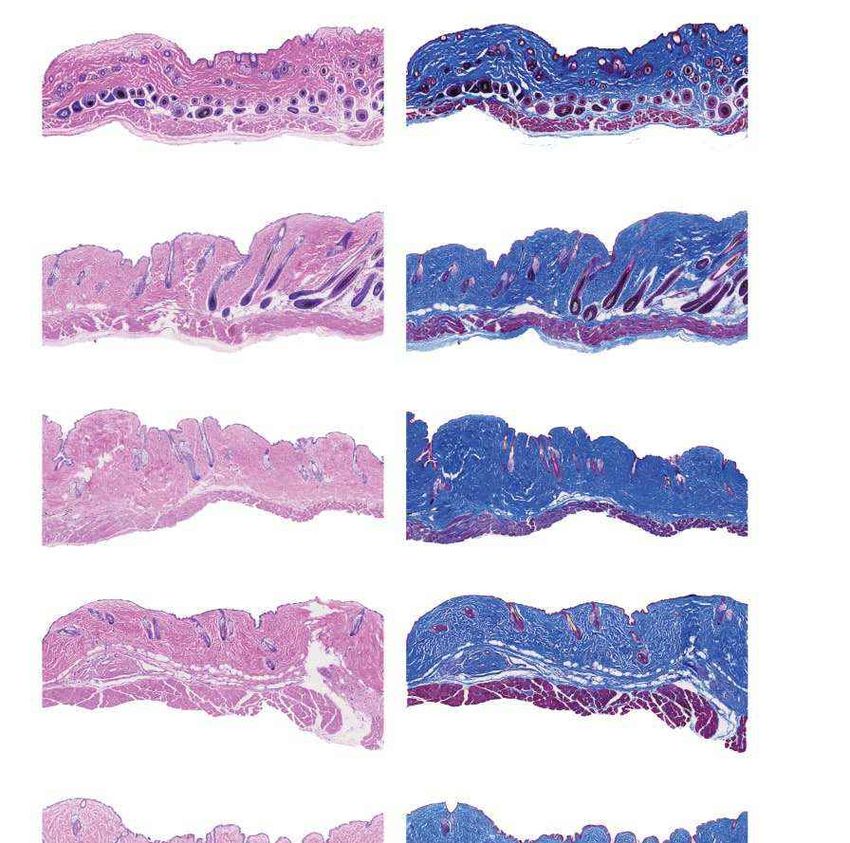
In vivo base editing rescues Hutchinson-Gilford progeria syndrome in mice - Gwern.net
Article
                                     a                         Exon 11          Intron   Exon 12                                       LMNA mRNA
                                                                                                                                                                 Lamin A     b                                                       HGADFN167                                                               HGADFN188

                                                                                                                                                                                                                                                                  Base identity at c.1824 (%)
                                                                                                                                                                           Base identity at c.1824 (%)
                                                                                                                                                                                                         100                                                                                    100
                              ¥ &$**7***&¥&$* *7¥                                    ¥ $* $*&&¥                                  ¥ &$**7***&¥&$* $*&&¥                                                                                                                                                                        C
                                                                                                                                                                                                                                                                                                                              T
                                                                                                                                                                                                         80                                                                                     80
                                                                         Base editing by
                                                                         ABE7.10max-VRQR                                                                        Progerin                                 60                                                                                     60
                                                               Exon 11          Intron   Exon 12                                     LMNAΔ150 mRNA                                                       40                                                                                     40
                              ¥&$**7***7¥&$* *7¥                                     ¥ $* $*&&¥                                       ¥&$* $*&&¥
                                                                                                                                                                                                         20                                                                                     20

                                                                                                                                                                                                           0                                                                                     0
                                 c                                     Untreated HGADFN167                                               Untreated HGADFN188                                              Untreated
                                                                                                                                                                                                                    10 d 20 d
                                                                                                                                                                                                                                                                                                 Untreated
                                                                                                                                                                                                                                                                                                           10 d 20 d
                                                                       ABE7.10max-VRQR, 10 d                                             ABE7.10max-VRQR, 10 d                                                      ABE7.10max-                                                                            ABE7.10max-
                              Normalized gene expression

                                                                                                    Normalized gene expression
                                                           7           ABE7.10max-VRQR, 20 d                                     7       ABE7.10max-VRQR, 20 d                                                        VRQR                                                                                   VRQR
                                                                       Unaffected parent                                                 Unaffected parent
                                                                                                                                                                             d

                                                                                                                                                                                                                                          HGADFN167

                                                                                                                                                                                                                                                      HGADFN167

                                                                                                                                                                                                                                                                                                 HGADFN188

                                                                                                                                                                                                                                                                                                             HGADFN188
                                                           6                                                                     6

                                                                                                                                                                                                                        Unaffected
                                                           5                                                                     5

                                                                                                                                                                                                                        parent

                                                                                                                                                                                                                                                      + ABE

                                                                                                                                                                                                                                                                                                             + ABE
                                                           4                                                                     4                                **
                                                           3       *                                                             3
                                                                                                                                         *
                                                           2                                                                     2
                                                                                                                                                                                                                                                                                                                         Lamin A
                                                           1                   ****                                              1                 ****                                                                                                                                                                  Progerin
                                                                                 ****                                                                ****                                                                                                                                                                Lamin C
                                                           0                                                                     0
                                                                  LMNA        Progerin       LMNC                                      LMNA        Progerin    LMNC
                                                                                                                                                                                                                                                                                                                         Histone
                                     e                     Lamin A/C              Progerin          Nuclei                                     Merge                                                                                                                                                                     H3

                                                                                                                                                                             f                                                       80
                                                                                                                                                              Unaffected

                                                                                                                                                                                                          Morphologically abnormal
                                                                                                                                                              parent
                                                                                                                                                                                                                                     60

                                                                                                                                                                                                                nuclei (%)
                                                                                                                                                                                                                                     40                                                                      ****
                                                                                                                                                              HGADFN167
                                                                                                                                                                                                                                     20

                                                                                                                                                                                                                                      0

                                                                                                                                                                                                                                           re ed

                                                                                                                                                                                                                                                 7

                                                                                                                                                                                                                                           AB 67
                                                                                                                                                              HGADFN167

                                                                                                                                                                                                                                              16
                                                                                                                                                                                                                                             nt

                                                                                                                                                                                                                                              E
                                                                                                                                                                                                                                         + N1
                                                                                                                                                                                                                                         pa ect

                                                                                                                                                                                                                                           FN
                                                                                                                                                              + ABE

                                                                                                                                                                                                                                           F
                                                                                                                                                                                                                                          ff

                                                                                                                                                                                                                                        AD

                                                                                                                                                                                                                                        AD
                                                                                                                                                                                                                                        na
                                                                                                                                                                                                                                       U

                                                                                                                                                                                                                                      G

                                                                                                                                                                                                                                      G
                                                                                                                                                                                                                                     H

                                                                                                                                                                                                                                     H
Fig. 1 | ABE-mediated correction of the LMNA c.1824 C>T mutation in                                                                                           technical replicates. d, Western blot of unaffected parent cells, untreated
fibroblasts derived from patients with progeria. a, The LMNA c.1824 C>T                                                                                       patient-derived cells or patient-derived cells 20 days after ABE lentiviral
mutation potentiates a cryptic splice site in exon 11 of the LMNA gene, resulting                                                                             treatment using the JOL2 antibody specific for human lamin A, progerin and
in an in-frame deletion of 150 nt (LMNAΔ150) and production of the pathogenic                                                                                 lamin C. Complete blots with molecular weight markers are available in
progerin protein. b, LMNA c.1824 nucleotide identity in untreated                                                                                             Supplementary Fig. 1. Additional independent biological replicates are
patient-derived HGADFN167 and HGADFN188 cells and in cells 10 or 20 days                                                                                      provided in Extended Data Fig. 1. e, Nuclear morphology of unaffected parent
after treatment with ABE7.10max-VRQR lentivirus. Data are mean ± s.d. of five                                                                                 cells, untreated HGADFN167 cells or HGADFN167 cells 20 days after treatment
technical replicates. c, Quantification by digital droplet PCR (ddPCR) of LMNA,                                                                               with ABE-expressing lentivirus, stained with a lamin-A-specific antibody, a
progerin and LMNC (a normal alternative splice form) transcripts in untreated                                                                                 progerin-specific antibody or DAPI. Scale bars, 20 μm. Additional replicates
patient-derived cells, cells 10 or 20 days after treatment with ABE lentivirus and                                                                            were not performed. f, Frequency of morphologically abnormal nuclei in
cells from an unaffected parent. Gene expression levels were normalized to                                                                                    samples of cells shown in e. Data are mean ± s.d. from three counts of
transferrin receptor (TFRC) expression levels. Data from the unaffected parent                                                                                independent images from the experiment in e. *P < 0.05, **P < 0.01, ***P < 0.001,
are shown in both graphs for comparison. Data are mean ± s.d. of three                                                                                        ****P < 0.0001 by Student’s unpaired two-sided t-test.

insertion and deletion (indel) products at the target locus together                                                                                          and protein levels. Mice treated at postnatal day 14 (P14) showed a
with the risk of disrupting the wild-type LMNA allele, which differs only                                                                                     notable improvement in vascular disease compared to saline-injected
at a single base pair from the pathogenic allele19,20, pose challenges                                                                                        controls, with aortic VSMC counts and adventitial fibrosis indistin-
to clinical translation of gene disruption strategies to treat progeria.                                                                                      guishable from those of wild-type mice, as well as reduced numbers of
   Base editors are genome editing agents that directly convert tar-                                                                                          progerin-positive VSMCs and increased numbers of lamin A or lamin C
geted base pairs without making double-strand DNA breaks6. Cytosine                                                                                           (lamin A/C)-positive VSMCs. The median lifespan of ABE-treated mice
base editors (CBEs)21 convert C•G to T•A, whereas adenine base editors                                                                                        with progeria was up to 2.4-fold longer than that of saline-injected con-
(ABEs)5 convert A•T to G•C. Base editors function in many mitotic and                                                                                         trols. These findings suggest a potential therapeutic strategy for HGPS
post-mitotic cell types and in a wide array of organisms6. ABEs use a                                                                                         that directly corrects the causative mutation in vivo, and will inform
laboratory-evolved deoxyadenosine deaminase to convert adenine to                                                                                             applications of base editors in the treatment of other genetic diseases.
inosine (which base pairs like guanine) within a small window of around
4–5 nucleotides at a Cas-protein-specified locus, and induce the cell
to replace the complementary thymine with cytosine by nicking and                                                                                             ABE corrects the HGPS mutation in patient cells
stimulating repair of the non-edited strand5,6.                                                                                                               To position the pathogenic LMNA c.1824 C>T mutation within the activ-
   Here we report ABE-mediated correction of the LMNA c.1824 C>T                                                                                              ity window of an ABE5 (positions 4–7, where the protospacer-adjacent
mutation in fibroblasts derived from children with HGPS and in a mouse                                                                                        motif (PAM) is positions 21–23), we chose a target site with an NGA PAM
model in which mice contain two genomically integrated copies of the                                                                                          that places the mutation at protospacer position 6. To target this PAM,
human LMNA c.1824 C>T progeria allele22. In cultured patient-derived                                                                                          we used ABEmax-VRQR23, which combines an optimized ABE7.10 vari-
cells, we observed efficient (around 90%) genomic DNA correction that                                                                                         ant24 with an engineered SpCas9-VRQR variant25 that targets NGA PAMs.
ameliorates pathogenic mis-splicing of the LMNA transcript, reduces                                                                                              We tested the ability of ABEmax-VRQR to correct the mutation in two pri-
the abundance of progerin protein and restores normal nuclear mor-                                                                                            mary fibroblast cell lines derived from patients with progeria—HGADFN167
phology. When delivered into a mouse model of human progeria by                                                                                               and HGADFN188 (Methods)—using a lentivirus to deliver ABEmax-VRQR
single retro-orbital injection of therapeutically relevant doses of                                                                                           and the sgRNA targeting the LMNA c.1824 C>T mutation. After lentiviral
AAV9 encoding the ABE and single-guide RNA (sgRNA), the ABE cor-                                                                                              transduction and puromycin selection of HGADFN167 and HGADFN188
rected the LMNA c.1824 C>T allele in various tissues at the DNA, RNA                                                                                          cells, we observed 84% and 85% correction of the pathogenic mutation

2 | Nature | www.nature.com
In vivo base editing rescues Hutchinson-Gilford progeria syndrome in mice - Gwern.net
versus diseased
 a                                      100                                                                 HGADFN167
                                                                                                                                   d                                                                                                             e

                                                                                                                                                                                                                                                     ABE-treated

                                                                                                                                                                                                                                                     ABE-treated
                                                                                                                                                                  Raw z-score
     Total sequencing reads with
                                                                                                                                                                                                                                                                            log2(FDR)
      A•T converted to G•C (%)

                                                                                                                                                                                                                                                     WT versus

                                                                                                                                                                                                                                                     WT versus
                                             80                                                             HGADFN188

                                                                                                                                                                                                                                                     diseased
                                                                                                                                                                                                                                                                          10 0 –10 –20
                                             60                                                                                                        2      1               0                   –1                 –2
                                             40
                                                                                                                                                                                                                                                                       APC-dependent
                                             20                                                                                                                                                                                                                        catabolism
                                                                                                                                                                                                                                                                       Cell−cell adhesion
                                             0.3                                                                                                                                                                                                                       plas. mem.
                                             0.2                                                                                                                                                                                                                       Chromosomal region
                                             0.1                                                                                                                                                                                                                       Chromosome
                                               0 //                                                                                                                                                                                                                    centromeric region
                                              On-target                All adenine bases (89) within the protospacers
                                               editing             of 32 candidate off-target DNA sites from CIRCLE-seq                                                                                                                                                Chromosome localization
 b                                                                                                                                                                                                                                                                     Chromosome
                                              50                                                                                                                                                                                                                       segregation
                                                                                                           HGADFN167
                                                                                                                                                                                                                                                                       Condensed
          Frequency of uncorrected

                                                                                                           HGADFN167 + ABE, 10 d                                                                                                                                       chromosome
             LMNA transcript (%)

                                              40
                                                                                                           HGADFN167 + ABE, 20 d                                                                                                                                       DNA recombination
                                              30
                                                                                                           HGADFN188                                                                                                                                                   DNA replication
                                              20                                                           HGADFN188 + ABE, 10 d                                                                                                                                       Homophilic cell
                                                                                                                                                                                                                                                                       adhesion plas. mem.
                                                                                                           HGADFN188 + ABE, 20 d                                                                                                                                       Kinetochore
                                              10
                                                                                                           Unaffected parent                                                                                                                                           Lysosomal lumen
                                                  0
                                                      Untreated ABE-     Untreated ABE-     Untreated                                                                                                                                                                  Mitotic cell cycle
 c                                                             treated            treated
                                             0.20                                                                                                                                                                                                                      Organelle fission
           frequency of A-to-I editing (%)

                                                                                                           HGADFN167
                                                                                                                                                                                                                                                                       Bone development reg.
                Transcriptome-wide

                                             0.15                                                          HGADFN167 + ABE, 10 d
                                                                                                                                                                                                                                                                       Chromosome
                                                                                                           HGADFN167 + ABE, 20 d                                                                                                                                       segregation reg.
                                                                                                                                                                                                                                                                       Response to
                                             0.10                                                          HGADFN188                   1 2 31 2 3 1 21 2 31 2 31 2 31 2 31 2 31 2 3                                                                                    mercury ion
                                                                                                                                                                                                                                                                       Response to

                                                                                                                                          Untreated
                                                                                                                                        HGADFN167
                                                                                                                                                        Untreated
                                                                                                                                                      HGADFN188

                                                                                                                                                         (Mateos)
                                                                                                                                                       Unaffected
                                                                                                                                                          (Coriell)
                                                                                                                                                                      Untreated parent
                                                                                                                                                                        (HGADFN168)
                                                                                                                                                                                         HGADFN167
                                                                                                                                                                                          + ABE 10 d
                                                                                                                                                                                                       HGADFN167
                                                                                                                                                                                                        + ABE 20 d
                                                                                                                                                                                                                     HGADFN188
                                                                                                                                                                                                                      + ABE 10 d
                                                                                                                                                                                                                                   HGADFN188
                                                                                                                                                                                                                                    + ABE 20 d
                                                                                                                                                       Unaffected
                                                                                                           HGADFN188 + ABE, 10 d                                                                                                                                       xenobiotic stimulus
                                             0.05
                                                                                                           HGADFN188 + ABE, 20 d                                                                                                                                       Spindle

                                                                                                           Unaffected parent
                                                  0
                                                      Untreated ABE-     Untreated ABE-     Untreated
                                                               treated            treated

Fig. 2 | Analysis of off-target DNA and RNA editing and gene expression                                                                Methods) and untreated or lentiviral-ABE-treated patient-derived cells.
changes after treating fibroblasts derived from patients with progeria                                                                 Expression z-scores across each gene are scaled so that mean expression = 0
with ABEmax-VRQR. a, DNA sequencing for the top 32 CIRCLE-seq-identified26                                                             and s.d. = 1. Samples and genes are ordered by hierarchical clustering.
candidate off-target loci from HGADFN167 and HGADFN188 cells 20 days after                                                             Patient-derived cells treated with lentiviral ABE for 10 and 20 days cluster with
treatment with ABE-expressing lentivirus. b, Uncorrected LMNA transcript                                                               unaffected fibroblasts. e, Gene ontology molecular function analysis of
frequency by RNA-seq in unaffected parental cells, untreated patient-derived                                                           differentially expressed genes. The 19 most significantly enriched gene sets in
cells and patient-derived cells 10 or 20 days after treatment with                                                                     the Broad Institute molecular signatures database were identified between
ABE-expressing lentivirus. Data are mean ± s.d. of three technical replicates.                                                         differentially expressed genes in wild-type (WT) cells (Mateos47, Coriell and
c, Transcriptome-wide cellular levels of A-to-I RNA editing in unaffected parental                                                     unaffected parent), diseased cells (untreated HGADFN167 and HGADFN188)
cells, untreated patient-derived cells and patient-derived cells 10 or 20 days                                                         and treated cells (lentiviral-ABE-treated HGADFN167 and HGADFN188 at 10 and
after treatment with ABE-expressing lentivirus. Data are mean ± s.d. of three                                                          20 days). A heat map of log 2-transformed FDR values for these 19 gene sets is
technical replicates. d, Heat map of z-scores for the top 100 differentially                                                           shown, with overexpressed gene sets in red and underexpressed gene sets in
expressed genes between unaffected control fibroblasts (obtained from the                                                              blue. Plas. mem., plasma membrane; reg., regulation.
Coriell Cell Bank (Coriell) and from a previously published dataset47 (Mateos);

at 10 days, and 87% and 91% correction at 20 days, respectively (Fig. 1b). A                                                           RNA editing6. Cas-independent off-target DNA editing by ABE7.10
low frequency (1.1–2.2%) of bystander editing was observed at the A•T at                                                               has been reported to be minimal or undetectable6. To identify candi-
protospacer position 10, which results in V690A (Extended Data Fig. 1a).                                                               date Cas-dependent off-target DNA editing sites associated with the
Indel frequencies were minimal (0.15% or lower) for both cell lines (Extended                                                          sgRNA and Cas9-VRQR variant used to correct the HGPS mutation,
Data Fig. 1b). These results indicate that an ABE can efficiently correct the                                                          we performed CIRCLE-seq26 on genomic DNA from HGADFN167 and
HGPS mutation to wild type with few editing by-products at the target locus.                                                           HGADFN188 cells treated in vitro with Cas9-VRQR nuclease and the
   Consistent with genomic correction of LMNA c.1824 C>T, we observed                                                                  LMNA-targeting sgRNA (Extended Data Fig. 2). We performed targeted
an 8.1-fold and a 4.4-fold reduction in the levels of mis-spliced LMNA                                                                 sequencing of genomic DNA at the top 32 candidate off-target loci
mRNA in ABE-lentivirus-transduced HGADFN167 and HGADFN188 cells,                                                                       identified by CIRCLE-seq in HGADFN167 and HGADFN188 cells 20 days
respectively, 20 days after treatment, compared to untreated cells                                                                     after ABE lentivirus transduction. We observed no detectable off-target
(Fig. 1c). ABE treatment also reduced the levels of progerin protein by                                                                DNA editing (0.1% or less) at the 32 tested candidate off-target loci in
6.1- and 15-fold, respectively, relative to untreated cells, and modestly                                                              either cell line, despite 87–91% on-target editing (Fig. 2a).
increased lamin A abundance (Fig. 1d). Nuclear morphology improved                                                                        To assess off-target RNA editing, we performed transcriptome-wide
in ABE-treated cells, which had 1.8-fold fewer abnormal nuclei com-                                                                    RNA sequencing (RNA-seq) on ABE-lentivirus-treated or untreated
pared to untreated cells (Fig. 1e, f, Extended Data Fig. 1c–e). Together,                                                              HGADFN167 and HGADFN188 cells, measuring the frequency of
these results show that base editing to correct the LMNA c.1824 C>T                                                                    adenine-to-inosine RNA deamination, which naturally occurs through-
mutation in cells derived from patients with HGPS rescues the molecu-                                                                  out the transcriptome from endogenous cellular deaminases27. The
lar and phenotypic consequences of the mutation.                                                                                       on-target nucleotide within the LMNA transcript was efficiently (more
                                                                                                                                       than 80%) corrected from U to C in ABE-treated cells (Fig. 2b). The aver-
                                                                                                                                       age frequency and distribution of A-to-I conversion in the transcrip-
Off-target editing analysis in patient cells                                                                                           tome of ABE-treated cells were similar to those of untreated cells (Fig. 2c,
Canonical ABE7.10 editors can induce Cas-dependent off-target                                                                          Extended Data Fig. 1h). Notably, ABE treatment of cells derived from
DNA editing and transient, low-level Cas-independent off-target                                                                        patients with HGPS restored the transcriptome to a state resembling

                                                                                                                                                                                                                                                 Nature | www.nature.com | 3
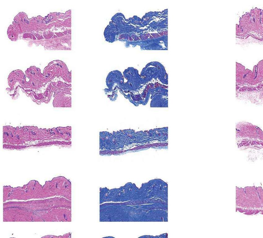
In vivo base editing rescues Hutchinson-Gilford progeria syndrome in mice - Gwern.net
Article
 a                                       ABE-AAV9 injection                                                                                    transgene that includes the complete human LMNA c.1824 C>T allele
                                                                                                                           Homozygous
                                         P3 retro-orbital
                                         P14 retro-orbital                                                               LMNA c.1824 C>T       (C57BL/6-tg(LMNA*G608G)HClns/J, previously used as heterozygous
                                                                                                                          (progeria) mice
                                         P14 intraperitoneal                                                                                   mice22); these homozygous mice hereafter are referred to as ‘progeria
                                   ITR Promoter      WT TadA
                                                    deaminase
                                                                 32-aa Evolved TadA 32-aa Cas9 nickase-VRQR-N Intein-N Terminator ITR
                                                                 linker deaminase linker
                                                                                                                                               mice’. Phenotypically, this model recapitulates hallmark symptoms
                                                                                                                                               of HGPS including VSMC defects, hair loss, lack of subcutaneous fat,
                        ITR Promoter Intein-C
                       ITR                                            Cas9 nickase-VRQR-C                    Terminator LMNA C>T sgRNA ITR
                                                                                                                                               musculoskeletal abnormalities and shortened lifespan3,4,22. Given the
 b                                                 P3 retro-orbital injection                             P14 retro-orbital injection          diverse tissues affected by progeria, we sought systemic in vivo delivery
 C (corrected) DNA at c.1824 (%)

                                    80                                     6 weeks         80                                     6 weeks
                                                                                                                                               of the ABE and sgRNA characterized above.
                                    70                                     (n = 4)         70                                     (n = 5)         We recently developed a strategy for base editor delivery in vivo using
                                                                           6 months                                               6 months
                                    60
                                                                           (n = 12)
                                                                                           60
                                                                                                                                  (n = 12)     adeno-associated virus (AAV)28, a delivery modality that is approved
                                    50                                                     50                   **
                                    40
                                              **                                           40                                                  by the US Food and Drug Administration (FDA). This approach uses
                                                                                                     ***
                                    30                   *                                 30                                                  trans-splicing inteins to reconstitute the full-length base editor in cells
                                                                           **        ***
                                    20                                                     20                                                  from a pair of AAVs, each expressing one half of the base editor28,29
                                    10                                                     10
                                     0                                                      0
                                                                                                                                               (Fig. 3a). We adapted this system to deliver ABEmax-VRQR and the LMNA
                                          Heart     Quad      Liver    Aorta       Bone           Heart     Quad      Liver    Aorta    Bone
                                                                                                                                               c.1824-targeting sgRNA. We chose the AAV9 capsid for its broad tissue
 c                                                                                  d                                                          tropism, clinical validation and ability to transduce progeria-relevant
                                              Liver
                                                                                            Liver, females (n = 6 saline, n = 6 ABE-AAV9)
                                                                                                                                               tissues including heart and muscle30,31.
 ddPCR count per 50 ng RNA

                                   10,000     (n = 12)
                                                                                                     P14 saline      P14 ABE-AAV9 WT
                                    8,000
                                                                                     Lamin A
                                                                                     Progerin                                                     To compare the effects of injection route and timing on in vivo edit-
                                                                                     Lamin C                                                   ing, we performed retro-orbital injection of P3 (3-day-old) mice (n = 4)
                                    6,000                                               Actin
                                                                                                                                               and P14 (2-week-old) mice (n = 5), and intraperitoneal injection of P14
                                    4,000
                                                       **                                       Liver, males (n = 6 saline, n = 6 ABE-AAV9)    mice (n = 5). P3 injections used 5 × 10¹⁰ viral genomes (vg) of each AAV
                                                                            ****
                                    2,000
                                                                                     Lamin A            P14 saline      P14 ABE-AAV9 WT
                                                                                                                                               for a total of 1 × 10¹¹ vg per mouse. Both P14 injections used 5 × 10¹¹ vg
                                                                                     Progerin
                                         0
                                                LMNA                 Progerin        Lamin C                                                   of each AAV for a total injection of 1 × 10¹² vg per mouse. Mice were
                                             Saline ABE-           Saline ABE-          Actin                                                  euthanized at the age of six weeks and editing was evaluated in various
                                                    AAV9                  AAV9
                                              Heart
                                                                                                                                               tissues (Fig. 3, Extended Data Fig. 3a–c).
                                                                                           Heart, females (n = 6 saline, n = 6 ABE-AAV9)
                                                                                                                                                  At six weeks of age, P14 retro-orbital injection resulted in the high-
 ddPCR count per 50 ng RNA

                                              (n = 12)
                                                                                     Lamin A        P14 saline       P14 ABE-AAV9 WT
                                    6,000
                                                                                     Progerin                                                  est editing efficiencies in aorta and bone among the tested injection
                                                                                     Lamin C
                                    4,000                                               Actin                                                  routes (Fig. 3b, Extended Data Fig. 3a). Editing efficiencies in bulk heart
                                                       **                                                                                      tissue (excluding aorta) were similar for the three injection routes. P14
                                    2,000                                                       Heart, males (n = 6 saline, n = 6 ABE-AAV9)    injections generally achieved higher base-editing efficiencies than P3
                                                                                     Lamin A
                                                                                                       P14 saline         P14 ABE-AAV9 WT      injections, possibly owing to the tenfold higher dose of AAV that could
                                         0
                                                LMNA                 Progerin
                                                                                     Progerin
                                                                                     Lamin C
                                                                                                                                               be injected into P14 mice or increased expression of AAV9 receptors
                                             Saline ABE-           Saline ABE-          Actin                                                  in the older mice28,32,33. Together, these data reveal that a single in vivo
                                                    AAV9                  AAV9
                                                                                                                                               injection of ABE-encoding AAV results in modest to high levels of correc-
Fig. 3 | DNA, RNA and protein levels after a single in vivo injection of
                                                                                                                                               tion (10–60%) of the causative LMNA point mutation in various organs.
ABE-expressing AAV9 in a mouse model of human progeria. a, Dual AAV9
encoding split-intein ABE7.10max-VRQR base editor halves28 and the
LMNA-targeting sgRNA were injected into progeria mice. P3 retro-orbital
                                                                                                                                               Long-term ABE treatment of progeria mice
injections (5 × 10¹⁰ vg of each AAV, 1 × 10¹¹ vg total), P14 retro-orbital injections
(5 × 10¹¹ vg of each AAV, 1 × 10¹² vg total) or P14 intraperitoneal injections                                                                 We performed long-term studies in mice using both P3 and P14
(5 × 10¹¹ vg of each AAV, 1 × 10¹² vg total) were administered. ITR, inverted                                                                  retro-orbital AAV injections to assess the relationship between
terminal repeats. b, DNA-editing efficiencies for correcting LMNA c.1824 from                                                                  long-term in vivo outcomes and editing efficiencies in disease-relevant
T (pathogenic) to C (wild type) in 6-week-old or 6-month-old mice that were                                                                    tissues such as the aorta, in which editing levels in P14-injected mice
retro-orbitally injected with ABE-expressing AAV9 (ABE-AAV9) at P3 (left) or                                                                   were 3.8-fold higher relative to P3-injected mice at six weeks of age.
P14 (right). Editing efficiencies in P14 intraperitoneally injected mice are shown                                                             We retro-orbitally injected 24 progeria mice at P3 with 1011 total vg of
in Extended Data Fig. 3a. c, ddPCR counts for the RNA transcript abundance of                                                                  dual AAV9, and 24 mice at P14 with 1012 total vg as before. As controls,
human LMNA (grey bars) and progerin (red bars) in the liver and heart of mice                                                                  24 mice at P3 and 24 mice at P14 were injected retro-orbitally with
(6 months old) that were retro-orbitally injected with saline or ABE-AAV9 at P14.                                                              saline. All groups contained equal numbers of male and female mice. At
See Extended Data Fig. 4 for additional data. d, Western blot analysis of human
                                                                                                                                               6 months of age, when untreated progeria mice typically show pheno-
lamin A, progerin and lamin C proteins in the liver and heart of mice that were
                                                                                                                                               typic decline but are not yet at the end of their lifespan, 24 P3-injected
retro-orbitally injected with saline or ABE-AAV9 at P14. Each lane shows tissue
                                                                                                                                               mice and 24 P14-injected mice (half AAV9-treated, half saline controls)
from a different mouse. ‘WT’ indicates a C57BL/6 mouse that lacks the
transgene, showing that the antibody is specific to human lamin proteins. See
                                                                                                                                               were euthanized and analysed for DNA base-editing efficiency, LMNA
Extended Data Fig. 5 for additional data. Data are mean ± s.d. for the indicated                                                               RNA splicing, levels of human progerin and lamin proteins, and tis-
numbers of biological replicates. *P < 0.05, **P < 0.01, ***P < 0.001,                                                                         sue histology. We placed the remaining 24 P3-injected mice and 24
****P < 0.0001 by Student’s unpaired two-sided t-test.                                                                                         P14-injected mice (half AAV9-treated, half saline controls) in a longevity
                                                                                                                                               study to assess lifespan.

that of cells from an unaffected parent (Fig. 2d, e). These results col-
lectively show that treating cells with the LMNA-targeting sgRNA and                                                                           DNA, RNA and protein analysis at six months
ABEmax-VRQR did not result in detected off-target DNA or RNA editing                                                                           Analysis of the DNA base-editing outcomes in six-month-old mice
using the above analysis methods, despite high levels of on-target editing.                                                                    revealed notable differences between the P3- and P14-injected cohorts.
                                                                                                                                               Both cohorts showed increases in DNA-editing efficiency in several
                                                                                                                                               tissues compared to the six-week time point (Fig. 3b). For example,
In vivo ABE delivery in mice with progeria                                                                                                     editing in the aorta rose from 4.5 ± 2.5% to 10 ± 3.4% in P3-injected mice,
Encouraged by these findings, we applied base editing in vivo to correct                                                                       and from 17 ± 5.2% to 23 ± 8.1% in P14-treated mice. Modest editing was
a mouse model of progeria. We used C57BL/6 mice homozygous for a                                                                               observed in the lung, skin, visceral fat and white adipose tissue (WAT),

4 | Nature | www.nature.com
In vivo base editing rescues Hutchinson-Gilford progeria syndrome in mice - Gwern.net
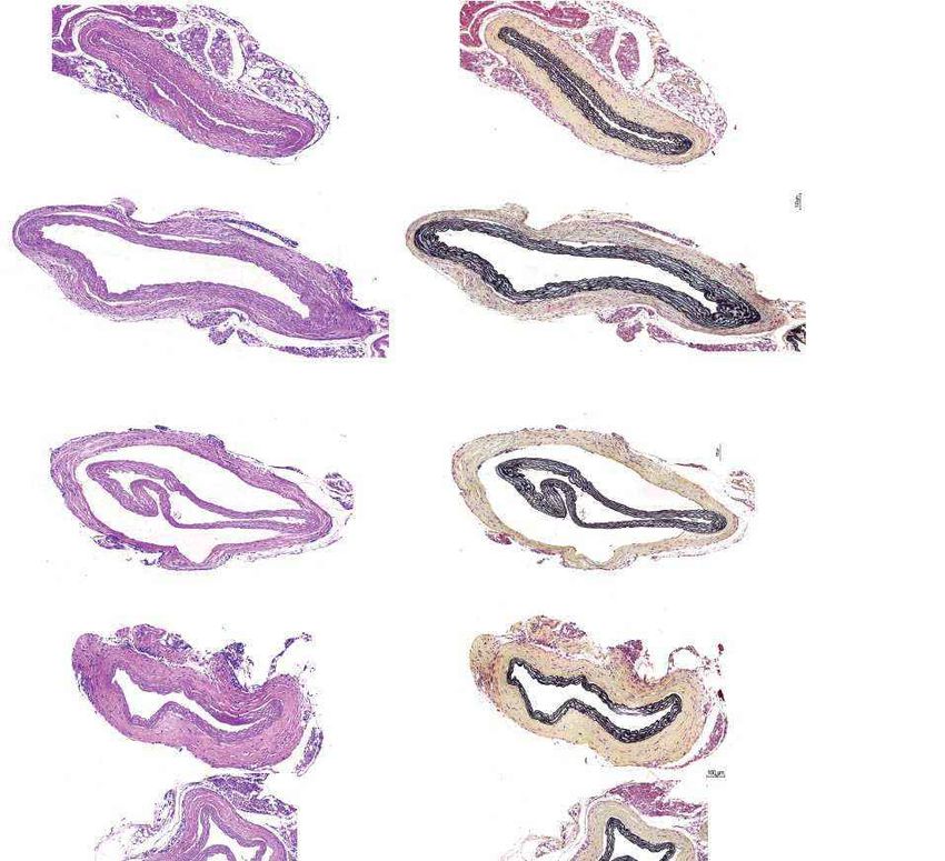
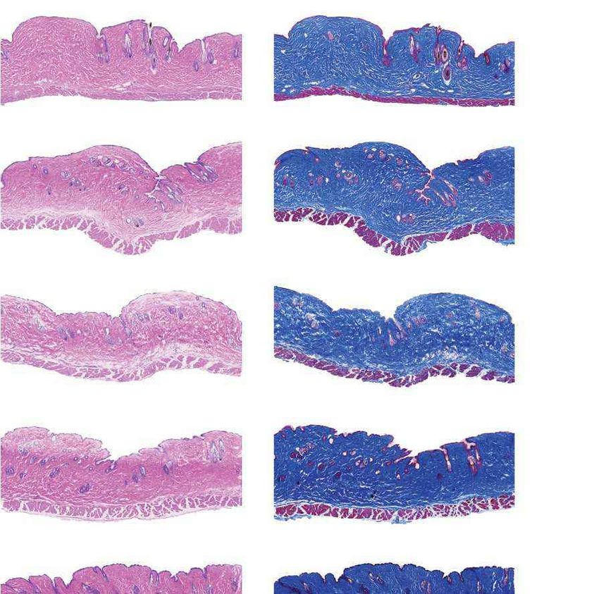
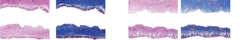
a                                                                  Females                                                       Males                                    b                                                           Fig. 4 | Aortic histopathology and lifespan of
                                                                                                                                                                                         VSMCs per mm2                                progeria mice after a single injection of ABE-AAV9.
                                                                                                                                                                                   0    1,000 2,000 3,000 4,000
                                                                                                                                                                                                                                      a, Representative aorta cross-sections from

                                                                                                                                                                H&E
                                                                                                                                                                          Saline
                                                                                                                                                                                                                                      six-month-old mice showing VSMC nuclei and

                                                                                                                                                                                                        *
                                                                                                                                                                           ABE
                    P3 injection

                                                                                                                                                                                                                ****
                                                                                                                                                                                                                       P3-injected
                                                                                                                                                                              WT                                                      adventitia in mice that were retro-orbitally injected
                                                                                                                                                                                       Adventitial area (mm2)                         with saline or ABE-AAV9 at P3 or P14. Top images were
                                                                                                                                                                                   0    0.2 0.4 0.6 0.8 1.0                           stained with haematoxylin and eosin (H&E); bottom

                                                                                                                                                                Movat’s
                                                                                                                                                                          Saline                                                      images were stained with Movat’s pentachrome. Red
                                                                                                                                                                           ABE
                                                                                                                                                                                                                                      arrows indicate decreased VSMC counts and adventitial

                                                                                                                                                                                                  **
                                                                                                                                                                              WT
                                                                                                                                                                                                                                      fibrosis; green arrows indicate preserved VSMC counts
                                                                                                                                                                                          VSMCs per mm2                               and reduced adventitial fibrosis after AAV9-ABE
                                                                                                                                                                                   0    1,000 2,000 3,000 4,000                       treatment. Scale bars, 100 μm (P3 and wild type);

                                                                                                                                                                H&E
                                                                                                                                                                          Saline                                                      200 μm (P14). Additional replicates are shown in

                                                                                                                                                                                                            ****
                      P14 injection

                                                                                                                                                                           ABE
                                                                                                                                                                                                                                      Extended Data Figs. 6, 7. b, Quantification of VSMC

                                                                                                                                                                                                                       P14-injected
                                                                                                                                                                              WT
                                                                                                                                                                                       Adventitial area (mm2)
                                                                                                                                                                                                                                      nuclei counts and adventitial area in mouse cohorts.
                                                                                                                                                                                   0    0.2 0.4 0.6 0.8 1.0                           Data are mean ± s.d. of n = 12 (P3 saline), n = 10 (P3

                                                                                                                                                                Movat’s
                                                                                                                                                                          Saline                                                      ABE-AAV9, P14 saline and P14 ABE-AAV9) or n = 8 (wild

                                                                                                                                                                                                 ****
                                                                                                                                                                           ABE
                                                                                                                                                                                                                                      type) replicates. Data from wild-type samples are
                                                                                                                                                                              WT
                                                                                                                                                                                                                                      shown in both graphs for ease of comparison.
                                                        Saline               ABE-AAV9              Saline                                          ABE-AAV9
                                                                                                                                                                                                                                      Replicates analysed are provided in Extended Data
                                                                                                   c                  Untreated                         WT control        Saline-injected ABE-AAV-injected
                                                                                                                                                                                                                                      Figs. 6, 7. c, Representative fixed aortas stained for
                                                                                                                    (28 days old)                     (6 months old)      (6 months old) (6 months old)
                                                                                                                                                                                                                                      human lamin A/C + DAPI and progerin + DAPI for
                                                                                         H&E
                        Unaffected WT control

                                                                                                                                                                                                                                      untreated progeria mice at P28 (far left), wild-type
                                                                                                        Lamin A/C
                                                                                                          + DAPI

                                                                                                                                                                                                                                      C57BL/6 mice at 6 months (middle left), saline-injected
                                                                                                                                                                                                                                      progeria mice at 6 months (middle right) and
                                                                                                                                                                                                                                      ABE-treated progeria mice at 6 months (far right).
                                                                                         Movat’s

                                                                                                                                                                                                                                      Autofluorescent elastin fibres in the tunica media
                                                                                                        Progerin
                                                                                                        + DAPI

                                                                                                                                                                                                                                      appear as wavy lines. Scale bars, 10 μm. Additional
                                                        Females                Males
                                                                                                                                                                                                                                      replicates are shown in Extended Data Fig. 8. d, Kaplan–
                                                                                                                                                                                                                                      Meier curve for mice that were retro-orbitally injected
d                                                           Saline                                                  e                                  Saline
                                                            ABE AAV9                                                                                   ABE AAV9                                                                       with saline (n = 12) or ABE-AAV9 (n = 11) at P3. Median
                       1.00                                                                                                             1.00
                                                                                                                                                                                                                                      lifespans: saline-injected mice, 189 days;
                                                                                                                                                                                                                                      ABE-AAV9-injected mice, 337 days (1.8-fold longer,
                       0.75                                                                                                             0.75                                                                                          P < 0.0001). e, Kaplan–Meier curve for mice that were
Fraction survival

                                                                                                                    Fraction survival

                                                                                                                                                                                                                                      retro-orbitally injected with saline (n = 12) or ABE-AAV9
                       0.50                                                                                                             0.50                                                                                          (n = 10) at P14. Median lifespans: saline-injected mice,
                                                                                                                                                                                                                                      215 days; ABE-AAV9-injected mice, 510 days (2.4-fold
                       0.25                                                                                                             0.25                                                                                          longer, P < 0.0001). *P < 0.05, **P < 0.01, ***P < 0.001,
                                                                                                                                                                                                                                      ****P < 0.0001 by Student’s unpaired two-sided t-test (b)
                                                                                                                                                                                                                                      or Mantel–Cox test (d, e).
                                                0                                                                                         0
                                                    0            100       200          300            400                                     0       100     200          300           400      500      600
                                                                         Age (d)                                                                                          Age (d)

whereas editing was minimal in the kidney and spleen (Extended Data                                                                                                                     Finally, we noted increases in the abundance of correctly spliced LMNA
Fig. 3d). Bystander V690A editing and indels varied by tissue but were                                                                                                                  transcripts among ABE-treated mice in a variety of tissues compared
generally observed at low frequencies compared to on-target editing                                                                                                                     with saline-injected controls (Extended Data Fig. 4).
(Extended Data Fig. 3e, f). These results suggest that base editing may                                                                                                                    We evaluated the protein levels of progerin and lamin A in
continue in vivo from six weeks to six months of age, consistent with the                                                                                                               six-month-old mice by western blot. P14 ABE-treated mice showed
known persistence of AAV in mammals34,35, or that edited cells may have                                                                                                                 robust reductions of progerin protein in the liver (87 ± 14% reduction),
a survival advantage over uncorrected cells in some organs, increasing                                                                                                                  heart excluding aorta (86 ± 9.1% reduction) and aorta (49 ± 19% reduc-
the prevalence of edited alleles over time.                                                                                                                                             tion) compared to saline-injected controls (Fig. 3d, Extended Data
   Editing efficiencies at six months in most tissues remained higher                                                                                                                   Fig. 5). P3 ABE-treated mice showed similar reductions in progerin levels
in the P14-injected cohort than the P3-injected cohort, including by                                                                                                                    (Extended Data Fig. 5). Levels of progerin protein were generally reduced
2.2-fold in the aorta, 2.1-fold in skeletal muscle, 1.7-fold in bone and                                                                                                                more than those of progerin mRNA in the same organ, suggesting that
1.4-fold in lung (Extended Data Fig. 3d). These results indicate that                                                                                                                   corrected cells may be translationally more active than uncorrected
injecting mice with 1012 total vg at P14 results in higher levels of LMNA                                                                                                               cells. Together, these findings indicate that in vivo ABE-mediated correc-
correction 6 months after treatment, compared to injecting mice with                                                                                                                    tion of the pathogenic human LMNA c.1824 C>T allele in mice can reduce
1011 total vg at P3.                                                                                                                                                                    progerin RNA and protein levels in several clinically relevant tissues.
   Next, we quantified the effects of in vivo ABE treatment on tran-
script abundance and protein levels for progerin and human lamin A in
six-month-old mice. Base editing led to decreases in progerin transcript                                                                                                                ABE treatment improves vascular pathology
abundance (Fig. 3c, Extended Data Fig. 4) that were sometimes larger                                                                                                                    To assess the physiological consequences of base editing the patho-
than DNA correction levels; for example, in P14 WAT we observed a                                                                                                                       genic LMNA c.1824 C>T allele, we performed histological analysis of
31% reduction in the levels of progerin mRNA despite only 4.0 ± 3.6%                                                                                                                    aorta and adipose tissue. Patients with progeria exhibit a loss of VSMCs
DNA correction. These findings suggest that corrected cells may be                                                                                                                      in aortic vessel walls and periadventitial thickening, which together
more transcriptionally active than uncorrected cells or that cells with                                                                                                                 contribute to aortic stiffening and the impairment of cardiac func-
higher transcriptional activity may be more efficiently edited in vivo.                                                                                                                 tion7–11. We observed these hallmark vascular features of progeria

                                                                                                                                                                                                                                                           Nature | www.nature.com | 5
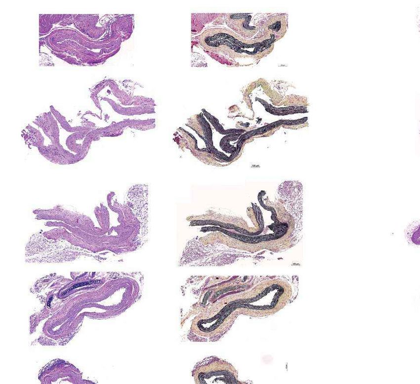
In vivo base editing rescues Hutchinson-Gilford progeria syndrome in mice - Gwern.net
Article
in saline-injected control mice22 (Fig. 4a). Aortas from P3-injected        Data 1). Although samples from liver tumours showed similar numbers
ABE-treated mice at 6 months of age showed 3.3-fold higher average          of AAV integration events as samples from non-tumour liver tissue, pre-
VSMC counts per cross-sectional area compared with saline-injected          vious observations of AAV integration at these regions in liver tumours
controls, but similar (1.2-fold lower) adventitial thickness (Fig. 4a, b,   from AAV-injected mice37 suggest that AAV integration may have
Extended Data Figs. 6, 7). Notably, P14 injection of ABE completely         contributed to liver tumour formation. We note that AAV-associated
rescued both aortic VSMC counts (11-fold increase) and adventitial          liver tumours, although documented in mice, have thus far not been
thickness (5.5-fold decrease) compared to saline-injected controls, such    reported in humans treated with therapeutic AAV vectors38.
that P14 ABE-treated mice were indistinguishable from wild-type mice           The fraction of A•T-to-G•C point mutations among all point muta-
in these two parameters (Fig. 4a, b, Extended Data Figs. 6, 7).             tions detected by whole-genome sequencing was not significantly
   Progerin-induced VSMC death is a key driver of mortality in patients     different among saline-injected mouse liver tissue, ABE-treated mouse
with progeria7,11. The improvements in aortic pathology in ABE-treated      liver tissue and ABE-treated mouse tumour tissue (false discovery rate
mice prompted us to examine the protein levels of human progerin            (FDR)-adjusted P = 0.28–0.50), suggesting that ABE treatment had no
and lamin A/C in aortic VSMCs. Sections of aorta from saline- and P14       apparent effect on the relative genome-wide abundance of A•T-to-G•C
ABE-treated mice were fixed and stained with antibodies specific to         point mutations (Extended Data Fig. 12a, b, Supplementary Data 2). We
human lamin A/C and human progerin (Fig. 4c, Extended Data Fig. 8).         also searched for A•T-to-G•C mutations and indels in the exons, introns
As expected given their young age, VSMC nuclei from four-week-old           and regulatory regions defined by assay for transposase-accessible
progeria mice stained robustly for both proteins (Fig. 4c, far left). No    chromatin using sequencing (ATAC-seq) within 100 kb of 84 genes
staining for human lamin A/C or progerin was observed in wild-type          recurrently mutated in liver cancer (Methods), and found no evident
C57BL/6 aortic VSMCs, demonstrating antibody specificity (Fig. 4c,          patterns distinguishing saline-injected mouse liver tissue, ABE-treated
middle left). Consistent with VSMC counts, aortas from six-month-old        mouse liver tissue or ABE-treated mouse tumour tissue (Extended Data
saline-injected progeria mice contained virtually no VSMCs (Fig. 4c,        Fig. 12c, Supplementary Data 3). These data collectively suggest no
middle right). By contrast, ABE-treated progeria mice maintained            apparent role of base editing in liver tumour formation. Finally, we note
much higher numbers of VSMCs that expressed human lamin A/C,                that the progeria mice in this study contain two copies of human RAB25
with minimal progerin (Fig. 4c, far right). These observations suggest      downstream of LMNA, and RAB25 overexpression promotes human
that DNA base editing of around 25% in the aorta rescues two key vas-       hepatocellular carcinoma39, raising the possibility that expression of
cular defects of progeria—VSMC loss and periadventitial fibrosis–while      transgenic RAB25 in these mice (confirmed in Extended Data Fig. 12d)
preserving the expression of lamin A/C and reducing the abundance           may contribute to liver tumorigenesis that manifests only once lifespan
of progerin in VSMCs.                                                       is greatly extended. Additional studies are needed to understand the
   Lipodystrophy (reduced subcutaneous body fat) is a clinical feature      potential roles of uncorrected LMNA c.1824 C>T alleles, RAB25, AAV
of patients with progeria4. ABE treatment modestly rescued the loss of      transduction, ABE treatment and mouse age in the long-term health
the hypodermal fat layer in both P3- and P14-injected mice relative to      of these mice.
saline-injected controls (Extended Data Figs. 9, 10). Both ABE-treated
and saline-injected mice exhibited moderate dermal hypoplasia com-
pared to wild-type C57BL/6 mice (Extended Data Figs. 9, 10).                Discussion
                                                                            Correcting pathogenic alleles that cause devastating diseases is a long-
                                                                            standing challenge in medicine. Base editing provides an opportunity
ABE treatment extends progeria mouse lifespan                               to directly correct point mutations that drive many disorders without
We conducted longevity studies on 24 P3-injected and 24 P14-injected        requiring double-strand DNA breaks. Here we demonstrate direct cor-
progeria mice (half ABE-injected, half saline-injected, equal numbers       rection of the mutation that underlies most HGPS cases using an ABE. In
of both sexes). Over the 1.5-year duration of the longevity studies, 3      patient-derived cells, base editing efficiently corrects the pathogenic
of 48 mice were excluded after health issues that were deemed to be         allele, substantially reduces RNA mis-splicing, decreases the abundance
unrelated to progeria or treatment (Supplementary Note 1).                  of progerin protein and rescues nuclear morphology abnormalities.
   Body weights were maintained in both ABE-treated cohorts                 Characterization of off-target DNA and RNA editing suggests a low
(Extended Data Fig. 11). The median survival of P3 saline-injected mice     degree of off-target editing in patient-derived cells, despite on-target
(n = 12) was 189 days, whereas that of P3 ABE-treated mice (n = 11) was     editing of 87–91%.
337 days (1.8-fold longer, P < 0.0001 by Mantel–Cox test) (Fig. 4d).           Systemic injection of a single dose of dual AAV9 encoding the ABE
The median survival of P14 saline-injected mice (n = 12) was 215 days,      and sgRNA into a mouse model of human progeria resulted in durable
whereas that of P14 ABE-treated mice (n = 10) was 510 days (2.4-fold        correction of the pathogenic allele, amelioration of RNA mis-splicing,
longer, P < 0.0001 by Mantel–Cox test) (Fig. 4e), which corresponds         reduction of progerin protein in various tissues and greatly improved
approximately to the beginning of old age in wild-type C57BL/6 mice36.      aortic health, fully rescuing VSMC counts and adventitial fibrosis in
P14-injected mice exhibited normal behaviour and vitality at ages well      progeria mice treated with ABE at P14, which in the course of normal
beyond the lifespan of saline-injected mice (Supplementary Videos           mouse maturation corresponds to approximately year 5–6 in humans36.
1–5). One of the P14 ABE-treated mice remains in apparent good health       These findings are particularly encouraging because arterial pathol-
at the time of writing. Complete data from the mouse longevity study        ogy is a major determinant of morbidity and mortality in children with
are in Supplementary Table 1.                                               progeria4,7,8.
   Among the nine deceased P14-injected ABE-treated mice, necropsy             These results also establish that single injections of an ABE pack-
revealed gastrointestinal necrosis in one, liver tumours in five and no     aged in a clinically relevant AAV capsid can greatly extend the lifespan
apparent abnormalities in three (Supplementary Table 1). To investigate     of an animal model of progeria, with median lifespan increasing in
the potential origins of the liver tumours, we performed whole-genome       P14-treated mice from 215 to 510 days—approaching old age in healthy
sequencing of liver tissue from two P14 saline-injected mice and of         C57BL/6 mice36. The marked improvements in aortic pathology, lifespan
livers and liver tumours from three P14 ABE-treated mice that showed        and vitality among P14-injected mice long after a single ABE treatment
liver tumours. Samples from all of the AAV-injected mice, but none          collectively suggest that this strategy has the potential to improve tis-
from saline-injected mice, showed evidence of rare AAV integration in       sue function and lifespan in patients with progeria.
genomic regions where AAV integration has been previously associated           Although single administration with possible transient immunosup-
with liver tumours in mice (Abr, Alb, Focad and Ksr1)37 (Supplementary      pression may help to mitigate potential immunological responses to

6 | Nature | www.nature.com
In vivo base editing rescues Hutchinson-Gilford progeria syndrome in mice - Gwern.net
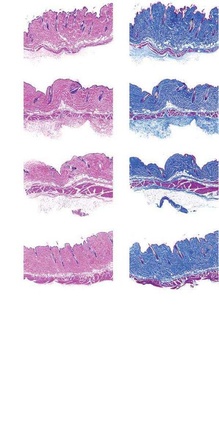
editing agents, efforts to translate these findings into patients must                              16.   Santiago-Fernández, O. et al. Development of a CRISPR/Cas9-based therapy for
                                                                                                          Hutchinson–Gilford progeria syndrome. Nat. Med. 25, 423–426 (2019).
closely monitor immune responses to treatment. AAV integration and                                  17.   Suzuki, K. et al. Precise in vivo genome editing via single homology arm donor mediated
liver tumours observed in some of the longest-lived mice—consist-                                         intron-targeting gene integration for genetic disease correction. Cell Res. 29, 804–819
ent with previous reports of AAV-integration-induced liver tumours in                                     (2019).
                                                                                                    18.   Scaffidi, P. & Misteli, T. Reversal of the cellular phenotype in the premature aging disease
mice37,40–43—further highlights the importance of vector and dose optimi-                                 Hutchinson–Gilford progeria syndrome. Nat. Med. 11, 440–445 (2005).
zation as well as post-treatment monitoring, even though liver tumours                              19.   Jiang, F. & Doudna, J. A. CRISPR–Cas9 structures and mechanisms. Annu. Rev. Biophys.
have not yet been observed in humans treated with AAV vectors38.                                          46, 505–529 (2017).
                                                                                                    20.   Pattanayak, V. et al. High-throughput profiling of off-target DNA cleavage reveals
   In some tissues, modest DNA editing resulted in disproportionately                                     RNA-programmed Cas9 nuclease specificity. Nat. Biotechnol. 31, 839–843 (2013).
large benefits at the RNA, protein or tissue levels. For example, edit-                             21.   Komor, A. C., Kim, Y. B., Packer, M. S., Zuris, J. A. & Liu, D. R. Programmable editing of a
ing of around 25% in the aorta of P14-injected mice resulted in 11-fold                                   target base in genomic DNA without double-stranded DNA cleavage. Nature 533,
                                                                                                          420–424 (2016).
higher VSMC counts, a 5-fold decrease in adventitia fibrosis and a lack of                          22.   Varga, R. et al. Progressive vascular smooth muscle cell defects in a mouse model of
observed progerin-positive VSMC nuclei. The outsized benefits of DNA                                      Hutchinson–Gilford progeria syndrome. Proc. Natl Acad. Sci. USA 103, 3250–3255
editing suggest that edited cells may contribute disproportionately to                                    (2006).
                                                                                                    23.   Huang, T. P. et al. Circularly permuted and PAM-modified Cas9 variants broaden the
the health of tissues in this animal model. Further studies are needed                                    targeting scope of base editors. Nat. Biotechnol. 37, 626–631 (2019).
to understand the molecular basis of this phenomenon.                                               24.   Koblan, L. W. et al. Improving cytidine and adenine base editors by expression
   A number of additional studies may further advance base-editing                                        optimization and ancestral reconstruction. Nat. Biotechnol. 36, 843–846 (2018).
                                                                                                    25.   Kleinstiver, B. P. et al. High-fidelity CRISPR–Cas9 nucleases with no detectable
treatments for progeria towards clinical application. We recently                                         genome-wide off-target effects. Nature 529, 490–495 (2016).
reported ABE variants with much higher editing activity than                                        26.   Tsai, S. Q. et al. CIRCLE-seq: a highly sensitive in vitro screen for genome-wide CRISPR–
ABE7.10max44,45. These variants could further increase editing efficiency                                 Cas9 nuclease off-targets. Nat. Methods 14, 607–614 (2017).
                                                                                                    27.   Eisenberg, E. & Levanon, E. Y. A-to-I RNA editing—immune protector and transcriptome
and phenotypic rescue, or might reduce the required dosage. The tim-                                      diversifier. Nat. Rev. Genet. 19, 473–490 (2018).
ing of treatment may also need to be further optimized for best out-                                28.   Levy, J. M. et al. Cytosine and adenine base editing of the brain, liver, retina, heart and
comes, taking into account the time to diagnosis. Finally, ABE editing                                    skeletal muscle of mice via adeno-associated viruses. Nat. Biomed. Eng. 4, 97–110 (2020).
                                                                                                    29.   Villiger, L. et al. Treatment of a metabolic liver disease by in vivo genome base editing in
has the potential to synergize with emerging progeria treatments3,14–16                                   adult mice. Nat. Med. 24, 1519–1525 (2018).
including farnesyltransferase inhibitors3, other small-molecule drugs14                             30.   Zincarelli, C., Soltys, S., Rengo, G. & Rabinowitz, J. E. Analysis of AAV serotypes 1-9
or antisense oligonucleotides that target the mutant LMNA allele18,46.                                    mediated gene expression and tropism in mice after systemic injection. Mol. Ther. 16,
                                                                                                          1073–1080 (2008).
                                                                                                    31.   Inagaki, K. et al. Robust systemic transduction with AAV9 vectors in mice: efficient global
                                                                                                          cardiac gene transfer superior to that of AAV8. Mol. Ther. 14, 45–53 (2006).
Online content                                                                                      32.   Bostick, B., Ghosh, A., Yue, Y., Long, C. & Duan, D. Systemic AAV-9 transduction in mice is
                                                                                                          influenced by animal age but not by the route of administration. Gene Ther. 14,
Any methods, additional references, Nature Research reporting sum-                                        1605–1609 (2007).
maries, source data, extended data, supplementary information,                                      33.   Wang, L., Wang, H., Bell, P., McMenamin, D. & Wilson, J. M. Hepatic gene transfer in
                                                                                                          neonatal mice by adeno-associated virus serotype 8 vector. Hum. Gene Ther. 23,
acknowledgements, peer review information; details of author contri-
                                                                                                          533–539 (2012).
butions and competing interests; and statements of data and code avail-                             34.   Kessler, P. D. et al. Gene delivery to skeletal muscle results in sustained expression and
ability are available at https://doi.org/10.1038/s41586-020-03086-7.                                      systemic delivery of a therapeutic protein. Proc. Natl Acad. Sci. USA 93, 14082–14087
                                                                                                          (1996).
                                                                                                    35.   Nathwani, A. C. et al. Long-term safety and efficacy of factor IX gene therapy in
1.    Eriksson, M. et al. Recurrent de novo point mutations in lamin A cause Hutchinson–Gilford           hemophilia B. N. Engl. J. Med. 371, 1994–2004 (2014).
      progeria syndrome. Nature 423, 293–298 (2003).                                                36.   Hagan, C. When are mice considered old? The Jackson Laboratory https://www.jax.org/
2.    De Sandre-Giovannoli, A. et al. Lamin A truncation in Hutchinson–Gilford progeria.                  news-and-insights/jax-blog/2017/november/when-are-mice-considered-old (2017).
      Science 300, 2055 (2003).                                                                     37.   Chandler, R. J., Sands, M. S. & Venditti, C. P. Recombinant adeno-associated viral
3.    Gordon, L. B. et al. Impact of farnesylation inhibitors on survival in Hutchinson–Gilford           integration and genotoxicity: insights from animal models. Hum. Gene Ther. 28, 314–322
      progeria syndrome. Circulation 130, 27–34 (2014).                                                   (2017).
4.    Gordon, L. B., Brown, W. T. & Collins, F. S. Hutchinson–Gilford Progeria Syndrome             38.   Nault, J.-C. et al. Wild-type AAV insertions in hepatocellular carcinoma do not inform
      (GeneReviews, 2019).                                                                                debate over genotoxicity risk of vectorized AAV. Mol. Ther. 24, 660–661 (2016).
5.    Gaudelli, N. M. et al. Programmable base editing of A•T to G•C in genomic DNA without         39.   Geng, D., Zhao, W., Feng, Y. & Liu, J. Overexpression of Rab25 promotes hepatocellular
      DNA cleavage. Nature 551, 464–471 (2017).                                                           carcinoma cell proliferation and invasion. Tumour Biol. 37, 7713–7718 (2016).
6.    Anzalone, A. V., Koblan, L. W. & Liu, D. R. Genome editing with CRISPR–Cas nucleases,         40.   Donsante, A. et al. Observed incidence of tumorigenesis in long-term rodent studies of
      base editors, transposases and prime editors. Nat. Biotechnol. 38, 824–844 (2020).                  rAAV vectors. Gene Ther. 8, 1343–1346 (2001).
7.    Olive, M. et al. Cardiovascular pathology in Hutchinson–Gilford progeria: correlation with    41.   Donsante, A. et al. AAV vector integration sites in mouse hepatocellular carcinoma.
      the vascular pathology of aging. Arterioscler. Thromb. Vasc. Biol. 30, 2301–2309 (2010).            Science 317, 477 (2007).
8.    Gerhard-Herman, M. et al. Mechanisms of premature vascular aging in children with             42.   Embury, J. E., Charron, C. C., Poirier, A. E. & Zori, A. Long term portal vein administration
      Hutchinson–Gilford progeria syndrome. Hypertension 59, 92–97 (2012).                                of AAV-WPRE vector results in increased incidence of neoplastic disease and hepatic
9.    Rivera-Torres, J. et al. Cardiac electrical defects in progeroid mice and Hutchinson–               pathology. Mol. Ther. 13, S83 (2006).
      Gilford progeria syndrome patients with nuclear lamina alterations. Proc. Natl Acad. Sci.     43.   Sands, M. S. AAV-mediated liver-directed gene therapy. Methods Mol. Biol. 807, 141–157
      USA 113, E7250–E7259 (2016).                                                                        (2011).
10.   Prakash, A. et al. Cardiac abnormalities in patients with Hutchinson–Gilford progeria         44.   Richter, M. F. et al. Phage-assisted evolution of an adenine base editor with enhanced Cas
      syndrome. JAMA Cardiol. 3, 326–334 (2018).                                                          domain compatibility and activity. Nat. Biotechnol. 38, 883–891 (2020).
11.   Stehbens, W. E., Wakefield, S. J., Gilbert-Barness, E., Olson, R. E. & Ackerman, J.           45.   Gaudelli, N. M. et al. Directed evolution of adenine base editors with increased activity
      Histological and ultrastructural features of atherosclerosis in progeria. Cardiovasc.               and therapeutic application. Nat. Biotechnol. 38, 892–900 (2020).
      Pathol. 8, 29–39 (1999).                                                                      46.   Osorio, F. G. et al. Splicing-directed therapy in a new mouse model of human accelerated
12.   Gordon, L. B. et al. Clinical trial of a farnesyltransferase inhibitor in children with             aging. Sci. Transl. Med. 3, 106ra107 (2011).
      Hutchinson–Gilford progeria syndrome. Proc. Natl Acad. Sci. USA 109, 16666–16671              47.   Mateos, J. et al. Next-generation sequencing and quantitative proteomics of Hutchinson–
      (2012).                                                                                             Gilford progeria syndrome-derived cells point to a role of nucleotide metabolism in
13.   Capell, B. C. & Collins, F. S. Human laminopathies: nuclei gone genetically awry. Nat. Rev.         premature aging. PloS One 13, e0205878 (2018).
      Genet. 7, 940–952 (2006).
14.   Lai, W.-F. & Wong, W.-T. Progress and trends in the development of therapies for              Publisher’s note Springer Nature remains neutral with regard to jurisdictional claims in
      Hutchinson–Gilford progeria syndrome. Aging Cell 19, e13175 (2020).                           published maps and institutional affiliations.
15.   Beyret, E. et al. Single-dose CRISPR–Cas9 therapy extends lifespan of mice with
      Hutchinson–Gilford progeria syndrome. Nat. Med. 25, 419–422 (2019).                           © The Author(s), under exclusive licence to Springer Nature Limited 2020

                                                                                                                                                            Nature | www.nature.com | 7
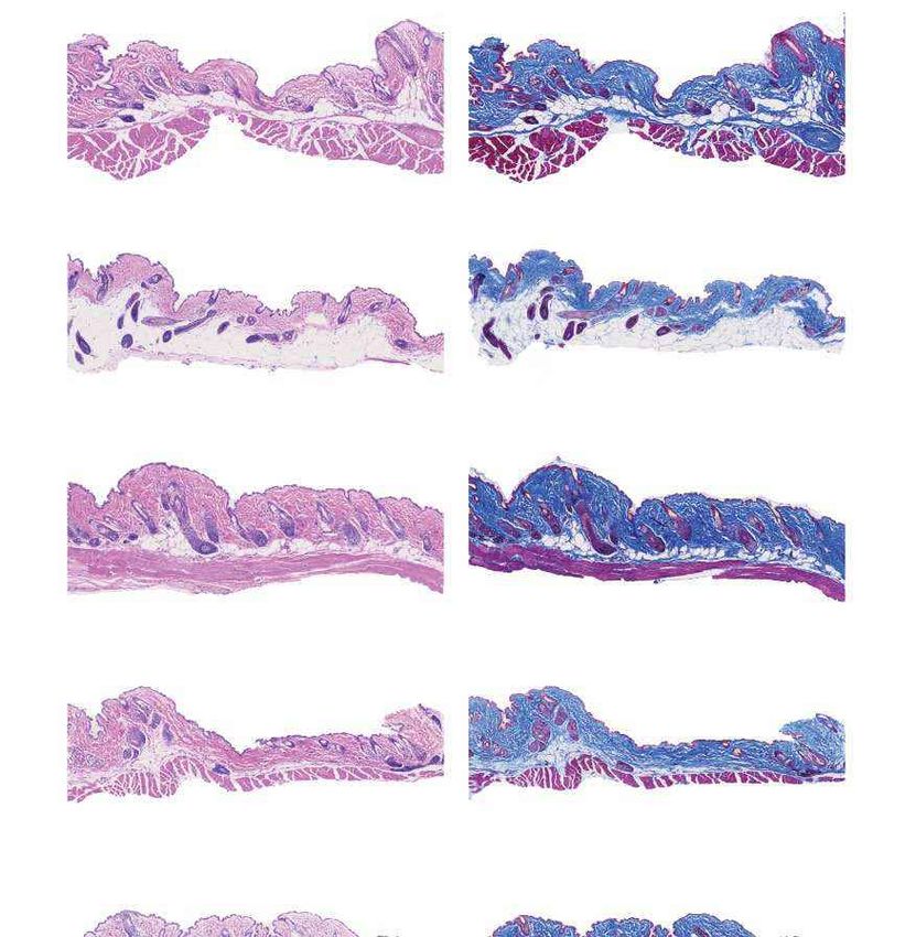
In vivo base editing rescues Hutchinson-Gilford progeria syndrome in mice - Gwern.net
Article
Methods                                                                       changed on cells and cells were transfected using FuGENE HD accord-
                                                                              ing to the manufacturer’s protocol. Transfection mix included 9 μg
Data reporting                                                                of transfer vector (the packaging genome of interest), 9 μg of psPAX2
Sample sizes were determined on the basis of literature precedence for        (encoding the viral packaging proteins) and 6 μg of pVSV-G (encoding
genome editing experiments, and were justified by power calculation           the VSV-G envelope protein). Transfection mix was then supplemented
estimating 90% power to detect differential ages in longevity studies.        with 70 μl of room temperature equilibrated FuGENE and brought
The experiments were not randomized and the investigators were not            to a final volume of 1,500 μl per flask with Opti-MEM. Two days after
blinded to allocation during experiments and outcome assessment.              transfection, medium was collected and spun at 3,000g for 15 min to
                                                                              remove remaining cells. Centrifuged supernatant was passed through
Cell culture                                                                  a 0.45-μm PVDF filter to eliminate all non-viral debris. Supernatant was
HGADFN167 and HGADFN188 cells (Progeria Research Founda-                      transferred directly to target cells.
tion) were maintained in antibiotic-free DMEM (Thermo Fisher Sci-                The human non-targeting control sgRNA sequence was used from a
entific10569044) supplemented with 20% (v/v) fetal bovine serum               previous study48. Oligos containing these non-targeting sgRNAs with
(Thermo Fisher), at 37 °C with 5% CO2. HEK239T/17 (ATCC CRL-11268)            5′ overhang BsmBI digestion sites were synthesized by Integrated DNA
cells were maintained in antibiotic-free DMEM (Thermo Fisher Sci-             Technologies. The oligos were first annealed and inserted into the
entific 10569044) supplemented with 10% (v/v) fetal bovine serum              lentiCRISPR v2 plasmids (a gift from F. Zhang, Addgene plasmid 52961)
(Thermo Fisher Scientific), at 37 °C with 5% CO2.                             as previously described49. The fragments containing these non-targeting
                                                                              sgRNAs were digested from the recombinant lentiCRISPR v2 plasmids
Lentiviral vector cloning                                                     by restriction enzymes KpnI and NheI. These fragments were then
The ABEmax-VRQR gene was inserted into the lentiCRISPRv2 back-                ligated into the ABEmax7.10 backbone, which was extracted from the
bone (Addgene 52961) via restriction cloning. Backbone plasmid was            digests of KpnI and NheI. The sequences of the recombinant plasmids
digested using AgeI and BamHI according to the manufacturer’s pro-            were confirmed by Sanger sequencing. Control sgRNA oligonucleotide
tocol. ABEmax-VRQR was amplified from (Addgene 119811) using the              sequences: ctrl sgRNA F 5′-CACCGGCCTGCCCTAAACCCCGGAA-3′; ctrl
primers LWK901 and LWK902. Gibson assembly was performed using                sgRNA R 5′-AACTTCCGGGGTTTAGGGCAGGCC-3′.
a 3:1 molar ratio of insert to vector backbone according to the manu-
facturer’s protocol. The LMNA c.1824 C>T targeted sgRNA was installed         HGADFN167 and HGADFN188 lentiviral transduction
by digesting the cloned backbone with BsmBI and gel-extracting the            Lentivirus was generated as described above. HGADFN167 and
resulting cleaved backbone. DNA oligonucleotides encoding the                 HGADFN188 cells were cultured in 75-cm flasks in antibiotic-free DMEM
sgRNA were ordered to match the corresponding overhangs gener-                supplemented with 20% FBS. Supernatant was removed from cells
ated by BsmBI digestion (5′-CACCGGTCCACCCACCTGGGCTCC-3′                       and medium was replaced with 15 ml filtered lentiviral medium per
and 5′-AAACGGAGCCCAGGTGGGTGGACC-3′). Oligonucleotides were                    plate, supplemented with 5 ml of regular DMEM. Cells were grown for
annealed and phosphorylated using T4-PNK according to the manu-               3 days before medium change with DMEM + 20% FBS including 2 μg/ml
facturer’s instructions and ligated into the digested backbone as pre-        puromycin to select for cells expressing full-length editor. Cells were
viously described24. LWK901: 5′-TTTGCCGCCAG AACACAGGACCGGT                    maintained in selective medium for 10 and 20 days post-infection
GCCACCATGAAACGGACAGCCGACG-3′; LWK902: 5′-GGGAAAAGTT                           before collecting genomic DNA, RNA and protein while also isolating
GGTGGCCCCGGATCCGACTTTCCTCTT CTTCTTGGGCTCG-3′.                                 cells at 20 days of age for histological analysis.

AAV vector cloning                                                            HGADFN167 and HGADFN188 genomic DNA isolation
N-terminal virus was as previously reported28. C-terminal virus required      Genomic DNA for DNA sequencing analysis was isolated first by trypsi-
the installation of the VRQR mutations (D1135V, G1218R, R1335Q,               nizing cells and centrifugation of one 15-cm dish per cell line per time
T1337R) as well as the sgRNA sequence targeting LMNA c.1824 C>T.              point. Trypsinized cells were resuspended in medium and spun gently
The VRQR mutations were installed by Gibson assembly and ligation             at 100g for 10 min. Cell pellets were resuspended in 200 μl of lysis buffer
of the ABEmax-VRQR C terminus. The sgRNA targeting LMNA c.1824                (10 mM Tris-HCl, pH 7.5, 0.05% SDS, 25 μg/ml proteinase K (NEB)). Lys-
C>T was installed by BsmBI digestion of the backbone as described             ing cells were incubated at 37 °C for 1 h. Proteinase K was inactivated
for the lentiviral backbone and ligation of the same top and bottom           by 30-min incubation at 80 °C.
oligonucleotides into the cut vector.
   To generate the C-terminal AAV genome encoding Npu-ABEmax                  HGADFN167 and HGADFN188 RNA extraction and ddPCR
(VRQR) and the sgRNA targeting LMNA G608G, we first subcloned                 Total RNA from the cell lines was extracted with Trizol (Life Technolo-
Npu-ABEmax(VRQR) in a mammalian (CMV) expression plasmid by                   gies) and purified using the RNeasy mini kit (Qiagen) per the manu-
deleting the UGI domains from an Npu-BE4max(VRQR) intermediate.               facturer’s instructions. The total RNA yield was determined using the
We then amplified by PCR the Npu-ABEmax(VRQR) gene using primers              NanoDrop 2000 spectrophotometer. One microgram of total RNA per
877(fwd)/670(rev) and cloned by Gibson assembly into AgeI/BglII-cut           condition was converted to cDNA using the iScript Select cDNA Synthe-
AAV plasmid. In a subsequent cloning step, annealed oligonucleo-              sis Kit (Bio-Rad). PCR cycling conditions consisted of an initial enzyme
tides encoding the sgRNA targeting LMNA G608G were ligated into               activation step for 10 min at 95 °C, followed by 40 cycles of 94 °C for 30 s
BsmBI-cut plasmid. 877(fwd): 5′-TCACTTTTTTTCAGGTTGGACCGGT                     and 59 °C for 30 s with a 2 °C/second ramp rate, and a 10-min enzyme
GCCACCATGAAACGGACAGCCGACGG-3′; 670(rev): 5′-AATCCAGAGGTT                      deactivation step at 98 °C for 10 min. Each reaction was duplexed with
GATTATCAGATCTTAGACTTTCCTCTTCTTCTTGGGCTCG AATTCGC-3′.                          the Human TFRC PrimePCR Probe Assay (assayID qHsaCIP0033292,
                                                                              HEX) and performed in triplicate. After completion of reactions,
Lentiviral production                                                         samples were analysed on a QX200 droplet reader (BioRad) to obtain
HEK239T/17 (ATCC CRL-11268) cells were maintained in antibiotic-free          expression levels relative to mouse Hprt and transcript-specific copy
DMEM (Thermo Fisher Scientific 10569044) supplemented with 10%                number, then further analysed using Excel software.
(v/v) fetal bovine serum (Thermo Fisher Scientific), at 37 °C with 5% CO2.
Cells were verified to be free of mycoplasma by ATCC upon purchase.           Cell line qPCR analysis
On day 1, cells were split 1:3 from rapidly dividing HEK293T/17 flasks that   RT–qPCR was performed in triplicate using SYBR Green Supermix
had been split 1:10 three days prior. The following day, the medium was       (Bio-Rad) on CFX96 real-time system (C1000 Thermal Cycler; Bio-Rad).
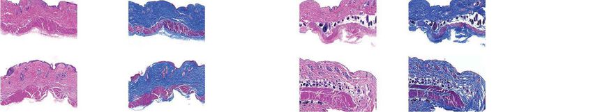
In vivo base editing rescues Hutchinson-Gilford progeria syndrome in mice - Gwern.net In vivo base editing rescues Hutchinson-Gilford progeria syndrome in mice - Gwern.net
You can also read