Drug absorption in the lungs - studies in the isolated perfused rat lung model combined with physiologically based biopharmaceutics modelling ...
←
→
Page content transcription
If your browser does not render page correctly, please read the page content below
Digital Comprehensive Summaries of Uppsala Dissertations
                from the Faculty of Pharmacy 292

                Drug absorption in the lungs
                studies in the isolated perfused rat lung
                model combined with physiologically based
                biopharmaceutics modelling

                JOHANNA ERIKSSON

    ACTA
UNIVERSITATIS
 UPSALIENSIS    ISSN 1651-6192
                ISBN 978-91-513-1086-2
  UPPSALA       urn:nbn:se:uu:diva-426023
    2021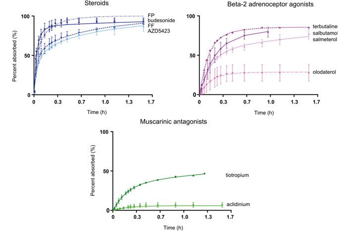
Dissertation presented at Uppsala University to be publicly examined in A1:111a, BMC,
Husargatan 3, Uppsala, Friday, 5 February 2021 at 09:15 for the degree of Doctor of
Philosophy (Faculty of Pharmacy). The examination will be conducted in English. Faculty
examiner: Professor Ben Forbes (Institute of Pharmaceutical Science, King’s College
London).

Abstract
Eriksson, J. 2021. Drug absorption in the lungs. studies in the isolated perfused rat lung model
combined with physiologically based biopharmaceutics modelling. Digital Comprehensive
Summaries of Uppsala Dissertations from the Faculty of Pharmacy 292. 56 pp. Uppsala: Acta
Universitatis Upsaliensis. ISBN 978-91-513-1086-2.

Pulmonary delivery of drugs is the preferred route of administration for treatment of local lung
diseases like asthma and chronic obstructive pulmonary disease. Recently, there has also been
increased interest in systemic delivery of drugs via the lungs to avoid problems with low and/
or variable gastrointestinal absorption, and as a needle-free alternative for drugs that cannot
be ingested. Both the pharmacological and the potentially adverse effects of inhaled drugs
depend on the drug’s local and systemic concentrations, which in turn depend on the pulmonary
absorption of the drug. Pulmonary drug absorption is governed by the dissolution, permeability,
tissue retention, and non-absorptive clearance of the drug in the lungs. Predicting systemic and
local exposure is necessary for developing an inhaled drug product, and these predictions can
be based on data obtained from both in vitro and ex vivo methods, such as cell lines, solubility
measurements, and the isolated perfused lung (IPL) model. Data obtained by these methods
can then be used to inform physiologically based biopharmaceutics (PBB) models about drug-
specific absorption parameters.
   The overall aim of this thesis was to increase the mechanistic understanding of pulmonary
drug absorption, with a special focus on obtaining and analyzing ex vivo absorption parameters
for different inhalation drugs and formulations, and evaluating the predictive power of these
parameters in simulations of pulmonary drug absorption. In the first two papers of the thesis,
drugs were formulated as solutions, suspensions, and dry powders, and pulmonary absorption of
these were measured using the IPL model. The data from these experiments were then analyzed
to obtain absorption parameters for each drug using a PBB model. Tissue retention was shown to
be an important parameter for describing drug absorption in IPL, and particle wetting was shown
to greatly affect the absorption of dry powders. Permeability in IPL correlated well with intrinsic
permeability measured in cell monolayers, suggesting that passive transcellular transport is the
main transport mechanism in the lungs. In the second two papers, the absorption parameters
obtained from IPL data were used to simulate rat and human pulmonary drug absorption. The
simulations predicted systemic exposure after inhalation well for both rat and human, suggesting
that ex vivo parameters can be used to predict rat in vivo and human plasma concentrations.
This thesis deepens our understanding of absorption parameters involved in pulmonary drug
absorption, and suggests applications for these parameters in predictions of local and systemic
exposure after inhalation.

Keywords: Pulmonary drug absorption, physiologically based biopharmaceutics modeling,
pulmonary drug delivery

Johanna Eriksson, Department of Pharmaceutical Biosciences, Box 591, Uppsala University,
SE-75124 Uppsala, Sweden.

© Johanna Eriksson 2021

ISSN 1651-6192
ISBN 978-91-513-1086-2
urn:nbn:se:uu:diva-426023 (http://urn.kb.se/resolve?urn=urn:nbn:se:uu:diva-426023)
List of Papers

This thesis is based on the following papers, which are referred to in the text
by their Roman numerals.

      I      Eriksson, J., Sjögren, E., Thörn H., Rubin K., Bäckman P., Len-
             nernäs H. (2018) Pulmonary absorption – estimation of effective
             pulmonary permeability and tissue retention of ten drugs using
             an ex vivo rat model and computational analysis. European Jour-
             nal of Pharmaceutics and Biopharmaceutics, 124:1–12
      II     Eriksson J., Thörn H., Sjögren E., Holmstén L., Rubin K., Len-
             nernäs H. (2019) Pulmonary dissolution of poorly soluble com-
             pounds studied in an ex vivo rat lung model. Molecular Pharma-
             ceutics, 16:3053-3064
      III    Eriksson J., Sjögren E., Lennernäs H., Thörn H. (2020) Drug
             absorption parameters obtained using the isolated perfused rat
             lung model are predictive of rat in vivo lung absorption. The
             AAPS Journal, 22(3):71
      IV     Eriksson J., Thörn H., Lennernäs H., Sjögren E. (2020) Pulmo-
             nary drug absorption and systemic exposure in human: predic-
             tions using physiologically based biopharmaceutics modeling.
             European Journal of Pharmaceutics and Biopharmaceutics,
             156:191-202

Reprints were made with permission from the respective publishers.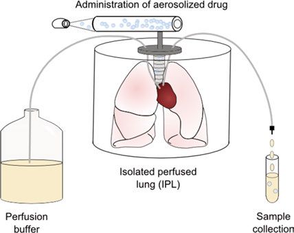
Contents

Introduction ..................................................................................................... 9
   Background ................................................................................................ 9
   Lung anatomy and physiology ................................................................. 10
      The human lung ................................................................................... 10
      The rat lung .......................................................................................... 12
   Pulmonary drug absorption ...................................................................... 12
      Dissolution ........................................................................................... 13
      Permeability ......................................................................................... 14
      Tissue retention.................................................................................... 15
      Metabolism .......................................................................................... 16
      Clearance mechanisms......................................................................... 16
   Models for measuring pulmonary drug absorption .................................. 16
      In vitro models ..................................................................................... 17
      Ex vivo models ..................................................................................... 18
      In vivo models ...................................................................................... 19
      In silico models .................................................................................... 20
Aims of the thesis.......................................................................................... 21
Methods ........................................................................................................ 22
  Study compounds ..................................................................................... 22
  Absorption studies in the isolated perfused lung model ........................... 22
     Study animals ...................................................................................... 23
     Drug formulations................................................................................ 23
     Experimental set-up ............................................................................. 24
  Physiologically based biopharmaceutics model ....................................... 25
     Lung deposition ................................................................................... 26
     Parameter estimation ........................................................................... 27
  Simulations of in vivo and clinical plasma concentration ........................ 27
     Study design ........................................................................................ 27
     Lung absorption models ...................................................................... 28
  Statistical evaluation ................................................................................ 29
Results and discussion .................................................................................. 31
  Ex vivo pulmonary drug absorption.......................................................... 31
     Experimental absorption data .............................................................. 31
     Absorption parameters derived using the IPL PBB model .................. 33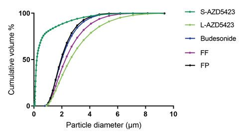
Simulation of rat and human pulmonary drug absorption ........................ 37
      High solubility APIs ............................................................................ 37
      Low solubility APIs ............................................................................. 39
      IPL as a predictive experimental model .............................................. 40
      Predictive PBB models for assessing pulmonary drug absorption ...... 41
Conclusions ................................................................................................... 42
Populärvetenskaplig sammanfattning ........................................................... 44
Acknowledgements ....................................................................................... 46
References ..................................................................................................... 48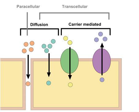
Abbreviations

AAFE        absolute average fold error
ACI         Andersen cascade impactor
AFE         average fold error
API         active pharmaceutical ingredient
AUC         area under the curve
CL          plasma clearance
Cmax        maximum concentration
Cs          solubility
CV%         coefficient of variance
d10         10th percentile of size distribution
d90         90th percentile of size distribution
DPI         dry powder inhaler
ELF         epithelial lining fluid
FF          fluticasone furoate
FP          fluticasone propionate
fu,tissue   Fraction unbound drug in lung
GSD         geometric standard deviation
IPL         isolated perfused lung
kin         association rate constant
kout        dissociation rate constant
MMAD        mass median aerodynamic diameter
MVD         median volume diameter
NGI         next generation pharmaceutical impactor
PBB         physiologically based biopharmaceutics
PBPK        physiologically based pharmacokinetics
Papp        apparent permeability
Peff        effective permeability
Pmem        membrane permeability
pMDI        pressurized meter dose inhaler
SD          Standard deviation
tmax        time when maximum concentration occurs
wf          wetting factor
Introduction

Background
Both local and systemic conditions can be treated by drugs administered
through oral inhalation, but the most common use of this method is with lo-
cally-acting drugs intended to treat pulmonary diseases such as asthma,
chronic obstructive pulmonary disease, and cystic fibrosis 1. There are two
primary benefits to using local treatment for pulmonary diseases: first, there
is a rapid onset of drug action because the drug is directly available at site of
target, and second, a lower dose can be applied because of the high concen-
tration at site of target, which in turn lowers the risk for adverse systemic ef-
fects 2. More recently, commercially available inhalation therapies have
emerged for treating systemic diseases; for example InbrijaTM for treating Par-
kinson’s disease and Afrezza® for treating diabetes 3, 4. The pulmonary route
of administration for systemic active drugs is often chosen as a needle-free
alternative for drugs with low intestinal permeability or high gastro-intestinal
degradation 5. Pulmonary permeability has been shown to be sufficient even
for high molecular weight molecules with very low permeabilities 6-8. In addi-
tion, enzymatic activity is often much lower in the lungs compared to the gas-
tro-intestinal tract and so first pass metabolism is avoided 5. Knowledge about
pulmonary drug absorption, and thus the local and systemic drug concentra-
tions, is important to be able to assess the desired and adverse effects of drug
candidates for both local and systemic treatments 2.
    Drugs are administered to the lung by an inhalation device that creates an
inhalable aerosol 9. The aerodynamic diameter of the droplets or particles in
the aerosol need to be between 0.5–5 µm to deposit in the lungs. Larger drop-
lets or particles will deposit in the throat and smaller ones will be exhaled 10.
There are different types of devices available on the market, and these are
categorized into nebulizers, dry powder inhalers (DPIs) and pressurized meter
dose inhalers (pMDIs). Solutions or suspensions can be administered using
nebulizers or pMDIs, while DPIs are used to deliver dry powders 9.

                                                                               9
Lung anatomy and physiology The human lung Humans have two lungs, with the left lung consisting of two lobes, the upper and lower lobe, and the right lung consisting of three lobes, the upper, middle and lower lobe 11. In men, the left lung weighs 395 ± 147 g and the right 445 ± 159 g, while in women, the lungs each are approximately 100 g smaller 12, 13 . The function of the lungs is gas exchange: oxygen is provided to the circu- lating blood and carbon dioxide is eliminated from the blood. The lungs also protect the body from airborne pathogens. The trachea connects the lungs with the atmospheric air via the oral and nasal cavity 14. The tracheobronchial tree The lungs are made up of a branching system in which, one branch, or airway, divides into two smaller airways. For each division, the branches are called a generation. The human lung consists of 24 generations, where the trachea is generation 0 and the alveolar sacs are generation 23. The number of airways in each generation is twice the number in the previous generation. As a result, in each generation, the airways has a smaller cross-sectional diameter but a larger total cross-sectional area than the previous generation. The generations can be divided into the conducting zone (which humidifies incoming air) and the respiratory zone (where gas exchange takes place). The conducting zone includes generations 0–16, comprising the trachea, bronchi, bronchioles and terminal bronchioles, and the respiratory zone includes generations 17–23, which consists of respiratory bronchioles, alveolar ducts and alveolar sacs (Figure 1) 15. To ensure efficient gas exchange, the respiratory zone has a large surface area (approximately 140 m2) and is well connected with capillary beds 16 . Blood is supplied from the pulmonary circulation, resulting in the whole cardiac output passing the respiratory zone 14. The conducting zone receives blood from the systemic circulation and has a much smaller absorptive area (1–2 m2) 11, 17. 10
Figure 1. Schematic figure of the lung regions and corresponding airway genera-
tions in humans.

Lung epithelium
The epithelium in the respiratory zone, i.e. the alveolar epithelium consists of
two types of cells, alveolar type I and alveolar type II cells 18. Alveolar type I
cells are flat and cover most of the alveolar epithelial surface (93 ± 1%), mak-
ing up the structure of the alveoli, but there are approximately half as many of
these as there are alveolar type II cells. Alveolar type II cells are cuboidal in
shape, produces alveolar surfactant, and have a surface of microvilli. The
thickness of the epithelium is approximately 0.35 µm (Table 1) 18.
    The epithelium in the conducting zone, i.e. the bronchial epithelium, con-
sists of five types of cells: ciliated, goblet, serous, basal and Clara cells. The
ciliated cells’ main function is to transport mucus towards the throat. Goblet
and serous cells produce and secret mucus into the air space. The basal cells
are progenitor cells for renewal of the airway epithelium. The Clara cells are
also thought to be a progenitor cell and to produce lung surfactant 19, 20. The
thickness of the bronchial epithelium ranges from 10–58 µm from the terminal
bronchioles up to the bronchi (Table 1) 7.

Epithelial lining fluid
The epithelial lining fluid (ELF) consists of a watery layer and surfactant
(which lowers surface tension) in the respiratory zone and of a periciliary layer
and a gel mucus layer in the conducting zone 21. The surfactant in the respira-
tory zone hinders collapse of the lung during expiration by lowering the sur-
face tension 22. The periciliary layer surrounds the cilia and consists mostly of
water 21. The gel mucus layer contains mucins which gives the layer its viscous
consistency. This layer lies on top of the cilia and is moved with the cilia
movement 21. The pH of the ELF is around 6.6 23. The thickness of ELF varies

                                                                                  11
along with the generations in the lungs. In the bronchi, the thickness is re-
ported to be between 2–10 µm and in the alveoli between 0.01–0.2 µm (Table
1) 2, 24, 25. The total volume of ELF is thought to be between 10–40 mL (Table
1) 21.
The rat lung
The rat is a common laboratory animal and it was used in this research to study
pulmonary absorption parameters 26. Rats also have two lungs, but in contrast
to humans, a rat’s right lung is divided into 4 lobes, while the left lung consists
of one single lobe 27. The weight of a rat lung is approximately 1.1–1.2 g 28, 29.
The morphology of the branching system of the rat lung is a little different
than the human lung: in the rat lung, starting after generation 0, a main branch
persists all the way through the generations but with smaller outgrowths at
each generation (recall that in the human lung, that the split produces two
equally sized branches at each generation) 30. Thickness, volume and area of
the lung epithelia and ELFs are given in Table 1.
Table 1. Thickness, volume and area of the lung epithelium and epithelial lining
fluid (ELF) for human and rat.
Human Rata
ELF tracheobronchial alveolar tracheobronchial alveolar
Volume (mL) 10–30b 7–20b 0.054c 0.027c
Thickness (µm) 2–10d 0.01–0.2d 1–10e 0.07f
Lung epithelium
Area (m2) 2g 140g 0.011 0.387
Thickness (µm) 10–58h 0.35i 9–24 0.38
aValues obtained from Yu and Rosania if not stated otherwise 31.
bObtained from Hastedt et al. 32.
cCalculated as thickness of ELF × area of epithelium.
dObtained from Olsson et al., Wauthoz and Amighi, Patton and Byron 2, 24, 25.
eObtained from Widdicombe 33.
fObtained from Boger and Fridén 34.
gObtained from Eixarch et al. 17.
hObtained from Patton 7.
iObtained from Crapo et al. 18.
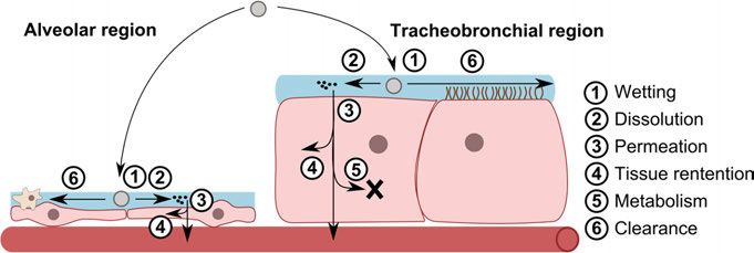
Pulmonary drug absorption
Orally inhaled drugs intended for local effect need to be absorbed into the
tissue in the lungs in order to reach the pharmacological target 35. The drug
particles need to be wetted and dissolved in the ELF and then permeate the
cell membrane to enter the cell (Figure 2) 2. Drug particles also need to escape
the clearance mechanisms in the lungs in order to be available for absorption
122
. For orally inhaled drugs intended for systemic effect (or for systemic ad-
verse effects of local acting drugs), the drug needs to be transported across the
cell into the blood stream 25. After entering the cell, the drug needs to escape
tissue retention and metabolism and then permeate the basolateral membrane
of the cell to enter the systemic circulation (Figure 2) 2.
Figure 2. An overview of the absorption processes in the alveolar and tracheobron-
chial region. The schematic maps the potential fates of an inhaled particle (grey circle).
Dissolution
The dissolution rate (dm/dt) of particles in the lungs is dependent on the diffu-
sivity of the drug molecule in ELF (D), the surface area of the particle in contact
with ELF (A(t)), the length of diffusion (h) (i.e. the distance a particle must
travel) and the difference between the solubility and the concentration in ELF
(Cs – Cb), as described by the modified Nernst-Brunner equation (Eq. 1) 36:
( )
= ×( ) (Eq. 1)
The diffusivity of the drug molecule in ELF depends on the size of the mole-
cule and the viscosity of ELF 37. The composition of the ELF regulates its
viscosity, and therefore the diffusivity of the drug molecule can be quite dif-
ferent in the two different lung regions.
The length of diffusion under well-stirred conditions is considered equal to
the thickness of the unstirred water layer surrounding the particle 38.
The available surface area of the particle depends on the particle size dis-
tribution and the shape of the particles 39. A smaller particle size distribution
gives a larger available surface area, resulting in a higher dissolution rate. The
particle will be wetted in ELF, which will also affect the available surface area
of the particle 38. A less wetted particle will have a lower available surface
area. Wetting in the respiratory zone is facilitated by the presence of surfac-
tant, which decreases the surface tension and thus increasing the wetting rate
40
.
13The solubility of the drug in ELF depends on the composition of ELF. In
the respiratory zone, ELF contain surfactant, which can increase the solubility
of lipophilic drugs 22, 41. For ionizable drug molecules, the solubility is further
affected by the pH in ELF, where ionized molecules have a higher solubility
than a non-ionized 42.
The drug concentration in ELF depends on the volume of ELF and the re-
moval of drug molecules by absorption into the lung epithelium 43. The absorp-
tive clearance is controlled by the epithelial membrane permeability of the drug,
which will be discussed in the following section. If the concentration in ELF is
less than 10% of the solubility of the drug in ELF, sink conditions (Cb is negli-
gible) are considered applicable and thus resulting in a higher dissolution rate
44
. Considering the small volume of ELF, sink conditions might not be ensured
for poorly soluble drugs unless the absorptive clearance is high 2.
Permeability
Permeability (P) is used to describe the ability of a drug molecule to be trans-
ported (dm/dt) across a cell membrane (mem) according to Equation 2 45:
= × ×∆ (Eq. 2)
where Amem is the surface area of the epithelial membrane and ΔC is the con-
centration difference between the donor and receiver side. The total permea-
bility (Ptot) across the epithelium, i.e. both the apical and basolateral mem-
brane permeability, is described by Equation 3:
= + (Eq. 3)
Permeation can be transcellular, i.e. transported across the cell membrane, or
paracellular, i.e. transported in the space between the epithelial cells (Figure
3) 46. Transcellular permeation can be either passive or active, while paracel-
lular transport can only be passive 47. Passive transport takes place along a
concentration gradient, from high to low concentration 47. Passive transcellu-
lar transport can be by diffusion across the epithelium or by carrier-mediated
transport, while primary and secondary active transport is facilitated by direct
and indirect energy-dependent carrier-mediated transport 48. Transport by dif-
fusion is thought to be the most common route of transport for drug molecules,
but the lungs also have transport proteins which enables carrier-mediated
transport 49, 50. Lipophilic drug molecules have high preference for diffusion
across the lipophilic cell membrane, therefore small lipophilic drugs are often
high permeable 51.
14Figure 3. Schematic representation of the different drug transport mechanism across
an epithelial membrane. These processes often co-exist in drug uptake52.
Paracellular transport takes place trough aqueous channels in between the ep-
ithelial cells and this route is thought to mainly transport small hydrophilic
drug molecules 46, 53.
The drug transport rate across the epithelium would, according to Equation
2, be different in the respiratory and conducting zones due to the difference in
epithelial surface area between the two regions (Table 1). But this difference
only pertains to passive transcellular diffusion, since this is the only route that
depends on the total surface area 54. For carrier-mediated transport, the avail-
ability of transport proteins is most important, and for paracellular transport,
the total area of aqueous pores determines the rate of transport 54. The expres-
sion of transport proteins differs in the two lung regions, where some catego-
ries of transporters are more expressed in the conducting zone and some are
more expressed in the respiratory zone 55. The conducting zone is thought to
have a larger area of aqueous pores than the respiratory zone due to the large
surface area and low number of alveolar type I cells in the respiratory zone,
thus reducing the amount of space available between cells 56.
Tissue retention
Tissue retention in the lungs, which may prolong the duration of action of
inhaled drugs used for local treatment, is suggested to occur by several mech-
anisms 57-60. Lipophilicity is suggested to be one factor determining the tissue
retention in the lungs, where more lipophilic drugs achieve a higher retention
58, 61
. Intracellular processes, such as reversible esterification of drug com-
pounds, has also been proposed as contributing to high tissue retention 57, 60, 62,
63
. Esterification increases the lipophilicity of drugs, for example, this process
increases the lipophilicity of budesonide by 500–1000 times, thus increasing
15retention of the drug 57. Another proposed mechanism of tissue retention is lysosomal trapping, which is thought to predominantly affect basic drug com- pounds 59, 64. In this process, drug molecules that are intracellularly distributed to lysosomes get trapped there, because the higher pH in the lysosome ionizes the drug molecule, which subsequently hinder distribution of the drug from the lysosome back to the cytosol 59, 65. Tissue retention can be described by rate constants as a dynamic process, or as an equilibrium process, using frac- tion unbound drug in lung 66, 67. Metabolism Drug metabolism can occur in the lungs 68. The lungs expresses most metab- olizing enzymes that are expressed in the main metabolizing organ, the liver, but the activity of the enzymes in the lungs is often less than in the liver 69. Drug metabolism in the lungs can be used for activation of prodrugs, as it is in the prodrug beclomethasone dipropionate, where metabolism gives rise to the active drug beclomethasone-17-monopropionate 70. Clearance mechanisms The two zones of the lungs have different clearance mechanisms. In the res- piratory zone, foreign entities are removed by alveolar macrophages, while in the conducting zone, removal is by mucociliary clearance 25. Mucociliary clearance transports all particulate matter towards the throat with a half-life of 1–1.5 hours 71. In the respiratory zone, clearance is particle size dependent, where particles in the diameter size range of 0.1–10 µm can be engulfed by alveolar macrophages while larger particles are believed to escape phagocy- tosis 72, 73. Models for measuring pulmonary drug absorption To assist decision making during all parts of drug development of an inhaled product it is important to measure the pulmonary drug absorption. Measure- ments of pulmonary drug absorption can be used to predict the dose, desired and adverse effects, duration of effect, and administration frequency 74, 75. Pul- monary drug absorption can be measured with models ranging in complexity, cost and time consumption (Figure 4) 76. 16
Figure 4. Models for measuring pulmonary drug absorption and bioavailability pre-
sented according to complexity and capacity, modified from Tronde et al. 76.

In vitro models
Cell lines and primary cell cultures
Cell lines and primary cell cultures are commonly used during drug develop-
ment to assess the permeability of a candidate drug 77. The transport rate of a
drug is measured over a cell monolayer and an apparent permeability can be
calculated 78, 79. By using cell lines and cultures, the main mechanism of drug
transport across the epithelium can be investigated, i.e. passive/carrier-medi-
ated and transcellular/paracellular transport 79. For assessing intestinal perme-
ability, the human colon carcinoma epithelial cell line Caco-2 is a standardized
permeability model; however, there are no standardized cell lines/cultures or
methodologies for assessing apparent pulmonary permeability 50, 79. Some cell
lines derived from lung airway tissue and cell cultures derived from lung res-
piratory tissue have recently been used 50, 80-82, although, whether there is a
benefit from measuring permeability using these lung specific cell lines and
cultures (over more standardized and investigated cell lines with other tissue
origin) still remains to be established 83, 84. Cell lines or cultures for permea-
bility measurements is an ethically more sound method compared to animal
studies, and is suitable for an early stage of drug development 50. Compared
to predictions based on physicochemical properties, cell lines and cultures can

                                                                               17
give more information on transport processes, although they still lack the im- portant physiological aspects that are included by the use of ex vivo/in vivo methods 79. Tissue slices The lung slice method is used to measure the tissue retention of a drug in the lungs 85. This method examines the equilibrium distribution between a buffer and a lung slice, and from this, a value of fraction unbound drug in lung (fu,tis- sue) can be calculated. Dissolution testing As with in vitro permeability measurements there is no standardized method for measuring the dissolution rate in vitro of orally inhaled drugs 74, 86. Im- portant factors that must be accounted for when measuring the dissolution rate in vitro are deagglomeration of particles, deposition, dose, volume and com- position of available dissolution medium and sink/non-sink conditions 43. There are several different methods described in the literature for achieving deagglomeration and deposition of particles and for measurements of dissolu- tion rate 41, 43, 87-90. In these methods, the Andersen Cascade Impactor (ACI) and Next Generation Pharmaceutical Impactor (NGI) are used for deposition and deagglomeration of particles, and the Transwell system, flow-through dis- solution apparatus and modified paddle apparatus are used for measurements of dissolution rate. A well-developed in vitro dissolution method has the pos- sibility to give important information when assessing the expected dissolution rate and residence time in the lungs 74. Ex vivo models The isolated perfused animal lung (IPL) model is an ex vivo lung model that can be used for measurements of pulmonary drug absorption 76. The lungs, trachea and heart are removed from a euthanized animal and set up in a hu- midified enclosed chamber (Figure 5) 76. Different animals have been used in the IPL model, the most common being rat, rabbit and guinea pig 91-97. The lungs are connected to the atmospheric air via the trachea. The pulmonary circulation is cannulated via the heart and perfused with a buffer at a constant flow rate. A constant volume of buffer can be re-circulating in the lungs or the buffer can be single-pass perfused through the lungs. The buffer composition mimics the blood regarding albumin, glucose and salt concentrations. The lungs can be ventilated by varying either a positive or negative pressure, where negative pressure is considered beneficial because it lowers the risk for pul- monary edema 98. Drugs can be administered to the lungs as an aerosol in a spacer air chamber from where the lungs are breathing, or via a syringe into the trachea 76, 99-102. Drug content in the perfusion buffer is analyzed to deter- mine the pulmonary drug absorption. Thus, the IPL model gives the sum of 18
all processes involved in pulmonary drug absorption as it is not possible to get
direct measurements of each process separately (see Figure 2).

Figure 5. Schematic illustration of the isolated perfused lung model.

The IPL model has several advantages over in vivo studies: first, the nose is
bypassed and the drug is delivered as an aerosol by breathing directly into the
trachea; second, the lung delivered dose can be determined by mass balance
calculations; third, this method avoids the effect of systemic disposition 79, 103.
The disadvantages with IPL on the other hand are that the tissue is dying and
has a limited viability (2–4 h), and that only the pulmonary circulation is per-
fused, i.e. the conducting zone is not perfused 98.

In vivo models
Rats are the animal most commonly used for inhalation in vivo studies, but
dogs, pigs and sheep can also be used 79. The drug is administered by either
nose-only inhalation in a chamber or intra-tracheally to the animal’s lungs 104.
In nose-only inhalation, the animal is breathing the aerosol from a chamber.
The advantage of this method is that it mimics physiological conditions, but
the disadvantage is that the lung deposited dose is difficult to assess because
a large portion will deposit in the nose. In contrast, the advantage of intra-
tracheal intubation allows better control of the dose administered to the lungs,
but does not mimic normal physiological conditions because the animal is un-
der anesthesia 79, 104. In both methods, blood samples are taken at several time
points during the experiment, and the lungs are harvested at the end of exper-
iment to determine the amount of drug remaining in the lungs. To obtain con-
tinuous data on the lung amount, an animal needs to be sacrificed at each time-
point to harvest the lungs 79. Like with the IPL model, the data from in vivo
studies capture the sum of all absorption processes, but (if blood samples are

                                                                                19
taken) also the systemic disposition. It is therefore important to also measure the systemic disposition alone by intravenous administration to be able to de- convolute the data from blood samples and in that way assess the pulmonary drug absorption 79. In silico models In silico models can be used to predict and simulate the pulmonary absorption of a drug based on the drug physicochemical properties and/or data from in vitro, ex vivo or in vivo studies 31, 34, 74, 105-109. One commonly used in silico approach is compartmental modelling, in which the body is described with compartments and the transport of drug between compartments is described by mass transport equations 110. Physiologically based pharmacokinetic (PBPK) modelling is a type of compartmental modelling where physiological entities of the different tissues, such as plasma, intracellular space and inter- stitial space, are described by physiologically relevant values for volume and surface area 111. Recently, a new term has been coined to define a subfield of PBPK modelling, namely physiologically based biopharmaceutics (PBB) modelling, which focuses on the absorption process 112. 20
Aims of the thesis

The overall aim of this thesis was to increase the mechanistic understanding
of pulmonary drug absorption, with a special focus on obtaining and analyzing
absorption parameters for different inhalation drugs and formulations and
evaluating the predictive power of these parameters in simulations of pulmo-
nary drug absorption.

The specific aims of the papers included in the thesis were as follows.

      •   Paper I: The two aims here were, first, to obtain pulmonary absorp-
          tion data of solutes by using the rat IPL and to develop a PBB model
          to describe the absorption process in the IPL in order to retrieve
          absorption parameters. The second aim was to increase the mecha-
          nistic understanding of the absorption process by comparing the
          IPL absorption parameters to in vitro absorption parameters.
      •   Paper II: The primary aims were to obtain pulmonary absorption
          data of drugs formulated as suspensions and dry powders by using
          the rat IPL and to further develop the PBB model to include de-
          scription of the dissolution process. This allows for comparison of
          the absorption processes of the different formulations.
      •   Paper III: The aim of this paper was to investigate the potential to
          use pulmonary absorption parameters obtained from rat IPL to pre-
          dict rat in vivo pulmonary drug absorption.
      •   Paper IV: To investigate the potential for using pulmonary absorp-
          tion parameters obtained from the rat IPL to predict human pulmo-
          nary drug absorption and to develop an in silico lung model (in
          MoBi®), which will enable simulation and prediction of human pul-
          monary absorption in an open source whole-body framework.

                                                                           21
Methods Study compounds In total ten active pharmaceutical ingredients (APIs) were used in the studies. All APIs were studied in Paper I, AZD5423, budesonide, fluticasone furoate (FF) and fluticasone propionate (FP) were studied in Paper II, AZD5423, FF, FP, salbutamol and salmeterol were studied in Paper III and AZD5423, budesonide, FF, FP, salbutamol, terbutaline and tiotropium were studied in Paper IV. These APIs were chosen based on their range in physicochemical properties (Table 2). A total of ten APIs for which IPL data was found in the literature (two of these were also experimental APIs) were also analyzed in the PBB model in Paper I. Table 2. Physicochemical properties of the study compounds. All values were ob- tained from the chemical library MicroSource US Drugs found in the database ZINC if not otherwise stated 113. API Paper MW (g/mol) cLogD7.4 cLogP tPSA NRB aclidinium I 485 1.3 0.6 56 10 AZD5423 I-IV 487 3.5 5.1 65 8 budesonide I, II, IV 431 3.2 3.2 93 4 FF I-IV 539 3.4 4.9 93 6 FP I-IV 501 3.1 4.9 93 6 olodaterol I 386 0.9 2.1 100 7 salbutamol I,III,IV 240 -1.5 1.4 77 5 salmeterol I,III 417 1.9 3.9 87 16 terbutaline I,IV 226 -1.5 1.1 77 4 tiotropium I,IV 393 -1.1 -1.9 59 5 API = active pharmaceutical ingredient; MW = molecular weight; cLogD7.4 = logarithm of the calculated octanol/water partitioning coefficient at pH 7.4, calculated with ACD/ChemSketch® (Berkshire, UK); cLogP = logarithm of the calculated octanol/water partitioning coefficient; tPSA=topological polar surface area; NRB=number of rotatable bonds; FF = fluticasone fu- roate; FP = fluticasone propionate. Absorption studies in the isolated perfused lung model The isolated perfused rat lung (IPL) model was used in Paper I and II to study the pulmonary drug absorption for APIs in solutions, suspensions and dry powders. 22
Study animals
The animals used in Paper I and II were male Han Wistar rats (Charles River
Germany), weighing 275–300 g. The experiments were conducted at the pre-
clinical laboratory at AstraZeneca (Gothenburg, Sweden) and were approved
by the Animal Ethics Committee of Gothenburg (135-2014). The animals
were allowed to acclimatize for at least 5 days prior to experiments and had
free access to food pellets and water.

Drug formulations
Composition
The APIs were formulated in solution in Paper I and as suspensions and dry
powder in Paper II. The solutions were prepared in water for aclidinium (0.1
mg/mL), olodaterol (0.03 mg/mL), salbutamol (0.1 mg/mL), salmeterol (0.1
mg/mL), terbutaline (0.1 mg/mL), and tiotropium (0.1 mg/mL); in 19% etha-
nol in water for budesonide (0.03 mg/mL) and salmeterol (0.1 mg/mL); and
in 38% ethanol in water for AZD5423 (0.03 mg/mL), FF (0.03 mg/mL) and
FP (0.03 mg/mL). The suspensions were prepared by dispersing 1.0 mg/mL
of each micronized batch (two different batches for AZD5423 and one batch
each for budesonide, FF and FP) of the APIs in an aqueous solution. The so-
lution contained 0.138 mg/mL anhydrous citric acid, 0.08 mg/mL trisodium
citrate dehydrate, 0.3 mg/mL polysorbate 80, and 9 mg/mL NaCl. Ultrasoni-
cation for at least 30 min at room temperature was used to efficiently disperse
the API particles in the aqueous media. The dry powders were administered
as pure micronized API (same batches as for the suspensions) to the IPL
model.

Volume and aerodynamic size distribution
Aerodynamic size distribution of the droplets and dry powder particles admin-
istered to the IPL model was measured to be able to calculated the lung re-
gional deposition, while the volume size distribution of the particles in the
suspension and dry powders was used to assess the dissolution rate.
    The median volume diameter (MVD) and span of the aerosolized solution
droplets were measured using the Malvern Spraytec® laser diffraction ana-
lyzer (Malvern Instruments Ltd, Worcestershire, UK). The solutions were
nebulized using a PARI eFlowTM nebulizer. The focal length of the Spraytec®
lens was 100 mm, and all measurements were made with a media refractive
index of 1.00+0.00i and a particulate refractive index of 1.33+0.00i. A flow
rate of 0.35 L/min was used in the measurements. Data was reported as MVD
and the 10th (d10) and 90th (d90) percentiles of the volume diameter, assum-
ing a lognormal distribution. MVD was converted to mass median aerody-
namic diameter (MMAD) for the solutions and suspensions by applying Equa-
tion 4:

                                                                            23
= × (Eq. 4)
where ρ is the density of the droplet and ρ0 is the unit density (1.0 kg/L). The
drug content in the droplets was assumed to not affect the density and shape
of the droplets.
The MMAD and geometric standard deviation (GSD) of the aerosolized
dry powders were measured using a NGI (MSP, Minneapolis). 0.5 mg of the
dry powders were aerosolized into a 300 mL chamber using the Dust Gun
system (Dust Gun Technologies AB, Sweden) by applying a flow rate of 90
L/min. A flow rate through the impactor of 30 L/min was applied for 30 s to
draw the aerosol from the chamber into the NGI. A Brij/glycerol solution
(23.5/76.5 v/v) was added to each cup of the NGI to prevent the dry powder
particles from bouncing in the impactor [0.5 mL for the small cups (stages
2−7) and 1 mL for the large cups (stages 1 and 8)]. All measurements were
performed in duplicate. The amount of API in each sample extracted from the
impactor was quantified using ultra performance liquid chromatography−ul-
traviolet spectroscopy (UPLC-UV). The MMAD and GSD were calculated by
applying linear regression to the size distribution obtained from the NGI data.
The MVD and span of the particles in the nebulized suspensions was meas-
ured with a Malvern Mastersizer laser diffraction analyzer (Malvern Instru-
ments Ltd., Malvern, U.K.). All measurements were performed in triplicate
with an obscurity of 3%. Data was reported as a particle size distribution.
The MVD and span of the aerosolized dry powders were calculated from
the MMAD and GSD by applying Equation 4.
Experimental set-up
The IPL model set-up used in Paper I and II was adopted from Gerde et al.
100
. The animal was anesthetized with an intraperitoneal injection of pentobar-
bital (200 mg/kg). The heart and lungs were surgically removed from the rat
and set up in a humidified chamber held at 37 °C. The lungs were single-pass
perfused with a rate of 20 mL/min using a modified Krebs Ringer buffer con-
taining 4% w/v bovine serum albumin. The buffer was kept at 37 °C and pH
7.35 ± 0.05. Negative pressure was applied to mechanically ventilate the lungs
at a rate of 75 breaths per minute. Prior drug administration, the lungs were
left to stabilize for 10–15 min. After the stabilization period, the drug was
administered to the lungs. In Paper I, the drug solutions were nebulized
(PARI eFlowTM) into a spacer from which the lungs inhaled air over 1 min. In
Paper II, the drug suspensions were nebulized like the solutions in Paper I,
but the lungs inhaled air from the chamber over 30 sec. The dry powders were
aerosolized using the Dust Gun system into a 300 mL chamber from where
the lungs were left to inhale air for 60 sec. Mechanistic physiological lung
data, such as tidal volume and dynamic compliance, were monitored continu-
ously to check the viability of the tissue. Samples were collected from the
24perfusing buffer throughout the experiment at predetermined time points by
an automatic sampler. Directly after the experiment, the lung lobes were cut
from the heart and trachea, and the weight of the lobes were noted. Quantita-
tive analysis of the APIs in the samples and lungs were performed at Astra-
Zeneca using an ultraperformance liquid chromatography triple quadrupole
(UPLC-MS/MS). The lung deposited dose of the drugs was calculated as cu-
mulative amount in samples and the amount in lung lobes at end of experi-
ment.
Physiologically based biopharmaceutics model
A physiologically based biopharmaceutics (PBB) model was developed to
mechanistically describe the absorption process in the IPL model and estimate
absorption parameters, such as wetting, dissolution, permeability and tissue
retention, based on the IPL drug absorption data. In Paper I, the PBB model
was developed to describe the absorption of solutes in the lungs and was fur-
ther developed in Paper II to account for the dissolution process so that the
absorption of suspensions and dry powders could be described. The PBB
model was also used in Papers III and IV to retrieve input parameters for
prediction software to simulate rat and human plasma concentrations.
Figure 6. Illustration of the physiologically based biopharmaceutics model describ-
ing drug absorption from the isolated perfused rat lung.
The PBB model consists of regionally (alveolar and tracheobronchial) specific
descriptions of the deposited dose, the ELF, and the intracellular and vascular
spaces in the lung tissue, including intra- and extracellular drug binding (Fig-
ure 6). Drug transport (dm/dt) between ELF and intracellular space and be-
tween the intracellular and interstitial space was described by the following
equation:
25× ×
= (Eq. 5)
where m, A and V are the amount, surface area and volume of the donor com-
partment and Pmem is the membrane permeability. The effective epithelial per-
meability (Peff) was calculated from Pmem according to Equation 6:
= (Eq. 6)
The intra- and extracellular drug binding was described by drug transport to
(Eq. 7) and from (Eq. 8) a tissue retention compartment:
= × (Eq. 7)
= × (Eq. 8)
where kin is the association rate constant and kout is the dissociation rate con-
stant.
In Paper II, the dissolution rate (kdiss) in the lungs was described by a mod-
ified Nernst-Brunner equation:
⁄
= (Eq. 9)
where α is × ÷ ℎ, m is the amount of solid drug and r is the par-
ticle radius. To account for the slower dissolution rate for dry powders com-
pared to the suspensions, a wetting factor (wf) was added:
⁄
= (Eq. 10)
( × )
In Paper IV, the full Nernst-Brunner equation (Eq. 1) was used in the PBB
model to describe the dissolution rate of both suspensions and dry powders.
Eight bins were used to describe the particle size distribution for suspen-
sions and nine bins were used for the dry powders. The size distribution was
obtained from the MVD and MMAD measurements as described in the “Drug
formulations” section above.
Lung deposition
In Papers I and II, the fraction of the dose ending up in each lung region
(alveolar and tracheobronchial) was estimated using a previously reported
deposition model 114. The applied deposition model was modified to exclude
the extra-thoracic region (i.e. the nose), resulting in a model consisting of 24
26airway generations. The deposition model included expressions for sedimen-
tation, diffusion and impaction, which gives a probability of drug deposition
for each generation depending on the aerodynamic size distribution of the
droplets or particles. The MMAD and GSD used in the deposition model were
obtained from the Malvern SprayTec® measurements for the solutions and
suspensions and from the NGI measurements for the dry powders (both meth-
ods are described above in the section “Drug formulations”).
Parameter estimation
Absorption parameters were estimated from the IPL absorption data for solu-
tions, suspensions and dry powders using the PBB model and non-linear re-
gression (Phoenix® WinNonlin® 8.1, WinNonlin, Certara, New Jersey, USA).
In Paper I, Pmem, kin and kout were estimated from the IPL absorption data for
solutions. These estimated parameters were used as input values in Paper II,
when estimating α and Cs from the IPL absorption data for suspensions. In
addition to Pmem, kin, and kout, the values for α and Cs were applied when esti-
mating wf from the IPL absorption data for dry powders.
Simulations of in vivo and clinical plasma concentration
Study design
In Papers III and IV, absorption parameters obtained from IPL data (ex vivo
input parameters) or in vitro models (in vitro input parameters) were combined
with systemic distribution and elimination to simulate plasma concentrations
and lung amounts after pulmonary administration. This was performed using
the AstraZeneca in-house developed PBB model Lung-Sim (Paper III) or a
lung model developed in MoBi® (Open Systems Pharmacology: Bayer AG,
Leverkusen, Germany) (Paper IV). The absorption input parameters (Perme-
ability, solubility and tissue retention) differed between the two settings (ex
vivo and in vitro) while formulation specific parameters (particle size and dep-
osition) and pharmacokinetic parameters (plasma clearance, volume of distri-
bution, fraction unbound in plasma and distribution parameters) were the same
for both settings. The predictive performance of the simulations was validated
by comparing the simulated plasma concentration to observed concentrations
obtained from AstraZeneca’s in-house rat in vivo inhalation studies (Paper
III) and from published (human) clinical studies (Paper IV).
27Lung absorption models Both the lung model in MoBi® (which was developed based on the spatial structure available in PK-Sim®) and Lung-Sim consist of the same compart- ments, sub-compartments and drug transport descriptions as the IPL PBB model described above. In addition, the two models also include systemic dis- tribution and elimination, and mucociliary clearance. The applied volumes and areas for the rat lung compartments differ in Lung-Sim compared to the IPL PBB model, and in the lung model in MoBi®, the volumes and areas rep- resents the human lung instead of the rat lung. Lung absorption input parameters Ex vivo input parameters IPL data from Papers I and II were used to obtain ex vivo lung absorption input parameters. Pmem, kin and kout was used as reported in Paper I in MoBi®, while the values were re-estimated based on the IPL data in Paper I using the same areas and volumes as applied in Lung-Sim for input into Lung-Sim. Cs was estimated from the IPL data in Paper II by applying Eq. 1 for input into MoBi® and estimated in Lung-Sim based the dissolution concentration-time profiles obtained from IPL data and the IPL PBB model for input to Lung- Sim. In vitro input parameters The in vitro input parameters used in MoBi® were, for Pmem, the intrinsic (ma- jor transporters inhibited) Caco-2 apparent permeability (Papp) as reported in Paper I multiplied by two according to Eq. 6, and for Cs, the solubility in phosphate buffer was used as reported in Paper II. No tissue retention was applied for the simulations using in vitro input parameters in MoBi®. In Lung-Sim, the in vitro input value for Peff was an intestinal Peff value calculated from a Caco-2 Papp -intestinal Peff correlation. The tissue retention was applied as an equilibrium process, using volume unbound (Vu,lung) meas- ured in lung slices as described by Bäckström et al. where fu,tissue = 1/ Vu,lung 85. The in vitro input value for solubility was the solubility in phosphate buffer as reported in Paper II. Formulation parameters For the simulations of human plasma concentrations, the lung-delivered dose (LDD) was obtained from the literature or based on literature values of fine particle fraction, gamma scintigraphy or impactor sized mass. Regional lung deposition (i.e. the peripheral to central lung ratio, P/C), was obtained from the literature or estimated based on literature values of mass median aerody- namic diameter (MMAD) and breathing data using Mimetikos Preludium™ Software 1.1.7.1 (Emmace Consulting AB, Lund, Sweden). The impact of dis- solution rate on the overall absorption rate was assumed to be negligible for 28
the high solubility APIs (salbutamol, terbutaline and tiotropium), and there-
fore these APIs were simulated as solutions. For the low solubility APIs
(AZD5423, budesonide, FF and FP), administered as particles, the volume
particle size distribution was either obtained from the literature or calculated
from the MMAD according to Eq. 4. The MMAD of the deposited particles
in each region was obtained from the deposition estimations performed using
Mimetikos Preludium™.
    For the simulations of rat plasma concentrations, the P/C ratio was obtained
from the literature. Measurements of particle size distributions were per-
formed in-house at AstraZeneca. The median particle size and span of the par-
ticles were adjusted to represent the lung-delivered particle size distribution
by only considering particles ≤ 1µm to deposit in the lungs. This assumption
was based on the study by Schmid et al., who found that particles ≥ 1µm de-
posit to a large extent in the nose and extrathoracic region 114.

Pharmacokinetic parameters
Systemic distribution and elimination of all APIs were obtained from intrave-
nous (i.v.) plasma concentration time profile for both rat and human. Human
i.v. data were obtained from the literature, and an i.v. simulation of the API
was fitted to observed i.v. plasma concentrations by estimating plasma clear-
ance (CL) and lipophilicity (cLogP) in PK-Sim® to describe the systemic dis-
position.
    I.v. plasma concentration-time profiles in rats were obtained from Astra-
Zeneca’s in-house database. The rat i.v. profiles were analyzed by applying a
two-compartment model to obtain pharmacokinetic parameters describing
systemic distribution and elimination, i.e. volume of distribution (Vd), rate of
distribution (k12 and k21) and CL (Phoenix® WinNonLin® 8.1, Certara USA,
New Jersey, USA).

Statistical evaluation
Values (cumulative percent absorbed, amount recovered, MVD, d10 and d90)
were reported as mean ± standard deviation (SD). The size distributions were
reported as median (GSD) or as median, d10 and d90. Model evaluation was
performed by comparing Akaike information criterion (AIC), where a lower
value indicates a better result, and the correlation of the estimated curve to
experimental data. The statistical certainty of the estimated parameter was as-
sessed using the coefficient of variance (CV%) where a value over 100% was
considered less reliable. Comparison between groups were performed using
the Student’s t-test with a significance level of 95% (p < 0.05). Similarity of
absorption profiles in the IPL model was evaluated using the similarity factor
(f2), where a value over 50 represents similarity between two profiles. Simi-
larity between predicted and observed data was evaluated by comparing area

                                                                             29
under the curve (AUC), maximum concentration (Cmax), time when Cmax oc-
curred (tmax), absolute average fold error (AAFE) and average fold error
(AFE). AUC, Cmax and tmax were calculated using GraphPad Prism 8®
(GraphPad Software, San Diego, USA). AAFE and AFE were defined as fol-
lows:
∑| ( )|/
= 10 (Eq. 11)
∑ ( )/
= 10 (Eq. 12)
where pred is the predicted data, obs is the observed data and N is the number
of data points included. An AAFE value of 1 indicates a perfect agreement
between simulated and experimental data, while a value of 2 indicates an av-
erage two-fold difference between the compared data. AAFE can therefore be
used to evaluate the difference between two data sets and AFE evaluates
whether the simulation over- or underestimates the experimental data.
30Results and discussion
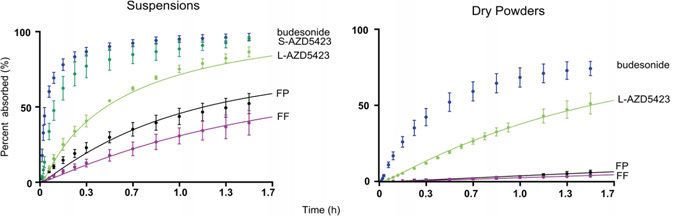
Ex vivo pulmonary drug absorption
Experimental absorption data
Ex vivo pulmonary drug absorption of inhalation APIs in solution, suspension
and dry powder were measured using single-pass IPL (Papers I and II).
Figure 7. Cumulative drug absorption profiles (means ± SD) measured in the IPL
for the ten investigated APIs (dots) and the curve-fitted drug absorption profiles ob-
tained from the IPL PBB model (lines). The graphs are divided by pharmacological
class.
In Paper I, ten APIs with a range in physicochemical properties (Table 2)
were administered as solutions to the single-pass IPL model. An agreement
between pharmacological class and initial drug absorption rate was observed
for the solutions, where the steroids were most rapidly absorbed into the per-
fusing buffer, followed by the beta-2 adrenoceptor agonists (except for olodat-
erol), and finally the muscarinic antagonists (Figure 7). The steroids have a
high permeation rate across the lipoidal epithelial membrane because of their
31You can also read