An introductory
guide for assessing
and understanding
common wounds
with people who
inject drugs
←
→
Page content transcription
If your browser does not render page correctly, please read the page content below
An introductory guide for assessing and understanding common wounds with people who inject drugs
Preface                                                                                  Contents
    This guide was created for harm reduction medical staff and volunteers as a                      1. Abscesses.....................................................2–7
resource about the types of wounds common with injection drug use and also
to increase knowledge about treatment modalities for this population. Skin and                       2. Missed Shots ...............................................8–11
soft-tissue infections are the most common cause of hospitalization among people
                                                                                                     3. Cellulitis......................................................12–13
who inject drugs.1 One study reported that 32% of active injection drug users
had a current soft-tissue infection, and this number is most likely higher in areas                  4. Phlebitis, Track Marks & Scarring....................14–15
where tar heroin is prevalent.2 Effectively treating skin and soft-tissue infections
is an imperative component of harm reduction, as these infections can lead to                        5. Chronic Wounds..........................................16–19
catastrophic conditions such as sepsis and endocarditis and can also negatively
impact injection drug users’ social and employment status.                                                  Venous Ulcers..........................................20–23
    Due to concerns about finances, lack of health insurance, and stigmatization
by health care providers, people who inject drugs typically seek professional                               Arterial Ulcers........................................24–25
medical care as a last resort. A study done in Washington DC in 2009 noted
that 81% of those who inject drugs have reported having had an injection-related                     6. Muscling & Skin Popping.................................26-27
wound, and that 93.9% of them reported self-management of wounds, including
                                                                                                     7. Dangerous Conditions
such potentially dangerous practices as self-incision and drainage and the use of
nonprescribed antibiotics.3 Though self-management of wounds among injection                                Sepsis ....................................................28–29
drug users presents dangers, it is also potentially an advantage, as this community
shows a strong history of resilience and self-care. With this background of self-                           Cotton Fever...........................................30
reliance, given the right tools and information, people who inject drugs may be
able to safely and effectively prevent minor infections from becoming catastrophic.                         Necrotizing Fasciitis ................................31
This guide was created as a starting point for assessing, educating, triaging, and
providing care in harm reduction settings.                                                                  Endocarditis.............................................32

                                                              Alec Dunn and Tim Gauthier                    Osteomyelitis.........................................33
                                                                              April 2020
                                                                                                     8. Puffy Hand Syndrome ..................................34
The information presented here is based on research, experience, mentorship, and                     9. HIV & Hep C................................................35
multiple guidebooks. We have attempted to cite references to current research when
appropriate, especially when there seems to be new or conflicting information. We                    10. Wound Healing............................................36–37
are always open to feedback, criticism, and questions.
                                                                                                     11. Assessment Basics.......................................38

                                                                                                     12. Referrals.....................................................39

                                                                                                     13. Pearls for Basic Wound Care........................40–41
1. Brown, P.D., & Ebright, J.R. (2002). Skin and Soft Tissue Infections in Injection Drug Users.     14. Phlebotomy Tips .........................................42–43
Current Infection Disease Reports, 4(5), 415–419. doi: 10.1007/s11908-002–0008-0.
2. Grau, L., Arevalo, S., Catchpool, C., & Heimer, R. (2002). Field Action Reports. Expanding Harm
Reduction Services through a Wound and Abscess Clinic. American Journal of Public Health, 92(12),
                                                                                                     15. Notes on Nursing.........................................44–45
1915–1917. doi:10.21t05/AJPH.92.12.1915.
3. Roose, R., Hayashi, A., & Cunningham, C. (2009). Self-Management of Injection
Related Wounds Among Injecting Drug Users. Journal of Addictive Diseases, 28(1), 74–80.
doi:10.1080/10550880802545200.
                                                                                                                                                                                1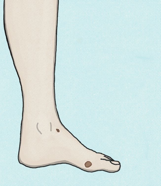
Abscesses
Definition                                                                                Fig. 1: An open abscess. Photo by
      An abscess is an encapsulation of pus that builds up within tissue, caused by       Alec Dunn, 2015.
a localized reaction against pathogens. Although abscesses can occur anywhere
within the body, they represent one of the most common skin and soft-tissue
infections (SSTIs) among people who inject drugs. Abscesses are composed of
blood, dead tissue, pathogens, and white blood cells. Abscesses are the body’s de-
fense against infectious substances and pathogens; by walling off the area and
flooding the area with white blood cells, the body is attempting to contain the
infection.
      The act of injecting drugs places people at greater risk for acquiring an abscess
by passing the body’s surface bacteria into deeper tissue, and they may occur more
frequently as an SSTI when a vein is missed. Harm reduction strategies are essen-
tial in reducing the risk of acquiring an abscess (hand hygiene and the availability
of alcohol swabs are absolute necessities), and careful recognition and appropriate       Fig. 2: An abscess that has spon-
management of these infections are essential in preventing these infections from          taneously opened and is draining
                                                                                          clear fluid.
advancing into deeper tissues or disseminating systemically.
                                                                                          Photo: Centers for Disease Control
                                                                                          and Prevention’s Public Health
Risk Factors                                                                              Image Library, public domain,
• Not cleaning injection site and hand before injecting. As nurses it is our re-          2014.
    sponsibility to advocate for transportable cleaning resources to be available
• Missing an injection
• Injecting drugs cut with damaging substances (pathogens, irritants, vasocon-
    strictors)
• Injecting cocaine or amphetamines, as they are vasoconstrictive and more
    inflammatory to tissue
• Reusing needles, which makes venous access more difficult and damages                   Fig. 3: An open abscess.
    tissue                                                                                Photo: Centers for Disease Control
• Reusing cookers and cottons, which increases the microbial burden                       and Prevention’s Public Health
• Diminished immune function                                                              Image Library, public domain,
• Skin/mucosa colonized with aggressive pathogens (e.g., MRSA, IGAS)                      2014.
• Injecting into areas that show signs of damage and inflammation
• Injecting into areas with poor circulation (e.g., venous stasis, diabetic foot)
• Epidemiology—it is important to know pathogens that are endemic to
    your area that may be useful in the early detection of rapidly disseminating
    pathogenic wounds
• Available drug supply—tar heroin is, in general, more damaging to veins
    than powder heroin

2                                                                                                                          3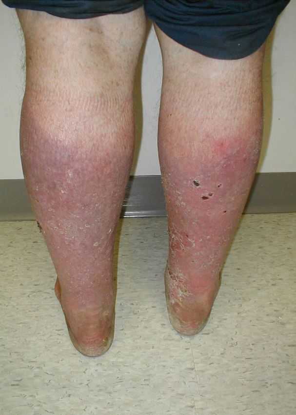
Abscesses (cont’d)
Signs & Symptoms
     Abscesses are encapsulations, so by nature they will begin as closed wounds.
When they occur near the skin’s surface, they will be raised, red, swollen, and may
be tender or painful. Deep-seated infections (muscular, internal...) may present in
ways that are more subtle with regards to heat, redness, and swelling, but there
will often be a brilliant pain associated with these wounds. Skin may be noticeably
warmer around the site due to increased blood flow and inflammatory modulators.
When deep-seated abscesses/infections are suspected, it is important to refer to a
primary care provider who will likely make a decision to treat empirically with
antibiotics or order imaging to determine if there is a collection of fluid beneath
the tissue.
     Abscesses may open up over time due to mechanical manipulation (picking,
scratching, squeezing), or they may swell and open up to the tissue’s surface as the
body forces the contents to the surface. Other times, abscesses will heal without
opening as the body deals with this issue internally, or they may require external
intervention to open up and release their contents (incision and drainage). In most
states and provinces, incision and drainage are outside of the scope of nurses ex-
cept for nurse practitioners, so it is important to understand the scope of practice
in the area in which you are practicing.
     An open abscess may drain any combination of purulence, clear/serous fluid,
or blood. They may dry and scab over and, depending on the person’s immune sys-
tem and circulation, may take a considerable amount of time to heal. Depending         Fig. 4: Cross section of an encapsulated abscess—note the possibility of releasing abscess
                                                                                       contents into bloodstream from manipulation.
on the size of the wound, they will need to heal by primary or secondary intention,
so wound care may be ongoing and will need to adapt to whatever changes occur
within the wound bed and more generally for the person. Age, stress, nutrition
status, drug of choice, homelessness, comorbidities, and immunodeficiency may
all delay wound healing.

Prevention
• Hand and skin hygiene before and during injection process
• Alcohol swabs prior to injection, but avoid the use of alcohol swabs post-in-
    jection as this decreases clotting factors and damages the injection site
• Use clean/sterile equipment (cottons, cookers, rigs should not be reused)
• Exclusive use of one’s own supplies: tourniquets, cookers, needles, filters. Any
    supplies used for injection that have been used by another person carries a
    significant risk in terms of infectious disease
• Phlebotomy: Avoid missed shots and venous rupture. Choose injection sites
    that are easy to see and easy to feel and that have good perfusion. Avoid sites
    that are close to important structures (arteries, nerves, tendons…)
                                                                                       Figs. 5 & 6: Two closed abscesses. Photo from San Francisco General Hospital Emergency
                                                                                       Services, no date.

4                                                                                                                                                                               5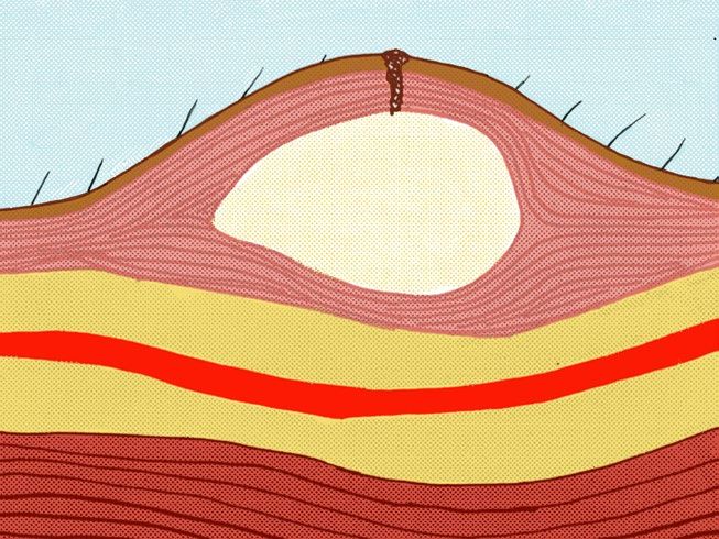
Abscesses (cont’d)
Secondary Prevention
• Apply warm compresses to small abscesses to promote blood flow and healing
• Avoid picking or squeezing of the wound, which may exacerbate the
inflammatory response, promote the spread of infection to surrounding tissues,
and delay healing
• Monitor for the spread of inflammation and infection into surrounding tissues
(advancing erythema/redness, heat, tenderness, induration)
• Assess for systemic illness: fever, chills, myalgia/arthralgia (muscle/joint aches),
lymphadenopathy, lymphangitis (streaking), tachycardia, and tachypnea
• In caring for people who have significant (or seemingly insignificant) wounds,
we have an immense responsibility to rule out red flags that may indicate a
catastrophic infection or death stemming from their wounds.
Treatment Information
Many abscesses may heal on their own over time. They may also spread
beyond their border and become more serious infections. Keeping the area
clean with soap and water, applying warm compresses, and not injecting in
that site are primary first steps. Hot soaks or Epsom salts may help improve
circulation to the area and support the body in drawing out pus as well as
relieving irritation and surface tension. A New England Journal of Medicine
Fig. 7: An abscess with lymphangitis. Photos from Google Image search, unattributed, re-
review recommended that the size threshold for incisions and drainage of a trieved 2017.
closed abscess is greater than or equal to 2 cm.1 Below that size, the patient
can apply local heat and follow it closely. Above that, the best treatment is in- target the bacterial infection in the tissues, and wound care can help manage the
cision and drainage is the best treatment, with or without systemic antibiotics. infection occurring on the wound’s surface.
If an abscess needs to be incised, drained, and debrided, it should only be
done by a medical professional, as there is real potential to inadvertently spread Complications
the infection to surrounding tissues, cause serious injury, and delay healing if done As mentioned above, abscesses may resolve on their own, though they may
incorrectly. Abscesses that require incision and drainage may require packing to also develop into more serious infections. Warning signs that require medical
keep the wound open to enable continued drainage. Without packing, the body attention include: red or black streaks running along the veins away from the
may try to close the incision prematurely, with subsequent encapsulation of the abscess (streaking or lymphangitis), a swollen and red area growing widely (and
abscess being a possible outcome. Topical or systemic antibiotics may be required, possibly rapidly) around the abscess (cellulitis), and the general signs and
depending on the pathogen, the extent of the wound, due to any signs of systemic symptoms of systemic infection (malaise, myalgia, fever), which could indicate
infection (fever, chills, feeling unwell, swollen glands, muscle aches and pains) and bacteremia or septicemia (a life-threatening blood infection).
the individual’s immune function. It may be necessary to educate or remind people While this chapter focuses primarily on abscesses involving the skin and
that antibiotics need to be prescribed to treat specific infections, and that using surrounding tissues, please keep in mind that abscesses can also occur internally
the wrong antibiotics will be ineffective, may promote resistance, and can cause (e.g., brain, spine, psoas muscle), as pathogens injected directly into the blood stream
serious adverse reactions. Systemic antibiotics treat most infections but leave the may cause infections wherever they find an opportunity. Staff should have a high
individual immunocompromised and should be used cautiously. Antibiotics will index of suspicion for anyone who injects drugs and presents with systemic signs
1 Singer, A. & Talan, D. (March 2014). Management of Skin Abscesses in the Era of Methicil-
of infection and new onset of focal pain. Referral to an emergency department is
lin-Resistant Staphylococcus Aureus. New England Journal of Medicine (370), 1039–1047. DOI: essential in preventing death and disability for these deep-seated infections.
10.1056/NEJMra1212788
6 7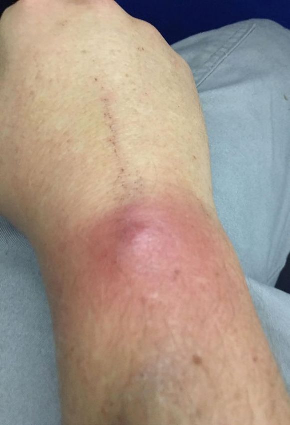
Missed Shots
Definition
     A missed shot is a local site of inflammation that occurs when someone misses
while injecting into a vein. The presentation of the missed injection depends on
where the miss occurs. Missed injections most commonly occur in subcutaneous
or muscle tissue, but they can also involve nerves and arteries. Injecting into an artery,
aside from being quite painful, causes the substances to travel directly to the
distal tissues, where an inflammatory and histamine cascade is usually provoked
distally to the injection site. Arterial injections can threaten life and limb through
thromboembolism, hemorrhage or compartment syndrome, with a high degree of
risk when major arteries are accessed by mistake (e.g., femoral, carotid).
     Nerve injections are often described as sharp, electric, or burning, and pain
will often radiate proximal and distal to the injection site. Complications from
nerve involvement can lead to loss of sensation and function, and chronic pain to
the tissues innervated by the nerve.
     Venous ruptures are not true misses, as the majority of the drug is contained
within the vein, but the leaked drugs/contaminants may still cause pain,
inflammation, or infection.

Risk Factors
• Missing a vein: dull or damaged needle, wrong needle type (too long, large
    gauge), rushed injection, poor lighting, injecting in a public space (fear of
    police, security, public), tremor/shakes, poor eyesight, misidentifying a vein,
    insertion angle too steep or too low, speed of insertion too fast or too slow
    (veins typically roll when the injection is slow)
• Venous rupture: injecting into small/fragile veins (hands/feet), tourniquet
    too tight, injecting too quickly
• Dehydration
• Stimulants like cocaine (powder and freebase/crack) and methamphetamines are
    vasoconstrictive and irritating to body tissues, which provokes inflammation
    and delays healing. Crack cocaine also requires the use of an acid to free the
    cocaine from the base it is bound to for it to be injected. Vinegar (acetic
    acid) is commonly used and is corrosive to the vessels and inflammatory and
    irritating to the tissues. Lemon juice carries the risk of fungal infections. It is
    best to use ascorbic acid (prepared in the smallest amount possible) to break
    down the drugs until they are fully dissolved in water
• Black tar heroin is inflammatory and also carries the risk of exposing the user
    to anaerobic pathogens such as botulism. Tar heroin may also be difficult to
    break down for injection and may require the use of an acid (e.g., ascorbic acid)

                                                                                             Fig. 8 Missed shot. Photo by u/muaDeeeb from reddit.com/r/opiates, retrieved 2019.

8                                                                                                                                                                                 9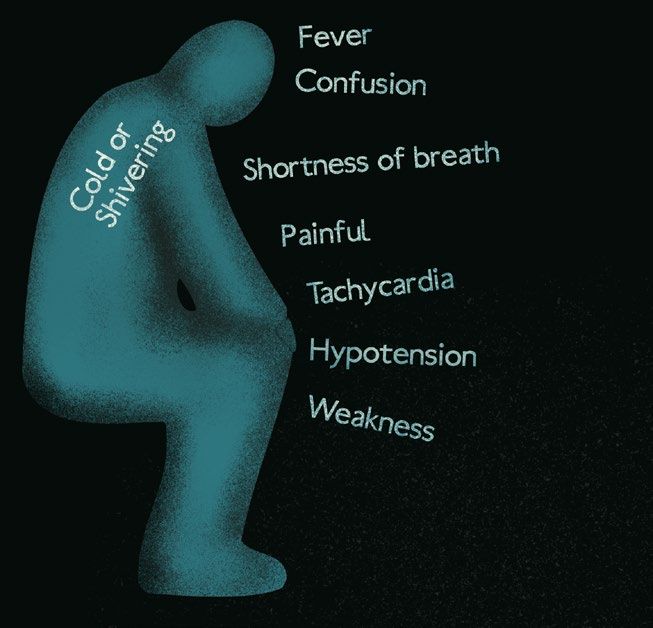
Missed Shots (cont’d)
Signs and Symptoms of Interstitial Injections
     Most of the time interstitial injections will present as a small area of redness,
which may be flat or raised and warm to the touch. Some drugs are numbing
to the tissues (e.g., cocaine is a powerful anesthetic) but even then bright and
intense pain at the injection site at the time of injection is almost always reported.
Disseminating erythema, pain, heat, and swelling are all signs of complications
from a missed injection. It is important to continue monitoring for skin and
soft-tissue infections (erysipelas, cellulitis, abscess) and their complications
(lymphangitis, bacteremia, nerve compression, pain, disability, and disseminating
infections).

Treatment
• Monitor closely for complications. Assess and treat for ulcerations,
    abscesses, cellulitis, or other signs of infection
• Warm compresses can help increase circulation to the site of a missed
    injection, which can promote absorption of the missed drugs and an
    improved immune response
• Provide education about safer injection practices: tourniquets, vein care and
    location (arms vs. hands, arms vs. legs and feet, avoiding the neck, wrist, and
    groin), injection hygiene
• Advocate for safer spaces for people to use drugs
• Advocate for and provide unlimited harm reduction supplies
• Allow access to sinks to wash hands and injection sites
• Arterial injections: due to the high-risk nature of these events, it is
    recommended that anyone showing signs and symptoms of an arterial
    injection seek prompt medical care
• Treatment may include anti-inflammatories, pain control, and
    anticoagulation to prevent a thromboembolic event

Missed Shot vs. Abscess
     A missed shot may present with similar properties to an unruptured abscess
(red, painful, swollen). The symptoms from uncomplicated missed shots (injected
into subcutaneous or muscular tissue) will, in most cases, ebb within 12 hours.
Abscesses will typically take longer to develop. Any raised and swollen area that
spontaneously opens or leaks fluid should not be considered a missed shot.

                                                                                         Figs. 9 & 10: Missed shot? Developing abscess? Cellulitis? Photos from Google
                                                                                         Image search for “missed shot,” unattributed.

10                                                                                                                                                                       11
Cellulitis
Definition                                                                                 Complications
     Cellulitis is an inflammation of the deeper layers of the skin (the dermis and             Cellulitis may resolve unassisted, though typically it will need a course of anti-
subcutaneous fat layers) caused by bacterial infection. Cellulitis may occur               biotics. Cellulitis leaves one at a high risk of developing more serious complications
secondarily to an abscess or other wound type, or independently. It may appear             including sepsis, endocarditis, necrotizing fasciitis, and infections of the bones and
in areas where there is no history of injecting but may still be related to injection      lymphatic system. Cellulitis in the leg may be confused for deep vein thrombosis
hygiene or contaminated drug supply.                                                       (clots in the large veins of the legs), which also presents as a unilateral red, swollen,
                                                                                           painful extremity.
Risk Factors
• Not cleaning skin before injecting                                                                                                                Fig. 11: Cellulitis in the arm.
• Reusing needles, cookers, and cottons                                                                                                             Photo: Poupou l’quourouce, 2006,
• Immunosuppression                                                                                                                                 licensed under the Creative Com-
• Poor circulation in extremities                                                                                                                   mons, retrieved from Wikimedia
                                                                                                                                                    Commons.
Signs & Symptoms
     Cellulitis will appear as a red, swollen, area with diffuse, patchy, or loose
borders. It is generally very painful and tender to the touch. The skin may be warm
or hot. Erysipelas (an infection of the more superficial dermis) is often confused
with cellulitis. Like cellulitis, erysipelas will present with pain, heat, erythema, and
tenderness but the borders will be well defined or clearly demarcated. Erysipelas                                                                  Fig. 12: Cellulitis in the left leg.
can overlap with cellulitis, making it difficult to distinguish between these two                                                                  Photo: Colm Anderson, 2006,
syndromes. Over time, erysipelas can extend into the deeper tissues (cellulitis) or                                                                licensed under the Creative Com-
can disseminate and become systemic. Therefore, the assessment and management                                                                      mons, retrieved from Wikimedia
of erysipelas remain the same the same as for cellulitis.                                                                                          Commons.

Treatment Information
      Cellulitis moves outside the scope of localized wound/nursing care, as there is
no topical treatment or care that will help resolve this type of infection. Suspected
cellulitis should be referred to a primary care provider for diagnosis and antibiot-
ics. It may be helpful to trace the border of the reddened area with a marker. It is
important to regularly assess for signs and symptoms of systemic dissemination for                                                                 Fig. 13: Cellulitis in the hand
anyone with cellulitis and to refer to an urgent care or emergency department when                                                                 and wrist. Photo by Alec Dunn,
systemic signs and symptoms are present, as this may indicate bacteremia or sepsis.                                                                2015.
Systemic signs and symptoms may include fever, chills, malaise, increased respira-
tion rate, tachycardia, and lymphangitis. Even when systemic antimicrobial therapy
is initiated, it is important that wound care is still provided when open wounds
are present. Systemic antimicrobials address the infection within the tissues, while
wound care addresses the infection (and other issues) occurring in the wound bed
and on the wound surface.

12                                                                                                                                                                                   13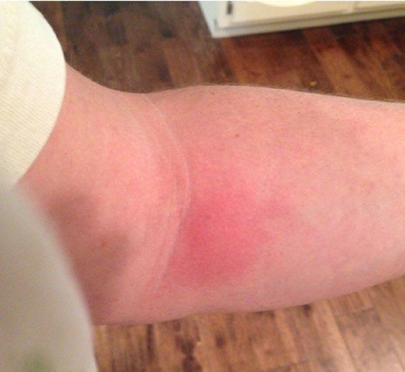
Phlebitis, Track Marks                                                                   & Vein Scarring
Definition                                                                               fection, and increase the risk of thrombus formation. It is also recommended that
      Phlebitis is an acute, temporary, inflammation of a vein resulting from injury     injections sites immediately distal to the site of phlebitis be avoided for injection.
or infection. Sometimes phlebitis is caused by, or provokes, a thrombus at the site of   Conservative management of simple phlebitis includes elevation, hot or cold com-
infection, in which case the term thrombophlebitis is used. Suppurative phlebitis is     presses (to improve blood flow or decrease inflammation), and NSAIDs (for pain
a type of phlebitis that occurs in the context of systemic infection or bacteremia and   and inflammation). Most of the time phlebitis is self-limiting, but it is important
is serious in nature and should be suspected when someone presents with symptoms         to monitor phlebitis carefully as it may lead to more serious complications (sup-
of phlebitis and a high fever. Track marks are small scabs and scarring (possibly with   purative phlebitis, thrombophlebitis, lymphangitis) and may co-occur with other
localized inflammation) along areas of current injection. Scarring of veins may          serious pathological processes (e.g., deep vein thrombosis, cellulitis). Scarring and
occur with recurrent usage of certain veins for injection.                               track marks may be lessened by topical treatments such as cocoa butter, aloe vera,
                                                                                         or vitamin E oil or cream. Scarred veins have a much higher incidence of infection
Signs & Symptoms                                                                         and should not be used for injection.
     Phlebitis is characterized by raised and hardened (often described as “ropey”)
veins. Phlebitis will be painful and tender, and may also be red. Signs and symp-
toms of suppurative phlebitis are consistent with phlebitis as described above,
plus erythema to surrounding tissue and a high fever. Pay specific attention to
signs and symptoms of sepsis (increased resp. rate, pale/mottled skin, fever, tachy-
cardia and hypotension in later stages). Scarring is characterized by raised and
hardened veins, which may also be ropey and roll easily. Scarred veins without
inflammation (nonphlebitic) will not be tender. Track marks will be a series of
scabs or scars along frequently used veins for injection.

Prevention
• Accurate phlebotomy and access to appropriate equipment
• New needles for each injection (to prevent blunting of needles and to reduce
    infection)
• Use of alcohol swabs prior to injection, but not after (alcohol damages healthy
    tissue, provokes inflammation/scarring and delays coagulation and wound              Fig. 14: Phlebitis. Photo by Alec Dunn, 2015.
    closure)
• Adequate compression of injection site post injection to promote hemostasis
    and immediate wound closure
• Rotating injection sites, especially avoiding injection sites distal to previous
    damage

Treatment Information
      Moderate to severe phlebitis should be referred to a primary care provid-
er. If the person has signs of systemic infection or a thrombus, they should be
referred to an emergency room. Prevention measures for phlebitis include edu-
cation around injection hygiene, the rotation of injection sites, and phlebotomy
techniques. Avoid injecting into veins that show signs and symptoms of phlebitis,
as this will provoke an inflammatory response, delay healing, increase risk of in-
                                                                                         Fig. 15: Track marks. Photo by Alec Dunn, 2015.
14                                                                                                                                                                          15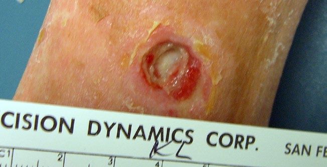
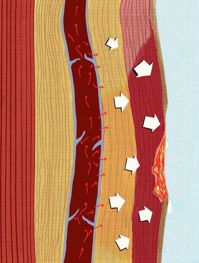
Chronic Wounds
Introduction
      Many IV drug users have preexisting conditions that make them more sus-
ceptible to acute and chronic wounds. Common preexisting conditions that reduce
immune function include poor circulation, hepatitis B & C, and HIV. Suscepti-
bility to injection related infections and wounds may be compounded by common
chronic diseases like diabetes, hypertension, cancer, heart failure, and peripheral
vascular disease. It is also believed that heroin may suppress some immune system
functions (specifically T cell activity and wound healing).1
      The damage to veins and vein valves from injecting drugs in one’s extrem-
ities may exacerbate preexisting problems for people with poor circulation. Poor
circulation may prevent wounds from healing, and chronic wounds and chronic
infections may develop. According to studies, the most common type of chronic
wound in longtime injection drug users is venous leg ulcers.2 Arterial ulcers re-
lated to peripheral artery disease (PAD) should be understood, as well. A brief
introduction follows.

     Peripheral vascular disease (PVD) is a catchall term that is generally applied
to blood circulation problems in the legs. There are two main categories of PVD:
peripheral artery disease and venous insufficiency (also known as venous stasis).
Understanding the difference between these two conditions is crucial when assess-
ing the different types of wounds and possible treatment plans. If we misidentify
peripheral arterial disease as venous insufficiency and proceed with applying com-
pression therapy to the leg (a standard of care for venous insufficiency), we run
the risk of occluding the arteries, which may lead to necrotic injuries to the affected
limb.
     Both venous insufficiency and peripheral artery disease can lead to chronic
wounds that do not easily heal because of impaired circulation, increased pressure
within the tissues, and prolonged inflammation. It is possible to have mixed arteri-
al and venous insufficiencies, so a detailed lower leg assessment is imperative when
making a nursing diagnosis.

     Venous insufficiency describes the poor return of blood from the extremities
and almost always applies to the lower limbs. This is especially common with peo-
ple with cardiac issues like heart failure (and of note for people who inject drugs,
this can be a result of having a history of endocarditis, septic emboli/valve failure,
and liver disease). When assessing IV drug users, have a high index of suspicion

1 Ebright, J.R. & Pieper, B. (2002). Skin and Soft-Tissue Infections in IV Drug Users. Infectious
Disease Clinics of North America 16(3), 697–712.                                                     Fig. 16: Mixed venous and arterial insufficiency. Notable signs of venous insufficiency
2 Birk T.J., Kirsner R.S., Pieper B., & Templin T.N. (2007). Injection Drug Use: An Underestimated   include leg swelling, stasis dermatitis, and ulcer development. Arterial insufficiency indi-
Cause of Venous Disease. Archives of Dermatology 143(10), 1305–1309.                                 cated by dry ulcers. Photo: Charlie Goldberg, MD, ©2005

16                                                                                                                                                                                              17
Chronic Wounds
Venous Insufficiency Symptoms Peripheral Artery Disease Symptoms
• Pain or cramps while standing • Pain with walking and activity (intermit-
• Dull ache in legs tent claudication)
• Relief with raising of the legs • Absence of hair on the legs with dry skin
• Edema/swelling of the legs • Pain when legs are raised
• Leg heaviness • Numbness
• Varicose veins • Cold extremities
• Itchy skin • Thready or absent pulse
• Shiny, thickened skin • Ulcers occur more typically in areas of
• Discoloration/darkening of the skin in the feet, ankles, bony prominences
legs (stasis dermatitis)
• Pulse present but may be difficult to find
due to edema
• Ulcers most likely found along ankle,
shin, and calf areas
for infective endocarditis when they present with new or sudden onset of heart is important to consider agents that may further constrict the vessels (e.g., stimulants
failure symptoms, especially if they have a history of valvular damage or previous like cocaine, methamphetamine and nicotine all possess vasoconstrictive properties
infective endocarditis. Furthermore, venous insufficiency may be made worse that may exacerbate venous/arterial wounds or delay wound healing). Peripheral
by damage to valves and vein walls secondary to injection (if injection occurs in artery disease may be asymptomatic in an estimated 50% of people, so any intensive
the lower legs). When venous return is compromised in the lower legs, blood pools compression therapies should only be initiated under the guidance of a certified
in the big veins in the lower extremities and begins to leak out into the tissues of wound care clinician or primary care provider. The standard of care for diagnosis of
the legs. Eventually, if the area becomes overwhelmed with the amount of fluid, the peripheral artery disease includes the ankle-brachial pressure index (a comparison
tissue will open and lead to weepy ulcers. of pressures between brachial and lower-leg arteries) and in severe cases, imaging/
Peripheral artery disease describes the poor perfusion of blood to the extremities vascular studies.
because of narrowed arteries (secondary to hypertension and atherosclerosis), and it
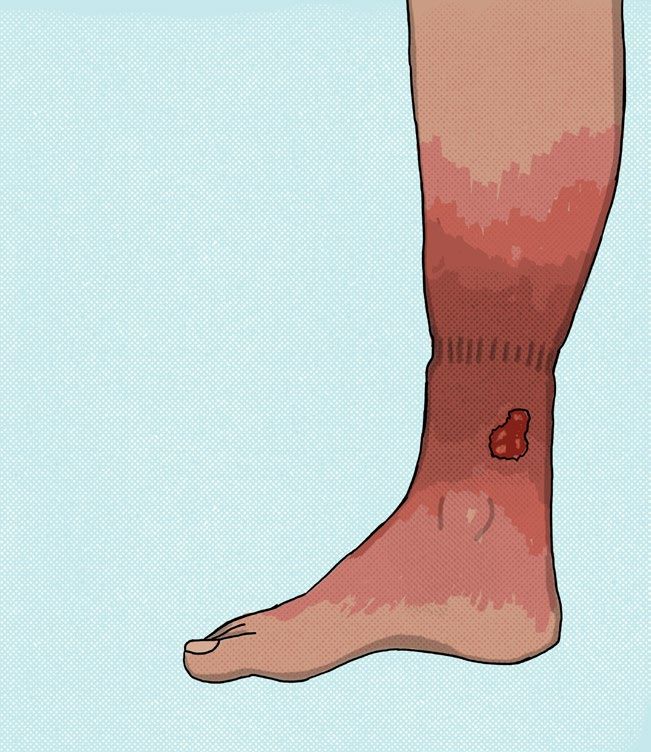
18 19Venous Ulcers
Fig. 17: Lower arm
Definition venous ulcer. Photo from
Venous leg ulcers are the most common type of chronic wound in injection San Francisco General
drug users. These ulcers occur when poor venous return leads to venous pooling, Hospital Emergency
venous engorgement, and venous rupture/leakage. These events lead to an under- Services, no date.
whelming immune response and to decreased oxygen and nutrients delivery, which
becomes compounded by more and more fluid pooling in the legs. Eventually the
tissues can no longer compensate for the increased pressure, and the legs ulcerate
and begin to weep fluid.
Risk Factors
• Venous insufficiency
• Injecting into the veins of the legs
• History of frequent phlebitis
• Hypertension
• Smoking Fig. 18: Venous leg
• Work that requires standing or sitting for long hours ulcer. Photos from Google
Images, unattributed,
• Diabetes
November 9, 2014.
• Family history
• Chronic conditions such as heart failure, liver disease, and diabetes
Signs & Symptoms
Venous leg ulcers are irregularly shaped, may be of any size (and may be quite
large), and may be painless. Wound bed presentation will vary depending on chronic-
ity, inflammation, microbial burden, necrosis, trauma, and adaptive changes. Healthier
wound beds are red and granulated; chronic ulcers may have yellow to brown fibrin;
dead tissue may present as loose and stringy (slough) or as dry and attached (eschar).
Dead tissue significantly increases the risks for infection and should be removed in
the safest way possible (autolytic, sharp, or mechanical debridement). This may require
antimicrobial therapy (topical, systemic, or both) depending on whether signs and Fig. 19: Large venous
symptoms of infection are present. Venous ulcers are typically wet and weepy, and stasis ulcers. Photo:
typically exudate a clear (serous) or pink (serosanguinous) fluid. Milorad Dimic MD,
The surrounding skin may be dark (hemosedrin staining is present in most 2009, licensed under
limbs chronically affected by venous insufficiency) and the leg may be very edema- the Creative Commons,
tous (assess for pitting edema). Prolonged venous engorgement will lead to varicos- retrieved from Wikimedia
ities in the lower leg, and a general inspection of both legs ought to be performed Commons.
to support the assessment and diagnosis of venous insufficiency. It is essential to
assess for tissue perfusion to the lower limbs, in order to distinguish between venous
and arterial insufficiency—assess for color, warmth, movement, and sensation in the
lower limb. Palpate pedal pulses of both legs and note any differences between limbs.
Assess for hair growth of the lower legs, and note the absence of hair to the toes, feet,
ankles, and legs, and whether this marks a status change for the individual.
20 21Venous Ulcers
Prevention
People with any type of peripheral vascular disease should avoid injecting into
the legs. For those who are unable or unwilling to cease injecting in their legs, it is
important to communicate the benefits of rotating sites (to prevent phlebitis, scle-
rosis, repeated valve damage, and collapse), changing needles between injections (to
prevent infection and dulling), and using the smallest gauge possible (to minimize
trauma). Compression therapy (unless contraindicated due to arterial disease) is
useful in supporting venous return, to prevent venous engorgement and pooling,
and should be used even in the absence of active disease for anyone with venous
stasis, especially with a history of venous ulcers. Compression stockings are costly
but are easy to apply once an acute exacerbation has been resolved.
Treatment
Venous leg ulcers take a long time to heal. For some ulcers, debridement
of the wound bed may be necessary to clear away necrotic or nonviable tissue.
Compression therapy is the standard of care for venous insufficiency and pro-
motes wound healing by improving venous return. Compression stockings may
be worn over the top of dressed ulcers when there are no signs of active infection
or when the infection is managed appropriately. For acute exacerbations of ve-
nous ulcers, or an increase in lower leg edema, more aggressive/reactive forms of
compression may be recommended (e.g., compression wraps like Coban) until
the edema is under control.
When the lower leg edema is back to baseline, it is recommended to resume
consistent compression therapy through stockings or stocking-like therapies such
as Tubigrip. It may take weeks to months to regain control of an acute exacerba-
tion of venous insufficiency and to fully approximate the ulcer. Please note that
the healed/scarred tissue is not as durable as the original tissue and that venous
ulcers may reopen easily if the venous insufficiency is not kept under control.
Gentle exercise such as walking can help improve circulation through mechanical
support of the lower venous system, primarily through the calf-muscle pump.
Nursing care for venous ulcers may include gentle washing and antimicrobial
support of the ulcer along with a topical dressing appropriate to the level of drain-
age (with dry gauze or alginates recommended for very wet wounds). Care should
be taken not to compromise poor circulation when wrapping wounds on the legs
or arms. Again, it is crucial that one is assessed for mixed arterial and venous com-
promise and that one does not apply any compression to limbs affected by arterial
disease. Even mild compression applied to diseased arteries may cut off circulation Complications
and cause ischemic injury, including death to compromised tissues. Compression Infection is the major risk of chronic open wounds with poor circulation.
therapy should only be suggested or used when the person has clear signs of poor People with venous stasis/venous leg ulcers are also at a higher risk for developing
venous return, and when there are signs of arterial health (color, warmth, good cap- deep vein thrombosis (DVT), which can lead to a pulmonary embolism.
illary refill, movement, sensation, hair growth, palpable pedal pulses).
22 23Arterial Ulcers
Definition
Arterial ulcers are caused by poor circulation in the extremities due to
arterial constriction. Poor circulation leads to inadequate oxygen and nutrient
delivery to the area and will lead to chronic nonhealing wounds. Though they are
most common in the lower extremities, they may also occur on other parts of the
body. Arterial ulcers may also be referred to as ischemic wounds.
Risk Factors
• Hypertension
• Nicotine use
• Cocaine/methamphetamine use
• Injecting into the veins of the legs
• Diabetes
• Stress
• High cholesterol
• Family history
Fig. 20: Arterial ulcer. Photo: Wound, Ostomy, and Continence Nurses Society, ©2007.
Signs & Symptoms
Arterial ulcers will typically be round, pale, and sunken. They are generally As noted above, debridement may not be
small to midsize, though they may be quite deep. The surrounding skin may be appropriate for arterial ulcers. Nursing
black (necrotic) or gangrenous. They will typically be dry, and there may be no care for arterial ulcers involves keeping
surrounding inflammation. General conditions that point to arterial disease may the area clean, though covering the
help in diagnosing these wounds, including: pain with walking or elevation, tight- wound may not be necessary. Meticulous
looking skin, little to no hair on the legs, and signs of compromised circulation foot care should be encouraged and
(e.g., cool to touch, poor capillary refill, weak pulses). Pain may be blunted with exercised. Healing will only occur with
diabetics due to neuropathies. improvement of circulation to the
Ischemic wounds and arterial ulcers may be covered in a substance referred extremities, so prevention and education
to as eschar, which is a scab-like formation of hard necrotic fiber over the wound. is a key strategy.
Though treatments of most other wound types involve removing dead tissue to
promote healing, in arterial wounds it is typically recommended to leave the Complications
eschar in place, as it can work as a protective layer against opportunistic infection. People with PAD are at a high risk
for serious infections in their lower
Prevention extremities. Infections can easily lead
Treat high blood pressure and avoid injecting into the legs. A referral to a to gangrenous or black (necrotic) tissue,
primary care provider should be recommended to begin treatment as long-term amputations, catastrophic infections, and
peripheral artery disease can lead to gangrene, amputation, and death. death.
Treatment
Peripheral artery disease is difficult to treat. Lifestyle changes can help increase Fig. 21: Arterial ulcers. Small, round,
circulation in the legs, including mild exercise, healthy diet, and treating high “punched-out” appearance. Photos: Alex
blood pressure. There are surgical and medical options for treating PAD as well. Banger, © 2011
24 25Muscling & Skin Popping
Definition Complications
Skin popping and muscling refer to two different injection practices. Skin Infections are the primary danger of muscling, particularly deep infections
popping is the injection of drugs into subcutaneous tissue or intradermal spaces, that may not present themselves until they are well developed. These can be
and muscling is the injection of drug intramuscularly. Due to the inflammatory difficult to identify and treat. Any deep abscesses, cellulitis, muscular pain will
nature of most drugs and their adjuvants, skin popping and muscling are generally most likely require imaging and antibiotic therapy and may also require surgical
damaging to body tissues. This is especially true for most stimulants, which also debridement depending on the severity of the insult.
provoke vasoconstriction that in turn prevents absorption, prolongs inflammation,
and delays wound healing. Heroin and morphine, on the other hand, are readily
absorbed through intramuscular and subcutaneous routes and can significantly
decrease risks of fatal overdose through delayed absorption. People may choose
these injection practices for a variety of reasons, most commonly due to having
collapsed, scarred, or difficult-to-find veins. People may also muscle as it offers an
easier choice of sites to hide injection use, for a longer and more sustained high, or
due to a lack of knowledge about basic injection/phlebotomy techniques.
Signs & Symptoms
Wounds related to skin popping and muscling are not a specific type of wound
in and of themselves but may be an assortment of abscesses, cellulitis, and scarring
from repeated and multiple injections.
Prevention
If the injector would prefer to mainline, then education should be provided
about phlebotomy and injection technique. All parenteral routes (IV, IM, SC)
carry risks for infection and overdose events, so one should always be ready to
offer support and education around other routes of ingestion that may minimize
these risks. If muscling is preferred, there are several recommended practices:
extremely thorough hygiene of the injection site, rotating injection sites, and the
proper filtering of drugs will substantially reduce the risks of infection and injury,
including wounds, phlebitis, vascular collapse, and scarring. Whenever possible, it
is always recommended that drugs be heated/cooked prior to injection to reduce
the risk of infection, which is especially true if using a wash (or washes) due to the
opportunity for microbes to flourish between uses.
Using the right size and length of needle is recommended to allow penetration
into the highly vascularized muscle tissue–muscling needles are most commonly
1–1.5” long and generally a little bit larger, between 22 and 28 gauge. Muscling
needles should be injected at a 90-degree angle to the skin. Because muscling
needles go deep, they carry the risk of damaging or encountering nerves, arteries,
and connective tissue. The safest sites for muscling are the vastus lateralis, the
deltoid, and the ventrogluteus muscles. Figs. 22 & 23: Photos from Powell, G. (2011). Wound Care for Injecting Drug Users: Parts 1 &
2. Nursing Standard, 25(46); Hennings, C., & Miller, J. (2012). Illicit Drugs: What Dermatolo-
gists Need to Know. Journal of the American Academy of Dermatology
26 27Sepsis
Definition
Sepsis is a systemic and life-threatening inflammatory response due to an
infection. It can cause a dangerous and drastic drop in blood pressure (septic
shock) and, if untreated, will lead to death. Though uncommon, people who inject
drugs are at a higher risk of developing sepsis secondary to localized infection and
is much more common among people who inject drugs due to regular exposure of
pathogens to the circulatory system. Sepsis may be easy to miss, so it is important
to adequately assess anyone who may be at risk.
Initially, the body mounts a response referred to as the systemic inflammatory
response syndrome (SIRS) that is easy to confuse with other milder illnesses. SIRS
is characterized by fever, tachycardia, and tachypnea, along with an elevated white
blood cell count. As nurses working in the community, we may be unable to order
lab work to assess for the white blood cell count (and waiting for those results
could result in serious morbidity and mortality). So anyone with a risk for infection
(effectively all people who are actively injecting), feel ill, and are tachycardic,
tachypnic, and febrile should be encouraged to seek urgent or emergency care.
SIRS symptoms have some notable similarities to opioid withdrawal. Sepsis and
SIRS are frequently underdiagnosed in people who inject drugs. People who
inject drugs are often very resilient and may appear to be in less distress than they
actually are. Care should be exercised with any assessment and advice.
Sepsis can develop quickly into septic shock, and once someone is in shock
emergency care is essential to prevent imminent death. Visible signs of active
septic shock include a decreased level of consciousness, a weak and thready pulse,
and pale/gray skin.
As a rule, if something (or someone) feels wrong or scary, trust your gut. Find
another person to talk about it if you need a second opinion, or just take the person
to the ER. Treatment
Count the heart and respiratory rate, and, if possible, take the person’s blood
Sepsis Signs & Symptoms pressure, temperature, and oxygen levels. Having 2 of the following 4 criteria
• Fever and chills meets the definition of systemic inflammatory response syndrome:
• Shortness of breath • HR > 90
• Malaise and weakness • RR > 20
• Confusion • Temp > 38 C (100.4 F) or < 36 (96.8)
• Myalgia (muscle pain) • Elevated WBC
• Severe abdominal pain
• Hypotension SIRS criteria are sensitive but not specific, so people meeting SIRS criteria
• Tachycardia should raise flags for further investigation. Additionally, if the individual is
• Sudden swelling in legs/abdomen hypotensive (SBPCotton Fever Necrotizing Fasciitis
Definition Definition
Cotton fever is a rare complication from aspirating drugs (prior to injection) through Necrotizing fasciitis (sometimes referred to as the “flesh-eating bacteria”) is a
cotton filters. Among drug users, cotton fever was commonly thought to be a reaction rare infection of the deep layers of the skin and connective tissue, which typically
to loose threads of cotton floating in the bloodstream. With the increasing availability of affects immunocompromised individuals and has a higher prevalence among
sterile single-spun cotton filters at harm reduction sites in the last two decades, cotton people who inject drugs. With necrotizing fasciitis, toxins released by bacteria
fever was regarded by professionals (even in the harm reduction community) as an out- destroy tissue while also causing severe localized inflammation that isolates the
dated myth. Many health care professionals believed that IV drug users suffered from a area from the body’s immune response. This can lead to a cascading infection that
short-term inflammatory reaction that was thought to be the result of contaminants in destroys skin, fat, muscle, and connective tissue and that spreads throughout the
the drug supply, not related to cotton. body. To clarify, the bacteria involved are common bacteria with other infections
New research about cotton fever is inconclusive but points to evidence that cotton (e.g., Staph. aureus, Strep. pyogenes, E. coli), so necrotizing fasciitis describes an
fever is a real (though uncommon) reaction to injecting drugs filtered through cotton. infectious process, not a specific type of flesh-eating bacteria.
Research has proposed that cotton, commonly colonized by Enterobacter anaerobic bac-
teria, carries endotoxins that can trigger an inflammatory response (as has been seen in Signs & Symptoms
workers in the cotton industry). Cotton fever then is thought to be a result of the release The surrounding tissue may exhibit red, pink, yellow, brown, black, and green
and denaturing of endotoxins from these bacteria. It is also theorized that the heating of tones. These wounds may be large and weepy or may not be open at all. They will
drugs during preparation releases the endotoxin complex, leading to more pronounced be painful and may be swollen and shiny. Systemic symptoms include a high fever,
symptoms in people who inject drugs.1 dehydration, weakness, and other septic-like symptoms. Necrotizing fasciitis may
develop secondary to untreated/unresolved cellulitis or abscess.
Signs & Symptoms
Cotton fever will present with symptoms similar to sepsis and endocarditis: fever, Treatment
weakness, tachycardia, nausea, dyspnea, hypotension, and pain. People with suspected necrotizing fasciitis should be referred to an emergency
room. This needs prompt, serious medical attention and may lead to sepsis,
Treatment gangrene, amputation, and death. Treatment will involve IV antibiotics to resist
Cotton fever is difficult to diagnose from endocarditis or sepsis. Time may be the the spreading of the infection and will involve surgical debridement of necrotic
only significant differential in that cotton fever may come on very shortly after injection. tissue as well as a long course of healing.
Regardless, if the participant presents with these symptoms, they should be referred to
Figs. 24: Ad-
an emergency room immediately due to the difficulty in differentiating the diagnosis of vanced necrotiz-
cotton fever from life-threatening conditions such as sepsis or endocarditis. ing fasciitis. Photo:
If the participant is admitted to a hospital, their workup may reveal an elevated Smuszkiewicz, P.,
white blood cell count but ultimately with negative blood cultures and with normal heart Trojanowska, I.,
function. Time will help differentiate the diagnosis, as cotton fever will typically resolve and Tomczak, H.,
within 12–24 hours. 2008.
Complications
Once again, due to the possibility that the participant may be septic or have endo-
carditis (which have increased prevalence among people who inject drugs), cotton fever
should never be considered to be the first diagnosis.
1 Torka, P. & Gill, S. (2013) Cotton Fever: An Evanescent Process Mimicking Sepsis in an Intrave-
nous Drug Abuser. Journal of Emergency Medicine (44), 385–387.
30 31Endocarditis Osteomyelitis
Definition Definition
Endocarditis is an infection of the inner lining of the heart (the endocardium), Osteomyelitis (OM) is an infection of the bone. It is difficult to diagnose based
in which bacterial microbes adhere to heart valves, eventually causing permanent on overt signs and symptoms. Osteomyelitis should be suspected for any wound
damage. People who inject drugs are at a higher risk of developing infective where bone or tendon is exposed or where it is possible to probe directly to the bone
endocarditis due to the frequent exposure of external bacteria into the circulatory during wound care. Deep wounds that are slow to heal and that recur frequently
system, especially when they have preexisting endocardial/valvular damage, as in the same site should raise some suspicion for OM. Acute or persistent pain and
these increase the risk of microbial attachment. Bacteria that grow within the tenderness along a bony prominence or within and around a joint in the context of
heart and break off in clumps are called septic emboli. These may cause organ an infectious process should also warrant a high degree of suspicion.
damage, pulmonary embolism, and stroke. Osteomyelitis of the peripheral bones, the spine, and the hip are common
sites for infection among people
Signs & Symptoms who inject drugs. It is important to
The early stages of endocarditis may present with several ominous but regularly assess vital signs for anyone
inconclusive symptoms including: with a diagnosis or suspicion of OM
• Fever (may be on/off in chronic infections) as there is an increased potential for
• Weakness/fatigue these infections to become systemic.
• Anorexia Several comorbidities carry a high
• Dyspnea association with OM, particularly
• Persistent cough diabetes, chronic immunosuppression,
• Pulmonary crackles and peripheral vascular disease.
• Chest discomfort
• Myalgias/arthralgias Signs & Symptoms
• Night sweats • Fever and chills
• Splinter hemorrhages and petechiae • Malaise and weakness
• Sudden or progressive swelling in the legs/abdomen • Bone pains
• A new heart murmur • Loss of function of the affected
• Late stage endocarditis may present with symptoms similar to septic shock extremity
(endocarditis is often accompanied by sepsis) • Localized swelling, deformity, or
pain
Treatment
As with sepsis, if endocarditis is suspected the person should be immediately Treatment
referred to emergency care. Treatment will most likely involve long-term Osteomyelitis will be diagnosed
IV antibiotics, and if there is damage to the heart valves, then surgical valve by imaging, biopsy, and lab work.
replacement may be indicated and necessary. Treatment will usually require long-
term IV antibiotics and possible
Prevention surgical intervention. People who
Prevention and education are very important for people with a history of have experienced OM in the past,
endocarditis, as they are at a higher risk of reinfection and, as has been documented, or who have hardware in situ, are
many surgeons may refuse to treat repeat infections. Prevention includes meticulous more at risk for developing future
hygiene prior to injection and not reusing needles for injection. Education should infections at the same site. Treatment
be clarified about the early symptoms for endocarditis, particularly for people with typically requires antibiotics and
a history of this type of infection. possible surgery.
32 33Puffy Hand Syndrome HIV & Hep C
Definition There are many resources within harm reduction addressing hepatitis C and
Puffy hand syndrome is a condition in which long-term IV drug users may develop HIV/AIDS. Exploring these topics is beyond the intention of this guide, but par-
chronic, nonpainful hand swelling. It is a complication of injecting into one’s distal ticipants at harm reduction services may come in with questions about physiologic
extremities (injecting into the hands or feet may result in puffy hand or puffy feet, changes, some of which may be concerning and should lead to further testing and
respectively). The exact cause is unknown, but research speculates that repeated injec- diagnosis. As the sharing of injection gear increases the risk of bloodborne dis-
tions into the extremities lead to the breakdown of the lymphatic system in that area, eases, it is prudent to remember some of these possibly benign symptoms of these
causing lymphatic fluid to back up and lead to the puffy appearance. The destruction two common diseases.
of the lymphatic networks in the hands and feet is exacerbated by missed shots, but
infection and inflammation may also contribute to this process. It is also speculated HIV
that the use of quinine in cutting drugs may also contribute to puffy hand syndrome.1 Early signs typically involve signs of ongoing immune dysfunction and may in-
Other research has shown a correlation between staph infections and puffy hand syn- clude:
drome.2 Puffy hand syndrome tends to be more common in women than men. • Persistent illness
• Ongoing swollen lymph nodes
Signs & Symptoms • Unexplained weakness and myalgias
Chronic, nonpainful swelling of the extremities. • Thrush
• Ulcers around the mouth or genitals
Prevention • Night sweats
• Avoiding injection into hands or feet
• Hygiene before and during injection Hepatitis C
• Accurate vein identification Early signs typically point to liver dysfunction and may include:
• Using a tourniquet • Dark urine
• Hitting the vein correctly • Fatigue
• Jaundice
Treatment Information • Nausea and vomiting
Some success has been found using long- • Prolonged bleeding
term compression therapy (the same basic • Easy bruising
treatment modality as used to treat lymph- • Ascites and leg edema
edema). Compression stocking gloves are • Rashes, itchiness, hives
available at many drug stores (and, as noted
elsewhere, compression therapy should not Health care workers in harm reduction settings should strive for testing to be
be used with extremities compromised by made available on site. If that is not possible, information and referrals to free or
arterial disease). Elevating the extremity will low-cost testing should be made available and kept current.
most likely not help. Puffy hands will contin- Fig. 25: Photo from Google Images,
unattributed, retrieved 10/10/18.
ue to remain swollen even with abstinence
from injection drug use.
1 Chouk, M., Vidon, C., Deveza, E., Verhoeven, F., Pelletier, F., Prati, C., & Wendling, D. (2017).
Puffy Hand Syndrome. Joint Bone Spine, 84(1), 83–85. doi:10.1016/j.jbspin.2016.05.001.
2 Amode, R., Bilan, P., Sin, C., Marchal, A., Sigal, M.-L., & Mahé, E. (2013). Puffy Hand Syndrome
Revealed by a Severe Staphylococcal Skin Infection. Case Reports in Dermatological Medicine, 2013,
376060. http://doi.org/10.1155/2013/376060.
34 35You can also read