COVID-19: Famotidine, Histamine, Mast Cells, and Mechanisms - Research Square
←
→
Page content transcription
If your browser does not render page correctly, please read the page content below
COVID-19: Famotidine, Histamine, Mast Cells, and
Mechanisms
Robert W. Malone ( RWMaloneMD@gmail.com )
RW Malone MD LLC, Madison, VA https://orcid.org/0000-0003-0340-7490
Philip Tisdall
Medical School Companion LLC, Marco Island, FL
Philip Fremont-Smith
MIT Lincoln Laboratory, Lexington, MA
Yongfeng Liu
Department of Pharmacology, University of North Carolina, Chapel Hill, Chapel Hill, NC
Xi-Ping Huang
Department of Pharmacology, University of North Carolina, Chapel Hill, Chapel Hill, NC
Kris M. White
Department of Microbiology, Icahn School of Medicine at Mount Sinai, New York, NY
Lisa Miorin
Global Health and Emerging Pathogens Institute, Icahn School of Medicine at Mount Sinai, New York,
NY
Elena Moreno Del Olmo
Global Health and Emerging Pathogens Institute, Icahn School of Medicine at Mount Sinai, New York,
NY
Assaf Alon
Department of Biological Chemistry and Molecular Pharmacology, Blavatnik Institute, Harvard Medical
School, Boston, MA
Elise Delaforge
McGill University, Department of Chemistry, Montreal, Quebec, Canada
Christopher D. Hennecker
McGill University, Department of Chemistry, Montreal, Quebec, Canada
Guanyu Wang
McGill University, Department of Chemistry, Montreal, Quebec, Canada
Joshua Pottel
Molecular Forecaster Inc, Montreal, Quebec, Canada
Robert Bona
Frank H. Netter MD School of Medicine – Quinnipiac University, Hamden, CT
Nora Smith
MIT Lincoln Laboratory, Lexington, MA
Page 1/37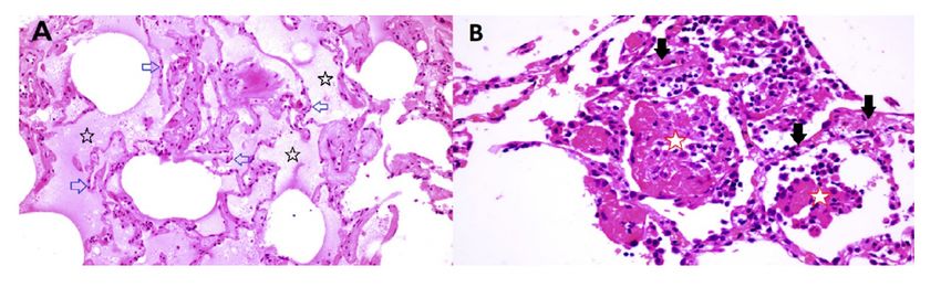
Julie M. Hall
Frank H. Netter MD School of Medicine – Quinnipiac University, Hamden, CT
Gideon Shapiro
Pharmorx LLC, Gainesville, FL
Howard Clark
University College London, London, UK
Anthony Mittermaier
McGill University, Department of Chemistry, Montreal, Quebec, Canada
Andrew C. Kruse
Department of Biological Chemistry and Molecular Pharmacology, Blavatnik Institute, Harvard Medical
School, Boston, MA
Adolfo García-Sastre
Global Health and Emerging Pathogens Institute, Icahn School of Medicine at Mount Sinai, New York,
NY
Bryan L. Roth
Department of Pharmacology, University of North Carolina, Chapel Hill, Chapel Hill, NC
Jill Glasspool-Malone
RW Malone MD LLC, Madison, VA
Victor Francone
Frank H. Netter MD School of Medicine – Quinnipiac University, Hamden, CT
Norbert Hertzog
Frank H. Netter MD School of Medicine – Quinnipiac University, Hamden, CT
Maurice Fremont-Smith
Frank H. Netter MD School of Medicine – Quinnipiac University, Hamden, CT
Darrell O. Ricke
MIT Lincoln Laboratory
Research Article
Keywords: COVID-19, Famotidine, Histamine, Mast Cell, GPCR
DOI: https://doi.org/10.21203/rs.3.rs-30934/v2
License: This work is licensed under a Creative Commons Attribution 4.0 International License.
Read Full License
Page 2/37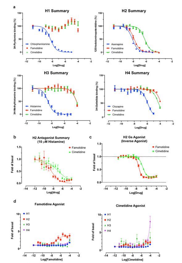
Abstract
SARS-CoV-2 infection is required for COVID-19, but many signs and symptoms of COVID-19 differ from
common acute viral diseases. Currently, there are no pre- or post-exposure prophylactic COVID-19 medical
countermeasures. Clinical data suggest that famotidine may mitigate COVID-19 disease, but both
mechanism of action and rationale for dose selection remain obscure. We explore several plausible
avenues of activity including antiviral and host-mediated actions. We propose that the principal
famotidine mechanism of action for COVID-19 involves on-target histamine receptor H2 activity, and that
development of clinical COVID-19 involves dysfunctional mast cell activation and histamine release.

Introduction
SARS-CoV–2 is a highly infectious and pathogenic betacoronavirus rst detected in human infections
during December 2019 1–3. COVID–19 is a disease spectrum causally associated with infection by SARS-
CoV–2. De nitive COVID–19 diagnosis requires the presence of the virus, which can be isolated, grown,
or otherwise detected as unique SARS-CoV–2 viral nucleic acid sequences. There are SARS-CoV–2 virus
shedding or nucleic acid positive patients that do not manifest clinical COVID–19 4–9. 13–20% of
patients with symptoms develop severe respiratory compromise requiring oxygenation, with radiological
 ndings of ground glass opacities and consolidation 10–12. Between 5 and 46% of SARS-CoV–2 positive
patients are asymptomatic and do not appear to progress to COVID–19 13–16. Therefore, SARS-CoV–2
infection is necessary but not su cient for development of clinical COVID–19 disease.

Patients with COVID–19 disease can present with a range of mild to severe non-speci c clinical signs
and symptoms which develop two to fourteen days after exposure to SARS-CoV–2. These symptoms
include cough or shortness of breath, and at least two of the following; fever, chills, repeated rigor,
myalgia, headache, oropharyngitis, anosmia and ageusia 17,18. More severe symptoms warranting
hospital admission include di culty breathing, a persistent sense of chest pain or pressure, confusion or
di culty to arouse, and central cyanosis. Of hospitalized patients, 20–42% develop ARDS, the most
common cause for admission to the ICU. 39–72% of patients admitted to the ICU will die 19.

Early clinical data from a variety of sources indicate that famotidine treatment may reduce morbidity and
mortality associated with COVID–19. A retrospective cohort study of 1,620 hospitalized COVID–19
patients indicates that 84 propensity score matched patients receiving famotidine during hospitalization
(oral or IV, 20mg or 40mg daily) had a statistically signi cant reduced risk for death or intubation
(adjusted hazard ratio (aHR) 0.42, 95% CI 0.21–0.85) and also a reduced risk for death alone (aHR 0.30,
95% CI 0.11–0.80) 20. In contrast, proton pump inhibitor use was not associated with reduced risk for
these outcomes. A preceding anecdotal report from Wuhan, China is purported to have indicated that
famotidine may be partially protective for COVID–19, but that neither cimetidine nor proton pump
inhibitors were protective 21. Together, these data have been interpreted as indicating that this increased
                                                   Page 3/37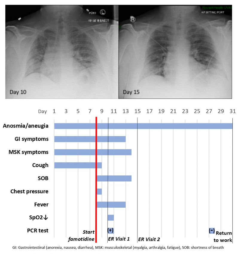
survival pattern is due to an off-target, non-histamine receptor-mediated property of famotidine that is not
shared with cimetidine. Famotidine is currently being tested under an IND waiver for treating COVID–19 in
a double blind randomized clinical trial at high intravenous doses in combination with either
hydroxychloroquine or remdesivir (ClinicalTrials.gov Identi er: NCT04370262).

Herein we aim to investigate how famotidine may act to relieve early phase COVID–19 clinical
symptoms. The most likely mechanisms of actions include: via antiviral activity, via novel human targets,
or via the on-target mechanism described in the current FDA market authorization—famotidine is a
histamine receptor H2 antagonist (and inverse agonist).

Results
Antiviral activity

Famotidine does not bind to SARS-CoV–2 proteases
The idea to test the usefulness of famotidine as a medical countermeasure for COVID–19 emerged from
a computational molecular docking effort aimed at identifying inhibitors of the papain-like protease
(PLpro) of SARS-CoV–2 22,23. In addition to processing the viral polyprotein, the papain-like protease from
coronaviruses (PLpro) is known to remove the cellular substrates ubiquitin and the interferon stimulated
gene 15 (ISG15) from host cell proteins by cleaving the C-terminal end of the consensus sequence LXGG,
a process termed deISGylation 24,25. Here, we used the enzymatic reaction of SARS-CoV–2 PLpro on
ISG15 to assess the potential inhibition of PLpro by famotidine. The cleavage of the 8 C-terminal amino
acids of ISG15 by PLpro is clearly detected by SDS-PAGE (Figure 1, lanes 2 and 3). However, the addition
of 1 to 100 µM famotidine to the reaction does not signi cantly reduce the amount of ISG15 cleaved
during the assay (Figure 1, lanes 4 to 6), thus suggesting that famotidine does not inhibit SARS-CoV–2
PLpro. A previous virtual screening report 26 suggested that famotidine might bind to the 3 chymotrypsin-
like protease (3CLpro), more commonly referred to as the main protease (Mpro), however this mechanism
was recently discounted 27.

Famotidine does not directly inhibit SARS-CoV–2 infection
To assess the possibility that famotidine may inhibit SARS-CoV–2 infection by other routes, a Vero E6
cell-based assay was performed to compare median tissue culture infectious doses (TCID50/mL) of
famotidine, remdesivir, and hydroxychloroquine (Figure 2). While both remdesivir and hydroxychloroquine
demonstrated antiviral activity, no inhibition of SARS-CoV–2 infection was observed with famotidine.

Human receptors

                                                 Page 4/37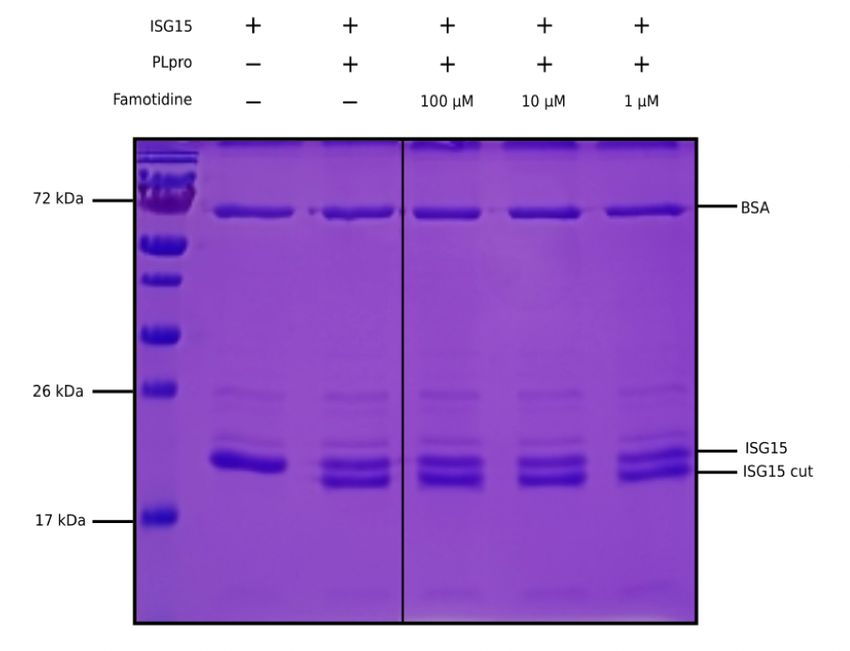
Famotidine does not act via sigma–1 or –2 receptor binding
A wide-ranging study recently presented a map of interactions between viral and host proteins 28. It was
shown that regulation of the sigma–1 and sigma–2 receptors had antiviral effects. Sigma and histamine
receptors share several ligands in common, like the antipsychotic haloperidol, the antihistamines
astemizole and clemastine, the antidepressive clomipramine, and many more. As such, we tested for
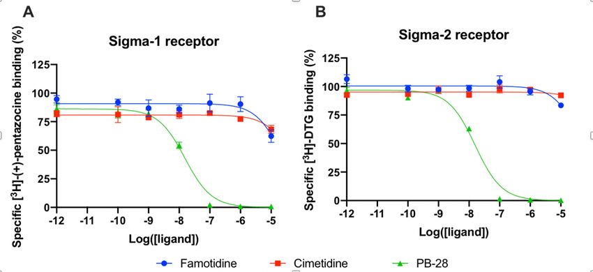
possible interaction between famotidine and sigma–1 or sigma–2 receptors (Figure 3). We performed
radioligand competition binding experiments using cloned sigma receptors, following established
procedures 29 30. In these assays, famotidine showed no detectable displacement of radioligand probes
for either sigma–1 or sigma–2 receptors at famotidine concentrations up to 10 μM. Hence, famotidine’s
binding to sigma–1 and sigma–2 receptors is likely negligible at physiologically relevant concentrations.
Famotidine is selective for receptor H2
As is well-known 31, famotidine is a selective blocker of the histamine H2 receptor with a nity of
approximately 14 nM, substantially more active than the 590 nM cimetidine (Figure 4A). Here we nd it to
have highly e cacious inverse agonist activity (reducing basal activity by 75%) with a potency of 33 nM
(Figure 4C). Intriguingly, and unlike cimetidine, while famotidine acts to block Gs protein signaling it
actually acts as a partial agonist of arrestin recruitment, with an e cacy of about 15% that of histamine,
and an EC50 of 105 nM (Figure 4D), suggesting that the molecule promotes arrestin-scaffolded signaling
—such as through the ERK pathway, 32 and promotes internalization of the receptor and further non-
canonical signaling once internalized 33,34 through an arrestin-biased mechanism. These features
distinguish famotidine certainly from cimetidine, and potentially from other H2 blockers, as such biased
activation of arrrestin recruitment for GPCR antagonists, while not unprecedented, is not common.
Famotidine may activate other GPCRs
Finally, we note that a screen for activation of 318 receptors of the GPCR-ome reveals only seven
receptors with an average fold of basal increase above 3.0, including H2 (Figure 5). In all cases, the
quadruplicate replicates were not in agreement and require follow-up studies. Chief among these are the
CCR2L and CXCR3 chemokine receptors 35–38. Such activity would be intriguing because these receptors
would be expected to activate immune cell mobilization and may plausibly have a role in famotidine’s
bene cial activities, especially at the high systemic concentrations it is expected to reach in the clinical
studies. This would also be consistent with famotidine’s lack of direct anti-viral activity in the Vero cell
direct infectivity assays, where immune cells are not present.
Famotidine reaches functionally relevant systemic concentrations,
whereas cimetidine does not
Page 5/37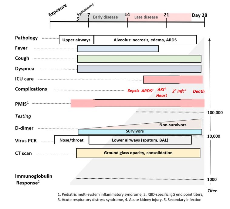
We calculated predicted steady state concentrations of famotidine and cimetidine at different doses
based on published pharmacokinetic and biodistribution data 39–41. This modeling demonstrated that the
different clinical outcomes exhibited by COVID–19 patients taking famotidine vs. cimetidine could be
readily explained by the distinctive pharmacokinetic and pharmacodistribution properties of the two
agents.

Therapeutic e cacy of a pharmacological antagonist requires that it achieves a steady-state
concentration that substantially exceeds the half maximal inhibitory concentration (IC50) for its target.
Thus, in order to evaluate the relative systemic effects of famotidine and cimetidine, the IC50 values of
each agent for the H2 receptor were compared to the steady-state plasma concentrations (Css) predicted
at standard clinical doses. As demonstrated above, famotidine binds to H2 with Ki of 14 nM, whereas
cimetidine binds to H2 with Ki 586 nM. Previous reports suggest functional IC50s are approximately 3x
higher, and these data were used for the current analyses 39,41. In these reports, the IC50 for the H2
receptor were reported as 13 g/L (0.039 M) for famotidine and 400–780 g/L (1.59–3.09 M) for
cimetidine. Css values were calculated using pharmacokinetic data for dosing, clearance, bioavailability,
and volume of distribution as summarized previously 41. Table 1 lists the Css values for both famotidine
and cimetidine.

In primary human neutrophils and eosinophils, H2 activation by histamine inhibits neutrophil effector
functions including O2- release 42,43, platelet-activating-factor induced chemotaxis 44 and leukotriene
biosynthesis 45. Eosinophil functions are also inhibited by H2 activation; histamine binding diminishes
eosinophil peroxidase release 46 and, at high concentrations, inhibits eosinophil chemotaxis 47,48.
Famotidine is one of the most effective antagonists of these H2-mediated histamine effects on
neutrophils and eosinophils49. IC50 for two measures that relate to these phenotypes are also listed in
Table 1. Mast cells express histamine H2 and H4 receptors, and histamine-induced increase of cAMP in
mast cells is inhibited by famotidine 50. 10 M famotidine pre-incubation blocks histamine-induced cAMP
increase in human skin mast cells, however, the IC50 for this effect has not been determined 50.

At all dosing regimens, the Css for famotidine exceeds the general IC50 value for the H2 receptor, and at
the twice daily (b.i.d.) and thrice daily (t.i.d.) dosing of 20 mg and 40 mg, the Css for unbound famotidine
is 2–5 fold greater than the H2 IC50. Also calculated and summarized is the Css for the intravenous
dosage currently being administered in clinical trial NCT04370262 and that dose exceeds famotidine IC50
by greater than 20-fold. In contrast, unbound cimetidine levels at standard doses of 200 or 300 mg daily
(q.d.), achieve a Css that is a fraction of the reported IC50 range of 400–780 µg/L.

Table 1: Steady-state concentrations (Css) of Famotidine and Cimetidine at standard doses compared to
the half maximal inhibitory concentration (IC50) value of Famotidine or Cimetidine for histamine H2
receptor antagonism

                                                   Page 6/37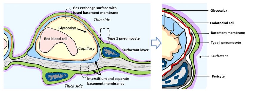
Page 7/37
IC50 or Css              Concentration        Concentration
                        (mass/volume)          (molarity)
                                              Famotidine
IC50 Histamine            13 mg/L             0.039 mM
H2
IC50                      67 mg/L              0.201 mM
Neutrophil H2 O
    -
  assay
2
IC50                       8 mg/L              0.024 mM
Neutrophil H2 c
AMP assay
IC50 Eosinophil          53.6 mg/L             0.158 mM
H2 cAMP assay
IC50       Mast                                        Not determined
Cell H2 cAMP
increase
                      Total Concentration   Total Concentration     Free drug        Free drug
                        (mass/volume)         (mass/volume)       Concentration3   Concentration3
                                                                  (mass/volume)      (molarity)
Css      (20    mg       17.7 mg/L             0.053 mM            14.2 mg/L        0.042 mM
tablet p.o. q.d.)
Css      (20    mg       18.4 mg/L             0.055 mM            14.7mg/L         0.044 mM
capsule        p.o.
q.d.)
Css      (20    mg       35.4 mg/L             0.105 mM            28.3 mg/L        0.084 mM
tablet         p.o.
b.i.d.)
Css      (20    mg       36.8 mg/L             0.109 mM            29.4 mg/L        0.087 mM
capsule        p.o.
b.i.d.)
Css      (20    mg       53.1 mg/L             0.157 mM            42.5 mg/L        0.126 mM
tablet p.o. t.i.d.)
Css      (20    mg       55.3 mg/L             0.164 mM            44.2 mg/L        0.131 mM
capsule        p.o.
t.i.d.)
Css      (40    mg       55.4 mg/L             0.164 mM            44.3 mg/L        0.131 mM
tablet         p.o.
t.i.d.)1
Css      (40    mg       80.8 mg/L             0.239 mM            64.6 mg/L        0.192 mM
tablet         p.o.
t.i.d.)2
Css      (60    mg      144.3 mg/L             0.425 mM           115.4 mg/L        0.340mM
tablet         p.o.
t.i.d.)
Css (120mg IV           1,290 mg/L             1.092 mM           1,032 mg/L        0.874 mM
every 8 hours)
                                               Cimetidine
IC50 Histamine        400-780 mg /L          1.59-3.09 mM
H2
Css (200 mg              175 mg /L              0.69 mM            140 mg/L         0.055 mM
tablet p.o. q.d.)
Css (300 mg              226 mg /L              0.90 mM           180.1 mg/L        0.720 mM
tablet p.o. q.d.)

                                                 Page 8/37
1
calculated using pK data reported by Lin et al 1987 39
2
calculated using pK data reported by Yeh et al 1987 40
3
Both famotidine and cimetidine are approximately 20% protein bound in systemic circulation 51,52
Case history, Severe COVID–19 Outpatient Treatment with
Famotidine
Patient JM is a 47 year old male who received PCR diagnosis of COVID–19 after 8 days of complaints of
diarrhea, abdominal cramping, eructation, low energy, dry cough, arthralgia, myalgia, anosmia and
ageusia. The patient has a history of hypertension (10y), Type II diabetes (4y), hypercholesterolemia (3y)
and gout (10y). Current medications included Metformin, Allopurinol, Lisinopril, and Atorvastatin. He is
employed as a hospital maintenance worker in the hospital to which he presented.
Contact tracing revealed that his son (same household) had developed COVID–19 symptoms 12 days
prior. Receipt on day 8 of positive PCR diagnosis (from a prior outpatient intranasal swab sample)
coincided with onset of fever (102oF), night sweats, shortness of breath and a feeling of chest pressure.
Famotidine (“PEPCID AC ®" 60mg p.o. t.i.d. = 2.24mg/ft2 t.i.d) was initiated upon receiving the PCR
diagnosis due to symptoms meeting FDA criteria for severe COVID–19, combined with high risk pre-
existing conditions. The famotidine drug regime was continued for 30 days. After initiating famotidine in
the evening, the patient was able to sleep through the night and reported complete relief from the chest
pressure sensation, reduction in cough, but continued to be febrile (101.6 oF).
On day 10, he presented to the emergency room (ER) with continuing complaints of diarrhea, abdominal
cramping, eructation, low energy, dry cough, arthralgia, myalgia, anosmia and ageusia and shortness of
breath on exertion. Day 10 ER physical examination, including the chest, was unremarkable and vital
signs were normal. The patient BMI was 36 (Du Bois BSA 26.78 ft2). SpO2 was 93%, rising to 97% and
99% on 3 L/min by nasal cannula over the next 30 minutes. An intranasal sample was obtained for
SARS-CoV–2 rtPCR diagnostic analysis. Comprehensive metabolic panel showed a mild decrease in
serum sodium and chloride with hyperglycemia (260 mg/dL). Complete blood count (CBC) was normal,
speci cally including the lymphocyte count. Urinalysis showed a speci c gravity of 1.025 but was
otherwise normal. A portable chest X-ray had poor inspiration but was interpreted as showing “bibasilar
areas of airspace disease” consistent with COVID–19 (Figure 6, CXR day 10). The patient was diagnosed
as dehydrated, given ondansetron IV, 1 L IV of normal saline and discharged home with a hospital pulse
oximeter. At the time of departure, he had an SpO2 of 94% on room air that did not drop with ambulation.
The patient again presented to the emergency room on day 15 after experiencing near-syncope during
showering. Physical examination was unremarkable. Vital signs were normal. SpO2 showed values of
98%, 93% and 97% on room air over the 2 hour period. Basic metabolic panel showed only hyperglycemia
(266 mg/dL). CBC was normal except for a mild lymphopenia (0.96; reference range 1.00–3.00 X10³/μL)
Page 9/37and mild monocytosis (0.87; 0.20–0.80 X10³/μL). Chest X-ray was interpreted as showing “Faint patchy
consolidation of lung bases bilaterally, similar to perhaps minimally improved at the lower left lung base
compared to prior” (Figure 6 CXR day 15). The patient was placed on azithromycin and discharged to
home.
On days 27 and 28 after initial symptoms, he tested negative (2x, successive days) for SARS-CoV–2
nucleic acid by PCR (intranasal swab) and returned to his work at the local hospital 31 days after initial
symptoms. 47 days after rst developing COVID–19 symptoms he continues to note a lack of ability to
taste or smell, but otherwise considers himself largely recovered from COVID–19 (Figure 6 timeline).
Use of famotidine in this patient was recommended due to meeting FDA criteria for severe COVID–19 and
his COVID–19 risk factors: male, 47yo, hypertension, obesity and diabetes mellitus Type 2. Although this
is an anecdotal example, the patient experienced relief of symptoms overnight upon initiating use of
famotidine. While not su cient to demonstrate proof of cause and effect, this case does provide context
for typical COVID–19 presentation and symptoms, as well as support for additional well-controlled
famotidine therapeutic clinical trials in an outpatient setting.
Discussion
Famotidine is an off-patent drug available as either branded (“PEPCID ®") or generic medicines in tablet,
capsule or intravenous forms. The general pharmacology of famotidine is well-characterized, with an
excellent absorption, distribution, metabolism, excretion and toxicology pro le 53. Famotidine is unique
among the drugs currently being tested for treatment of COVID–19, in that it is an H2 receptor antagonist
(and inverse agonist). Famotidine is currently being tested for treating COVID–19 in a double blind
randomized clinical trial at high intravenous doses in combination with either hydroxychloroquine or
remdesivir (ClinicalTrials.gov Identi er: NCT04370262). A recent retrospective cohort study of 1,620
hospitalized COVID–19 patients indicates that 84 propensity score matched patients receiving
famotidine during hospitalization (oral or IV, 20mg or 40mg daily) had a statistically signi cant reduced
risk for death or intubation (adjusted hazard ratio (aHR) 0.42, 95% CI 0.21–0.85) and also a reduced risk
for death alone (aHR 0.30, 95% CI 0.11–0.80) 20. In contrast, proton pump inhibitor use was not
associated with reduced risk for these outcomes. Anecdotal reports and undisclosed data indicating that
famotidine provided protection from COVID–19 mortality while neither cimetidine nor proton pump
inhibitors were similarly protective lead to an initial inference that the bene cial effects of famotidine
were not related to the known on-target activity of the drug 21. Studies detailed in this report and others,
however, indicate that famotidine does not act by directly inhibiting either of the principal SARS-CoV–2
proteases (PLpro or Mpro) 27. Vero E6-based cell assays also indicate that famotidine has no direct
antiviral activity in this cell line, although antiviral activity in cells that express H2 has not been tested.
Additional hypotheses that famotidine may act via binding either the sigma–1 or –2 receptors have not
been supported by the studies summarized herein.
Page 10/37The most straightforward explanation of the apparent famotidine activity as a COVID–19 therapy is that
the drug acts via its antagonism or inverse-agonism of histamine signaling and via its arrestin biased
activation—all a result of its binding to histamine receptor H2. If true, then it is reasonable to infer that a
SARS-CoV–2 infection that results in COVID–19 is at least partially mediated by pathologic histamine
release. The anecdotal lack of protection provided by oral administration of the H2 antagonist cimetidine
can be accounted for by insu cient systemic drug levels after oral administration and does not
contradict potential bene t provided by famotidine H2 binding. Intravenous cimetidine at su cient doses
may achieve levels high enough for clinical bene t and would further support this hypothesis. Failure to
achieve clinical COVID–19 responses with cimetidine may indicate that inverse agonism or other GPCR-
mediated effects of famotidine may play an important role in the (preliminary) observed clinical bene ts.
Analysis of famotidine activity in histamine receptor competition assays indicate that, over the range of
clinical steady state famotidine drug levels being tested, famotidine is speci c for H2. Therefore, off-
target antagonism of histamine H1 receptor, H3 receptor, or H4 receptor is unlikely to contribute to
famotidine-mediated effects.
Steady state famotidine concentrations su cient to elicit H2 antagonism (and inverse agonism) are
readily achieved using inexpensive oral tablets and safe dosage levels. The famotidine dosage employed
in the only retrospective hospital study currently available examining famotidine effects on COVID–19
outcomes appears to have employed dosages (20mg to 40mg daily) which are unlikely to fully inhibit
histamine-mediated effects at the H2 receptor 20. In contrast, study NCT04370262 administers
intravascular famotidine doses that are more than 20-fold greater than the IC50 for antagonism of H2.
The data presented herein provides a rationale for famotidine dose selection to maintain a steady state
concentration at a reasonable multiple of the IC50 for systemic antagonism of H2 and indicate that oral
tablet dosages of between 40mg every eight hours to 60mg every eight hours should be su cient to
insure maximal H2 target effects. As famotidine is primarily cleared by the kidney, adequate renal
function is required for higher dosages 53.
In addition to H2 antagonism, famotidine may also act as an inverse agonist thereby lowering the
concentration of cyclic-Adenosine Monophosphate (c-AMP) 32. Endothelial cell permeability has been
attributed to histamine H2 activation and is blunted by famotidine pretreatment 54. Histamine, bradykinin
and des-arg-bradykinin receptor engagements can lead to increased endothelial permeability through a
common pathway that results in AKT–1 activation 55. The H2 receptor also signals through Gq/11
proteins, resulting in inositol phosphate formation and increases in cytosolic Ca2+ concentrations which
may account for the increased endothelial cell uid permeability 56.
One alternative hypothesis is that famotidine may not only inhibit signaling through the H2 receptor but
may also engage in cross talk with the kinin B1 receptor, which moderates the response of endothelial
cells to DABK and DAKD ligands. Data provided here in are not consistent with this hypothesis; no
activation of bradykinin receptor B1 or B2 were observed in quadruplicate replicate TANGO assay.
Page 11/37While COVID–19 symptoms affect multiple organ systems, respiratory failure due to acute respiratory
distress syndrome (ARDS) is the most common cause of death. Examination of RNA expression pro les
of the cells which contribute to lung anatomy and function demonstrate the presence of multiple
ACE2/TMPRSS2 positive cell types susceptible to SARS-CoV–2 infection in the lung. In addition, these
and other associated lung cells that are positive for histamine receptors H1 and H2 could respond to local
histamine release following mast cell degranulation 57, and therefore those cells positive for H2 may be
responsive to famotidine effects.
To understand how famotidine may act to reduce pulmonary COVID–19 symptoms requires an
understanding of COVID–19 lung pathophysiology, which appears to have two principal disease phases.
In turn, this requires an appreciation of pulmonary tissue and cell types. Pulmonary edema results from
loss of a regulation of uid transfer that occurs at several levels in the alveolus, as diagrammed in Figure
7. In the capillary wall, there are the glycocalyx, the endothelial cell with associated tight junctions, and
the basement membrane. In the epithelium there is a surfactant layer on the alveolar lining uid,
manufactured and secreted by the Type II pneumocyte, and the Type I pneumocyte itself with its tight
junctions and negatively charged basement membrane which restricts albumin. The pulmonary pericytes
located in the terminal conducting airway region play a critical role in synthesizing the endothelial
basement membrane and regulating blood ow in the precapillary arteriole, the capillary and the
postcapillary venule. Disruption of any of these cells or layers can lead to edema. This edema uid may
be a transudate in milder dysfunctions or an exudate when in ammation or necrosis develop. Two
possible pathologies that could result in edema of the alveolar wall and space include infection of cells
by SARS-CoV–2 and mast cell degranulation with release of hundreds of compounds that can impact on
cellular and basement membrane functions, glycocalyx and tight junction integrity. These compounds
include histamine, bradykinin, heparin, tryptase and cytokines.
Gene expression patterns of these pulmonary cells provide insight into which cells are likely to be
infected, and which express the H2 receptor that could be directly impacted by famotidine treatment and
resulting H2 antagonism or inverse agonism (Figure 8). These patterns suggest that epithelial cells and
endothelial cells are more likely to be infected based on ACE2 and TMPRSS2 expression patterns in those
cell types. The cells most likely to show a famotidine effect include Type 2 pneumocytes, smooth muscle
cells, pericytes, and myeloid granulocytes (which includes mast cells, neutrophils and eosinophils).
The limited tissue pathology available from early COVID–19 cases seems to support both viral infection
as well as histamine effects in the lung. In a singular study of early COVID–19, Sufang Tian et al 59
describe the viral lung pathology of early COVID–19 in tissue resected for cancer. Their
photomicrographs show two different patterns of disease. As shown in Figure 9 panel B, some samples
of this lung tissue demonstrate the usual mononuclear in ammatory pattern of interstitial pneumonitis
and brinous exudate that one would associate with a viral infection. It is striking that no neutrophils or
eosinophils are observed in the in ammatory in ltrate. One explanation is that H2 activation of
neutrophils inhibits neutrophil effector functions including O2- release 42,43, platelet-activating-factor
Page 12/37induced chemotaxis 44 and leukotriene biosynthesis 45. Eosinophil functions are also inhibited by H2
activation; histamine binding diminishes eosinophil peroxidase release 46 and, at high concentrations,
inhibits eosinophil chemotaxis 47,48.
The reports of Tian et al 59 and Zeng et al 60 also include images in which there is interstitial and alveolar
edema while the alveolar septae retain normal architecture (Figure 9 panel A). This is not a pattern
typically observed in viral infection, as there is no in ammation, and the uid appears to be a transudate.
It is consistent with dysregulation of the uid barrier due to the effect of histamine or other mast cell
products on endothelial cells, pericytes or Type II pneumocytes. Increased endothelial permeability due to
histamine is driven by H1 receptor activation, and so if any potential famotidine treatment effect on these
cells occurs it would most likely be indirect by inhibition of mast cell degranulation. Forskolin activates
the enzyme adenylyl cyclase and increases intracellular levels of cAMP, and can be used to inhibit the
release of histamine from human basophils and mast cells 61. Histamine may act as an autocrine
regulator of mast cell cytokine and TNF-a release in a PGE2-dependent fashion. Based on in vitro studies,
this autocrine feedback appears to be mediated by H2 and H3 (88). Endothelial cells are also susceptible
to infection by SARS-CoV–2. Mast cell degranulation-related pulmonary edema could correlate with the
early phase silent hypoxia and the high compliance non-ARDS ventilation pattern associated with
shortness of breath 62. The image in Figure 9 panel B does not permit evaluation for microvascular
thrombi.
These ndings are supported in a separate autopsy case report of a patient dying 5 days after onset of
COVID–19 symptoms. In this case, photomicrographs also show a non-in ammatory transudative-type
edema 63. In both of these studies, the observed non-in ammatory edema in early-stage COVID–19
pulmonary disease is consistent with histamine release by mast cells.
Mast cell degranulation correlates with the COVID–19 natural history that progresses through
functionally and clinically different early and later phases. Most SARS-CoV–2 infections follow the
typical early phase pattern of any lower respiratory virus, in which a majority of patients have
asymptomatic or minimal disease, while a minority go on to later phase acute respiratory distress
syndrome (ARDS). Within this spectrum typical of any severe viral disease, COVID–19 has a number of
distinctive features. In the out-patient setting, early COVID–19 is usually indistinguishable from other
“in uenza-like illnesses”, presenting with various non-speci c symptoms ranging from sore throat,
headache and diarrhea to fever, cough, and myalgias. In these rst few days however, COVID–2 may also
be associated with anosmia, a unique feature 64. It is towards the end of the rst week of symptoms that
COVID–19 patients develop shortness of breath (SOB). This follows cough and fever by several days, a
feature not typical of other viruses 65. On physical examination of COVID–19 patients with SOB, the
oxygen saturation drops dramatically on exertion. CT scan will usually show bilateral bibasilar ground
glass opaci cations consistent with pulmonary edema. Nasopharyngeal swabs test positive for SARS-
CoV–19. This SOB correlates with a distinctive clinical phenotype of hypoxia with near normal
compliance (i.e. >50 mLcmH2O). Some authors attribute this to a loss of pulmonary vasoconstriction,
Page 13/37one cause of which could be histamine effect on the H2 receptors of pericytes and/or vascular smooth
muscle. H1-related edema and microthrombosis of lung vessels could also be causes. These are the
patients that PEEP ventilation will not help, as there are no recruitable alveoli. These patients are helped
by lying prone 66. It is at this stage that the patient is at greatest risk to progress onto the serious
complications of later disease, especially ARDS with its 60–80% mortality if ventilation is required.
Patients may also present with additional neurological symptoms and complications including ischemic
stroke 67–69. Cardiac complications of later COVID–19 include myocarditis, acute myocardial infarction,
heart failure, dysrhythmias, and venous thromboembolic events 70,71.
Multiple studies have demonstrated a hypercoagulable state in COVID–19 patients requiring
hospitalization. Results from a recent large autopsy study suggests that there is also a novel lung-centric
coagulopathy that manifests as a small vessel microthrombosis. Based on this study, there are
indications that over 50% of patients dying of COVID–19 have pulmonary microthrombosis 72. This
thrombosis is not only in arterial vessels, but also can be found in alveolar capillaries in the absence of
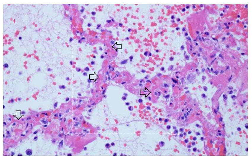
in ammation and ARDS, as seen in Figure 10 73.
There is widening of the alveolar septae by extensive brinous occlusion of capillaries (open black
arrows). There is alveolar space edema with red blood cell extravasation. Septae show a mild
mononuclear in ltrate. Alveolar edema shows neutrophils in proportion to the blood.
Capillary wall disruption accompanied by brin deposition and red cell extravasation, with neutrophils in
the septa and within the alveolar spaces. (Hematoxylin and eosin, 1000x). For further discussion of
microvascular coagulation associated with COVID–19, see 73.
Because small microthrombi are di cult to identify on CT scan even with iodinated contrast 74, pre-
mortem diagnosis is di cult. Laboratory coagulation tests have typically shown normal or mildly
prolonged Prothrombin time (PT) and activated partial thromboplastin time (aPTT), normal to increased
or slightly decreased platelet counts, elevated brinogen levels and very elevated D-dimers 75. Although
referred to by some authors as a DIC-like state, this pulmonary microthrombosis does not appear as a
typical coagulation factor consumptive bleeding condition typical of overt DIC, but instead more closely
resembles hypercoagulable thrombosis. This coagulopathy appears to be a core pathophysiology of
COVID–19 as rising D-dimer levels, correlate with a poor prognosis, as do rising levels of IL–6 and CRP.
IL–6 levels have been correlated to brinogen levels in one study, possibly through the acute phase
reactant response 76. The pathogenesis of microthrombosis of the lung in COVID–19 is not known. There
are multiple working hypotheses concerning this nding currently being assessed 77. Damage to the
vascular endothelial glycocalyx can be caused by TNF‐α, ischemia and bacterial lipopolysaccharide. As
well, activated mast cells release cytokines, proteases, histamine, and heparinase, which degrade the
glycocalyx 78 and may thereby contribute to microthrombosis. Disruption of the glycocalyx exposes
endothelial cell adhesion molecules, triggering further in ammation, rolling and adhesion of white blood
cells and platelets 79. Glycocalyx components measured in serum positively correlate with increased
Page 14/37mortality in septic patients 80. Other causes of hypercoagulability include direct damage to ACE2 positive
endothelial cells by viral invasion or secondary damage from the in ammatory response to the infection.
Mast cells release heparin which activates the contact system, producing plasmin and bradykinin.
Plasmin activation could account for the singular rise in D-dimer levels. Activation of platelets also seems
likely as part of the thrombo-in ammatory response but their precise role in thrombus formation remains
to be elucidated 81. A more complete understanding awaits further study.
In addition to the usual features of a viral infection, early COVID–19 often presents with anosmia,
ageusia, skin rashes including pruritis and urticaria, neuropsychiatric symptoms (including altered dream
states), and silent hypoxia. These symptoms are all consistent with histamine signaling. Anosmia,
ageusia, and other symptoms relating to cachexia are often reported in both COVID–19 and mast cell
degranulation syndrome, and the potential role of histamine signaling in driving the pathophysiology of
cachexia has been reviewed 82,83. As summarized in Figure 11, the distinctive later ndings of abnormal
coagulation, involvement of other organ systems and ARDS occur in the second week after the
appearance of symptoms. This is coincidental with a rising immunoglobulin response to SARS-CoV–2
antigens. For a subset of patients, disease progress may suddenly worsen at days 7–10, and this
correlates with the onset of SARS-CoV–2 spike protein neutralizing antibody titers 84. In this study, it was
shown that IgG starts to rise within 4 days post-symptoms, inconsistent with a rst antigenic exposure 84.
Rapid onset of speci c neutralizing antibody responses beginning less than seven days after exposure to
SARS-CoV–2 implies a recall rather than primary B cell response, and therefore the response is being
driven by a pre-existing memory cell population. These memory cells may have been educated by prior
exposure to another coronavirus (e.g. circulating alphanumeric coronaviruses), raising concerns that this
second phase of COVID–19 disease progression could share an immunologic basis with Dengue
hemorrhagic fever 85. Antibodies produced from this early rapid humoral response may drive further mast
cell degranulation. During this phase rising D-dimer levels correlate with poor prognosis, as do measured
levels of CRP and IL–6.
Current reviews seek to explain COVID–19 clinical and pathologic ndings based on standard models of
antiviral innate and adaptive immune responses which do not consider the potential role of mast cell
activation and degranulation. Reviews emphasize the in ammatory cell response cascade associated
with monocytes, macrophages 86, and adaptive T and B cell helper and effector responses 87. These
types of immune responses are also invoked to explain the novel microvascular pulmonary intravascular
coagulopathy associated with COVID–19 88.
We propose an alternative paradigm; SARS-CoV–2 infection-induced mast cell activation could account
for some of the core pathologic cascade and much of the unusual symptomatology associated with
COVID–19 89. Many of the unique clinical symptoms observed during the early phase of COVID–19 are
consistent with known effects of histamine release. Histamine may act as an autocrine regulator of mast
cell cytokine and TNF-a release in a PGE2-dependent fashion and based on in vitro studies the autocrine
feedback appears to be mediated by H2 and H3 90. This model is consistent with the histopathologic
Page 15/37ndings seen at surgery, autopsies, and is supported by clinical pharmacologic ndings suggesting
potential bene ts of histamine H2 receptor blockade using famotidine. This model is also supported by
the signi cant overlap in the clinical signs and symptoms of the initial phase of COVID–19 disease and
those of mast cell activation syndrome (MCAS) 91–94 as well similarities to Dengue hemorrhagic fever
and shock syndrome (including T cell depletion) during the later phase of COVID–19 85,95,96. The cardiac
events, stroke, and related outcomes associated with COVID–19 also appear consistent with the Kounis
syndrome 97–99.
If COVID–19 is partially driven by dysfunctional mast cell degranulation, then a variety of medical
interventions employing marketed drugs useful for treating mast cell-related disorders may help to reduce
death and disease associated with SARS-CoV–2 infection. Examples include drugs with mast cell
stabilizing activity, other histamine antagonists (for example H1 and H4 types), leukotriene antagonists
and leukotriene receptor antagonists 100, anti-in ammatory agents such as those developed for
in ammatory bowel diseases, and mast cell activation inhibitors 101. If such repurposed drugs are used in
combination with pharmaceuticals that directly inhibit SARS-CoV–2 infection or replication, it may be
possible to rapidly develop potent, safe and effective outpatient treatments for preventing or treating
COVID–19 until such time as a safe and effective SARS-CoV–2 vaccine becomes available.
Declarations
Acknowledgments
The authors acknowledge the Department of Defense (DoD), Defense Threat Reduction Agency (DTRA),
and the Joint Science and Technology O ce (JSTO) of the Chemical and Biological Defense Program
(CBDP) for funding under the Discovery of Medical countermeasures Against Novel Entities (DOMANE)
initiative. This work has also bene tted from advice, guidance, information and comments provided by
Drs. Revell Phillips, Howard Haimes, David Hone, and Roland Seifert. We appreciate the valuable input
provided by Dr. Frank Weichold of the O ce of Regulatory Science and Innovation (ORSI), O ce of the
Chief Scientist (OCS), O ce of the Commissioner (OC), FDA/HHS, and Dr. Lawrence Callahanof the O ce
of Health Informatics, O ce of the Chief Scientist (OCS), O ce of the Commissioner (OC), FDA/HHS. We
also thank Dr. Anton Yuryev from Elsevier for his assistance in literature mining and reconstruction of the
histamine signaling model. D. R. gratefully acknowledges the support of the MIT SuperCloud team.
DISTRIBUTION STATEMENT A. Approved for public release. Distribution is unlimited.
This material is based upon work supported under Air Force Contract No. FA8702–15-D–0001. Any
opinions, ndings, conclusions or recommendations expressed in this material are those of the author(s)
and do not necessarily re ect the views of the U.S. Air Force. Funding was also provided by grants from
the Defense Advanced Research Projects Agency HR0011–19–2–0020 (to A. G-S.); by CRIP (Center for
Research for In uenza Pathogenesis), a NIAID supported Center of Excellence for In uenza Research and
Page 16/37Surveillance (CEIRS, contract # HHSN272201400008C) and by supplements to NIAID grant U19AI135972
and DoD grant W81XWH–20–1–0270 to A. G.-S.
References
1.Zhu, N., et al. A Novel Coronavirus from Patients with Pneumonia in China, 2019. N Engl J Med 382,
727–733 (2020).
2.Wu, Z. & McGoogan, J. M. Characteristics of and Important Lessons From the Coronavirus Disease
2019 (COVID–19) Outbreak in China: Summary of a Report of 72314 Cases From the Chinese Center for
Disease Control and Prevention. JAMA (2020).
3.Wu, D., Wu, T., Liu, Q. & Yang, Z. The SARS-CoV–2 outbreak: What we know. Int J Infect Dis 94, 44–48
(2020).
4.Zou, L., et al. SARS-CoV–2 Viral Load in Upper Respiratory Specimens of Infected Patients. N Engl J
Med 382, 1177–1179 (2020).
5.Danis, K., et al. Cluster of coronavirus disease 2019 (Covid–19) in the French Alps, 2020. Clin Infect Dis
(2020).
6.Lai, C. C., et al. Asymptomatic carrier state, acute respiratory disease, and pneumonia due to severe
acute respiratory syndrome coronavirus 2 (SARS-CoV–2): Facts and myths. J Microbiol Immunol Infect
(2020).
7.Pan, X., et al. Asymptomatic cases in a family cluster with SARS-CoV–2 infection. Lancet Infect Dis 20,
410–411 (2020).
8.Furukawa, N. W., Brooks, J. T. & Sobel, J. Evidence Supporting Transmission of Severe Acute
Respiratory Syndrome Coronavirus 2 While Presymptomatic or Asymptomatic. Emerg Infect Dis 26(2020).
9.Ki, M. & Task Force for -nCo, V. Epidemiologic characteristics of early cases with 2019 novel coronavirus
(2019-nCoV) disease in Korea. Epidemiol Health 42, e2020007 (2020).
10.Huang, C., et al. Clinical features of patients infected with 2019 novel coronavirus in Wuhan, China.
Lancet 395, 497–506 (2020).
11.Wang, D., et al. Clinical Characteristics of 138 Hospitalized Patients With 2019 Novel Coronavirus-
Infected Pneumonia in Wuhan, China. JAMA (2020).
12.Shi, H., et al. Radiological ndings from 81 patients with COVID–19 pneumonia in Wuhan, China: a
descriptive study. Lancet Infect Dis 20, 425–434 (2020).
Page 17/3713.He, G., et al. The clinical feature of silent infections of novel coronavirus infection (COVID–19) in
Wenzhou. J Med Virol (2020).
14.Tian, S., et al. Characteristics of COVID–19 infection in Beijing. J Infect 80, 401–406 (2020).
15.Hu, Z., et al. Clinical characteristics of 24 asymptomatic infections with COVID–19 screened among
close contacts in Nanjing, China. Sci China Life Sci 63, 706–711 (2020).
16.Mizumoto, K., Kagaya, K., Zarebski, A. & Chowell, G. Estimating the asymptomatic proportion of
coronavirus disease 2019 (COVID–19) cases on board the Diamond Princess cruise ship, Yokohama,
Japan, 2020. Euro Surveill 25(2020).
17.Giacomelli, A., et al. Self-reported olfactory and taste disorders in SARS-CoV–2 patients: a cross-
sectional study. Clin Infect Dis (2020).
18.Lechien, J. R., et al. Olfactory and gustatory dysfunctions as a clinical presentation of mild-to-
moderate forms of the coronavirus disease (COVID–19): a multicenter European study. Eur Arch
Otorhinolaryngol (2020).
19.CDC. Interim Clinical Guidance for Management of Patients with Con rmed Coronavirus Disease
(COVID–19). Vol. 2020 (2020).
20.Freedberg, D. E., et al. Famotidine Use is Associated with Improved Clinical Outcomes in Hospitalized
COVID–19 Patients: A Retrospective Cohort Study. medRxiv, 2020.2005.2001.20086694 (2020).
21.Borrell, B. New York clinical trial quietly tests heartburn remedy against coronavirus. Vol. 2020
(Science Magazine, 2020).
22.Daczkowski, C. M., et al. Structural Insights into the Interaction of Coronavirus Papain-Like Proteases
and Interferon-Stimulated Gene Product 15 from Different Species. J Mol Biol 429, 1661–1683 (2017).
23.Baez-Santos, Y. M., St John, S. E. & Mesecar, A.D. The SARS-coronavirus papain-like protease:
structure, function and inhibition by designed antiviral compounds. Antiviral Res 115, 21–38 (2015).
24.Mielech, A.M., Chen, Y., Mesecar, A.D. & Baker, S. C. Nidovirus papain-like proteases: Multifunctional
enzymes with protease, deubiquitinating and deISGylating activities. Virus Research 194, 184–190
(2014).
25.Han, Y. S., et al. Papain-like protease 2 (PLP2) from severe acute respiratory syndrome coronavirus
(SARS-CoV): expression, puri cation, characterization, and inhibition. Biochemistry 44, 10349–10359
(2005).
26.Wu, C., et al. Analysis of therapeutic targets for SARS-CoV–2 and discovery of potential drugs by
computational methods. Acta Pharm Sin B (2020).
Page 18/3727.Anson, B. J., et al. Broad-spectrum inhibition of coronavirus main and papain-like proteases by HCV
drugs,. Research Square (2020).
28.Gordon, D. E., et al. A SARS-CoV–2 protein interaction map reveals targets for drug repurposing. Nature
(2020).
29.Schmidt, H. R., et al. Crystal structure of the human σ1 receptor. Nature 532, 527–530 (2016).
30.Alon, A., et al. Identi cation of the gene that codes for the sigma2 receptor. Proc Natl Acad Sci U S A
114, 7160–7165 (2017).
31.Bertaccini, G., Coruzzi, G., Poli, E. & Adami, M. Pharmacology of the novel H2 antagonist famotidine: in
vitro studies. Agents Actions 19, 180–187 (1986).
32.Alonso, N., et al. Physiological implications of biased signaling at histamine H2 receptors. Front
Pharmacol 6, 45 (2015).
33.Irannejad, R. & von Zastrow, M. GPCR signaling along the endocytic pathway. Curr Opin Cell Biol 27,
109–116 (2014).
34.Jean-Charles, P. Y., Kaur, S. & Shenoy, S. K. G Protein-Coupled Receptor Signaling Through beta-
Arrestin-Dependent Mechanisms. J Cardiovasc Pharmacol 70, 142–158 (2017).
35.Miao, M., De Clercq, E. & Li, G. Clinical signi cance of chemokine receptor antagonists. Expert Opin
Drug Metab Toxicol 16, 11–30 (2020).
36.Fantuzzi, L., Tagliamonte, M., Gauzzi, M. C. & Lopalco, L. Dual CCR5/CCR2 targeting: opportunities for
the cure of complex disorders. Cell Mol Life Sci 76, 4869–4886 (2019).
37.Zhou, Y. Q., et al. The Role of CXCR3 in Neurological Diseases. Curr Neuropharmacol 17, 142–150
(2019).
38.Nasi, A. & Chiodi, F. Mechanisms regulating expansion of CD8+ T cells during HIV–1 infection. J Intern
Med 283, 257–267 (2018).
39.Lin, J. H., et al. Effects of antacids and food on absorption of famotidine. Br J Clin Pharmacol 24,
551–553 (1987).
40.Yeh, K. C., et al. Single-dose pharmacokinetics and bioavailability of famotidine in man. Results of
multicenter collaborative studies. Biopharm Drug Dispos 8, 549–560 (1987).
41.Lin, J. H. Pharmacokinetic and pharmacodynamic properties of histamine H2-receptor antagonists.
Relationship between intrinsic potency and effective plasma concentrations. Clin Pharmacokinet 20,
218–236 (1991).
Page 19/3742.Burde, R., Seifert, R., Buschauer, A. & Schultz, G. Histamine inhibits activation of human neutrophils
and HL–60 leukemic cells via H2-receptors. Naunyn Schmiedebergs Arch Pharmacol 340, 671–678
(1989).
43.Gespach, C. & Abita, J. P. Human polymorphonuclear neutrophils. Pharmacological characterization of
histamine receptors mediating the elevation of cyclic AMP. Mol Pharmacol 21, 78–85 (1982).
44.Rabier, M., et al. Inhibition by histamine of platelet-activating-factor-induced neutrophil chemotaxis in
bronchial asthma. Int Arch Allergy Appl Immunol 89, 314–317 (1989).
45.Flamand, N., Plante, H., Picard, S., Laviolette, M. & Borgeat, P. Histamine-induced inhibition of
leukotriene biosynthesis in human neutrophils: involvement of the H2 receptor and cAMP. Br J Pharmacol
141, 552–561 (2004).
46.Ezeamuzie, C. I. & Philips, E. Histamine H(2) receptors mediate the inhibitory effect of histamine on
human eosinophil degranulation. Br J Pharmacol 131, 482–488 (2000).
47.Wadee, A. A., Anderson, R. & Sher, R. In vitro effects of histamine on eosinophil migration. Int Arch
Allergy Appl Immunol 63, 322–329 (1980).
48.Clark, R. A., Gallin, J. I. & Kaplan, A. P. The selective eosinophil chemotactic activity of histamine. J Exp
Med 142, 1462–1476 (1975).
49.Reher, T. M., Brunskole, I., Neumann, D. & Seifert, R. Evidence for ligand-speci c conformations of the
histamine H(2)-receptor in human eosinophils and neutrophils. Biochem Pharmacol 84, 1174–1185
(2012).
50.Lippert, U., et al. Human skin mast cells express H2 and H4, but not H3 receptors. J Invest Dermatol
123, 116–123 (2004).
51.Somogyi, A. & Gugler, R. Clinical pharmacokinetics of cimetidine. Clin Pharmacokinet 8, 463–495
(1983).
52.Echizen, H. & Ishizaki, T. Clinical pharmacokinetics of famotidine. Clin Pharmacokinet 21, 178–194
(1991).
53.Administration, U. F.a.D. PEPCID® (famotidine) tablets, for oral use. Vol. 2020 (1986).
54.Luo, T., et al. Histamine H2 receptor activation exacerbates myocardial ischemia/reperfusion injury by
disturbing mitochondrial and endothelial function. Basic Res Cardiol 108, 342 (2013).
55.Di Lorenzo, A., Fernandez-Hernando, C., Cirino, G. & Sessa, W. C. Akt1 is critical for acute in ammation
and histamine-mediated vascular leakage. Proc Natl Acad Sci U S A 106, 14552–14557 (2009).
Page 20/3756.Panula, P., et al. International Union of Basic and Clinical Pharmacology. XCVIII. Histamine Receptors.
Pharmacol Rev 67, 601–655 (2015).
57.Krystel-Whittemore, M., Dileepan, K. N. & Wood, J. G. Mast Cell: A Multi-Functional Master Cell. Front
Immunol 6, 620 (2015).
58.Du, Y., Guo, M., Whitsett, J. A. & Xu, Y. ‘LungGENS’: a web-based tool for mapping single-cell gene
expression in the developing lung. Thorax 70, 1092–1094 (2015).
59.Tian, S., et al. Pulmonary Pathology of Early-Phase 2019 Novel Coronavirus (COVID–19) Pneumonia
in Two Patients With Lung Cancer. J Thorac Oncol 15, 700–704 (2020).
60.Zeng, Z., et al. Pulmonary Pathology of Early Phase COVID–19 Pneumonia in a Patient with a Benign
Lung Lesion. Histopathology n/a(2020).
61.Marone, G., Columbo, M., Triggiani, M., Vigorita, S. & Formisano, S. Forskolin inhibits the release of
histamine from human basophils and mast cells. Agents Actions 18, 96–99 (1986).
62.Couzin-Frankel, J. The mystery of the pandemic’s ‘happy hypoxia’. Science 368, 455–456 (2020).
63.Schweitzer, W., et al. Implications for forensic death investigations from rst Swiss post-mortem CT in
a case of non-hospital treatment with COVID–19. Forensic Imaging 21, 200378 (2020).
64.Eliezer, M., et al. Sudden and Complete Olfactory Loss Function as a Possible Symptom of COVID–19.
JAMA Otolaryngology–Head & Neck Surgery (2020).
65.Cohen, P. A., Hall, L., Johns, J. N. & Rapoport, A. B. The Early Natural History of SARS-CoV–2 Infection:
Clinical Observations From an Urban, Ambulatory COVID–19 Clinic. Mayo Clinic Proceedings.
66.Gattinoni, L., Chiumello, D. & Rossi, S. COVID–19 pneumonia: ARDS or not? Critical Care 24, 154
(2020).
67.Mao, L., et al. Neurologic Manifestations of Hospitalized Patients With Coronavirus Disease 2019 in
Wuhan, China. JAMA Neurol (2020).
68.Filatov, A., Sharma, P., Hindi, F. & Espinosa, P. S. Neurological Complications of Coronavirus Disease
(COVID–19): Encephalopathy. Cureus 12, e7352 (2020).
69.Qureshi, A. I., et al. Management of acute ischemic stroke in patients with COVID–19 infection: Report
of an international panel. Int J Stroke, 1747493020923234 (2020).
70.Long, B., Brady, W. J., Koyfman, A. & Gottlieb, M. Cardiovascular complications in COVID–19. Am J
Emerg Med (2020).
Page 21/3771.Mahmud, E., et al. Management of Acute Myocardial Infarction During the COVID–19 Pandemic. J Am
Coll Cardiol (2020).
72.Carsana, L., et al. Pulmonary post-mortem ndings in a large series of COVID–19 cases from Northern
Italy. medRxiv, 2020.2004.2019.20054262 (2020).
73.Magro, C., et al. Complement associated microvascular injury and thrombosis in the pathogenesis of
severe COVID–19 infection: a report of ve cases. Transl Res (2020).
74.Oudkerk, M., et al. Diagnosis, Prevention, and Treatment of Thromboembolic Complications in COVID–
19: Report of the National Institute for Public Health of the Netherlands. Radiology, 201629 (2020).
75.Panigada, M., et al. Hypercoagulability of COVID–19 patients in Intensive Care Unit. A Report of
Thromboelastography Findings and other Parameters of Hemostasis. J Thromb Haemost (2020).
76.Ranucci, M., et al. The procoagulant pattern of patients with COVID–19 acute respiratory distress
syndrome. J Thromb Haemost (2020).
77.The Method of Multiple Working Hypotheses. Science 15, 92–96 (1890).
78.Alphonsus, C. S. & Rodseth, R. N. The endothelial glycocalyx: a review of the vascular barrier.
Anaesthesia 69, 777–784 (2014).
79.Becker, B. F., Chappell, D., Bruegger, D., Annecke, T. & Jacob, M. Therapeutic strategies targeting the
endothelial glycocalyx: acute de cits, but great potential. Cardiovasc Res 87, 300–310 (2010).
80.Nelson, A., Berkestedt, I., Schmidtchen, A., Ljunggren, L. & Bodelsson, M. Increased levels of
glycosaminoglycans during septic shock: relation to mortality and the antibacterial actions of plasma.
Shock 30, 623–627 (2008).
81.Jackson, S. P., Darbousset, R. & Schoenwaelder, S. M. Thromboin ammation: challenges of
therapeutically targeting coagulation and other host defense mechanisms. Blood 133, 906–918 (2019).
82.Zwickl, H., Zwickl-Traxler, E. & Pecherstorfer, M. Is Neuronal Histamine Signaling Involved in Cancer
Cachexia? Implications and Perspectives. Front Oncol 9, 1409 (2019).
83.Becker, S., et al. Olfactory dysfunction in seasonal and perennial allergic rhinitis. Acta Otolaryngol 132,
763–768 (2012).
84.Suthar, M. S., et al. Rapid generation of neutralizing antibody responses in COVID–19 patients.
medRxiv, 2020.2005.2003.20084442 (2020).
85.Mongkolsapaya, J., et al. Original antigenic sin and apoptosis in the pathogenesis of dengue
hemorrhagic fever. Nat Med 9, 921–927 (2003).
Page 22/37You can also read