Recent Advances in Monoclonal Antibody Therapy for Colorectal Cancers - MDPI
←
→
Page content transcription
If your browser does not render page correctly, please read the page content below
biomedicines
Review
Recent Advances in Monoclonal Antibody Therapy for
Colorectal Cancers
Kyusang Hwang † , Jin Hwan Yoon † , Ji Hyun Lee † and Sukmook Lee *

                                           Biopharmaceutical Chemistry Major, School of Applied Chemistry, Kookmin University, Seoul 02707, Korea;
                                           kyusang@kookmin.ac.kr (K.H.); yoonjinhwan8090@kookmin.ac.kr (J.H.Y.); 707jh@kookmin.ac.kr (J.H.L.)
                                           * Correspondence: Lees2018@kookmin.ac.kr; Tel.: +82-2-910-6763
                                           † These authors contributed equally to this work.

                                           Abstract: Colorectal cancer (CRC) is one of the leading causes of cancer deaths worldwide. Recent
                                           advances in recombinant DNA technology have led to the development of numerous therapeutic
                                           antibodies as major sources of blockbuster drugs for CRC therapy. Simultaneously, increasing
                                           numbers of therapeutic targets in CRC have been identified. In this review, we first highlight
                                           the physiological and pathophysiological roles and signaling mechanisms of currently known and
                                           emerging therapeutic targets, including growth factors and their receptors as well as immune
                                           checkpoint proteins, in CRC. Additionally, we discuss the current status of monoclonal antibodies in
                                           clinical development and approved by US Food and Drug Administration for CRC therapy.

                                           Keywords: colorectal cancer; monoclonal antibody; therapeutic target; therapy

                                           1. Introduction
                                                 Monoclonal antibody therapy is an effective therapeutic intervention to treat pa-

                                    tients with chronic (e.g., cancers and immunological disorders) as well as acute infec-
                                           tious diseases [1]. Since the discovery of hybridoma technology by Kohler and Milstein
Citation: Hwang, K.; Yoon, J.H.; Lee,
                                           in 1975 [2], OKT3, the first US Food and Drug Administration (FDA)-approved mouse
J.H.; Lee, S. Recent Advances in
                                           monoclonal antibody specific to CD3, was developed to prevent or reverse graft rejection
Monoclonal Antibody Therapy for
                                           by blocking T-cell activation [3]. However, it is not widely used in clinics because of the
Colorectal Cancers. Biomedicines 2021,
                                           immunogenicity issue observed in OKT3 treatment [4]. Consequently, the remarkable
9, 39. https://doi.org/10.3390/
biomedicines9010039
                                           development of recombinant DNA technology created many cutting-edge technologies for
                                           the development of therapeutic antibodies, including antibody library construction, phage
Received: 30 November 2020                 display, high-throughput-based antibody selection, affinity maturation, humanization, and
Accepted: 31 December 2020                 overproduction [5–10]. For example, phage display is the most common and practical tech-
Published: 5 January 2021                  nology for peptide or antibody selection that was initially developed by. Smith and Winter,
                                           the 2018 Nobel laureates in chemistry. Antibodies, such as antigen-binding fragments (Fab)
Publisher’s Note: MDPI stays neu-          or single-chain variable fragment (scFv), are displayed on a phage that confers antibod-
tral with regard to jurisdictional clai-   ies with the key properties of replicability and mutability [11]. Furthermore, Gregory P.
ms in published maps and institutio-       Winter and his team pioneered the humanization techniques to lower the immunogenicity
nal affiliations.                          elicited by nonhuman monoclonal antibodies [12]. Moreover, antibody humanization is
                                           a state-of-the-art technique to humanize the variable region of the antibodies obtained
                                           from nonhuman species including mice, rabbits, and chickens. Among several approaches,
                                           the complementarity-determining region (CDR) grafting method has been mostly used
Copyright: © 2021 by the authors. Li-
                                           in antibody humanization for the development of therapeutic antibodies including cetux-
censee MDPI, Basel, Switzerland.
This article is an open access article
                                           imab, rituximab, and infliximab. Humanized antibodies by CDR grafting are rendered by
distributed under the terms and con-
                                           transferring the CDRs of a variable region to a human antibody scaffold [10,13–15]. As of
ditions of the Creative Commons At-
                                           December 2019, 79 therapeutic monoclonal antibodies have been approved by the US FDA.
tribution (CC BY) license (https://        Furthermore, the global therapeutic monoclonal antibody market is the fastest-growing
creativecommons.org/licenses/by/           pharmaceutical industrial market and is expected to generate revenue of $300 billion by
4.0/).                                     2025 [14]. In addition, several antibody fragments such as Fab and scFv have also entered

Biomedicines 2021, 9, 39. https://doi.org/10.3390/biomedicines9010039                                  https://www.mdpi.com/journal/biomedicines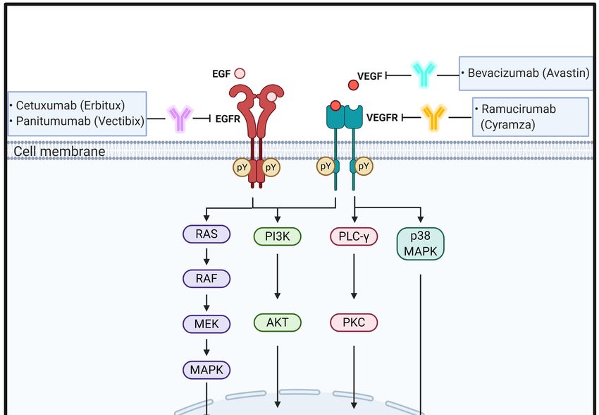
Biomedicines 2021, 9, 39                                                                                          2 of 24

                           clinical trials [16]. Antibody fragments retain the targeting specificity of whole monoclonal
                           antibodies and can be more economically produced. Furthermore, these fragments are
                           smaller and easily penetrate tissues and tumors more rapidly and deeply than monoclonal
                           antibodies having a higher molecular weight of 150 kDa. Antibody fragments have been
                           also forged into multivalent and multispecific reagents, linked to therapeutic payloads
                           (e.g., radionuclides, toxins, enzymes, liposomes, and viruses), and engineered for enhanced
                           therapeutic efficacy [16–19].
                                 Colorectal cancer (CRC) is the third most commonly occurring malignancy and sec-
                           ond leading cause of cancer death worldwide. The increasing prevalence of CRC across
                           the globe is one of the key factors driving the growth of the market [20]. According to
                           statistics, the global CRC therapeutic market is expected to reach $18.5 billion by 2023 from
                           $13.7 billion in 2018, at a compound annual growth rate of 6.1% from 2018 to 2023 [21].
                           Traditionally, 5-fluorouracil (5-FU)-based chemotherapeutic regimens, such as FOLFIRI
                           (irinotecan-containing regimen) and FOLFOX (oxaliplatin-containing regimen), have been
                           used clinically as standard therapies for treating CRC patients [22]. Recently, monoclonal
                           antibodies have been used in combination with standard chemotherapy to improve the
                           clinical outcomes of CRC patients. Compared with traditionally used chemotherapeutic
                           agents, monoclonal antibodies have fewer side-effects because their target specificity and
                           versatility are also being applied to next-generation antibody-based therapeutics, includ-
                           ing bispecific/multispecific antibodies, antibody-drug conjugates, and chimeric antigen
                           receptor T cells or natural killer cells [23–25].
                                 Over several decades, extensive in vitro and in vivo biochemical and molecular biol-
                           ogy studies have suggested many key signaling molecules closely associated with CRC
                           progression and metastasis. Among them, several growth factors, including epidermal
                           growth factor (EGF), vascular endothelial growth factor (VEGF), and hepatocyte growth
                           factor (HGF) and their cognate receptors, are proven therapeutic targets in CRCs for mon-
                           oclonal antibody therapy [26,27]. Further, human EGF receptor type 2 (HER2) is also
                           known as a monoclonal antibody target in CRCs [28]. More recently, Alison and Honjo, the
                           2018 Nobel prize winners in physiology, discovered immune checkpoint proteins, such as
                           programmed cell death protein-1 (PD-1) and cytotoxic T lymphocyte antigen 4 (CTLA-4),
                           which are key negative regulators of the immune system and cancer growth [27]. Presently,
                           these immune checkpoint proteins are drawing attention as the most promising therapeutic
                           targets in other types of cancers as well as CRCs for monoclonal antibody therapy. In addi-
                           tion, with infrastructural and technical advancement in monoclonal antibody development,
                           blockbuster humanized and fully human monoclonal antibodies, including cetuximab,
                           bevacizumab, and pembrolizumab, have received FDA approval and are widely used to
                           treat CRC patients [29].
                                 In this review, we first highlight recent studies of the roles and relevance of thera-
                           peutic targets in CRCs for monoclonal antibody therapy to understand the pathological
                           mechanism of CRCs governed by the target molecules. Simultaneously, presenting the
                           current status of FDA approved monoclonal antibodies in clinical development for CRC
                           therapy will provide insight into unmet medical needs in CRCs for monoclonal antibody-
                           based therapy.

                           2. Physiological and Pathophysiological Roles of Therapeutic Targets in CRCs
                           2.1. EGF/EGFR
                                 EGF is a 6 kDa growth factor with 53 amino acid residues; it binds to epidermal growth
                           factor receptor (EGFR) that is a single transmembrane glycoprotein with 1186 amino acids.
                           ErbB family members comprise ErbB1 (EGFR, HER1), ErbB2 (HER2), ErbB3 (HER3), and
                           ErbB4 (HER4). EGF binding to EGFR occurs within the 622-amino acid extracellular
                           domain (ECD), which is divided into four distinct domains: I–IV. Especially, domains I and
                           III are responsible for ligand binding, whereas domains II and IV have two cysteine-rich
                           regions that form disulfide bonds. Further, EGFR also has a 23-amino acid residue α-helical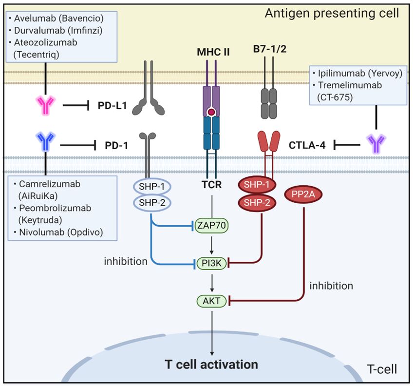
Biomedicines 2021, 9, 39                                                                                          3 of 24

                           transmembrane domain, 250-amino acid tyrosine kinase domain, and 229-amino acid
                           C-terminal tail with regulatory tyrosine residues [30–32].
                                 Under physiological conditions, EGFR activation proceeds sequentially by two steps.
                           First, prior to ligand binding, domain II is folded into domain IV via disulfide bonds in a
                           tethered conformation. Second, once EGF binds to domains I and III of EGFR monomers,
                           EGFRs promote domain rearrangement to expose dimerization arms in domain II, which
                           leads to receptor dimerization via domain II [31,33]. In turn, the EGFR dimers induce
                           trans-autophosphorylation by tyrosine kinase domains within the cytosolic parts of each
                           EGFR, resulting in activation of their downstream signaling cascades, such as the rat
                           sarcoma (RAS)/rapidly accelerated fibrosarcoma (RAF)/mitogen-activated protein kinase
                           (MAPK) and phosphoinositide 3-kinase (PI3K)/Akt pathways [33–35]. The RAS–RAF–
                           MAPK pathway is a major downstream signaling route of the ErbB family. EGF binding
                           to EGFR and consecutive tyrosine phosphorylation in EGFRs leads to activation of RAS,
                           a small GTP-binding protein, with the help of growth factor-bound protein 2 (GRB2), an
                           adaptor protein, and son of sevenless (SOS), a guanine nucleotide exchange factor. In
                           turn, activated RAS then activates downstream signaling molecules, including RAF and
                           MAPK [36]. Activated MAPKs phosphorylate specific transcription factors and participate
                           in regulation of cell migration and proliferation. EGFR activation also stimulates PI3K
                           composed of separate regulatory (p85) and catalytic (p110) subunits. The p85 regulatory
                           subunit directly binds to EGFR through the interaction of its Src homology domain 2
                           (SH2) with phosphotyrosine residues in activated EGFR. At the same time, the p110
                           catalytic subunit catalyzes phosphorylation of phosphatidylinositol 4,5-diphosphate to
                           generate phosphatidylinositol 3,4,5-triphosphate (PIP3 ), which in turn activates the protein
                           serine/threonine kinase, Akt [37–40] (Figure 1).
                                 Many previous reports have suggested the importance of EGFR as a therapeutic
                           target in CRC. For example, immunohistochemical studies have shown that EGFR was
                           highly overexpressed in 118 (80%) of 150 CRC patients, with a median follow-up of
                           40 months. In addition, Balb/c athymic nude mice subcutaneously injected with HCT116
                           or EGFR-knockout CT116 cells also revealed that depletion of EGFR in HCT116 cells
                           was associated with reduced tumor growth [41]. Osimertinib (Tagrisso® , AstraZeneca,
                           Cambridge, UK), a tyrosine kinase inhibitor of EGFR, was found to remarkably decrease
                           the tumor size and growth rate in a DLD-1, a colorectal adenocarcinoma cell line, xenograft
                           mouse model [42]. Administration of cetuximab, a human/mouse chimeric antibody,
                           exhibited significant growth suppression and inhibited the EGFR/MAPK pathway in a
                           HT29 xenograft mouse model [43]. Furthermore, another anti-EGFR antibody also showed
                           similar growth inhibition of CRCs to cetuximab in preclinical settings. GC1118, a novel fully
                           human anti-EGFR IgG1 antibody, exhibited potent inhibitory effects on EGFR signaling,
                           enhanced antibody-mediated cytotoxicity, and significantly inhibited tumor growth in a
                           CRC patient-derived xenograft (PDX) model [44]. Further, Ame55, an anti-EGFR IgG1
                           antibody, also inhibited tumor growth in a LoVo xenograft mouse model [45].
                                 In addition, some reports show the interrelationship of EGFR with other biomarkers.
                           Consequently, HER2 amplification has been implicated in therapeutic resistance to anti-
                           EGFR antibody therapy in preclinical studies in metastatic CRC (mCRC) [46]. HER2 am-
                           plification is seen in a small subset of mCRC, predominantly in KRAS wild-type tumors,
                           for which anti-EGFR antibodies are used as targeted therapies [47,48]. In these tumors,
                           aberrant HER2 signaling results in the bypass of the activation of the RAS/MEK/MAPK
                           signaling pathway, thereby blunting the effect of EGFR blockade [49,50]. Furthermore, the
                           EGFR-MET interaction induced by transforming growth factor-α, a specific EGFR ligand,
                           overexpression and concomitant phosphorylation of MET, and activation of MET down-
                           stream effectors have been proposed to be closely associated with the acquired resistance
                           to cetuximab in CRC cells [51,52]. Thus, these pieces of evidence demonstrate that the
                           combined inhibition of EGFR and other biomarkers can represent an effective strategy for
                           overcoming cetuximab resistance in patients with CRCs [53].
posed of separate regulatory (p85) and catalytic (p110) subunits. The p85 regulatory sub-
                 unit directly binds to EGFR through the interaction of its Src homology domain 2 (SH2)
                 with phosphotyrosine residues in activated EGFR. At the same time, the p110 catalytic
                 subunit catalyzes phosphorylation of phosphatidylinositol 4,5-diphosphate to generate
Biomedicines 2021, 9, 39                                                                                                    4 of 24
                 phosphatidylinositol 3,4,5-triphosphate (PIP3), which in turn activates the protein ser-
                 ine/threonine kinase, Akt [37–40] (Figure 1).

    Figure 1. Figure  1. Schematic
              Schematic              representation
                         representation               of the physiological
                                        of the physiological                rolespathways
                                                             roles and signaling  and signaling
                                                                                           of EGFpathways
                                                                                                  and VEGF of
                                                                                                            andEGF
                                                                                                                theirand
                                                                                                                      cognate
    receptors VEGF
              and theand  their
                      effect     cognate receptors
                             of antibodies           and the
                                           targeting these    effect of
                                                           signaling    antibodies
                                                                     molecules      targeting
                                                                                on CRC        these signaling
                                                                                        progression           molecules
                                                                                                    and metastasis. pY means
              on CRCresidues.
    phosphotyrosine    progression and metastasis. pY means phosphotyrosine residues.

                               2.2. VEGF/VEGFR
                                    VEGF is a disulfide-bonded dimeric glycoprotein of 45 kDa. The VEGF family has five
                               isotypes, including VEGF-A, placental growth factor, VEGF-B, VEGF-C, and VEGF-D [54].
                               Among them, VEGF-A is a glycosylated mitogen protein that is closely associated with
                               regulation of numerous pro-angiogenic functions, including endothelial cell growth and
                               migration, and vascular permeability in angiogenesis [55]. However, less is known about
                               the function and regulation of VEGF-B, -C, and -D. VEGFR receptor (VEGFR) is a member
                               of the receptor tyrosine kinase (RTK) family having multiple immunoglobulin-like ECDs
                               and tyrosine kinase activity. VEGFRs are divided into three types: VEGFR1 (Flt-1), VEGFR2
                               (KDR or Flk-1), and VEGFR3 (Flt-4). Especially, VEGFR2, a 200–230 kDa protein, mostly
                               interacts with VEGF-A and has a key role in angiogenesis at early embryogenesis and
                               mostly at lymph angiogenesis [54,56].
                                    Similar to that of other RTKs, the binding of VEGF to VEGFR forms a receptor dimer
                               and induces trans-autophosphorylation. Specifically, signaling of VEGFRs is initiated upon
                               binding of a covalently linked ligand dimer to the ECD of the receptor. This signaling
                               promotes receptor homodimerization followed by phosphorylation of specific tyrosine
                               residues located in the intracellular juxtamembrane domain, the kinase insert domain, and
                               the carboxy terminal tail of the receptor. Subsequently, a variety of signaling molecules
                               are recruited to VEGFR dimers [57,58]. These interactions next activate phospholipase
                               C (PLC)γ and protein kinase C (PKC) to induce transcription of genes necessary for
                               angiogenesis and cell proliferation [59]. Simultaneously, VEGFR-induced activation of
                               PI3K results in accumulation of PIP3, which induces phosphorylation of Akt to increase
                               endothelial cell survival and also induces systemic destruction of the entire basement
Biomedicines 2021, 9, 39                                                                                         5 of 24

                           membrane to increase vascular permeability [60,61]. Ligand binding to VEGFR-2 also
                           triggers the activation of the RAS pathway, initiating signaling through the RAF–MEK–
                           MAPK pathway known to be important in VEGF-induced cell proliferation [62]. In addition,
                           VEGF stimulates p38 MAPK to regulate the rearrangements of the actin cytoskeleton in
                           cell migration [63] (Figure 1).
                                 It has been reported that 70% of patients with stage IV CRCs had positive VEGF expres-
                           sion, whereas 50% and 47% of patients with stage II and III CRCs, respectively, had positive
                           VEGF expression. A statistically significant correlation was found between VEGF and
                           10-year disease-specific survival: VEGF-expressing tumors were more frequent in patients
                           who died of the disease than in those who survived for 10 years [64]. Further, other reports
                           have shown that the VEGF level also increased in CRC patients. In a prospective study by
                           Anastasios et al. that included 67 consecutive colorectal patients, VEGF was detectable in
                           all control subjects. Their median serum VEGF level was 186 pg/mL. Additionally, serum
                           VEGF levels were higher in 67 patients with newly diagnosed and histologically confirmed
                           primary CRC (492 pg/mL) than in the control subjects (186 pg/mL) [65].
                                 An increasing number of reports have suggested VEGF/VEGFR signaling as a promis-
                           ing therapeutic target in CRCs. First, Foersch et al. generated a conditional knockout for
                           VEGFR2 to investigate the functional role and underlying molecular mechanisms of the
                           signaling. Specific deletion of VEGFR2 was confirmed by qPCR of cDNA. Immunofluores-
                           cence staining revealed a lack of receptor expression relative to that of VEGFR2-expressing
                           control mice. Consequently, significantly fewer tumors developed in VEGFR2-knockout
                           mice than in control mice [66]. Second, regorafenib, a novel small-molecule multi-kinase
                           inhibitor, markedly slowed tumor growth in five of seven PDX models. The antitumor
                           effects of regorafenib were evaluated in seven PD CRC xenografts [67]. Third, bevacizumab,
                           a humanized antibody to VEGF, showed a significant delay in CRC tumor growth rela-
                           tive to that of the non-treated animals [68]. Fourth, intraperitoneal injection of DC101,
                           an anti-VEGFR mouse monoclonal antibody, inhibited tumor growth and induced apop-
                           tosis in CRC in a KM12L4 xenograft model. Further, treatment with DC101 decreased
                           tumor vascularity, growth, proliferation, and increased apoptosis [69]. Taken together, this
                           large number of studies shows the importance of VEGF/VEGFR signaling for developing
                           pharmaceutical anti-angiogenic drugs.

                           2.3. HGF/c-MET
                                HGF is synthesized as an inactive precursor (pro-HGF) that undergoes site-specific
                           proteolytic cleavage by extracellular serine proteinases into an active 90-kDa heterodimer
                           containing α and β chains. HGF is predominantly secreted by stromal cells and activates
                           mesenchymal-epithelial transition factor (c-MET) on adjacent epithelial cells [70]. HGF
                           contains two c-MET binding sites; a high affinity site in the N-terminal and first kringle
                           regions that binds to the immunoglobulin-like fold shared by plexins and transcriptional
                           factors (IPT) 3 and IPT4 domains in c-MET and a low-affinity site in the serine protease
                           homology domain that interacts with the semaphorin domain in c-MET [71]. As a tyrosine-
                           protein kinase Met or HGR receptor, c-MET is synthesized as a 170-kDa single-chain
                           precursor protein (pro-c-MET) that undergoes furin-mediated post-translational cleavage,
                           yielding a disulfide-linked heterodimer composed of an extracellular α-subunit and a
                           single transmembrane β-subunit [70,72].
                                In the physiological state, HGF binding to c-MET induces c-MET dimerization and
                           trans-autophosphorylation of the tyrosine residues Y1003 in the juxtamembrane domain
                           and Y1234 and Y1235 within the kinase activation loop, resulting in phosphorylation of
                           two tyrosine residues, Y1349 and Y1356, in the C-terminus. Tyrosine phosphorylation of
                           Y1349 and Y1356 residues creates docking sites for recruitment of key intracellular adaptor
                           proteins and signaling molecules through SH2-mediated interactions, including GRB2,
                           GRB2-associated binding protein 1 (GAB1), signal transducer and activator of transcription
                           3 (STAT3), the p85 subunit of PI3K, SRC, PLC-γ, and Shc [73,74]. The potency, duration, and
                           versatility of HGF/c-MET signaling are modulated by signaling amplifiers and co-receptors.
Biomedicines 2021, 9, x FOR PEER REVIEW 6 of 24
Biomedicines 2021, 9, 39 6 of 24
co-receptors. Recruitment and sustained phosphorylation of the multi-adaptor protein
GAB1 is an important
Recruitment hallmark
and sustained of sustained c-MET
phosphorylation signaling thatprotein
of the multi-adaptor can either
GAB1 bind is directly
an impor-
totant
c-MET through its unique 13-amino-acid c-MET binding site or indirectly
hallmark of sustained c-MET signaling that can either bind directly to c-MET through by association
with GRB2. 13-amino-acid
its unique The tyrosine-phosphorylated
c-MET binding site GAB1 protein serves
or indirectly as an auxiliary
by association with GRB2. signal
The
transduction platform through recruitment of various effector proteins,
tyrosine-phosphorylated GAB1 protein serves as an auxiliary signal transduction platform including PI3K.
Cell survival
through response of
recruitment is related
variousto the PI3K/Akt
effector proteins,pathway
including activated by c-MET
PI3K. Cell survival signaling
response
[75,76]. Furthermore, HGF-induced c-MET-dependent RAS/RAF/MAPK
is related to the PI3K/Akt pathway activated by c-MET signaling [75,76]. Furthermore, activation has
been found to require the co-receptor CD44v6, thus providing
HGF-induced c-MET-dependent RAS/RAF/MAPK activation has been found to require a platform for SOS recruit-
ment to the complex
the co-receptor and subsequently
CD44v6, thus providing triggering proficient
a platform for SOS activation of RAS
recruitment [32].
to the Inva-
complex
sion, branching morphogenesis, and tumorigenesis are mediated by
and subsequently triggering proficient activation of RAS [32]. Invasion, branching mor- STAT3 activation in
a phogenesis,
tissue-dependent manner. Typically, STAT3 activation is induced by
and tumorigenesis are mediated by STAT3 activation in a tissue-dependent phosphorylation
onmanner.
a criticalTypically,
tyrosine STAT3
residueactivation
(Y705) that is triggers
inducedSTAT3 dimerization owing
by phosphorylation to reciprocal
on a critical tyrosine
phosphotyrosine-SH2
residue (Y705) that triggers domain interactions.
STAT3 In addition
dimerization owing to tyrosine 705
to reciprocal phosphorylation,
phosphotyrosine-SH2
STAT3
domain is interactions.
also activated In through
addition serine (S727)
to tyrosine 705phosphorylation.
phosphorylation,Finally,STAT3 is the
alsoreversible
activated
acetylation of STAT3 by histone acetyltransferase on a single lysine
through serine (S727) phosphorylation. Finally, the reversible acetylation of STAT3 residue (K685) repre-by
sents a third mechanism of STAT3 activation. Acetylated STAT3 enhances
histone acetyltransferase on a single lysine residue (K685) represents a third mechanism the stability of
STAT3
of STAT3dimers, which are
activation. requiredSTAT3
Acetylated for DNA-binding
enhances theand transcriptional
stability of STAT3 activity. Cellular
dimers, which are
migration
required for andDNA-binding
adhesion areand mediated by a c-MET–SRC–focal
transcriptional activity. Cellularadhesion
migrationkinase (FAK) in-
and adhesion are
teraction.
mediated Within the complex, Src adhesion
by a c-MET–SRC–focal phosphorylates
kinase Y576
(FAK)and Y577 within
interaction. the the
Within kinase do-
complex,
main activation loop and
Src phosphorylates Y576Y861and and
Y577Y925
withinwithin the C-terminal
the kinase domain ofloop
domain activation FAK. The
and FAK–
Y861 and
Src complex further binds to and phosphorylates various adaptor proteins, such as
Y925 within the C-terminal domain of FAK. The FAK–Src complex further binds to and
phosphorylates
p130Cas various
and paxillin adaptor
[77–79] proteins,
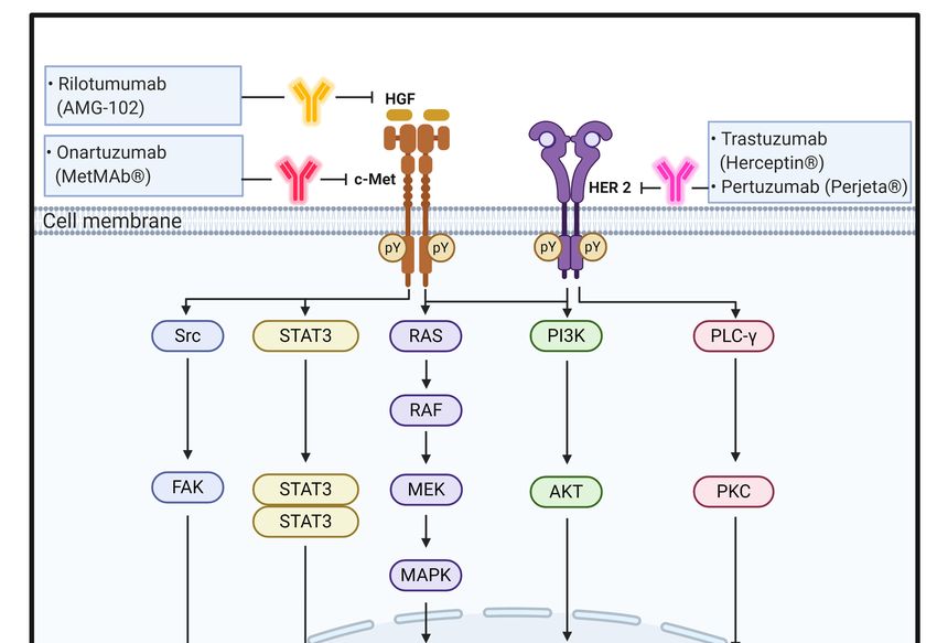
(Figure 2). such as p130Cas and paxillin [77–79] (Figure 2).
Figure 2. 2.
Figure The schematic
The representation
schematic ofof
representation the physiological
the roles
physiological rolesand
andsignaling
signalingpathways
pathwaysofofHGF/c-
HGF/c-
MET and HER2 and effect of antibodies targeting these signaling molecules on CRC progression
MET and HER2 and effect of antibodies targeting these signaling molecules on CRC progression and
and metastasis. pY means phosphotyrosine residues.
metastasis. pY means phosphotyrosine residues.
c-MET
c-METis isoverexpressed
overexpressed in in
CRCs
CRCs[80]. Specifically,
[80]. immunohistochemistry
Specifically, immunohistochemistrywithwith
23
cases of colorectal adenoma and 102 cases of primary colorectal carcinoma as well their
23 cases of colorectal adenoma and 102 cases of primary colorectal carcinoma as well their
corresponding
correspondingmetastases
metastases(44
(44lymph
lymphnodes,
nodes, 2121 peritoneal deposits, and
peritoneal deposits, and16
16liver
livermetastases)
metasta-
ses) showed that normal tissues had a negative or weak c-MET expression,
showed that normal tissues had a negative or weak c-MET expression, whereas c-MET whereas c-
was highly overexpressed in adenomas and primary CRC. Moreover, c-MET expression in
metastatic tissues was significantly higher compared with the primary tumor [81].Biomedicines 2021, 9, 39 7 of 24
Currently, HGF/c-MET signaling is one of the key therapeutic targets in CRC therapy.
Small hairpin RNA-mediated c-MET knockdown dramatically suppressed tumor growth in
a SW480 xenograft mouse model as well as SW480 cell proliferation in vitro [82]. SU11274,
an ATP-competitive inhibitor of c-MET, significantly inhibited tumor growth in a LoVo
xenograft mouse model. ARQ 197 (tivantinib), a non-ATP-competitive inhibitor of c-MET,
decreased tumor growth in a HT29 xenograft mouse model [83]. Antibody-based targeting
of c-MET also gave results similar to those of pre-existing chemical inhibitors. For example,
YYB-101, a humanized neutralizing antibody specifically binding to HGF, inhibits c-MET
activation and cell scattering in vitro and suppresses tumor growth in HCT116 xenograft
mouse models [84]. R13 and R28, two fully human antibodies against c-MET, synergisti-
cally inhibit HGF binding to c-MET and elicit antibody-dependent cellular cytotoxicity. The
combination of R13/28 significantly inhibited tumor growth in xenograft models of various
colon tumors, including OMP-C12, 27, and 28. Inhibition of tumor growth was associ-
ated with induction of hypoxia. Moreover, in an experimental metastasis model, R13/28
increased survival by preventing recurrence of otherwise lethal lung metastases [85].
2.4. HER2
HER2 is an ErbB family member with a molecular weight of 185 kDa comprising a 632-
amino acid ECD, 22-amino acid α-helical transmembrane domain, and a 580-amino acid
tyrosine kinase domain. Despite the many intensive studies on HER2, a ligand of HER2
has not been clearly identified yet. It is known that HER2 forms complexes with HER2 or
other ErbB family members, including EGFR, ErbB3, and ErbB4, to activate downstream
signaling pathways [86,87].
Similar to EGF/EGFR signaling pathways, it has been suggested that in normal cells,
the HER2 complex formation, such as through homodimerization or heterodimerization,
leads to continuous trans-autophosphorylation on tyrosine residues of HER2 and activates
downstream signaling pathways, including the RAS/RAF/MAPK pathway, PI3K/Akt
pathway, and PLC/PKC pathway. HER2 activation ultimately promotes cell growth,
proliferation, and survival. The RAS/RAF/MAPK and PI3K/Akt pathways are the two
most important and extensively studied downstream signaling pathways upon activation of
HER2 receptors. A third key signaling in the network is the PLC-γ/PKC pathway. Binding
of PLC-γ to phosphorylated HER2 stimulates PLC-γ activity and results in hydrolytic
cleavage of phosphatidylinositol-4,5-bisphosphate (PIP2 ) to yield inositol 1,4,5-triphosphate
(IP3 ) and 1,2-diacylglycerol. These second messengers are important for intracellular
calcium release and activation of PKC. As a result of these signaling pathways, different
nuclear factors are recruited and modulate the transcription of different genes involved in
cell-cycle progression, proliferation, and survival [32,33,88,89] (Figure 2).
In CRC, HER2 expression is varied because of many factors that influence the de-
termination of HER2 expression, especially of the intracellular fraction of HER2. One
report stated that HER2 overexpression was observed in 136 (11.4%) of 1195 CRC patients
with moderately to poorly differentiated tubular adenocarcinomas. Further, HER2 overex-
pression correlated with shorter mean overall survival (OS) [90]. Other studies have also
reported that membranous overexpression of HER2 occurs in only 5% of all CRC patients,
whereas cytoplasmic HER2 overexpression is observed in a significant proportion (30%)
of patients [91].
Several lines of evidence also support the idea that HER2 is a therapeutic target
in CRCs. Tucatinib, a reversible inhibitor that binds to the ATP pocket of the internal
domain of the HER2 receptor, prevents activation of HER2 signaling pathways [92]. Further,
administration of tucatinib in a CRC PDX model significantly reduced tumor volume.
Both H2Mab-19 and H2Mab-41, novel anti-HER2 IgG2 antibodies, significantly reduced
tumor development in Caco-2 xenograft mouse models [93,94]. Treatment with Herceptin®
(Genentech, San Francisco, CA, USA) caused a decrease in HER-2 protein levels in DLD-1,
HT-29, Caco-2, and HCA-7 colon cancer cells in vitro. Treatment of athymic mice engraftedBiomedicines 2021, 9, 39 8 of 24
with EGFR-dependent colon cancers, including HCA-7, DLD-1, and HT-29 with Herceptin®
showed tumor regression and decreased EGFR tyrosine phosphorylation in tumor cells [95].
2.5. Immune Checkpoint
2.5.1. CTLA-4
CTLA-4, also designated CD152, is a type I transmembrane T-cell inhibitory molecule
that functions as an immune checkpoint, downregulates immune responses and is found as
a covalent homodimer of 41–43 kDa [96]. CTLA-4 is a member of the IgG superfamily that
is expressed by activated T cells. CTLA-4 contains an ECD with one Ig-like V-type domain,
a transmembrane domain, and cytoplasmic tail. CTLA-4 is homologous to the T-cell co-
stimulatory protein, CD28, and both molecules bind to B7-1/B7-2 on antigen-presenting
cells [97]. CTLA-4 binds CD80 and CD86 with greater affinity and avidity than CD28, thus
enabling it to outcompete CD28 for its ligands. CTLA-4 transmits an inhibitory signal to
T cells, whereas CD28 transmits a stimulatory signal [98,99].
CTLA-4 is upregulated in a manner dependent on TCR stimulation. At the cell mem-
brane, CTLA-4 undergoes dimerization, and each CTLA-4 dimer can bind two independent
B7-1/B7-2 homodimers, forming a linear zipper-like structure between B7-1/B7-2 and
CTLA-4 homodimers [100]. Activated CTLA-4 binds to PI3K, the tyrosine phosphatases
(SHP1 and SHP2) and the serine/threonine phosphatase PP2A. SHP1 and SHP2 dephospho-
rylate TCR-signaling proteins, whereas PP2A targets phosphoserine/threonine residues
and is known to interfere with the activation of Akt [101].
CTLA-4 can inhibit T-cell responses by several mechanisms. One mechanism involves
antagonism of B7-CD28–mediated co-stimulatory signals by CTLA-4. The fact that CTLA-4
has a much higher affinity for B7 than CD28 supports the notion that the CTLA-4–mediated
sequestration of B7 is closely associated with negative regulation of T-cell signaling [102].
Another mechanism for the inhibitory activity of CTLA-4 is related to direct interaction
with the TCR–CD3 complex at the immunological synapse for negative regulation of
downstream signaling after TCR activation [103]. When CTLA-4 interacts with the ITAMs
present on the TCR–CD3 complex, the activated CTLA-4 binds to tyrosine phosphatases,
including SHP1, SHP2, and PP2A, and eventually deactivates various downstream signal-
ing molecules of activated T cells, including zeta-chain-associated protein kinase 70, spleen
tyrosine kinase, and proto-oncogene tyrosine-protein kinase [104–106] (Figure 3).
Many previous reports have shown that CTLA-4 is a key therapeutic target in CRCs [107].
First, Long et al. used the CRISPR-Cas9 system to generate CTLA-4 knockout cytotoxic T
lymphocytes (CTLs) and evaluated the effect on the antitumor activity of the CTLs [107].
The HCT-116 xenografted mice treated with CTLA-4 KO CTLs demonstrated repressed
tumor growth and prolonged survival relative to those in the control group. All of the
mice in the control group died from progressive tumors within 62 days. In contrast,
only 10% of CTLA-4 KO CTLs treated mice died within that time [108]. Second, Fu et al.
validated the efficacy of anti-CTLA-4 mouse monoclonal antibodies on tumor size in mice
inoculated with CT26 cells. The tumor volumes were 2106 ± 205 mm3 on day 17 in the
control group treated with vehicle only but were 23 ± 4 mm3 in the group treated with
the anti-CTLA-4 antibodies on day 5, which indicated a statistically significant difference
in antitumor activity between the treated and vehicle groups [109]. Third, Lute et al. also
reported that anti-human CTLA-4 human monoclonal antibodies-treated mice survived
longer than the control Ig-treated mice in a human peripheral blood leukocytes-SCID
mouse model. Fourth, administration of 9H10, an anti-murine CTLA monoclonal antibody,
as monotherapy moderately inhibited growth and metastatic spread of the colon cancer
cells in an orthotropically implanted CT26 xenograft mouse model [110]. Further, the sole
CTLA-4 inhibition significantly increased intratumoral CD8+ and CD4+ T cells and reduced
FOXP3+ /CD4+ Treg cells, which was associated with increased expression levels of the
pro-inflammatory Th1/M1-related cytokines IFN-γ, IL-1α, IL-2, and IL-12 [111].
In summary, the evidence from the studies above shows that CTLA-4 blockade exerts
inhibitory effects on growth and metastasis of CRCs.Biomedicines 2021, 9, 39 9 of 24
Biomedicines 2021, 9, x FOR PEER REVIEW 9 of 24
Figure 3.3. Schematic
Figure Schematic representation
representation of
of the
the physiological
physiological roles
roles and
and signaling
signaling pathways
pathways of
ofimmune
immune
checkpoint proteins of PD-1, PD-L1, CTLA-4, and B7-1/2 and effect of antibodies targeting
checkpoint proteins of PD-1, PD-L1, CTLA-4, and B7-1/2 and effect of antibodies targeting these these
signaling molecules on CRC progression and metastasis.
signaling molecules on CRC progression and metastasis.
2.5.2. Many
PD-1/PD-L1
previous reports have shown that CTLA-4 is a key therapeutic target in CRCs
[107].PD-1,
First,also
Long et al. usedCD279,
designated the CRISPR-Cas9
is a 55 kDasystem
membraneto generate
proteinCTLA-4 knockout
consisting of an ECDcyto-
toxic T lymphocytes
followed (CTLs) and
by a transmembrane evaluated
region and an theintracellular
effect on thetail
antitumor
containing activity of the CTLs
two phosphory-
[107]. sites
lation The HCT-116
located inxenografted
an immunoreceptor mice treated with CTLA-4
tyrosine-based KO CTLs
inhibitory demonstrated
motif (ITIM) and an re-
pressed tumor growth
immunoreceptor and prolonged
tyrosine-based survival
switch motif relative
(ITSM). to those incell
Programmed thedeath-ligand
control group. All
1 (PD-
of the
L1, mice in the
designated control
CD274, groupisdied
B7-H1) a 44 from progressive tumors
kDa transmembrane within
protein 62 days.on
expressed InTcontrast,
cells, B
only macrophages,
cells, 10% of CTLA-4 andKOdendritic
CTLs treated
cells asmice
welldied
as onwithin
tumorthat time
cells, [108]. Second,
including Fu et al.
CRCs [112,113].
validated
Underthe efficacy of anti-CTLA-4
physiological conditions, when mouseTmonoclonal antibodies
cells recognize antigens onon
tumor
majorsize in mice
histocom-
patibility
inoculated complex
with CT26 of thecells.
target
Thecell, inflammatory
tumor volumes cytokines,
were 2106such ± 205asmm tumor
3 onnecrosis
day 17 factor
in the
alpha
controland interferon
group treated gamma (IFNγ)only
with vehicle are produced
but were to 23initiate
± 4 mminflammatory
3 in the groupprocesses.
treated with These
the
cytokines
anti-CTLA-4 upregulate
antibodies theonexpression
day 5, which of PD-L1 on tissues
indicated and PD-1
a statistically on T cells.
significant In turn,
difference in
PD-1 directly interacts with PD-L1 to negatively regulate T-cell
antitumor activity between the treated and vehicle groups [109]. Third, Lute et al. alsoreceptor (TCR) signaling,
inhibits
reportedinterleukin-2
that anti-human (IL-2)CTLA-4
production humanin Tmonoclonal
cells, and increases T-cell apoptosis
antibodies-treated mice[114,115].
survived
Specifically,
longer than this the PD-1/PD-L1
control Ig-treated interaction
mice ininduces
a human lymphocyte-specific
peripheral blood protein tyrosine
leukocytes-SCID
kinase-induced
mouse model. Fourth, phosphorylation of twooftyrosine-based
administration motifs within
9H10, an anti-murine CTLAITIM and ITSM
monoclonal of
anti-
the cytoplasmic
body, tail of PD-1.
as monotherapy The recruitment
moderately inhibitedofgrowth
Src homology 2 (SH2) spread
and metastatic domain-containing
of the colon
protein tyrosine
cancer cells in anphosphatase
orthotropically 1 (SHP1) and SHP-2
implanted CT26phosphatase
xenograft mouse then induces dephosphory-
model [110]. Further,
lation of the TCR signalosome, including CD3ζ, ZAP70, and PI3K
the sole CTLA-4 inhibition significantly increased intratumoral CD8 and CD4+ T cells kinases,
+ resulting in and
the
deactivation of downstream signaling targets [116]. Moreover,
reduced FOXP3 /CD4 Treg cells, which was associated with increased expression levels
+ + the PD-1/PD-L1 interaction
also downregulates
of the pro-inflammatory the protein (casein) kinase
Th1/M1-related 2 expression
cytokines IFN-γ, that
IL-1α, phosphorylates
IL-2, and IL-12 the regula-
[111].
tory domain of phosphatase
In summary, the evidence andfrom
tensin thehomolog (PTEN)
studies above and inhibits
shows that CTLA-4phosphatase
blockade activity
exerts
to remove PIP3 produced by PI3K [112,117,118].
inhibitory effects on growth and metastasis of CRCs. Thus, PTEN can terminate PI3K activities
by dephosphorylating PIP3, which eventually leads to immune tolerance, a phenomenon
2.5.2. PD-1/PD-L1Biomedicines 2021, 9, 39 10 of 24
in which the immune system loses the control to mount an inflammatory response even in
the presence of actionable antigens [117,119] (Figure 3).
PD-L1 has been reported to be overexpressed in CRC. More specifically, among
the 80 tumor specimens, 22 (27.5%) showed high PD-L1 expression, 24 (30.0%) showed
moderate expression, and 34 (42.5%) showed weak or no PD-L1 staining. Furthermore, the
high PD-L1 expression in normal tissues was observed in four (6.3%) cases [120].
Many studies have suggested PD-1 and PD-L1 as promising therapeutic targets in
CRCs. First, in BALB/c Rag2−/−γc−/− mice engrafted with PD-L1-overexpressing and
PD-L1-knockout CT26 murine colon cancer cells, Gordon et al. found that after 3 weeks,
tumors were significantly smaller in the PD-L1-knockout group than in the PD-L1 over-
expression group [121]. Second, Cai et al. examined the efficacy of anti-mouse PD-1 rat
immunoglobulin (Ig) G2 antibodies on tumor growth in a CT26 colon cancer xenograft
mouse model. The antibody treatment showed significant inhibition of transplanted-tumor
growth in mice [122]. Third, in a humanized CRC PDX model established by Capasso et al.,
treatment with nivolumab, a fully human IgG4 (S228P) monoclonal antibody to PD-1, led
to significant tumor growth inhibition coupled with increased numbers of IFNγ-producing
human CD8+ tumor-infiltrating lymphocytes [123]. Fourth, Stewart et al. reported that
MEDI4736, a human IgG1 monoclonal antibody that binds with high affinity and specificity
to PD-L1, significantly inhibited the growth of human tumors in a novel CT26 xenograft
model containing co-implanted human T cells. This activity is entirely dependent on
the presence of transplanted T cells. Further, anti-mouse PD-L1 significantly improved
survival of mice implanted with CT26 CRC cells [124]. The antitumor activity of anti-PD-L1
was enhanced by combination with oxaliplatin, which resulted in increased release of
high-motility group box 1 within CT26 tumors [125].
3. Current Status of Monoclonal Antibodies for CRC Therapy
3.1. Cetuximab
Cetuximab (Erbitux® ), developed jointly by Merck KGaA (Darmstadt, Germany) and
Imclone Systems (New York City, NY, USA), is a monoclonal antibody that binds to the
ECD of the EGFR. It is a human/mouse chimeric IgG1 antibody that consists of the variable
fragments (Fvs) of a murine anti-EGFR antibody and human constant heavy and kappa
light chains [126,127].
Cetuximab was originally known as a blockade for inhibiting interactions between
EGFR and all known EGFR ligands by specifically binding to domain III of the EGFR
ECD [128]. Furthermore, its binding to EGFR is also able to promote receptor internaliza-
tion and concomitantly downregulate EGFR protein levels expressed on the cell surface,
resulting in suppression of EGFR-dependent downstream signaling pathways and tran-
scription. In addition to these specific modes of action, cetuximab indirectly attacks cancer
cells through antibody-dependent cell-mediated cytotoxicity (ADCC). After its binding
to EGFR, the IgG1 portion of cetuximab may be recognized by Fcγ receptors (FcγR) on
immune effector cells, such as natural killer cells and T cells, and participates in cancer cell
death. In general, FcγRs bind effectively to IgG1 and IgG3 antibodies. Thus, cetuximab is
speculated to more likely stimulate ADCC than panitumumab having IgG2. Consequently,
cetuximab reduces tumor angiogenesis, invasiveness, and metastatic spread [129–132].
In 2004, cetuximab received FDA approval for metastatic CRCs and head and neck
cancers, and its use is recommended in combination with standard chemotherapy for
treatment of patients with metastatic CRCs having EGFR-positive and wild-type KRAS
(Table 1). According to the CRYSTAL clinical trial (NCT00154102), compared with FOLFIRI
alone, cetuximab plus FOLFIRI improved progression-free survival (PFS) from 8.0 to 9.0
months and OS from 20 to 23.5 months for treatment of patients with KRAS wild-type [133].Biomedicines 2021, 9, 39 11 of 24
Table 1. Antibody therapeutics currently in clinical trials or approved by the US FDA.
Name Trade Name Company Target Format Clinical Stage
Cetuxumab Erbitux® Imclone Systems EGFR Chimeric IgG1 FDA approval in 2004
Panitumumab Vectibix® Abgenix Inc. EGFR Human IgG2 FDA approval 2006
Bevacizumab Avastin® Genentech VEGF-A Humanized IgG1 FDA approval in 2004
Ramucirumab Cyramza® ImClone Systems VEGFR2 Human IgG1 FDA approval in 2015
Rilotumumab AMG-102 Amgen Inc. HGF Human IgG1 P II
Onartuzumab MetMab Genentech c-MET Humanized IgG1, monovalent P II, failure
Trastuzumab Herceptin® Genentech HER2 Humanized IgG1 P II
Pertuzumab Perjeta® Genentech HER2 Humanized IgG1 P II
Ipilimumab Yervoy® BMS CTLA4 Human IgG1 FDA approval in 2018
Tremelimumab CT-675 Medimmune CTLA4 Human IgG2 P II
Pembrolizumab Keytruda® LifeArc PD-1 Humanized IgG4 FDA approval in 2020
Nivolumab Opdivo® Ono Phar. & Medarex PD-1 Humanized IgG4 P II
Camrelizumab AiRuiKa Jiangsu HengRui PD-1 Humanized IgG4 P II
Atezolizumab Tecentriq® Genentech PD-L1 Human IgG1 P II
Avelumab Bavencio® Merck KGaA PD-L1 Human IgG1 P III
Durvalumab Imfinzi® Medimmune PD-L1 Human IgG1 P II
Antibody information was obtained from the FDA Label database and the Drug Approval and Databases site maintained by the US FDA
(https://www.fda.gov/drugs/development-approval-process-drugs/drug-approvals-and-databases) or Clinical Trial Information Site
(https://clinicaltrials.gov).
3.2. Panitumumab
Panitumumab (Vectibix™) originally developed by Abgenix Inc. (Freemont, CA, USA)
is a fully human IgG2 monoclonal antibody that specifically binds to the ECD of EGFR.
Especially and different from cetuximab, it can also bind to a single-point mutation in
domain III of EGFR (S468R) that confers acquired or secondary resistance only to cetuximab-
treated patients [134].
In 2006, panitumumab was approved by the US FDA for treatment of patients with
EGFR-expressing metastatic CRCs with disease progression or following fluoropyrimidine-,
oxaliplatin-, and irinotecan-containing regimens (Table 1). Later, it was also approved for
treatment of patients with refractory metastatic CRCs having EGFR-positive and wild-type
KRAS. Currently, panitumumab was the first monoclonal antibody to use KRAS as a
predictive biomarker [135].
Panitumumab is being used in combination with chemotherapy in the first- and
second-line treatment of metastatic CRCs [136]. In the PRIME (NCT00364013) clinical
trial, compared with chemotherapy alone, a first-line treatment of panitumumab plus
FOLFOX improved PFS from 8.0 to 9.6 months and OS from 12 to 14 months. Further, in
the 20,050,181 clinical trial (NCT00339183), compared with chemotherapy, a second-line
treatment of panitumumab plus FOLFIRI improved PFS from 4 to 6 months and OS from
19 to 24 months [137,138].
3.3. Bevacizumab
Bevacizumab (Avastin® ) developed by Genentech (South San Francisco, CA, USA)
is a humanized IgG1 monoclonal antibody that binds to segment β5–β6 of VEGF165,
known as VEGF-A; its binding to VEGF-A inhibits angiogenesis by specifically inhibiting
the interaction between VEGF-A and VEGFR2. Thus, bevacizumab inhibits angiogenic
signaling caused by the interaction of VEGF-A and VEGFR2 [139–141].
In 2004, bevacizumab received FDA approval for first- or second-line treatment with 5-
FU-based therapy for patients with mCRCs (Table 1). As the first anti-angiogenic antibody
drug, bevacizumab is currently being used in clinics for treatment of patients with NSCLC,
mRCC, epithelial ovarian cancer, and recurrent glioblastoma as well as metastatic CRCs.
In the clinical trial ECOG3200 (NCT00069095) for treatment of patients with mCRCs,
compared with FOLFOX4 alone, bevacizumab plus FOLFOX4 improved OS from 10.8 to
12.9 months and PFS from 4.7 to 7.3 months [142–144].Biomedicines 2021, 9, 39 12 of 24
3.4. Ramucirumab
Ramucirumab (Cyramza® ) developed jointly by ImClone Systems (New York City,
NY, USA) and Dyax (Cambridge, MA, USA) is a fully human monoclonal IgG1 anti-
body that binds to the ECD of human VEGFR2, which is a key receptor that mediates
angiogenesis and is highly expressed in not only tumor microvessels but also malignant
tumors. Bevacizumab strongly neutralizes VEGF and blocks binding to VEGFR1/VEGFR2,
whereas ramucirumab specifically blocks the VEGF/VEGFR2 interaction by binding to
VEGFR2 [145–147].
Ramucirumab was isolated from a Dyax’s phage antibody library and developed as a
therapeutic antibody for treatment of solid tumors. In 2014, ramucirumab first received
FDA approval as a single-agent treatment for advanced gastric or gastro-esophageal junc-
tion adenocarcinoma after prior treatment with fluoropyrimidine- or platinum-containing
chemotherapy. In 2015, ramucirumab in combination with FOLFIRI was also approved by
the US FDA for the treatment of patients with mCRC with disease progression on or after
prior therapy with bevacizumab, oxaliplatin, and fluoropyrimidine (Table 1). In the RAISE
clinical trial (NCT01183780) for treatment of mCRC patients, compared with FOLFIRI
alone, ramucirumab plus FOLFIRI improved OS from 11.7 to 13.3 months and PFS from
4.5 to 5.7 months [148,149].
3.5. Rilotumumab
Rilotumumab (AMG-102) developed by Amgen Inc. (Thousand Oaks, CA, USA) is a
fully human monoclonal IgG2 antibody that binds to the beta chain of HGF. HGF, a scatter-
ing factor, influences cancer cell proliferation, survival, invasion, and metastasis through
interaction with c-MET. Therefore, specific binding of rilotumumab to HGF neutralizes the
interaction between HGF and c-MET and inhibits c-MET phosphorylation and downstream
signaling, resulting in inhibition of cancer cell proliferation, survival, and invasion through
partial antagonism of c-MET phosphorylation [150,151].
It has been reported that in the phase II clinical trial (NCT00788957) for treatment
of patients with wild-type KRAS metastatic CRCs, compared with panitumumab alone,
rilotumumab plus panitumumab combination therapy improved OS from 8.6 to 9.6 months
and PFS from 3.7 to 5.2 months [152] (Table 1).
3.6. Onartuzumab
Onartuzumab (MetMAb) developed by Genentech (South San Francisco, CA, USA)
is a humanized monoclonal antibody that binds to the semaphorin domain of c-MET; it
is a human IgG1 with a monovalent arm. c-MET is an RTK through which downstream
signals are transduced after dimerization or oligomerization by HGF binding. HGF/c-MET
signaling has also been implicated in the metastatic growth of multiple cancers, making
it an attractive target for various therapeutic agents [153]. When onartuzumab binds to
c-MET, it inhibits cell proliferation and survival, cell motility, migration, and invasion [154].
In a clinical trial (NCT01418222) of the MET inhibitor onartuzumab in combination with
mFOLFOX-6 plus bevacizumab for treatment of mCRC patients, no significant differences
existed in PFS or OS between onartuzumab combination therapy with mFOLFOX-6 plus be-
vacizumab and bevacizumab plus mFOLPOX-6 therapy. Therefore, the phase II clinical trial
of onartuzumab failed because it did not show any clinical improvement [155] (Table 1).
3.7. Trastuzumab
Trastuzumab (Herceptin® ) developed by Genentech (South San Francisco, CA, USA)
is a humanized IgG1 monoclonal antibody that binds to domain IV in the ECD of the
HER2. This antibody is composed of humanized Fvs of murine anti-HER2 antibody and
human constant heavy and kappa light chains. The antibody suppresses cancer growth
and proliferation [156].
Trastzumab binds to HER2 on the cell surface of CRCs and inhibits downstream
signaling pathways, such as RAS/RAF/MEK and PI3K/Akt pathways, thereby inhibitingBiomedicines 2021, 9, 39 13 of 24
the proliferation and survival of CRCs. This demonstrates that trastuzumab can be used
as a therapeutic agent in HER2-overexpressed CRCs. In addition, trastuzumab indirectly
attacks cancer cells through Fc effect functions, such as ADCC and complement-dependent
cytotoxicity (CDC) [157,158].
In 1998, trastuzumab first received US FDA approval for the treatment of patients
with metastatic HER2-overexpressing breast cancers, and its indications for numerous
cancer treatments have expanded [159,160]. Despite the low-expression pattern observed
in CRC patients, intriguingly, recent preclinical trials have shown that trastuzumab or
pertuzumab in combination with lapatinib significantly suppressed tumor growth in HER2-
amplified CRC tumor xenograft animal models. According to the HERACLES clinical
trial (NCT03365882), compared with chemotherapy alone, trastuzumab plus lapatinib
therapy in patients with HER2-positive, wild-type KRAS metastatic CRC improved PFS by
2.9 months and OS by 11.5 months [161] (Table 1).
3.8. Pertuzumab
Pertuzumab (Perjeta® ) developed by Genentech (South San Francisco, CA, USA) is a
humanized IgG1 monoclonal antibody that binds to HER2. The antibodies act as a blockade
to inhibit dimerization of HER2 with other HER receptors, especially HER3, by specifically
binding to domain II in the ECD of HER2, a different epitope for trastuzumab, resulting in
inhibition of cell growth and initiation of apoptosis [162]. In 2012, the US FDA approved
pertuzumab use in combination with trastuzumab and docetaxel for treatment of patients
with metastatic HER2-positive metastatic breast cancers. According to the HERACLES
study (NCT03365882), the randomized phase II trial studies is ongoing by evaluating the
efficacy of pertuzumab and tratuzumab in patients with HER-amplified mCRCs, compared
with cetuximab and irinotecan hydrochloride [163] (Table 1).
3.9. Ipilimumab
Ipilimumab (Yervoy® ) developed by Medarex (Princeton, NJ, USA), is a fully hu-
manized monoclonal antibody that binds to the ECD of CTLA-4, specifically blocks the
interaction between CTLA-4 and B7-1 or B7-2, and eventually maintains T-cell cytotoxicity
to attack cancer cells [164,165].
In 2011, ipilimumab first received FDA approval for melanoma treatment. Further-
more, it underwent clinical trials for the treatment of NSCLC, SCLC, and bladder cancer.
Moreover, it was approved by the FDA for the treatment of mCRC in 2018 [166] (Table 1).
This antibody is currently being evaluated in the CheckMate-142 clinical trial (NCT02060188)
for treatment of patients with dMMR/MSI-H mCRC. The CheckMate-142 trial that is eval-
uating the combination therapy of ipilimumab plus nivolumab is the same clinical trial
that is evaluating nivolumab [167].
3.10. Tremelimumab
Tremelimumab (CP-675) developed by AstraZeneca (Cambridge, UK) is a fully human
monoclonal IgG2-kappa antibody that binds to the ECD of CTLA-4 and has an epitope
similar to that for tremelimumab and ipilimumab. Its binding to CTLA-4 specifically blocks
the interaction between CTLA-4 and B7-H1 and B7-H2 and downregulates the immune
system. The function of tremelimumab is similar to that of ipilimumab, which maintains
T-cell cytotoxicity against cancer cells [166,168].
In 2015, tremelimumab first received US FDA approval as an orphan drug for the
treatment of patients with malignant mesothelioma. Currently, the indications that can
be treated through clinical trials are expanding. In clinical trials, tremelimumab has
been administered in various combination therapies with durvalumab and anti-cancer
drugs. In a phase II clinical trial (NCT02870920) that compared the combination therapy of
tremelizumab plus durvalumab with best supportive care for treatment of patients with
advanced mCRC, the PFS was estimated to have improved to 1.8 months and the OS toBiomedicines 2021, 9, 39 14 of 24
have improved to 6.6 months after a median follow-up of 15.2 months (Table 1). This
clinical trial is ongoing until December, 2020 [169,170].
3.11. Pembrolizumab
Pembrolizumab (Keytruda® ) developed by LifeArc (London, UK) is a humanized
monoclonal antibody that binds to PD-1. This antibody was generated by grafting the
variable region sequences of a high affinity mouse anti-human PD-1 antibody onto a human
IgG4-kappa isotype framework containing a stabilizing S228P Fc mutation for preventing
Fab-arm exchange. The antibody specifically binds the PD-1 C’D-loop and antagonizes the
interaction between PD-1 and its known ligands, PD-L1 and PD-L2 [171,172].
In 2014, pembrolizumab received FDA approval as the first PD-1/PD-L1 blockade
drug for the treatment of metastatic melanoma [173]. In 2020, the FDA also approved
pembrolizumab as the first-line treatment for patients with unresectable or metastatic
microsatellite high-instability or mismatch repair-deficient CRCs [174] (Table 1). Accord-
ing to the KEYNOTE-177 clinical trial (NCT02563002) for treatment of MSI-H/dMMR
CRCs, pembrolizumab improved PFS from 8.2 to 16.5 months compared with the standard
chemotherapy [175].
3.12. Nivolumab
Nivolumab (Opdivo® ) developed by Medarex (Princeton, NJ, USA) is a fully human
monoclonal antibody that binds to the IgV domain of PD-1. The antibody has a variable re-
gion grafted into the human kappa and IgG4-constant region containing an S228P mutation
in the hinge region. Especially, nivolumab specifically binds to the N-terminal loop of PD-1
different from an epitope for pembrolizumab. Nivolumab is also an immune checkpoint
inhibitor that potentiates the cytotoxicity of T cells to kill malignant tumors [176,177].
In 2014, nivolumab first received US FDA approval for treatment of patients with
advanced melanoma and was then approved to treat lung cancer in 2015. Currently,
immunotherapy using nivolumab is widely used to treat mCRC [178]. In the CheckMate-
142 phase II clinical trial (NCT02060188) for treatment of patients with dMMR/MSI-H
mCRC, the PFS was estimated to be 12 months and the OS rate improved from 73% to 85%
compared with that of nivolumab monotherapy as the primary endpoint after a median
follow-up of 13.4 months (Table 1). This CheckMate-142 clinical trial is ongoing until
primary completion in July 2022 [179,180].
3.13. Camrelizumab
Camrelizumab (AiRuiKa) developed by Jiangsu HengRui Medicine Co. Ltd. (Lianyun-
gang, Jiangsu, China) is a humanized monoclonal antibody that binds to the flexible N-
and C’D-loops of PD-1 and specifically blocks the interaction between PD-L1 and PD-1. It
has epitopes partly overlapped with epitopes that bind to nivolumab and pembrolizumab.
The cancer cell-killing mechanism of camrelizumab in immunotherapy is similar to that of
pembrolizumab and nivolumab [181,182].
In 2019, camrelizumab first received China Food & Drug Administration approval
for treatment of patients with classical Hodgkin’s lymphoma. The indications for camre-
lizumab are expanding through multiple ongoing clinical trials. A phase II clinical trial
(NCT03912857) for treatment of patients with advanced mCRC, the combination of camre-
lizumab plus apatinib, a VEGFR2 inhibitor, is expected to increase the overall response rate
of advanced mCRC after standard chemotherapy [183,184] (Table 1).
3.14. Atezolizumab
Atezolizumab (Tecentriq® ) developed by Genentech (South San Francisco, CA, USA)
is a monoclonal antibody that binds to the IgV domain of PD-L1, a ligand of the PD-1
receptor expressed on the surface of T cells. It is a humanized antibody in which the
variable region of atezolizumab is changed to a human germline sequence and Fc region
of atezolizumab engineered to reduce the Fc-mediated effector functions. PD-L1 acts as aYou can also read