Type 1 Nuclear Receptor Activity in Breast Cancer: Translating Preclinical Insights to the Clinic - MDPI
←
→
Page content transcription
If your browser does not render page correctly, please read the page content below
cancers
Review
Type 1 Nuclear Receptor Activity in Breast Cancer:
Translating Preclinical Insights to the Clinic
Sanjeev Kumar 1,2, * , Allegra Freelander 1,2                    and Elgene Lim 1,2

                                          1   Faculty of Medicine, St Vincent’s Clinical School, University of New South Wales,
                                              Darlinghurst 2010, Australia; a.freelander@garvan.org.au (A.F.); e.lim@garvan.org.au (E.L.)
                                          2   Garvan Institute of Medical Research, University of New South Wales, Darlinghurst 2010, Australia
                                          *   Correspondence: s.kumar@garvan.org.au; Tel.: +61-2-9355-5652; Fax: +61-9355-5602

                                          Simple Summary: Breast cancer is the most common cancer affecting women, and the importance
                                          of NR function in breast cancer biology has been recognized since the turn of the 20th century.
                                          The nuclear receptor family of transcription factors is associated with cancer development and
                                          progression, informs diagnostic and prognostic outcomes, and is an established therapeutic target.
                                          Across all subtypes of breast cancer, crosstalk between NR pathways and other signalling pathways
                                          has also been demonstrated. Recent critical findings into modulating these NRs, particularly Type
                                          1 NRs, have led to clinical trials and a greater understanding of their mechanism of action. Here, we
                                          reviewed the current preclinical insights into the role of Type 1 NRs in breast cancer that have served
                                          as a catalyst for clinical translation.

                                          Abstract: The nuclear receptor (NR) family of transcription factors is intimately associated with
                                the development, progression and treatment of breast cancer. They are used diagnostically and

                                          prognostically, and crosstalk between nuclear receptor pathways and growth factor signalling has
Citation: Kumar, S.; Freelander, A.;      been demonstrated in all major subtypes of breast cancer. The majority of breast cancers are driven by
Lim, E. Type 1 Nuclear Receptor           estrogen receptor α (ER), and anti-estrogenic therapies remain the backbone of treatment, leading to
Activity in Breast Cancer: Translating    clinically impactful improvements in patient outcomes. This serves as a blueprint for the development
Preclinical Insights to the Clinic.
                                          of therapies targeting other nuclear receptors. More recently, pivotal findings into modulating the
Cancers 2021, 13, 4972. https://
                                          progesterone (PR) and androgen receptors (AR), with accompanying mechanistic insights into NR
doi.org/10.3390/cancers13194972
                                          crosstalk and interactions with other proliferative pathways, have led to clinical trials in all of the
                                          major breast cancer subtypes. A growing body of evidence now supports targeting other Type 1
Academic Editors: Zeynep
Madak-Erdogan and Matthew Sikora
                                          nuclear receptors such as the glucocorticoid receptor (GR), as well as Type 2 NRs such as the vitamin
                                          D receptor (VDR). Here, we reviewed the existing preclinical insights into nuclear receptor activity in
Received: 15 September 2021               breast cancer, with a focus on Type 1 NRs. We also discussed the potential to translate these findings
Accepted: 28 September 2021               into improving patient outcomes.
Published: 3 October 2021
                                          Keywords: nuclear receptor; steroid hormone; oestrogen; progesterone; androgen; glucocorticoid;
Publisher’s Note: MDPI stays neutral      breast cancer
with regard to jurisdictional claims in
published maps and institutional affil-
iations.
                                          1. Nuclear Receptors in Breast Cancer
                                               An appreciation of the fundamental importance of nuclear receptor (NR) function
                                          in breast cancer biology can be initially traced back to Beatson’s pivotal work in 1896
Copyright: © 2021 by the authors.         demonstrating that oophorectomy in young women with unresectable breast cancer could
Licensee MDPI, Basel, Switzerland.        cause tumour regression [1]. Fast-forward to the 1960s, Jensen followed by Toft and
This article is an open access article    Gorski used murine models to confirm the existence of an intracellular hormone-binding
distributed under the terms and           entity that we now recognise as a subset of NR called the steroid hormone receptor [2].
conditions of the Creative Commons        Historically, the study of NR function in breast cancer has largely focused on characterising
Attribution (CC BY) license (https://     and therapeutically targeting the oestrogen (ER) and, to a lesser extent, the progesterone
creativecommons.org/licenses/by/
                                          (PR) and androgen receptors (AR). However, recognising that ER/PR-negative breast
4.0/).

Cancers 2021, 13, 4972. https://doi.org/10.3390/cancers13194972                                              https://www.mdpi.com/journal/cancers
Cancers 2021, 13, 4972 2 of 19
cancers, and indeed a subset of ER/PR-positive breast cancers, do not benefit from ER-
targeted therapies has provided a clinical imperative to evaluate the therapeutic impact
of modulating non-oestrogen NR-signalling pathways as well. A list of the main nuclear
receptors in breast cancer can be found in Table 1.
Table 1. Main nuclear receptors involved in breast cancer and their natural ligands.
Receptor Receptor Type Encoding Gene Ligand
ESR1 Oestradiol, oestrogens
Oestrogen Receptor Steroid Hormone
ESR2 Oestradiol, 5α-androstane-3β,17β-diol
Progesterone Receptor Steroid Hormone PGR Progesterone, progestogens
Androgen Receptor Steroid Hormone AR Androgens, 5α-dihydrotestosterone (DHT)
Glucocorticoid Receptor Steroid Hormone NR3C1 Glucocorticoids, cortisols
Mineralocorticoid Receptor Steroid Hormone NR3C2 Aldosterone
RARA
Retinoic Acid Receptor RXR Heterodimer RARB All-trans retinoic acid, 9-cis retinoic acid
RARG
Vitamin D Receptor RXR Heterodimer VDR Calcitriol, 1,25-dihydroxy vitamin D3
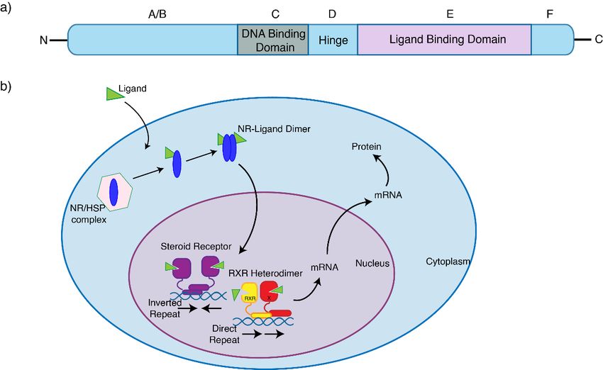
NRs are ligand-activated intracellular proteins or DNA-binding transcription fac-
tors, [3–5] that play a crucial role in key aspects of human physiology and pathophysiology,
such as breast cancer. The NR family of transcription factors consists of 48 different genes,
including the closely related steroid hormone receptors. Typically, nuclear hormone re-
ceptors consist of several domains, which share a similar structure and are differentially
conserved between each receptor (Figure 1a). Typically, ligands (such as hormones, vita-
mins, or nutrients [6]) bind to their cognate NRs, which in turn regulate the transcription
of a subset of genes expressed by a cell, resulting in what is typically considered ‘classical’
hormone signalling. Some NRs, however, can remain constitutively active. To enable
these pathways and provide an added layer of regulation, NRs recruit protein coregulators
into transcriptional complexes that bind to the DNA. These are either coactivators, which
enhance transcription, or corepressors, which repress transcription [4]. These coregulators
are essential to the underlying mechanism of NR action by mediating their transcriptional
potency [7] and therefore modulating gene expression [8].
NRs are classically divided into two subsets based on their subcellular location and
function [3]. Type 1 nuclear receptors include the sex steroid hormone receptors AR, ER
and PR, as well as the corticosteroids GR and the mineralocorticoid receptor (MR). Broadly,
sex steroids control the development of the urogenital tracts, secondary sexual characteris-
tics and behaviour, as well as gametogenesis in both males and females. Corticosteroids
control our physiological, developmental and behavioural responses to stress, as well as
play a critical role in regulating salt and water balance. Unliganded, Type 1 NRs share a
similar structure [6,9]. With classical direct genomic signalling, Type 1 NRs are located
in the cytoplasm, but translocate to the nucleus upon hormone binding as homodimers,
where they associate with chromatin and regulate gene transcription (Figure 1b). How-
ever, particularly with ER signalling, we now recognize that indirect regulation of gene
expression can also occur via genomic, nongenomic and ligand-independent pathways,
comprehensively reviewed by Fuentes et al. [10] and Siersbaek et al. [11]. These insights
into the interplay between intracellular kinases, transcription and growth factors, mem-
brane receptors, coregulators and both natural and synthetic ligands with NR signalling
have helped in the design of practice-changing therapeutic strategies. In contrast, Type 2
NRs, which include the thyroid hormone, vitamin D and retinoic acid receptors, reside in
the nucleus constitutively bound to DNA as RXR heterodimers, and typically act to repress
transcription in the absence of ligands [3,12].Cancers 2021, 13, x FOR PEER REVIEW 3 of 19
Cancers 2021, 13, 4972
While this review focuses predominantly on recent preclinical insights into Type 13 NRsof 19
with translational potential in ER+ breast cancer, this is a rapidly evolving space in molecular
biology and the clinic that is becoming increasingly relevant across all breast cancer subtypes.
Figure 1.
Figure 1. Nuclear
Nuclear receptors
receptors share
share the
the similar
similar structural
structural domains.
domains. (a)
(a) Most
Most NRs
NRs contain
contain an
an N-terminal
N-terminal region
region (A/B),
(A/B), aa
conserved DNA-binding domain (C), a variable hinge (D), a conserved ligand-binding domain (E), and a variable
conserved DNA-binding domain (C), a variable hinge (D), a conserved ligand-binding domain (E), and a variable C-
C-ter-
minal region (F). (b) The NRs involved in breast cancer are bound to heat-shock proteins (HSP) in the cytoplasm, and are
terminal region (F). (b) The NRs involved in breast cancer are bound to heat-shock proteins (HSP) in the cytoplasm, and
activated by binding of ligands, whereby canonical signalling is initiated. This NR–ligand complex will either form a
are activated by binding of ligands, whereby canonical signalling is initiated. This NR–ligand complex will either form a
heterodimer (steroid receptors), or form an RXR heterodimer, and translocate to the nucleus, binding to target genes and
heterodimer (steroid receptors), or form an RXR heterodimer, and translocate to the nucleus, binding to target genes and
initiating transcription.
initiating transcription.
2. Nuclear Receptor Autoregulation and Crosstalk
While this review focuses predominantly on recent preclinical insights into Type 1
NRs NR withautoregulation describesin
translational potential classical singlecancer,
ER+ breast nuclear receptor
this activity,
is a rapidly or the standard
evolving space in
regulation of expression of a NR gene by its hormone-bound
molecular biology and the clinic that is becoming increasingly relevant across protein product [3].
allThe au-
breast
toregulation
cancer subtypes. of NR genes can lead to induction (upregulation) or repression (downregu-
lation). Autoinduction leads to the cellular biosynthesis of more NRs and enhanced hor-
mone responsiveness,
2. Nuclear while autorepression
Receptor Autoregulation is a homeostatic mechanism that modulates
and Crosstalk
hormonal signalling by downregulating the hormone
NR autoregulation describes classical single nuclear receptor.
receptor Mechanisms for NR
activity, or the auto-
standard
regulation can be broadly transcriptional or posttranscriptional.
regulation of expression of a NR gene by its hormone-bound protein product [3]. The
In additionoftoNR
autoregulation ligands
genesautoregulating the expression
can lead to induction of their own
(upregulation) receptor, another
or repression (down-
important means for modulating cellular responsiveness to different
regulation). Autoinduction leads to the cellular biosynthesis of more NRs and enhanced hormonal signals is
through
hormonethe cross-regulation
responsiveness, of expression
while of other
autorepression is aNRs. This ‘crosstalk’
homeostatic mechanismbetweenthatnuclear
modu-
receptors can besignalling
lates hormonal defined as bythe interplay between
downregulating different receptor.
the hormone nuclear receptors
Mechanisms or even be-
for NR
tween their overlapping
autoregulation signalling
can be broadly pathways [6].
transcriptional DNA crosstalk mechanisms can be clas-
or posttranscriptional.
sifiedInasaddition
either indirect (where
to ligands NRs do not physically
autoregulating the expressioninteract) or direct
of their own(where
receptor,NRs phys-
another
ically interact to jointly regulate specific target gene subsets). Broadly,
important means for modulating cellular responsiveness to different hormonal signals when these NR-is
induced
through the signalling pathways
cross-regulation of either indirectly
expression of other orNRs.
directly
Thisinterplay,
‘crosstalk’this creates
between unique
nuclear re-
signalling
ceptors canand gene expression
be defined profiles.
as the interplay An understanding
between different nuclear of the different
receptors NR crosstalk
or even between
mechanisms
their overlappingprovides us with
signalling novel opportunities
pathways [6]. DNA crosstalk to therapeutically
mechanisms targetcan beoncogenic
classified
pathways. For deeper
as either indirect (wheremechanistic
NRs do not insights into co-regulators,
physically nuclear
interact) or direct receptor
(where signalling
NRs physically
interact
and to jointly
crosstalk, we regulate
direct thespecific
readertarget gene
to other subsets). Broadly,
comprehensive when
reviews these
from NR-induced
Bagamasbad et
signalling pathways either indirectly or directly interplay, this creates unique signalling
al. [3], Conzen et al. [5], De Bosscher et al. [6], O’Malley et al. [7], Sikora et al. [8], and Doan and
gene
et expression profiles. An understanding of the different NR crosstalk mechanisms pro-
al. [13].
vides us with novel opportunities to therapeutically target oncogenic pathways. For deeper
mechanistic insights into co-regulators, nuclear receptor signalling and crosstalk, we direct
the reader to other comprehensive reviews from Bagamasbad et al. [3], Conzen et al. [5],
De Bosscher et al. [6], O’Malley et al. [7], Sikora et al. [8], and Doan et al. [13].Cancers 2021, 13, 4972 4 of 19
Biological insights into the underlying mechanisms of this complex NR interplay, both
at the level of the cistrome and the interactome, have been gained from techniques such as
Chromatin Immunoprecipitation coupled with high-throughput Sequencing (ChIP-seq)
and Rapid Immunoprecipitation Mass spectrometry of Endogenous protein (RIME) [14–18].
How these insights impact the targeting of NRs as a therapeutic pathway in breast cancer,
and highlight potential combinatorial strategies, are addressed in this review.
3. Oestrogen Receptor Signalling
The action of oestrogen is mediated by two ERs encoded on different chromosomes
but sharing sequence homology, the Estrogen Receptor alpha (ERα, NR3A1) and Estrogen
Receptor beta (ERβ, NR3A2) nuclear hormone receptors [19]. Meanwhile, the role of ERβ
in breast cancer remains unclear and is not specifically assessed by immunohistochemistry
in the clinical setting [20]. ERα has been intensely studied due to its integral role in
breast tumorigenesis, where it can initiate gene expression changes that promote cell
cycle progression [21]. In modern-day epidemiological studies, exposure to oestrogen
(and progestogens) has been consistently linked to an increase in breast cancer risk [22],
particularly ER+ breast cancer [23]. Around 75% of breast cancers are defined and driven
by ERα transcriptional activity and for decades, anti-oestrogen therapies targeting ER
have formed the cornerstone of therapy for the management of ER+ breast cancer. ERα
inhibition is achieved either by the direct blockade of ERα activation through competitive
inhibition of oestradiol (selective ER modulators, SERMS, i.e. tamoxifen), degrading ER
(selective ER degraders, SERDs, i.e. fulvestrant) or by preventing peripheral oestrogen
synthesis using aromatase inhibitors (AIs). However, clinical outcomes vary considerably,
and a proportion of women with early breast cancer driven by ERα transcriptional activity
develop drug resistance and relapse with incurable, metastatic disease.
More recently, the targeting of cell cycle progression with cyclin-dependant kinase 4/6
(CDK4/6) inhibitors in combination with anti-oestrogen therapy has become the standard
first-line therapy in de novo or recurrent breast cancer [24,25]. While this approach has pro-
longed the progression-free and overall survival of patients with metastatic disease [26–29],
when patients develop resistance to this treatment combination, there are currently no
equally robust therapeutic strategies in the second line [30]. Second-line treatment options
include fulvestrant, cytotoxic chemotherapy, or anti-oestrogen therapy combined with
inhibitors of the mammalian target of rapamycin (mTORi). Additionally, growing evidence
supports the use of next-generation oral SERDs (Table 2) and combinatorial strategies
with alpha-selective phosphoinositide 3-kinase inhibitors (PI3Ki) in patients harbouring
somatic, activating PIK3CA mutations [31,32]. The optimal sequencing of this repertoire
of therapeutic strategies, however, remains the subject of ongoing clinical trials such as
the SONIA study (ClinicalTrials.gov identifier NCT03425838), and the growing use of
commercially available tissue and liquid biopsy-based companion diagnostic panels is
leading to a surge in biomarker-driven treatment strategies post-CDK4/6-inhibitor therapy
in ER+ metastatic breast cancer.
As a member of the nuclear receptor superfamily of transcription factors, ERα is
composed of functional domains and structural regions in common with other nuclear
receptors (Figure 1a). The A/B region represents the N-terminal domain, involved in gene
transcription transactivation, and containing a zinc finger that mediates binding to target
sequences [10]. Significant progress has been made in understanding how wild-type ERα
interacts with DNA. Using genomic technologies in breast cancer cell lines, it has been
shown that ERα is able to bind to specific DNA sequences known as estrogen response
elements (EREs, with a consensus motif GGTCAnnnTGACC) within the chromatin [33,34].
The C region of ERα is the DNA binding domain which contributes to ER dimerization
and binds to these canonical EREs. The D region is the hinge region that binds chaperone
proteins and allows for receptor–ligand complexes to translocate to the nucleus, and
the E/F region is the ligand binding domain, which binds oestrogen, coactivators and
corepressors (Figure 1a). Additional regulators of ER transcriptional activity known asCancers 2021, 13, 4972 5 of 19
activation function domains AF1 and AF2 are located in the N-terminal and DNA binding
domains, respectively [35].
Table 2. Clinical trials in progress that are investigating nuclear receptor-directed therapies in breast cancer.
NR Target Treatment Combination Stage ClinicalTrials.gov
Class Treatment Phase BC Subtype Identifier
N/A II Neoadjuvant NCT04436744
Giredestrant N/A II ER+, HER2- NCT04576455
Palbociclib III Advanced NCT04546009
Oral SERD Abemaciclib,
Trastuzumab,
LY3484356 Alpelisib, I Advanced ER+, HER2- NCT04188548
ER
Everolimus
Rintodestrant Palbociclib I Advanced ER+, HER2- NCT03455270
ZB716 Palbociclib I, II Advanced ER+, HER2- NCT04669587
Camizestrant Palbociclib III Advanced ER+, HER2- NCT04711252
N/A I NCT04568902
H3B-6545 SERCA Palbociclib I Advanced ER+, HER2- NCT04288089
N/A I, II NCT03250676
Megestrol Letrozole II Early, Window ER+, HER2- NCT03306472
PR acetate PR Agonist
Prometrium Letrozole II Early, Window ER+, PR+, HER2- NCT03906669
Enobosarm SARM N/A III Advanced ER+, AR+, HER2- NCT04869943
Severitonel-D Docetaxel I, II Advanced AR+, TNBC NCT04947189
AR AR
Enzalutamide Antagonist N/A II Advanced AR+, TNBC NCT01889238
Darolutamide Capecitabine II Advanced AR+, TNBC NCT03383679
Nab-Paclitaxel II NCT02788981
Mifepristone GR Advanced GR+, TNBC
GR Antagonist Pembrolizumab II NCT03225547
N/A II Prevention BRCA1/2mut TNBC NCT01898312
VDR Vitamin D3 VDR Agonist N/A II Advanced TNBC NCT04677816
NR: nuclear receptor; BC: breast cancer; ER: estrogen receptor; PR: progesterone receptor; AR: androgen receptor; GR: growth receptor;
VDR: vitamin D receptor; SERD: selective estrogen receptor degrader; SERCA: selective estrogen receptor covalent antagonist; SARM:
selective androgen receptor modulator; LHRH: luteinising hormone-releasing hormone; HER2: human epidermal receptor 2; BRCA1/2:
breast cancer gene 1/2; TNBC: triple-negative breast cancer.
Genomic analyses have shown that ERα rarely acts through associations with pro-
moter regions of target genes. Instead, genome-wide maps of ERα binding in breast cancer
confirm that ERα binding events mostly occur at distal cis-regulatory enhancer elements at
significant distances from the transcription start sites [33,36]. DNA-looping occurs, bring-
ing enhancers in spatial proximity to promoter regions of target genes, and transcription is
initiated [37]. It is also now clear that, in addition to oestrogens, ER function is modulated
by other steroid receptors, with the interaction between ER and both PR and AR being
the best-characterized models of nuclear receptor crosstalk in breast cancer. In addition,
multiple signalling pathways (e.g., growth factor and cytokine signalling pathways) may
also have a substantial impact on the efficacy of anti-oestrogen therapies [8,11].
It has been proposed that resistance to endocrine therapies may be the result of both
genetic and epigenetic factors [38–41]. While gain-of-function mutations in ESR1, the
gene encoding ERα, are relatively rare in primary breast cancer [42], 11–55% of metastatic
cancers have point mutations in the ligand-binding domain of ER, especially in amino acids
Y537 and D538 [42]. These mutations generate a constitutively active ER that is less depen-
dent on oestrogen for activity [43–45]. The highest prevalence of ESR1 mutations has been
reported in the cell-free DNA (cfDNA) of AI-resistant metastatic ER+ breast cancer patients
using droplet digital PCR [46–48]. At a cistromic level, not only are ESR1 mutants distinct
from oestrogen-stimulated wild-type ER, Y537S and D538G ESR mutants also have distinct
cistromes and transcriptomes [49]. These mutations cluster in the ligand-binding domain
of ER and lead to ligand-independent ER activity that promotes tumour growth, partial
resistance to endocrine therapy and enhanced metastatic capacity. However, tumours
bearing ESR1 mutations can retain relative sensitivity to SERDs. A retrospective analysis
of plasma samples from the phase III SoFEA study in patients resistant to nonsteroidal aro-
matase inhibitors confirmed significantly improved outcomes with fulvestrant-containing
regimens compared with exemestane in the 39.1% of patients found to be harbouring an
ESR1 mutation [47]. This highlights the imperative to accelerate the development andCancers 2021, 13, 4972 6 of 19
rollout of more effective, potent and orally bioavailable next-generation SERDs, which have
been shown to act through slowing intra-nuclear ER mobility, resulting in the limitation
of both chromatin accessibility and downstream proliferative activity [50]. A number of
these next-generation SERDs are currently being investigated in phase I–III clinical trials
in early- and advanced-stage breast cancer, both alone and in combination with CDK4/6
inhibitors (ClinicalTrials.gov identifiers NCT04647487, NCT03455270, NCT04669587 and
NCT04711252).
The novel selective estrogen receptor covalent antagonists (SERCAs) are another
therapeutic alternative in overcoming endocrine resistance. This class of drugs targets
the cysteine residue at amino acid 530 (C530) that exists only in ER, to promote a unique
antagonist conformation. Specifically, the SERCAs H3B-5942 and H3B-6545 have been
demonstrated to covalently bind to C530 of both wild-type and mutant ERα proteins,
and have been shown to be superior to standard-of-care therapies in in vitro and in vivo
models of endocrine resistance [51,52]. Currently, H3B-6545 is being investigated in phase
I–II clinical trials in advanced, metastatic breast cancer either alone or in combination
with CDK4/6 inhibitors (ClinicalTrials.gov identifiers NCT04568902, NCT04288089 and
NCT03250676).
Emerging data have revealed that alterations in the epigenome can result in ER-
directed therapy resistance. Aromatase inhibitor-induced DNA hypermethylation at
estrogen-responsive elements (EREs) is associated with a reduction in ER binding and
activity [41,53]. As a result, decreased gene expression of key ER regulators and reduced
ER binding cause the cell to become less dependent on oestrogen for survival, and therefore
less sensitive to ER-directed therapies [41]. Moreover, ER+ breast cancers that have re-
lapsed following endocrine therapy exhibit higher DNA methylation at enhancer loci [41].
ER-directed therapy resistance can also emerge from altered methylation patterns at pro-
moter regions of genes [54,55]. For example, hypermethylation at the PTEN, PIXT2 and
HOXC10 promoters have been found to be predictive biomarkers of resistance in both cell
lines and tumour tissues [55–57]. While demethylation therapies such as decitabine have
been demonstrated to be efficacious in reversing hypermethylation in preclinical models,
they have not yet been translated into clinical use [58].
Post-translational histone modifications have also been shown to induce chromatin
remodelling that favour the repression of ER and promotion of signalling pathways associ-
ated with endocrine resistance. Histone variants have been linked to oestrogen signalling
and endocrine resistance. For example, overexpression of the H2A variant H2A.Z has
been linked to oestrogen-independent proliferation, and thus ER-targeted therapy resis-
tance [59]. Another study demonstrated that the H2B variant HIST1H2BE is overexpressed
in both resistant cell lines and tumours treated with aromatase inhibitors derived from
patients [60]. Finally, variations in histones by histone deacetylases (HDACs) have also
been associated with the loss of ER expression, also conferring endocrine resistance. In
early-phase clinical trials, combinations of an aromatase inhibitor or tamoxifen with HDAC
inhibitors such as vorinostat or entinostat demonstrated improvements in overall survival,
with the potential to re-sensitize tumours to endocrine therapy in women with resistant
disease [61,62]. Disappointingly, results from the positive phase II ENCORE301 study were
not replicated in the phase III E2112 study of entinostat plus exemestane, which failed
to demonstrate improved survival compared to exemestane alone in aromatase inhibitor-
resistant patients with advanced disease [63]. Unlike ENCORE301, many patients in the
E2112 study had received prior fulvestrant and/or CDK4/6-inhibitors, likely impacting the
final outcome, while simultaneously strengthening the trial’s relevance in a more ‘current’
therapeutic context where CDK4/6-inhibitors are now the standard first-line treatment.
We are awaiting results from exploratory analyses to identify a predictive biomarker of
response to this class of therapy.Cancers 2021, 13, 4972 7 of 19
4. Progesterone Receptor Signalling
In combination with oestrogen, progesterone plays a major role in normal breast
development, as well as changes in the mammary gland during the menstrual cycle,
pregnancy and lactation [64]. Mouse mammary studies have revealed that PR (NR3C3) is
essential in the mammary epithelium for ductal side-branching and alveologenesis [65].
In the adult mouse, 17β-oestradiol induces the expression of PR, and stimulation with
progesterone (e.g., during the luteal phase of the menstrual cycle) triggers cell proliferation.
Progestogens, which are any natural or artificial substance that exerts progesterone-like
activity via activation of PR [66], have long been employed as contraceptive agents, or
components of Menopausal Hormonal Therapy (MHT) in combination with oestrogen as
they prevent hyperplastic or malignant consequences of chronic, unopposed oestrogen
exposure on the endometrium [67].
PR is both a member of the NR family and an ERα target gene, co-expressed in over
two-thirds of ER+ breast cancers [68]. PR exists in two isoforms, PR-A and PR-B [69].
Historically, the accepted explanation for PR activity in ER+ breast cancer cells was that
PR expression was a passive consequence of a functional oestrogen receptor. PR was
therefore established as a biomarker of ERα functionality in breast cancer and a predictive
marker of response to ERα-directed agents [70]. Therefore, functional studies into the role
of progesterone and its receptor have lagged significantly behind those of ERα and have
in fact been the subject of heated debate. Epidemiological studies and clinical MHT trials
have implicated that synthetic progestins were associated with an increased risk of breast
cancer [22,23,71,72]. Additionally, progesterone-initiated PR signalling has been shown
to contribute to mammary tumourigenesis in murine models [73]. On the other hand,
multiple studies have demonstrated the improved prognosis of PR+ breast cancers [74–79],
and a Combined Endocrine Receptor (CER) score averaging the Allred score of both ER and
PR has been demonstrated to be a more powerful discriminator of patient outcome than
either ER or PR alone [80]. In support of this, the heterozygous or homozygous deletion of
PR occurs more often in the luminal B breast cancer subtype that is associated with a higher
proliferation rate and poorer prognosis compared with luminal A breast cancers [81].
Importantly, the role of PR in breast cancer is context-dependent and highlights the
importance of the hormonal milieu. Pivotal studies have emphasised that the function of
PR in breast cancer has to be considered in the context of the presence of oestrogen and
ERα signalling [15,82]. In the absence of a functional oestrogen-activated ER complex, PR
activation might have modest pro-proliferative effects, and differing effects in malignant
and normal breast tissue contingent on oestrogenic status. This emphasizes the impor-
tance of further delineating the precise mechanisms through which PR regulates tumours
compared with mammary gland proliferation.
In breast cancer cell lines, the administration of progestogens has been shown to
inhibit ERα transcriptional activity and oestrogen-induced cell proliferation [83–85]. Addi-
tionally, progestogens alone oppose the oestrogen-induced proliferation of MCF7 and T47D
cell line and ER+/PR+ patient-derived xenografts [15,86]. Interestingly, the combination
of tamoxifen and progesterone had an even greater suppressive effect on tumour growth.
Clinically, benefits have been demonstrated from a single injection of progesterone adminis-
tered before surgery [87], and the use of a single agent progestogen has consistently shown
to be clinically beneficial either as a first-line therapy in de novo metastatic ER+ breast
cancer, or in advanced disease when ER-directed endocrine agents have failed [88–100].
Taken together, existing data imply that PR can play an anti-proliferative role in
ER+ breast cancer. Mechanistically, the stimulation of PR by progestins regulates dis-
tinct cistromes and transcriptomes in breast cancer cells compared to normal breast
cells [101,102], and can function as a molecular rheostat to control ERα binding and tran-
scriptional activity. In the presence of agonist ligands, insights gained from ChIP-seq
experiments have confirmed that PR causes the rapid redistribution or sequestration of
ERα away from its pro-proliferative gene targets in breast cancer cells, resulting in a unique
gene expression program that is associated with a good clinical outcome and culminatingCancers 2021, 13, 4972 8 of 19
in cell cycle arrest [15,103]. The potential clinical significance of exploiting this interaction
between ERα and PR signalling in breast cancer affords the possibility that the addition of
a progesterone agonist might enhance the anti-proliferative effect of anti-oestrogen thera-
pies and therefore provide a more effective combination therapy. Indeed, two short-term,
pre-operative window-of-opportunity studies, PIONEER and WinPRO (ClinicalTrials.gov
identifiers NCT03306472 and NCT03906669), are currently testing this hypothesis clinically
in early-stage breast cancer.
Paradoxically, PR antagonists have also proven to be antiproliferative in cell line and
murine models of ER+ breast cancer [104–111]. It has indeed been hypothesized that PR
antagonists may also interfere with ER transcriptional activity, similarly to agonists [66].
However, clinical trials of agents such as mifepristone and onapristone have recruited
poorly, have shown either a lack of reproducible efficacy or unacceptable hepatotoxic-
ity [112–114], and have therefore not progressed to routine clinical use.
5. Androgen Receptor Signalling
AR (NR3C4) is essential for the development of male reproductive organs, and it
is also expressed in the majority of breast cancers. Two isoforms and several alternative
splicing variants, encoded by the same gene, have been described. While AR is currently
not routinely assessed immunohistochemically in biopsies and surgical excision specimens
from our breast cancer patients, it is in fact the most widely expressed hormone receptor in
all stages of breast cancer [115,116]. AR expression varies between breast cancer subtypes,
and the function of AR in breast cancer is highly context-dependant, contingent on the
co-expression of ER, the AR:ER protein ratio, the menopausal status of the patient and the
hormonal milieu [8].
AR is expressed in up to 85% of ER+ breast cancer and is an independent clinico-
pathological prognostic factor associated with favourable outcomes in this setting [117,118].
AR and ER co-localize at select genomic loci within the nuclei of breast cancer cells [119],
and functional crosstalk between the hormone receptors has been well-described [120].
Controversially, both AR agonists and antagonists have been shown to inhibit growth
in ER+ preclinical models, by inhibiting ER function at a genomic level [119,121–124].
However, the bulk of positive evidence supports an anti-proliferative role of androgens in
ER+ breast cancers, with androgenic signalling via AR generally antagonistic of oestrogen
activity [116,119,125]. Indeed, the historic use of androgens as a treatment in breast cancer
clinically supports this [126,127]. Major reasons that have limited its clinical utility include
virilising side effects, concerns regarding the aromatisation of androgens to oestrogen and
the development of effective ER-directed approaches.
More recently, there has been a resurgence of interest in the role of the AR signalling
axis in ER+ breast cancer. The activation of AR by its natural ligand DHT in endocrine-
sensitive ER+ cell lines in vitro inhibited proliferation and ER signalling [119,128], consis-
tent with aforementioned clinical studies, supporting the premise that androgens inhibit
proliferation and induce tumour regression. AR agonism has also been shown to retain its
anti-ER signalling and growth inhibitory effects in endocrine therapy-naïve and -resistant
in vivo patient-derived xenograft (PDX) models [129], including those harbouring genomic
aberrations of ESR1 and CCND1 [16]. The anticancer effect was a result of an upregulation
of AR target genes, including tumour suppressors, the reprogramming of the binding of ER
and its co-activators on chromatin, and the redistribution of E2-stimulated p300 binding
sites, resulting in inhibition of the expression of critical ER-regulated cell cycle and survival
genes [16].
Therapeutically, the development of selective androgen receptor modulators (SARMs),
such as enobosarm and RAD140, now offers a novel approach for targeting AR in breast
cancer [16,130]. SARMs exhibit a high specificity of binding to AR and have the advantage
that they dissociate the anabolic from androgenic effects of AR and therefore lack the
virilising effects seen with the historical use of androgens [131]. A phase II study has
demonstrated that enobosarm was well tolerated and conferred clinical benefit in heavilyCancers 2021, 13, 4972 9 of 19
pre-treated patients with ER+/AR+ metastatic breast cancer [132]. Enobosarm in com-
bination with CDK4/6 inhibitors has also been shown to be efficacious in the setting of
endocrine or CDK4/6 inhibitor-resistant breast cancer models (which retain the expression
of AR), suggesting that AR agonism may partially restore sensitivity to CDK4/6 inhibitors,
thus positing that this combination may be an effective therapeutic strategy in the second-
line treatment of ER+ metastatic breast cancer [16]. Together, these findings provided the
motivation behind the phase III registration ARTEST study (ClinicalTrials.gov identifier
NCT04869943) of enobosarm in endocrine therapy and CDK4/6 inhibitor-resistant ER+
metastatic breast cancer patients [133].
A transdermal preparation of 4-OH-testosterone, CR1447, has also demonstrated
efficacy and a favourable toxicity profile in phase I/II studies [134]. Another alternative
AR agonist includes oral testosterone undecanoate, which utilises a self-emulsifying drug
delivery system to minimise pharmacokinetic variability experienced with other formu-
lations [135], maximising free testosterone levels and AR interaction while potentially
reducing the risk of androgenisation by more predictably maintaining testosterone levels
in the eugonadal range [136]. Window-of-opportunity studies using this compound are
also currently under development.
In contrast to ER+ breast cancer, high AR expression is associated with a poor
prognosis in triple-negative breast cancers (TNBC) [137]. AR is expressed in 15–35%
of TNBCs [138], primarily in the luminal androgen receptor (LAR) subtype. These tumours
demonstrate a gene expression signature resembling that of endocrine-responsive tu-
mours [139], are characterised by both AR expression and androgen-dependant growth [140],
and are classically less responsive to conventional cytotoxic chemotherapy. AR-antagonism
in this setting, with antiandrogens such as bicalutamide, have been investigated in clinical
trials [141]. The second-generation AR antagonist enzalutamide has been shown to be
well-tolerated in combination with exemestane, with clinical efficacy in this subset of
TNBC that expresses AR [142], as well as in a cohort of ER+ patients with high levels
of AR mRNA and low levels of ER mRNA on mRNA-sequencing [143]. This provides
a rationale to explore combinatorial strategies of AR antagonists with agents such as
CDK4/6i [144,145] and PI3K inhibitors [146]. Growing evidence also supports the utility
of other next-generation AR-targeted agents such as abiraterone acetate with prednisone in
AR+ TNBC, as well as the novel CYP17 lyase inhibitor and potent AR antagonist seviteronel
(VT-464/INO-464), which is about to enter clinical trials in combination with chemotherapy
in the phase I/II 4CAST study (ClinicalTrials.gov identifier NCT04947189). Furthermore,
seviteronel has been shown to induce DNA damage following radiation in AR+ TNBC
models, demonstrating a unique radiosensitizing effect [147].
6. Glucocorticoid Receptor Signalling
As with AR, GR (NR3C1) expression is not routinely evaluated in breast carcinomas.
GR is expressed in ~70% of ER+ breast cancers and >60% of all breast cancers [148]. Clini-
cally, synthetic glucocorticoids (GR agonists) such as dexamethasone are ubiquitously used
in breast cancer in conjunction with chemotherapy to mitigate hypersensitivity reactions,
and for their antiemetic, anti-inflammatory, as well as orexigenic properties. However,
dexamethasone triggers different effects depending on the breast cancer subtype [149].
Similarly to the trend observed with AR expression, retrospective meta-analyses of
patients with ER+ breast cancer have determined that high GR mRNA levels are associated
with low tumour grade [148] and better prognosis [150,151] compared to low or negligible
GR expression, independent of PR expression. Again, the opposite was observed in ER-
breast cancer where high tumour GR expression was associated with a worse progno-
sis [150]. Therefore, mirroring AR, the tumour suppressor vs. oncogenic potential of
GR is dependent upon ER expression. Mechanistically in ER+ disease, both ER and GR
undergo crosstalk upon co-treatment with oestradiol and dexamethasone [151,152], with
reciprocal reprogramming of both receptors occurring via the Hager lab’s well-described
assisted loading mechanism [152,153]. This model involves chromatin remodelling by oneCancers 2021, 13, 4972 10 of 19
receptor followed by recruitment of the other receptor. Upon co-stimulation of ER and
GR in MCF7 cells with oestradiol and glucocorticoids, transcriptional changes in genes
linked to cell proliferation and differentiation occur [151], inhibiting growth. Despite these
preclinical findings, clinical studies in ER+ breast cancer patients have demonstrated varied
effects of glucocorticoid use on breast cancer patient survival, with modest effects when
used as a single agent and no additive effect in combination with other drugs, including
anti-oestrogens [154,155]. However, given the widespread use of glucocorticoids in cancer,
further exploration of the full therapeutic potential of activating GR in ER+ breast cancer
warrants further investigation.
By contrast, GR activation with glucocorticoid has been shown to inhibit chemotherapy-
induced cell apoptosis [156], and drives the expression of pro-tumorigenic genes in
TNBC [157,158]. This raises the concern that the routine administration of high doses
of synthetic glucocorticoids as a chemotherapy premedication has the potential to activate
GR-mediated cell survival pathways and diminish the effectiveness of chemotherapy in
this setting. More recently, an extensive study using both patient-derived and TNBC cell
line-derived xenograft models also suggested that activation of GR with glucocorticoid
treatment increases tumour heterogeneity and promotes breast cancer metastasis [159].
The suggested mechanism was the upregulation of the expression of a receptor tyrosine
kinase-like orphan receptor-1 (ROR-1). With GR identified as a target in this setting, pre-
clinical findings of the potentiated cytotoxic efficacy of paclitaxel chemotherapy with the
addition of the GR antagonist Mifepristone [156] has now led to the translation of these
findings to the clinical research setting. Consistent with these observations, a randomised
phase I clinical trial combined mifepristone with nab-paclitaxel, confirming disease activity
and manageable toxicity [160], and a randomised phase II trial is currently recruiting
(NCT02788981).
7. Other Nuclear Receptors
The mineralocorticoid receptor (MR, NR3C2) is another receptor of interest, given that
it is most likely expressed in the majority of breast cancers (up to 90%) [161]. Aldosterone,
the primary ligand for MR, is typically used in the management of hypertension and
cardiac failure. As with GR, MR can undergo crosstalk with PR to induce significant
growth inhibition, and many glucocorticoids also bind to MR with high affinity [162]. An
interaction between ER and MR has not been directly explored in breast cancer models;
however, high MR and retinoic acid receptor (RAR) expression is associated with improved
ER+ breast cancer-specific survival. A tumour-suppressive relationship between these
nuclear receptors was illustrated by co-treatment with mineralocorticoids and retinoic acid
receptor-stimulating retinoids [163], highlighting another potential therapeutic pathway
worth characterising in the setting of breast cancer resistant to standard ER-targeted
therapies. Independently, RAR expression can predict for resistance to tamoxifen [164].
While the results of combinatorial therapeutic strategies with retinoids in patients with
breast cancer have been generally disappointing [165], retinoids have been shown to inhibit
the expansion of chemoresistant cytokeratin 5-positive (CK5+) cells through RAR/PR
crosstalk [166].
The relationship between Vitamin D and breast cancer remains controversial. A re-
cent meta-analysis demonstrated that while there is no relationship between nuclear VDR
(NR1I1) expression and overall survival in patients with breast cancer, high total nuclear
and cytoplasmic VDR expression was associated with improved survival outcomes [167].
Both the VDR and vitamin D 1-hydroxylase, the enzyme that generates the active Vitamin
D3 ligand 1,25-dihydroxycholecalciferol, are expressed in the human breast. Addition-
ally, VDR expression correlates with the expression of ER. Interest has been provoked by
growing evidence suggesting that adequate Vitamin D levels and intake inversely correlate
with breast cancer risk [168–170], and that low levels of vitamin D are associated with an
increased risk of recurrence or death in breast cancer patients [167,171]. This is arguably
enough evidence to suggest that early breast cancer patients should at least supplementCancers 2021, 13, 4972 11 of 19
vitamin D if found to be deficient. 1,25-dihydroxycholecalciferol inhibits the proliferation
of breast cancer cell lines and promotes differentiation and apoptosis in vitro [172–174], but
clinical data regarding tumour responsiveness to vitamin D are limited and remain incon-
clusive [174,175]. The impact of vitamin D supplementation in the neoadjuvant setting on
the rate of pathological complete response is being investigated in TNBC (ClinicalTrials.gov
identifier NCT04677816).
8. Conclusions
Given that the prominent nuclear receptors in breast cancer all share similar consensus
DNA binding motifs, as well as mechanisms of co-activation, it is not surprising that there
is substantial functional crosstalk between these receptors. Additionally, given that most
nuclear receptors are regulated by ligands and are often highly co-expressed, this renders
them susceptible to external control over their gene regulatory activities [6], and therefore
ideal druggable targets.
In ER+ breast cancer, steroid hormone receptors are certainly not bystanders in ER
signalling pathways. Modulation of their activity can alter or reprogram ER DNA binding
to dramatically modify target gene expression. Such a modulation of hormone receptor
function in combination with anti-oestrogen therapy can either modify the response or alter
the trajectory of developing resistance to therapy, and certainly highlights the imperative to
improve our understanding of how other steroid hormone receptors influence ER function
in the context of standard anti-oestrogen therapy. However, the fact that AR and GR
have opposite functions in the presence and absence of ER highlights the importance
of characterising the functional interplay between different steroid hormone receptor
signalling pathways in both luminal and nonluminal breast cancer subtypes, to fully
exploit their therapeutic potential. Overwhelmingly, the modulation of nuclear receptor
activity beyond targeting the ER provides us with novel approaches to manage patients
with breast cancer.
Given the crosstalk of pathways triggered between different nuclear receptors, it
may be possible that in the near future, breast cancer therapeutic decisions may require
consideration of the expression of all four major steroid hormone receptors in breast cancer—
ER, PR, AR and GR. This opens the prospect of the deeper characterisation of breast cancers
for both prognostic purposes and as predictive biomarkers of response to a new array of
endocrine therapies.
Author Contributions: Conceptualisation, S.K.; data curation, S.K.; writing, S.K., A.F. and E.L.;
funding acquisition, E.L. All authors have read and agreed to the published version of the manuscript.
Funding: E.L. is supported by a National Breast Cancer Foundation Endowed Chair (Grant number
NBCF EC17-02) and Love Your Sister. E.L. and S.K. are supported by the Balnaves Foundation. A.F.
is supported by the Australian Commonwealth Government Research Training Program.
Conflicts of Interest: The authors declare no conflict of interest.
References
1. Beatson, G.T. On the treatment of inoperable cases of carcinoma of the mamma. Lancet 1896, 2, 1–14.
2. Jensen, E.V.; Jacobson, H.I.; Walf, A.A.; Frye, C.A. Estrogen action: A historic perspective on the implications of considering
al-ternative approaches. Physiol. Behav. 2010, 99, 151–162. [CrossRef]
3. Bagamasbad, P.; Denver, R.J. Mechanisms and significance of nuclear receptor auto- and cross-regulation. Gen. Comp. Endocrinol.
2011, 170, 3–17. [CrossRef] [PubMed]
4. O’Malley, B.W. 90 YEARS of PROGESTERONE: Reminiscing on the origins of the field of progesterone and estrogen receptor
action. J. Mol. Endocrinol. 2020, 65, C1–C4. [CrossRef]
5. Conzen, S.D. Minireview: Nuclear Receptors and Breast Cancer. Mol. Endocrinol. 2008, 22, 2215–2228. [CrossRef] [PubMed]
6. De Bosscher, K.; Desmet, S.J.; Clarisse, D.; Estébanez-Perpiña, E.; Brunsveld, L. Nuclear receptor crosstalk—Defining the
mecha-nisms for therapeutic innovation. Nat. Rev. Endocrinol. 2020, 16, 363–377. [CrossRef] [PubMed]
7. O’Malley, B.W. Coregulators: From whence came these ‘master genes’. Mol. Endocrinol. 2007, 21, 1009–1013. [CrossRef] [PubMed]
8. Sikora, M.J. Family Matters: Collaboration and Conflict Among the Steroid Receptors Raises a Need for Group Therapy.
Endocrinology 2016, 157, 4553–4560. [CrossRef]Cancers 2021, 13, 4972 12 of 19
9. A Lamb, C.; I Vanzulli, S.; Lanari, C. Hormone receptors in breast cancer: More than estrogen receptors. Medicina 2019, 79,
540–545. [PubMed]
10. Fuentes, N.; Silveyra, P. Estrogen receptor signaling mechanisms. Adv. Protein Chem. Struct. Biol. 2019, 116, 135–170. [CrossRef]
11. Siersbæk, R.D.; Kumar, S.; Carroll, J. Signaling pathways and steroid receptors modulating estrogen receptor α function in breast
cancer. Genes Dev. 2018, 32, 1141–1154. [CrossRef] [PubMed]
12. Mangelsdorf, D.; Thummel, C.; Beato, M.; Herrlich, P.; Schütz, G.; Umesono, K.; Blumberg, B.; Kastner, P.; Mark, M.; Chambon, P.;
et al. The nuclear receptor superfamily: The second decade. Cell 1995, 83, 835–839. [CrossRef]
13. Doan, T.B.; Graham, J.D.; Clarke, C. Emerging functional roles of nuclear receptors in breast cancer. J. Mol. Endocrinol. 2017, 58,
R169–R190. [CrossRef]
14. Schmidt, D.; Wilson, M.D.; Spyrou, C.; Brown, G.D.; Odom, D.T. Europe PMC Funders Group ChIP-seq: Using high-throughput
sequencing to discover protein-DNA interactions. Methods 2014, 48, 240–248. [CrossRef]
15. Mohammed, H.; Russell, I.A.; Stark, R.; Rueda, O.M.; Hickey, T.; Tarulli, G.; Serandour, A.A.A.; Birrell, S.N.; Bruna, A.; Saadi,
A.; et al. Progesterone receptor modulates ERα action in breast cancer. Nat. Cell Biol. 2015, 523, 313–317. [CrossRef] [PubMed]
16. Hickey, T.E.; Selth, L.A.; Chia, K.M.; Laven-Law, G.; Milioli, H.H.; Roden, D.; Jindal, S.; Hui, M.; Finlay-Schultz, J.; Ebrahimie,
E.; et al. The androgen receptor is a tumor suppressor in estrogen receptor–positive breast cancer. Nat. Med. 2021, 27, 310–320.
[CrossRef] [PubMed]
17. Mohammed, H.; Taylor, C.; Brown, G.D.; Papachristou, E.; Carroll, J.; D’Santos, C.S. Rapid immunoprecipitation mass spec-
trometry of endogenous proteins (RIME) for analysis of chromatin complexes. Nat. Protoc. 2016, 11, 316–326. [CrossRef]
[PubMed]
18. Papachristou, E.K.; Kishore, K.; Holding, A.N.; Harvey, K.; Roumeliotis, T.I.; Chilamakuri, C.S.R.; Omarjee, S.; Chia, K.M.;
Swarbrick, A.; Lim, E.; et al. A quantitative mass spectrometry-based approach to monitor the dynamics of endogenous
chromatin-associated protein complexes. Nat. Commun. 2018, 9, 1–13. [CrossRef]
19. Gburcik, V.; Picard, D. The cell-specific activity of the estrogen receptor a may be fine-tuned by phosphorylation-induced
structural gymnastics. Nucl. Recept. Signal. 2006, 4, e005. [CrossRef]
20. Ström, A.; Hartman, J.; Foster, J.S.; Kietz, S.; Wimalasena, J.; Gustafsson, J. Estrogen receptor beta inhibits 17beta-estradiol-
stimulated proliferation of the breast cancer cell line T47D. Proc. Natl. Acad. Sci. USA 2004, 101, 1566–1571. [CrossRef]
21. Carroll, J.S.; Liu, X.S.; Brodsky, A.S.; Li, W.; Meyer, C.A.; Szary, A.J.; Eeckhoute, J.; Shao, W.; Hestermann, E.V.; Geistlinger,
T.R.; et al. Chromosome-wide mapping of estrogen receptor binding reveals long-range regulation requiring the fork-head
protein FoxA1. Cell 2005, 122, 33–43. [CrossRef]
22. Group, C.; Cancer, B. Type and timing of menopausal hormone therapy and breast cancer risk: Individual participant meta-
analysis of the worldwide epidemiological evidence. Lancet 2019, 394, 1159–1168.
23. Althuis, M.D.; Fergenbaum, J.H.; Garcia-Closas, M.; A Brinton, L.; Madigan, M.P.; E Sherman, M. Etiology of hormone receptor-
defined breast cancer: A systematic review of the literature. Cancer Epidemiol. Biomark. Prev. 2004, 13, 1558–1568.
24. Finn, R.S.; Martin, M.; Rugo, H.S.; Jones, S.; Im, S.-A.; Gelmon, K.; Harbeck, N.; Lipatov, O.N.; Walshe, J.M.; Moulder, S.; et al.
Palbociclib and Letrozole in Advanced Breast Cancer. N. Engl. J. Med. 2016, 375, 1925–1936. [CrossRef] [PubMed]
25. Cristofanilli, M.; Turner, N.C.; Bondarenko, I.; Ro, J.; Im, S.-A.; Masuda, N.; Colleoni, M.; DeMichele, A.; Loi, S.; Verma, S.; et al.
Fulvestrant plus palbociclib versus fulvestrant plus placebo for treatment of hormone-receptor-positive, HER2-negative metastatic
breast cancer that progressed on previous endocrine therapy (PALOMA-3): Final analysis of the multicentre, double-blind, phase
3 randomised controlled trial. Lancet Oncol. 2016, 17, 425–439. [CrossRef]
26. Turner, N.C.; Jungsil PALOMA3 Study Group; Andre, F.; Loi, S.; Verma, S.; Iwata, H.; Harbeck, N.; Loibl, S.; Bartlett, C.H.; Zhang,
K.; et al. Palbociclib in Hormone-Receptor–Positive Advanced Breast Cancer. N. Engl. J. Med. 2015, 373, 209–219. [CrossRef]
[PubMed]
27. Hortobagyi, G.; Stemmer, S.; Burris, H.; Yap, Y.-S.; Sonke, G.; Paluch-Shimon, S.; Campone, M.; Petrakova, K.; Blackwell, K.;
Winer, E.; et al. Updated results from MONALEESA-2, a phase III trial of first-line ribociclib plus letrozole versus placebo plus
letrozole in hormone receptor-positive, HER2-negative advanced breast cancer. Ann. Oncol. 2018, 29, 1541–1547. [CrossRef]
[PubMed]
28. Gao, J.J.; Cheng, J.; Bloomquist, E.; Sanchez, J.; Wedam, S.B.; Singh, H.; Amiri-Kordestani, L.; Ibrahim, A.; Sridhara, R.; Goldberg,
K.B.; et al. CDK4/6 inhibitor treatment for patients with hormone receptor-positive, HER2-negative, advanced or metastatic
breast cancer: A US Food and Drug Administration pooled analysis. Lancet Oncol. 2020, 21, 250–260. [CrossRef]
29. Schettini, F.; Giudici, F.; Giuliano, M.; Cristofanilli, M.; Arpino, G.; del Mastro, L.; Puglisi, F.; de Placido, S.; Paris, I.; de Placido,
P.; et al. Overall Survival of CDK4/6-Inhibitor-Based Treatments in Clinically Relevant Subgroups of Metastatic Breast Cancer:
Systematic Review and Meta-Analysis. J. Natl. Cancer Inst. 2020, 112, 1089–1097. [CrossRef]
30. Schettini, F.; Giuliano, M.; Giudici, F.; Conte, B.; De Placido, P.; Venturini, S.; Rognoni, C.; Di Leo, A.; Locci, M.; Jerusalem, G.; et al.
Endocrine-Based Treatments in Clinically-Relevant Subgroups of Hormone Receptor-Positive/HER2-Negative Metastatic Breast
Cancer: Systematic Review and Meta-Analysis. Cancers 2021, 13, 1458. [CrossRef]
31. Rugo, H.S.; Rugo, H.S.; Lerebours, F.; Ciruelos, E.; Drullinsky, P.; Ruiz-Borrego, M.; Neven, P.; Park, Y.H.; Prat, A.; Bachelot,
T.; et al. Alpelisib plus fulvestrant in PIK3CA-mutated, hormone receptor-positive advanced breast cancer after a CDK4/6
inhibitor (BYLieve): One cohort of a phase 2, multicentre, open-label, non-comparative study. Lancet Oncol. 2021, 22, 489–498.
[CrossRef]Cancers 2021, 13, 4972 13 of 19
32. Turner, S.; Chia, S.; Kanakamedala, H.; Hsu, W.; Park, J.; Chandiwana, D.; Ridolfi, A.; Yu, C.; Zarate, J.P.; Rugo, H.S. Effectiveness
of Alpelisib + Fulvestrant Compared with Real-World Standard Treatment Among Patients with HR+, HER2-, PIK3CA-Mutated
Breast Cancer. Oncologist 2021, 26, e1133–e1142. [CrossRef]
33. Carroll, J.; A Meyer, C.; Song, J.; Li, W.; Geistlinger, T.R.; Eeckhoute, J.; Brodsky, A.S.; Keeton, E.K.; Fertuck, K.C.; Hall, G.F.; et al.
Genome-wide analysis of estrogen receptor binding sites. Nat. Genet. 2006, 38, 1289–1297. [CrossRef]
34. Klinge, C.M. Estrogen receptor interaction with estrogen response elements. Nucleic Acids Res. 2001, 29, 2905–2919. [CrossRef]
35. Kumar, R.; Zakharov, M.N.; Khan, S.H.; Miki, R.; Jang, H.; Toraldo, G.; Singh, R.; Bhasin, S.; Jasuja, R. The Dynamic Structure of
the Estrogen Receptor. J. Amino Acids 2011, 2011, 1–7. [CrossRef] [PubMed]
36. Ross-Innes, C.S.; Stark, R.; Teschendorff, A.E.; Holmes, K.A.; Ali, H.R.; Dunning, M.; Brown, G.D.; Gojis, O.; Ellis, I.; Green,
A.; et al. Differential oestrogen receptor binding is associated with clinical outcome in breast cancer. Nature 2012, 481, 389–393.
[CrossRef] [PubMed]
37. Fullwood, M.; Liu, M.H.; Pan, Y.F.; Liu, J.; Xu, H.; Bin Mohamed, Y.; Orlov, Y.; Velkov, S.; Thoreau, H.; Mei, P.H.; et al.
An oestrogen-receptor-α-bound human chromatin interactome. Nature 2009, 462, 58–64. [CrossRef]
38. Ellis, M.J.; Ding, L.; Shen, D.; Luo, J.; Suman, V.J.; Wallis, J.W.; Van Tine, B.A.; Hoog, J.; Goiffon, R.; Goldstein, T.C.; et al.
Whole-genome analysis informs breast cancer response to aromatase inhibition. Nature 2012, 486, 353–360. [CrossRef]
39. Fuqua, S.A.W.; Gu, G.; Rechoum, Y. Estrogen receptor (ER) α mutations in breast cancer: Hidden in plain sight. Breast Cancer
Res. Treat. 2014, 144, 11–19. [CrossRef]
40. Jeselsohn, R.; Buchwalter, G.; DE Angelis, C.; Brown, M.; Schiff, R. ESR1 mutations—A mechanism for acquired endocrine
resistance in breast cancer. Nat. Rev. Clin. Oncol. 2015, 12, 573–583. [CrossRef] [PubMed]
41. Stone, A.; Zotenko, E.; Locke, W.; Korbie, D.; Millar, E.; Pidsley, R.; Stirzaker, C.; Graham, P.; Trau, M.; Musgrove, E.A.; et al.
DNA methylation of oestrogen-regulated enhancers defines endocrine sensitivity in breast cancer. Nat. Commun. 2015, 6, 7758.
[CrossRef]
42. Atlas, T.C.G. Comprehensive molecular portraits of human breast tumours. Nature 2012, 490, 61–70.
43. Merenbakh-Lamin, K.; Ben-Baruch, N.; Yeheskel, A.; Dvir, A.; Soussan-Gutman, L.; Jeselsohn, R.; Yelensky, R.; Brown, M.; Miller,
V.A.; Sarid, D.; et al. D538G Mutation in Estrogen Receptor-α: A Novel Mechanism for Acquired Endocrine Resistance in Breast
Cancer. Cancer Res. 2013, 73, 6856–6864. [CrossRef] [PubMed]
44. Robinson, D.R.; Wu, Y.-M.; Vats, P.; Su, F.; Lonigro, R.J.; Cao, X.; Kalyana-Sundaram, S.; Wang, R.; Ning, Y.; Hodges, L.; et al.
Activating ESR1 mutations in hormone-resistant metastatic breast cancer. Nat. Genet. 2013, 45, 1446–1451. [CrossRef]
45. Toy, W.; Weir, H.; Razavi, P.; Lawson, M.; Goeppert, A.U.; Mazzola, A.M.; Smith, A.; Wilson, J.; Morrow, C.; Wong, W.L.; et al.
Activating ESR1 Mutations Differentially Affect the Efficacy of ER Antagonists. Cancer Discov. 2016, 7, 277–287. [CrossRef]
[PubMed]
46. Chandarlapaty, S.; Chen, D.; He, W.; Sung, P.; Samoila, A.; You, D.; Bhatt, T.; Patel, P.; Voi, M.; Gnant, M.; et al. Prevalence of ESR1
Mutations in Cell-Free DNA and Outcomes in Metastatic Breast Cancer: A Secondary Analysis of the BOLERO-2 Clinical Trial.
JAMA Oncol. 2016, 2, 1310–1315. [CrossRef]
47. Fribbens, C.; O’Leary, B.; Kilburn, L.; Hrebien, S.; Garcia-Murillas, I.; Beaney, M.; Cristofanilli, M.; Andre, F.; Loi, S.; Loibl, S.; et al.
Plasma ESR1 Mutations and the Treatment of Estrogen Receptor–Positive Advanced Breast Cancer. J. Clin. Oncol. 2016, 34,
2961–2968. [CrossRef] [PubMed]
48. Spoerke, J.M.; Gendreau, S.; Walter, K.; Qiu, J.; Wilson, T.R.; Savage, H.; Aimi, J.; Derynck, M.K.; Chen, M.; Chan, I.T.; et al.
Heterogeneity and clinical significance of ESR1 mutations in ER-positive metastatic breast cancer patients re-ceiving fulvestrant.
Nat. Commun. 2016, 7, 1–10. [CrossRef] [PubMed]
49. Jeselsohn, R.; Bergholz, J.S.; Pun, M.; Cornwell, M.; Liu, W.; Nardone, A.; Xiao, T.; Li, W.; Qiu, X.; Buchwalter, G.; et al. Allele-
Specific Chromatin Recruitment and Therapeutic Vulnerabilities of ESR1 Activating Mutations. Cancer Cell 2018, 33, 173–186.e5.
[CrossRef] [PubMed]
50. Guan, J.; Zhou, W.; Hafner, M.; Blake, R.A.; Chalouni, C.; Chen, I.; De Bruyn, T.; Giltnane, J.M.; Hartman, S.; Heidersbach, A.; et al.
Therapeutic Ligands Antagonize Estrogen Receptor Function by Impairing Its Mobility. Cell 2019, 178, 949–963.e18. [CrossRef]
51. Puyang, X.; Furman, C.; Zheng, G.Z.; Wu, Z.J.; Banka, D.; B, K.A.; Agoulnik, S.; Bolduc, D.M.; Buonamici, S.; Caleb, B.; et al.
Discovery of Selective Estrogen Receptor Covalent Antagonists for the Treatment of ERαWT and ERαMUT Breast Cancer.
Cancer Discov. 2018, 8, 1176–1193. [CrossRef]
52. Hamilton, E.P.; Wang, J.S.; Pluard, T.; Johnston, S.; Morikawa, A.A.; Dees, C.E.; Jones, R.H.; Haley, B.; Armstrong, A.; Cohen, A.L.;
et al. Abstract PD8-06: Phase I/II trial of H3B-6545, a novel selective estrogen receptor covalent antagonist (SER-CA), in estrogen
receptor positive (ER+), human epidermal growth factor receptor 2 negative (HER2-) advanced breast cancer. Cancer Res. 2021,
81, PD8-06 LP.
53. Stone, A.; Valdes-Mora, F.; Gee, J.M.W.; Farrow, L.; McClelland, R.A.; Fiegl, H.; Dutkowski, C.; McCloy, R.; Sutherland, R.L.;
Musgrove, E.A.; et al. Tamoxifen-Induced Epigenetic Silencing of Oestrogen-Regulated Genes in Anti-Hormone Resistant Breast
Cancer. PLoS ONE 2012, 7, e40466. [CrossRef]
54. Fan, M.; Yan, P.S.; Hartman-Frey, C.; Chen, L.; Paik, H.; Oyer, S.L.; Salisbury, J.D.; Cheng, A.; Li, L.; Abbosh, P.H.; et al. Diverse
Gene Expression and DNA Methylation Profiles Correlate with Differential Adaptation of Breast Cancer Cells to the Antiestrogens
Tamoxifen and Fulvestrant. Cancer Res. 2006, 66, 11954–11966. [CrossRef]You can also read