In-Cell NMR: Analysis of Protein-Small Molecule Interactions, Metabolic Processes, and Protein Phosphorylation - MDPI
←
→
Page content transcription
If your browser does not render page correctly, please read the page content below
International Journal of
           Molecular Sciences

Review
In-Cell NMR: Analysis of Protein–Small Molecule
Interactions, Metabolic Processes,
and Protein Phosphorylation
Amit Kumar 1,2,3, *, Lars T. Kuhn 1              and Jochen Balbach 2,4, *
 1    Astbury Centre for Structural Molecular Biology, School of Molecular and Cellular Biology, University of
      Leeds, Leeds LS2 9JT, UK
 2    Institute of Physics, Biophysics, Martin–Luther–University Halle–Wittenberg, 06120 Halle, Germany
 3    Department of Diabetes, Faculty of Lifesciences and Medicine, King’s College London, Great Maze Pond,
      London SE1 1UL, UK
 4    Centre for Structure und Dynamics of Proteins (MZP), Martin–Luther–University Halle–Wittenberg,
      06120 Halle, Germany
 *    Correspondence: A.Kumar@leeds.ac.uk (A.K.); jochen.balbach@physik.uni-halle.de (J.B.);
      Tel.: +44-113-34-37036 (A.K.); +49-345-55-28550 (J.B.)

 Received: 30 November 2018; Accepted: 13 January 2019; Published: 17 January 2019

 Abstract: Nuclear magnetic resonance (NMR) spectroscopy enables the non-invasive observation of
 biochemical processes, in living cells, at comparably high spectral and temporal resolution. Preferably,
 means of increasing the detection limit of this powerful analytical method need to be applied when
 observing cellular processes under physiological conditions, due to the low sensitivity inherent to the
 technique. In this review, a brief introduction to in-cell NMR, protein–small molecule interactions,
 posttranslational phosphorylation, and hyperpolarization NMR methods, used for the study of
 metabolites in cellulo, are presented. Recent examples of method development in all three fields
 are conceptually highlighted, and an outlook into future perspectives of this emerging area of NMR
 research is given.

 Keywords: protein NMR; in-cell NMR; in-situ NMR; DNP; review

1. General Introduction to In-Cell NMR
     Most biological pathways are controlled by macromolecules. In order to study the structure and
function of biomolecules, in vitro studies are usually applied; then, the resulting data are extrapolated
to the native cellular environment. Although such approaches provide a wealth of information
surrounding the structure–function activity of biomolecules, they lack the context of a native-complex
environment [1,2]. The function of molecules in vivo may differ from that determined in vitro because
their native network of interactions within the cell is missing.
     In-cell NMR spectroscopy provides a direct readout of protein–protein and ligand–protein
interactions in the cellular environment. Therefore, this method gains added value to in vitro
based techniques such as cryo-electron microscopy, X-ray crystallography, and hydrogen–deuterium
exchange mass spectrometry, generating a wealth of information such as changes in structure and/or
dynamics between the free and bound forms. However, these in vitro methods may not fully reflect
the protein state in vivo, as the experimental conditions and protein constructs are optimized to obtain
the best resolution with each respective method. Electron microscopy provides cellular structural
features, but physiological temperatures and molecule sizes below 50 kDa are still challenging for
high-resolution studies [3]. In-cell NMR spectroscopy is an ideal technique to study (at atomic
resolution) the structural features of biomolecules, their function, and their interactions while

Int. J. Mol. Sci. 2019, 20, 378; doi:10.3390/ijms20020378                           www.mdpi.com/journal/ijms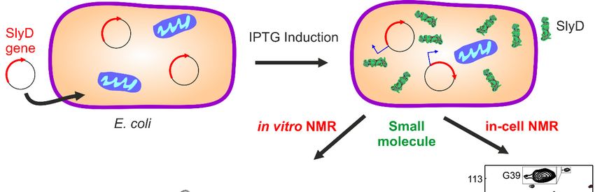
Int. J. Mol. Sci. 2019, 20, 378 2 of 23
they remain in their native cellular environment, as reviewed recently (e.g., References [1,2,4,5]).
This technique is non-invasive and provides structural and biochemical details of macromolecules
in solution, while applied in living cells over a wide range of parameters including temperature
and pH. NMR methods are ensemble methods, meaning that the outcome is sample-averaged
information Int.
originating from
J. Mol. Sci. 2018, 19, various
x FOR PEER REVIEWmolecules in many cells. Thus, the information 2 of 23 obtained
reflects the global properties of molecules without sub-cellular resolution. Historically, in vivo NMR
macromolecules in solution, while applied in living cells over a wide range of parameters including
started with temperature
studies of and small pH.molecules
NMR methods within living organisms/cells.
are ensemble methods, meaning that Onetheofoutcome
the firstis in-cell NMR
approaches tosample-averaged
obtain high-resolution information
information originating fromof various
biomacromolecules
molecules in many (e.g., proteins)
cells. Thus, the was described
information
by Serber et al. [6,7] inside obtained reflects
living the(Figure
cells global properties of molecules
1). In their study, thewithout sub-cellular
authors choseresolution.
two globular soluble
Historically, in vivo NMR started with studies of small molecules within living organisms/cells. One
proteins, the of
N-terminal
the first in-cell domain of bacterial
NMR approaches mercuric
to obtain ion reductase
high-resolution (Nmer)
information A and human calmodulin,
of biomacromolecules
to explore protein in-cellwas
(e.g., proteins) NMR. In by
described their approach,
Serber they living
et al. [6,7] inside utilized conventional
cells (Figure 1). In theirrecombinant
study, the protein
expression andauthors chose labeling
isotope two globular insoluble
bacterialproteins, thewhile
cells, N-terminal domain of other
considering bacterialparameters
mercuric ion such as cell
reductase (Nmer) A and human calmodulin, to explore protein in-cell NMR. In their approach, they
growth, induction
utilized time, cell viability,
conventional recombinantisotopic labelingand
protein expression type, and
isotope NMRin line
labeling broadening.
bacterial cells, while Expression
was optimized to obtain
considering sufficient
other parameters signal,
such asmuch aboveinduction
cell growth, that oftime,
other cellcellular
viability,proteins, to be detected by
isotopic labeling
type, and NMR line broadening. Expression was optimized to obtain sufficient signal, much above
NMR spectroscopy.
that of other cellular proteins, to be detected by NMR spectroscopy.
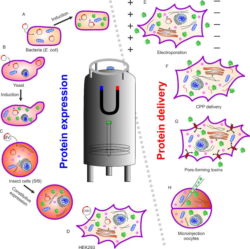
Figure 1. Schematic overview
Figure 1. Schematic of different
overview known
of different approaches
known approaches in in-cell
in in-cell NMR.
NMR. (Left) (Left) Endogenously
Endogenously
expressed and isotopically labeled protein can be achieved by transferring the expression vector
expressed and isotopically labeled protein can be achieved by transferring the expression vector
containing the gene of interest into (A) bacteria, (B) yeast, (C) insect cell lines, and (D) mammalian
containing the gene
cells. of An
(Right) interest into
alternate way(A) bacteria,
of in-cell NMR, (B) yeast,
where (C) insect
isotopically labeledcell lines,
protein and (D) mammalian
is exogenously
cells. (Right) An alternate way of in-cell NMR, where isotopically labeled protein
prepared followed by delivery into eukaryotic cells with different methods such as is
(E)exogenously
prepared followed by delivery into eukaryotic cells with different methods such as (E) electroporation,
(F) attaching protein with cell-penetrating peptides (CPP), (G) protein transport via pore-forming
toxins, and (H) microinjection-mediated delivery into Xenopus leavis oocytes.
Different types of isotope labeling of protein samples for in-cell NMR are available. Uniform
15 N labeling was found to be most useful and the first choice for most of the studies (Figure 1a,b).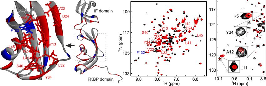
Int. J. Mol. Sci. 2019, 20, 378                                                                     3 of 23

The higher natural abundance of 13 C in biomolecules, compared to 15 N, renders this carbon isotope
as the sole modification unsuitable for in-cell NMR studies. An alternative approach to uniform 13 C
enrichment is the specific labeling of amino acids [7]. Here, methyl-13 C methionine labeling was a
successful strategy to detect side-chain carbons well above the cellular background [8]. Yet another
approach is the incorporation of non-natural amino acids containing 19 F. This approach turned out to
be a feasible means of investigating protein dynamics in the cellular environment. The advantage of
19 F-labeled protein is that the in-cell NMR spectrum is virtually free of background [9,10].

      Further developments of in-cell NMR led to methods such as structure interactions NMR
(STINT-NMR), cross-correlated relaxation-induced polarization transfer NMR (CRIPT-NMR),
and small-molecule interactor libraries NMR (SMILI-NMR). STINT-NMR allowed the study of
protein–protein interactions while two molecules are heterologously overexpressed at different
time points inside the same bacteria. Firstly, the 1 H–15 N HSQC spectrum of the 15 N-labeled
protein of interest is recorded within the cellular environment. Following this, the 15 N growth
medium is exchanged with an unlabeled medium to overexpress the interaction partner inside
the cell. The changes in the chemical environment of the 15 N nuclei are observed with time
as the concentration of unlabeled binding partner increases. Burz et al. first demonstrated
STINT-NMR applications by studying the interaction between a ubiquitin-binding peptide and the
signal transducing adaptor molecule 2 protein (STAM2) [11,12]. Subsequently, STINT-NMR was
applied to study the interactions between prokaryotic ubiquitin-like protein Pup-GGQ, mycobacterial
proteasomal ATPase, Mpa, and the Mtb proteasome core particle (CP). These studies addressed
the question of transient binding of Mpa to the proteasome CP that eventually controls the fate
of Pup [13]. CRIPT-NMR is yet another in-cell NMR method that allows the identification of
interacting surfaces presented on target 15 N-labeled proteins within eukaryotic cells, such as HeLa [14].
High-molecular-weight protein molecules can be studied in cells using relaxation optimized 15 N-edited
cross-relaxation enhanced polarization transfer (CRINEPT), heteronuclear multiple quantum coherence
(HMQC), transverse relaxation optimized spectroscopy (TROSY) (1 H-15 N CRINEPT–HMQC–TROSY)
experiments. This method is advantageous due to its relative insensitivity to unavoidable magnetic
field inhomogeneity and its high sensitivity to NMR signals. In the in-cell NMR experiment, proton
relaxation was minimized by exchanging α and β protons of the amino acids for deuterons called
reduced proton density (REDPRO) labeling. Thereafter, a calibration of the CRINEPT transfer time
is required to achieve maximum in-cell NMR peak intensities. The in-cell NMR spectrum of the
fully expressed protein is compared with its in vitro spectrum and its spectrum in cell lysate. Thus,
the interacting surfaces are mapped based upon the residues exhibiting the greatest change in peaks
position/intensity. SMILI-NMR was developed, by the same authors, to follow the interactions
of proteins with small molecules by in-cell NMR. This technique relies on complex formation of
isotope-labeled proteins with small molecules to screen in cellulo entire libraries. The protein of
interest gets uniformly labeled with NMR-active heteronuclei under in-cell NMR conditions. This is
followed by addition of cell-penetrable small molecules. Monitoring in-cell NMR protein spectra, thus,
allows direct observation of protein–small molecule complex formation, in addition to any possible
conformational changes [15].
      The comprehensive in-cell NMR methods described above to reveal protein–protein or
protein–small molecule interactions could potentially act as a bridge between structural and cellular
biology. These techniques, already providing excellent results within bacterial systems, unleashed
their full potential when applied to eukaryotic and mammalian cell systems. Yeast expression systems
provide a simple platform for the study of eukaryotic protein molecules (Figure 1b). This system
has the advantage of a unicellular organism with an established expression system and supplement
control. The study of proteins within different cellular compartments can be readily performed in
yeast [16]. Although the yeast expression system is quite valuable, it suffers from the short lifetime
of cells in the NMR sample tube, limiting the experimental observation of events to just a few hours.
Int. J. Mol. Sci. 2019, 20, 378                                                                      4 of 23

To overcome this limitation, micro-bioreactors are available for both bacteria/yeast and human cells,
which can supply fresh medium and air, and maintain a stable pH value [17,18].
      In-cell NMR was first performed in eukaryotic cells on the Xenopus laevis oocyte cell system
(Figure 1H) [19–21]. This was achieved by preparing protein, injecting into the oocytes, resulting in high
labeling selectivity and almost no cellular background. This method proved to be an excellent tool to
study posttranslational protein modifications. The Selenko group and others studied serine, threonine,
or tyrosine phosphorylation in physiological environments using reconstituted kinase reactions,
cell extracts, and intact cells [22]. Real-time monitoring provides additional information about
mechanistic insights into modification hierarchies. These may include inhibitory [23], sequential [24],
stimulatory [25,26], or “priming” events in phosphorylation cascades. The stepwise modifications
of adjacent casein kinase 2 binding sites in the SV40 large T antigen regulatory region, coupled to
intermediate substrate release, was disclosed using in situ NMR within Xenopus laevis egg extracts and
whole live oocyte cells [26].
      Inomata et al.       applied in-cell NMR spectroscopy in cultured human cells utilizing
the cell-penetrating peptide (CPP)2 derived from the human immunodeficiency virus (HIV-1)
trans-activator of transcription (Tat) protein. The method relies upon the fusion of (CPP)2 to the protein
of interest for internalization into cells (Figure 1F). It is also possible to covalently link (CPP)2 to the
protein of interest via a disulfide bond. Such a linkage is cleaved upon internalization in the reducing
cellular environment, releasing the peptide-free protein [27]. Using this approach, the folding of human
superoxide dismutase (SOD1) during individual steps of the maturation process was studied [28,29].
Its misfolding is implicated in Lou Gehrig’s disease, leading to fatal motor neuron impairments.
An alternate approach was provided by Ogino et al., who used pore-forming toxins (streptolysin O) to
permeabilize the plasma membrane, followed by resealing the plasma membrane with Ca2+ to prevent
cell death. This allows a sufficient amount of labeled protein to translocate into cultured human cells
(Figure 1G). Thymosin β4 (Tβ4) was delivered via this method to 293F cells. The authors observed
N-terminal acetylation of Tβ4, which occurred inside the cell as a posttranslational modification [30].
      Electroporation is an efficient and recently employed method to transfer isotope-labeled protein
into mammalian cells [5] The reversible permeabilization of the plasma membrane allows protein
internalization via passive diffusion (Figure 1E). The Banci group expressed protein intracellularly
within cultured human cells (Figure 1D) [31]. With this approach, it is possible to obtain
atomic-resolution information pertaining to protein folding and maturation processes occurring
immediately after protein synthesis in the cytoplasm, using NMR spectroscopy [32,33]. The expression
and subsequent NMR analysis of proteins in insect cells was also reported (Figure 1C). For example,
in-cell NMR spectra could be recorded using the sf9 cell/baculovirus system for four small model
proteins (Streptococcus protein G B1 domain, Thermus thermophilus HB8 TTHA1718, rat calmodulin, and
human HAH1 [34]). Electroporation was also successful in studying the phosphorylation pattern of
the intrinsically disordered tau protein by in-cell NMR [35]. Here, disease-associated phosphorylation
was immediately eliminated after delivery into human embryonic kidney (HEK)-293T cells. Further
examples of in-cell NMR studies of protein phosphorylation are discussed in detail below.
      More recently, interactions between unlabeled proteins and small molecules became accessible
under in-cell NMR conditions. Here, for the first time, the interaction of unlabeled anti-apoptotic
protein B-cell lymphoma 2 (Bcl-2) with the quercetin–alanine bioconjugate was studied in living
human cancer cells utilizing saturation transfer difference (STD) and transfer NOESY (Tr-NOESY)
NMR experiments [36].

2. In-Cell NMR and Small Molecules
      In recent years, in-cell NMR was utilized for probing protein structures, protein folding,
disulfide-bond formation, protein–protein and protein–small molecule interactions, and metal uptake
in living cells [27,37–40]. In general, it is quite challenging to probe protein–protein and protein–small
molecule interactions within living cells. Difficulties surrounding this include poor spectral quality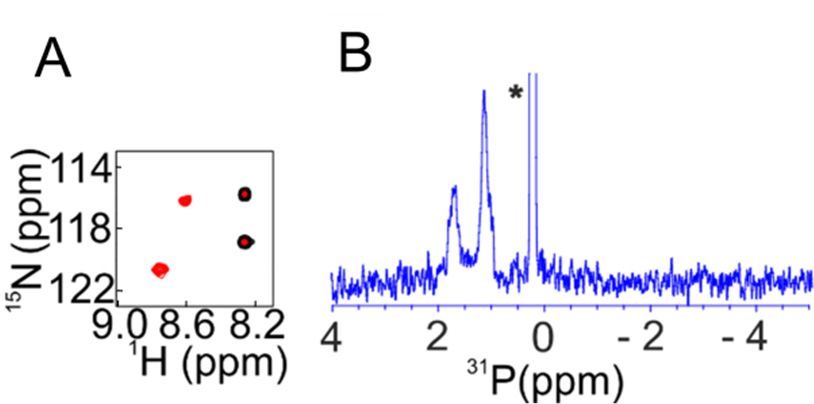
Int. J. Mol. Sci. 2019, 20, 378                                                                    5 of 23

caused by specific and non-specific interactions. However, recent developments showed the success of
in-cell NMR in probing protein–small molecule interactions.

2.1. Protein–Small Molecule Interactions
      SMILI-NMR is an exciting example [15]. Once the target protein shows detectable and
well-dispersed cross-peaks, NMR can be used to carry out target-engagement drug discovery
inside the cell. A second example is the interaction of 12-kDa FK506-binding protein (FKBP12)
within living cells with extracellularly administered immuno-suppressants [41]. Here, a cleavable
CPP–ubiquitin–FKBP12 construct was prepared and the 15 N-labeled fusion protein was expressed
and purified from Escherichia coli. The labeled protein was transduced into HeLa cells. Following
this, the CPP–ubiquitin was cleaved off by endogenous deubiquitinating enzymes of the HeLa cells.
Subsequently, CPP aggregated, leaving15 N FKBP12 as the sole soluble, labeled protein in the cytosol.
Its cross-peak pattern was similar to 15 N FKBP12 measured in vitro. This indicated that, in HeLa
cells, the three-dimensional structure of FKBP12 was maintained and, therefore, the interaction with
small molecules could be studied. Subsequently, in-cell NMR spectra were recorded after treatment
with the immune-suppressants FK506 or rapamycin. Significant spectral changes were observed after
administration of these drug molecules. Interestingly, the two spectra recorded in vitro and in cellulo
had both similar features and subtle changes. Thus, some interactions may be correlated with the
interaction observed only in living cultured cells [27,41].
      In contrast to classical in-cell NMR experiments, in many cases, the protein cannot be labeled
externally and then transduced into the cells of interest. Instead, two steps of production and
isotopic labeling need to be carried out simultaneously. Special medium and labeling protocols
are well established for bacteria and other organisms, including yeast and insect cells. The use
of bacteria to overexpress protein within cells may lead to heavy background signals, caused by
the concomitant labeling of other cellular components. To tackle this problem, the Dötsch group
developed a scheme which reduced the background noise observed when studying the bacterial
protein Nmer A. The activity of bacterial RNA polymerase can be inhibited by rifampicin but not by
bacteriophage T7. Since protein expression was under the control of the T7 promoter, production of all
endogenous bacterial proteins could be suppressed by rifampicin [6,7]. Nmer A plays a critical role in
the bacterial pathway involved with mercury detoxification. The addition of Zn2+ to the NMR tube
led to changes/disappearance of cross-peaks, thus highlighting the possible utility of this approach to
investigate metal and drug binding.
      Recently, we showed the targeting of the bacterial chaperone “sensitive to lysis” (SlyD) to
inhibit bacterial growth using a small molecule, with in-cell NMR spectroscopy [4]. Emergence
of dangerous multi-drug-resistant strains of bacteria is one of the biggest threats to human health
currently. With a continuous rise in antibacterial resistance, it was estimated that, by 2050, it may
result in the death of 10 million people per year [42]. Among Enterococcus faecium, Staphylococcus
aureus, Klebsiella pneumoniae, Acinetobacter baumannii, Pseudomonas aeruginosa, and Enterobacter species
(ESKAPE) pathogens, Gram-negative bacteria are of particular concern, due to their increased ability
to attain multi-drug resistance [43]. In the case of Gram-negative bacteria, many small molecules
including antibiotics become ineffective, due to the presence of an outer polysaccharide layer and
multi-drug efflux transporters. This led researchers to create a distinct class of novel antibacterial
agents. For example, the heat shock proteins (HSPs) were targeted as potential molecules in cancer
therapy [44,45]. Much attention was paid to target HSP90, leading to the identification of geldanamycin
and radicicol; however, HSP60, HSP70, or other chaperones are less studied so far [46]. Therefore,
we chose a small molecule, which was a metal-based coordination complex with a water-soluble organic
moiety capable of crossing the cell-wall barrier and selectively targeting the bacterial chaperone SlyD.
      SlyD is a bacterial chaperone, and all prokaryotes and archaea express homologous proteins [47,48].
Thus, being unique to prokaryotes and archaea, SlyD represents a potential target against which to
develop drug molecules. In these organisms, SlyD is involved in several biochemical pathways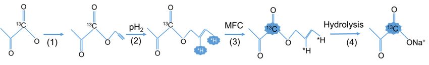
Int. J. Mol. Sci. 2019, 20, 378 6 of 23
including the biosynthesis of [NiFe] hydrogenases, twin-arginine-mediated translocation
Int. J. Mol. Sci. 2018, 19, x FOR PEER REVIEW 6 of 23
(Tat
transport), and metal storage/release. Additionally, SlyD exhibits both a peptidyl–prolylisomerase
(PPIase) andpathways
chaperone including the biosynthesis of [NiFe] hydrogenases, twin-arginine-mediated translocation
activity, which prevents protein aggregation by binding to hydrophobic
(Tat transport), and metal storage/release. Additionally, SlyD exhibits both a
patches [47–49]. The PPIase
peptidyl–prolylisomerase and molecular
(PPIase) chaperone
and chaperone activity,activities [50]protein
which prevents are located
aggregation onbytwo separate
domains, whose binding cooperative
to hydrophobic interplay
patches is required
[47–49]. for full
The PPIase andenzymatic activity [47,51–54].
molecular chaperone activities [50] The
are N-terminal
located on 2+ two separate domains, whose cooperative interplay is required for full enzymatic activity
tail contains the Ni binding site followed by the FKBP binding domain. The FKBP domain harbors
[47,51–54]. The N-terminal tail contains the Ni2+ binding site followed by the FKBP binding domain.
the active siteTheof FKBP
the PPIase, which is modulated by
domain harbors the active site of the PPIase, Ni2+ , and
whichthe chaperone
is modulated by function
Ni2+, and the is located on a
domain that is inserted into the FKBP domain (Figure 2) [47,51–54].
chaperone function is located on a domain that is inserted into the FKBP domain (Figure 2)
[47,51–54].
Figure NMR
Figure 2. In-cell 2. In-cellstudy
NMR study of protein–small molecule
of protein–small molecule interactions. Here, a Here,
interactions. “sensitive
a to lysis”
“sensitive to lysis”
(SlyD)-containing plasmid was transformed into Escherichia coli and protein expression was initiated
(SlyD)-containing plasmid was transformed into Escherichia coli and protein expression
by isopropyl β-D-1-thiogalactopyranoside (IPTG) induction. These cells were further incubated with
was initiated by
isopropyl β-D -1-thiogalactopyranoside
a small molecule (Cu2+ complex). The(IPTG) induction.
interaction of this small These
moleculecells werecould
with SlyD further incubated with a
be observed
small moleculeusing(Cu 2+ complex).
in-cell NMR, and correlated with corresponding
The interaction of thisinsmall
vitro NMR studies, with
molecule revealing the binding
SlyD could be observed
site in SlyD for this Cu2+ complex (adopted according to Reference [4]).
using in-cell NMR, and correlated with corresponding in vitro NMR studies, revealing the binding site
in SlyD for this Cu2+ complex
Complete (adopted
bacterial growth can beaccording to Reference
inhibited with [4]). (anthracenyl terpyridine Cu2+
a small molecule
complex) at 2 µM [4]. In order to evaluate the role of SlyD in bacterial growth, we transformed E. coli
Complete with the plasmid
bacterial containing
growth can bethe inhibited
SlyD gene, with
which aissmall
under molecule
the control (anthracenyl
of T7 promoter.terpyridine
The Cu2+
expression of the SlyD gene can, thus, be initiated by isopropyl β-D-1-thiogalactopyranoside (IPTG).
complex) at 2Overexpressed
µM [4]. In order to evaluate the role of SlyD in bacterial growth, we transformed E. coli
SlyD could restore bacterial growth, confirming that SlyD is involved in growth
with the plasmid containing
inhibition induced the
by theSlyD
Cu2+gene, which
complex. This is under
small the control
molecule binds toof T7 with
SlyD promoter. The expression of
a dissociation
constant (K D) ~50 µM with a 2:1 stoichiometry without inducing large conformational changes. In
the SlyD gene can, thus, be initiated by isopropyl β-D-1-thiogalactopyranoside (IPTG). Overexpressed
vitro NMR with 15N SlyD showed that this Cu2+ complex binds into the PPIase active site of the FKBP
SlyD could restore
domain bacterial
(Figure 2), asgrowth,
shown in confirming
earlier studiesthat
[54].SlyD
In-cellisNMR
involved in growth
spectroscopy confirmedinhibition
with induced
2+
by the Cu complex. This that
small
residue resolution the molecule
Cu complex
2+ binds to SlyD
also binds with
to SlyD a dissociation
inside constant
bacteria, verifying that the(KD ) ~50 µM
complex can penetrate
with a 2:1 stoichiometry withoutthe cell wall and bind
inducing to SlyD
large when inhibiting cell
conformational growth. Interestingly,
changes. In vitro NMRthe with 15 N
small molecule also2+ inhibited the growth of pathogenic bacteria from the category of ESKAPE
SlyD showedpathogens.
that thisThe Cuhalf maximal
complex binds concentration
inhibitory into the PPIase active
(IC50) of site
the small of thewas
molecule FKBP belowdomain
1 µM (Figure 2),
as shown in earlier studies [54]. In-cell NMR spectroscopy confirmed with residue resolution that
the Cu2+ complex also binds to SlyD inside bacteria, verifying that the complex can penetrate the cell
wall and bind to SlyD when inhibiting cell growth. Interestingly, the small molecule also inhibited the
growth of pathogenic bacteria from the category of ESKAPE pathogens. The half maximal inhibitory
concentration (IC50 ) of the small molecule was below 1 µM for Staphylococcus aureus and Pseudomonas
aeruginosa [4,55], showing the general applicability of the approach.
2.2. Small-Molecule Libraries
The Shekhtman group developed small-molecule interactor libraries NMR (SMILI-NMR).
The interactions between two or more components of a biomolecular complex may be disruptedInt. J. Mol. Sci. 2019, 20, 378 7 of 23
or enhanced by a few of these small molecules. The method provides atomic-level information and
relies on the formation of a defined complex under in-cell NMR conditions. The approach was
applied to the fujimycin (FK-506) binding protein (FKBP) and the FKBP rapamycin-binding domain
of the mammalian target of rapamycin (FRB), a well-studied system for heterodimer formation,
to screen those small molecules that can facilitate hetero-dimerization. In mammalian cells, one of
the immune-modulatory systems (mitogenic responses) is constituted by rapamycin–FKBP–FRB
interactions [56]. The labeled proteins were sequentially overexpressed using two compatible plasmids
in E. coli under in-cell NMR conditions. To observe the NMR spectrum of FKBP or FRB, bi-complex
(FKBP–FRB) formation was required. The X-ray structure of the FKBP–rapamycin–FRB ternary
complex indicated the limited availability of the FKBP–FRB interaction surface. While the interaction
between free FKBP and FRB is quite weak, with KD values >50 µM, a well-defined ternary complex
is formed when the FKBP–rapamycin complex binds to FRB (KD ~12 nM). In their in-cell NMR
experiments, either FKBP or FRB was labeled when the complex was formed inside the cells. This did
not result in any visible spectrum. Addition of rapamycin to the cell suspension resulted in the
appearance of FKBP resonances and, for 32 out of the 107 residues, changes with respect to free FKBP
could be detected. Many resonances in FRB residues also changed upon performing the reverse
experiment [15]. Ascomycin is a competitive inhibitor of rapamycin. It binds to FKBP with 1.4 nM
affinity and has no known affinity with FRB. However, the ascomycin–FKBP complex binds to FRB
with lower affinity [57]. Addition of ascomycin to the aforementioned dual-plasmid system resulted in
the appearance of the FKBP/FRB NMR spectrum. In the next step, the authors screened a library of
small molecules of 289 dipeptides with SMILI-NMR. The peptides were selected as drug candidates
from the literature, based on their facile and cost-effective preparation, as well as their ability to be
imported into prokaryotic and eukaryotic organisms through naturally occurring transport systems.
Screening of a matrix of 17 × 17 peptides resulted in the identification of various combinations that
showed completely different behavior when compared to the rapamycin-induced ternary complex.
Addition of these dipeptides resulted in extreme line broadening and the disappearance of some peaks
in the NMR spectrum. Later on, the addition of low concentrations of Ala–Glu resulted in the same
interactions with FKBP, suggesting that FKBP and FRB hetero-oligomerization can be facilitated by this
peptide. Thus, SMILI-NMR could screen the protein–small molecule interactions within the cellular
environment using high-resolution NMR as a readout [15].
3. In-Cell NMR Observation of Metabolic Processes Using Hyperpolarization
NMR spectroscopy provides considerable opportunities for collecting diverse and unique
information on cellular processes due to its non-invasive nature [58], rendering it highly suitable
for studying the fate of metabolites and biochemical pathways in situ. In order to raise the detection
limit of this inherently insensitive technique, hyperpolarization NMR methods—mainly dissolution
dynamic nuclear polarization (d-DNP) and para-hydrogen-induced polarization (PHIP)—were devised
as tracer techniques to follow the fate of detectable, small molecular probes for the visualization of
cellular functions that are not easily observable by other means. Furthermore, NMR signals from these
molecules can be enhanced by several orders of magnitude via the combined use of hyperpolarization
(HP) and isotope enrichment. The NMR signal enhancements achieved using these methods are
often sufficiently high to track endogenous molecules at physiological concentrations. Among all
the potentially suitable target molecules for these specialized experiments, hyperpolarized pyruvate,
a metabolite whose cell biochemistry lies at the interface between catabolic and anabolic metabolism,
is the most widely studied probe. This is mainly due to its (a) high hyperpolarizability, (b) rapid
cellular uptake, and (c) central biochemical position as a key intermediate in several biochemical
pathways. In addition, a host of other probes emerged in the meantime that can also be used to
characterize the phenotype of cells under a particular set of conditions (Table 1).Int. J. Mol. Sci. 2019, 20, 378 8 of 23
Table 1. Selection of endogenous target molecules used as reporter probes for hyperpolarization-
assisted in-cell NMR studies of metabolic processes, together with their associated field(s) of application.
Target Molecule/Probe Field of Application (FOA)/Observable
Pyruvate metabolism; cell permeability; cell lysis; drug efficacy; enzyme activity
and reaction fluxes; intracellular pH determination; oncogene signaling; indication
Pyruvate
of aerobic glycolysis; tricarboxylic acid (TCA) pathway activity; mono carboxylate
transporter level/activity; tumor grading
Fumarate Fumarate metabolism; cell permeability; cell lysis; drug efficacy
Lactate Enzyme activity and reaction fluxes; tumor grading
Alanine Enzyme activity and reaction fluxes; enzyme mechanistic studies; tumor grading
Gene expression/loss; glycolysis pathway activity; sulfite cytotoxicity; glucose
Glucose
transporter level/activity
Acetate Enzyme activity and reaction fluxes; intracellular pH determination
Glutamine Enzyme activity and reaction fluxes
Fructose Enzyme mechanistic studies
Experimentally, the method, which can lead to a nuclear sensitivity enhancement of up to five
orders of magnitude, works as follows: a frozen solution (T ~1.1 to 1.5 K) of the sample to be analyzed
is polarized in the presence of a radical molecule, e.g., trityl (triphenylmethyl, “Trityl OX063”) or
the nitroxide-based radical 2,2,6,6-tetramethylpiperidin-1-yl)oxyl (TEMPO) [59], with microwave
(MW) irradiation (spin polarization is transferred via the DNP mechanism from electrons to nuclei
upon microwave irradiation at or near the Larmor frequency of the radical electron [60,61]) using
a specifically designed DNP polarizer. Subsequently, the sample is thawed, dissolved in a suitable
hot solvent, and then transferred to a conventional liquid-state NMR spectrometer for detection [62].
In solids, DNP is known to occur via a number of different mechanisms known as the solid effect,
thermal mixing, and the cross effect [60]. Depending on the experimental conditions used in each
d-DNP experiment, e.g., radical type, substrate, and solvent (among others), the contribution of each
of these effects to the observed polarization enhancement differs.
The longitudinal relaxation time (T1 ) of the hyperpolarized nuclei is crucial in dissolution DNP.
To preserve the nuclear polarization acquired in the solid state, polarized samples need to be thawed
and transferred to the NMR spectrometer faster than nuclear T1 spin–lattice relaxation. Typical
dissolution DNP samples experience a so-called “transfer time” as they are moved from the polarizing
magnet to the NMR spectrometer. During this period, the sample is often exposed to low magnetic
fields, which are typically on the order of ca. 0.5 mT. Given that nuclear T1 times are generally shorter
at low magnetic fields—and even more so in the presence of radicals—a fast transfer of the sample, as
well as the elimination of stable free radicals in solution, is crucial to reduce polarization losses [63].
The use of a “magnetic tunnel” for transfer and/or the addition of a radical scavenging agent, e.g.,
ascorbate (vitamin C), to the sample during the dissolution step were shown to alleviate polarization
losses during and immediately after the transfer. Attempts to shorten the transfer time include
construction of a dedicated “hybrid” spectrometer, comprising a single dual-isocenter superconducting
magnet featuring regions of different magnetic field strengths suitable for both polarization and NMR
detection [64] or, alternatively, a “shuttle” DNP spectrometer comprising a two-center magnet [65].
A significant number of d-DNP-based investigations were carried out in recent years to probe
the metabolic behavior of tissues, e.g., heart, liver, and tumor cells, and to study the fate of individual
metabolites both in vitro and in vivo [66]. In addition, metabolite molecules hyperpolarized using
the d-DNP method were used to probe a variety of different biochemical pathways. For example,
the enzymatic conversion of pyruvate to lactate, acetylcarnitine, citrate, and glutamate was tracked
in real time employing [2-13 C] pyruvate, in isolated perfused heart tissue, to study healthy and
pathological states [67]. Hyperpolarized probes were also used to track intracellular pathways of
short-chain fatty acids and ketone body metabolism in real time. A butyrate probe visualized the flux of
fatty acids to acetoacetate and several tricarboxylic-acid-cycle intermediates in cardiac muscle cells [68].
In addition, hyperpolarized [1-13 C] pyruvate was used as a clinical diagnostic tool in metabolic imaging
to characterize differences between healthy tissue and tumor cells [69]. Further applications includeInt. J. Mol. Sci. 2019, 20, 378 9 of 23
the study of enzyme kinetics [70], biosynthetic pathways [70], and the detection of lowly populated
reaction intermediates [71]. More recently, the para-hydrogen-induced hyperpolarization method
was also introduced for the signal amplification of metabolites and the subsequent tracking of their
biochemical pathways in both healthy and pathological forms of tissue. Up to this point, however,
only a few very recent examples exist in the literature where the method was successfully employed to
hyperpolarize metabolites for in-cell NMR studies [72–74].
In the following subsections, we give an overview of hyperpolarization NMR methods—in
particular, dissolution dynamic nuclear polarization (d-DNP) and para-hydrogen-induced nuclear
polarization (PHIP)—and a few selected examples of hyperpolarization-assisted in-cell NMR
observations of metabolic processes are highlighted in a conceptual manner. The section concludes with
an outlook into future perspectives of this emerging, yet still relatively novel, area of NMR research.
3.1. Dissolution DNP Application to Metabolic Pathways and Biological Functionality
The number of applications of heteronuclear d-DNP for the study of living cellular systems is
vast [66]. For example, the real-time tracking of metabolic conversion using hyperpolarized NMR is
particularly suitable for the observation of metabolic reaction networks, provided that conversion rates
are high and that the obtained levels of hyperpolarization are significant. In this context, glycolysis
was identified as an adequate metabolic process to be studied with d-DNP, given its overwhelming
biochemical importance and its central role in a variety of different biochemical reaction routes.
For example, enzymatic reaction mechanisms, bottlenecks, and off-pathway reactions were probed
using hyperpolarized carbohydrates, i.e., [2-13 C] fructose and [U-13 C, U-2 H] glucose, as substrates [66].
Chemical detail in the observation of pathway reactions extends to the distinction of isomers and their
susceptibility to enzymatic turnover. The use of site-specifically labeled [2-13 C] fructose, for example,
permitted the real-time observation of probe flux during gluconeogenesis, as well as the formation
of non-productive off-pathway intermediates, such as dihydroxy acetone phosphate hydrate [75].
A recent approach combined hyperpolarized dynamic measurements with metabolite extraction,
isotopomer evaluation, and flow analysis [76]. This approach measured pyruvate metabolism in living
cells to obtain quantitative data of several biochemical pyruvate pathways in different cell types.
The existence of different interlocked pathways, all featuring a similar set of key metabolites,
makes it difficult to predict how cellular physiology and intracellular metabolism respond to
the modification of individual genes [77]. The use of hyperpolarized NMR spectroscopy to
study genetically well-defined and homogeneous cell suspensions shows promise in studying the
cellular response to genetic modifications. For example, the two E. coli strains, BL21 and K-12,
show strong differences in the reaction progression of their pentose phosphate pathways. In the
BL21 strain, a reactive intermediate accumulates, and is responsible for covalent modifications
observed for the recombinant proteins expressed within this strain [78]. Genome alignment
techniques prove that the gene encoding for lactonase—the enzyme which catalyzes the hydrolysis of
6-phosphogluconolactone—is absent in the BL21 strain due to a deletion. Such molecular phenotypes
can be observed in the absence of phenotypic variations [79]. Metabolic differences in different cell
types were recently compared in human cells by tracking the glycolytic pathway [80]. Ratiometric
measurements of lactate and pyruvate signals in two different proliferating cell types were used to
non-invasively detect differences in the cytosolic redox state. In the cytosol, lactate and pyruvate
form a redox pair, whose equilibration rate depends crucially on the ratio of oxidized to reduced
nicotinamide adenine dinucleotide (NAD+ /NADH) in the cytosol. PC3, a specific prostate cancer cell
line, showed a fourfold increase in the intracellular ratio of free cytosolic NAD+ /NADH in comparison
with breast cancer cells in an experiment that used hyperpolarized glucose as a reporter metabolite.
The increase in the ratio of NAD+ versus NADH reflects a distinct metabolic phenotype consistent
with previously reported alterations in the energy metabolism of prostate cells. In a relatively recent
study, the importance of hyperpolarized NMR probes as tools for functional studies involving the
human genome was underlined by observing human cell types differing only in the mutational statusInt. J. Mol. Sci. 2019, 20, 378 10 of 23
of the enzyme isocitrate dehydrogenase 1 (IDH1), using hyperpolarized [1-13 C] alpha-ketoglutarate
as a molecular reporter. IDH1 catalyzes the decarboxylation of cytosolic isocitrate to α-ketoglutarate.
Specific mutations in IDH1 result in its ability to catalyze the NAD phosphate (NADPH)-dependent
reduction of α-ketoglutarate to (R)-2-hydroxyglutarate, an onco-metabolite [81]. As a consequence,
isogenic glioblastoma cells, differing only in the status of IDH1, show differences in the conversion of
the hyperpolarized α-ketoglutarate to (R)-2-hydroxyglutarate, as probed by changing hyperpolarized
NMR signal intensities.
3.2. Following Metabolism in Living Microorganisms Using Hyperpolarized 1 H NMR
As mentioned before, most d-DNP-based in-cell NMR studies focus on hyperpolarizing nuclei
with low gyromagnetic ratios (γ), given their relatively long spin–lattice (T1 ) relaxation times, e.g.,
non-protonated 13 C or 15 N nuclei in small molecules exhibiting short rotational correlation times
(τ c ). Nevertheless, advantages can also result from observations based on hyperpolarizing and
observing high-γ nuclei, e.g., protons. For example, 1 H signal intensities should be, on average,
approximately 16-fold higher as compared with 13 C, given the fourfold higher gyromagnetic ratio of
protons. While this gain is moderated by a concomitant increase in spectral noise, the indisputable fact
that state-of-the-art 1 H-observation hardware is widely available and represents the most mature across
all in vivo NMR technologies, might make these observations more worthwhile. Lastly, instances may
arise where 1 H-based detection provides a better chemical discrimination than 13 C-based methods.
In fact, spontaneous enhancements in the 1 H-NMR spectra of hydrogen nuclei covalently bound to
hyperpolarized 13 C nuclei were reported a while ago [82,83]. Heteronuclear cross-relaxation effects
arising in rapidly tumbling small molecules were identified as the mechanism responsible for this
spontaneous polarization transfer. Given that, in such experiments, hyperpolarization can be stored
in a relatively slowly relaxing nucleus that shares its hyperpolarization with a neighboring proton,
opportunities arise from using these latter signals to monitor enzymatic turnover.
Using 1 H NMR detection, two such processes were recently studied by Frydman and co-workers
to yield successful results [84] using both solutions of purified enzymes in vitro and suspensions of
intact cells. The substrate in each of these studies was hyperpolarized [13 C] pyruvate, and the enzymatic
processes targeted were (a) the production of acetaldehyde following the addition of hyperpolarized
[U-2 H3 ,2-13 C] pyruvate either to samples containing pyruvate decarboxylase (PDC) purified from
Saccharomyces cerevisiae or, alternatively, to cultures of S. cerevisiae fermenting glucose, and (b) the
generation of formic acid due to the activity of pyruvate formatelyase (PFL), measured in cultures
of anaerobic E. coli following the addition of hyperpolarized [1-13 C] pyruvate. In these enzymatic
reactions, the formation of new covalent bonds between the hyperpolarized 13 C nucleus and protons in
the reaction products, i.e., acetaldehyde and formate, allowed the authors to transfer hyperpolarization
using either insensitive nuclei enhanced by polarization transfer (INEPT)-type pulse sequences
or by spontaneous cross-relaxation. Features that favored the execution of such reversed INEPT
experiments included (i) the possibility to transfer magnetization between heteronuclei separated
by multiple bonds (something that cross-relaxation is very inefficient at doing); (ii) the possibility of
incorporating coherence-selection pulsed field gradients (PFG) to efficiently eliminate background
1 H signals of endogenous metabolite molecules; and (iii) the possibility of acquiring 1 H NMR spectra
exhibiting a signal-to-noise ratio (SNR) which is approximately an order of magnitude higher than
the SNR obtained using solely spontaneous magnetization transfer by cross-relaxation. In particular,
using 1 H-detected INEPT spectroscopy allowed the detection of the acetaldehyde produced from
hyperpolarized pyruvate. The authors also observed, however, that the strong signal enhancement
achieved by the reversed INEPT experiment came, in part, at the expense of depleting all of the 13 C
polarization in a single acquisition. It was, hence, deduced that lower levels of polarization transfer
can be delivered using the INEPT sequence at the expense of not being able to employ more complex
procedures, which would otherwise “waste” hyperpolarization generated via the DNP mechanism.
It was suggested that a possible route to preserve the bulk 13 C hyperpolarization while using INEPTInt. J. Mol. Sci. 2019, 20, 378 11 of 23
would be to use sequences that selectively excite the carbon nuclei of the product while avoiding
excitation of the reactant 13 C coherences.
Subsequently, Frydman and co-workers explored the same biochemical process in cultures of
S. cerevisiae using proton detection. Although these microorganisms can be grown on pyruvate as the
sole source of carbon, pyruvate does not permeate the plasma membrane during glucose fermentation.
Undissociated pyruvic acid, however, rapidly crosses the plasma membrane of glucose-fermenting
S. cerevisiae. In this context, a recent d-DNP NMR study using carbon detection demonstrated that
rapid diffusion of undissociated HP [1-13 C] acetic acid into glucose-fermenting S. cerevisiae occurred,
in particular, at low extracellular pH [85]. After entering the cell, pyruvic acid is rapidly decarboxylated
by cytosolic PDC, a process which is pronounced during the exponential stages of cell growth.
The product, acetaldehyde, is present in a low equilibrium concentration and rapidly reduced to
ethanol. The accumulation of acetaldehyde in S. cerevisiae cultures was observed with acidification of
the cytosol, which was attributed to the inhibition of the enzyme alcohol dehydrogenase combined
with a shift in cytosolic pH to values closer to PDC’s pH optimum of 6.0 [86]. The 1 H NMR results
obtained by Frydman and co-workers were able to corroborate this feature via detection of a relatively
weak acetaldehyde 1 H signal found in NMR spectra of mid-exponential yeast cultures following
incubation in acetate buffer at pH 4.5 and prior exposure to hyperpolarized pyruvic acid.
In a final step, the wider applicability of spontaneous polarization transfer for investigating cell
metabolism was demonstrated in a study investigating the activity of PFL in E. coli cells. PFL rapidly
metabolizes pyruvate in anaerobic E. coli cultures, a process that is particularly heightened when
pyruvate is the main carbon source. Consistent with results obtained from direct 13 C detection,
Frydman and co-workers observed the rapid uptake and breakdown of pyruvate as a pronounced
formate 1 H signal. In contrast to the relatively weak acetaldehyde signal detected in S. cerevisiae
cultures after the addition of HP pyruvic acid, the build-up and decay of the formate signal was
observed on a single, scan-by-scan basis. The authors explained this higher signal-to-noise ratio by the
rapid uptake of pyruvate and the accumulation of formate as a metabolic end product, characteristic
of anaerobic E. coli cells.
In summary, Frydman and co-workers were able to demonstrate that 13 C-based dissolution DNP,
combined with both spontaneous and INEPT-driven polarization transfer from carbon to protons,
provides a clear 1 H signature of the enzymatic processes studied. They eventually concluded that
this feature can help decipher NMR-encoded metabolic information in spectra acquired in cellulo.
Even though metabolic fluxes from pyruvate to acetaldehyde in S. cerevisiae [85] and of pyruvate to
formate in E. coli [79] were measured in the past using d-DNP-enhanced 13 C NMR in vitro, close signal
proximity and subsequent signal overlap between the metabolic products and their direct precursors
complicated these measurements in vivo to a significant extent. Also, the spontaneous transfer to 1 H
turned out to be particularly superior to other methods, as it allowed the detection of hyperpolarized
1 H signals without the need for chemical manipulation of the probe molecule prior to the experiment,
or modifications of the dissolution method.
4. para-Hydrogen-Induced Hyperpolarization Side-Arm Hydrogenation (PHIP-SAH) Method for
the Detection of Cell Metabolism
As mentioned before, para-hydrogen-induced polarization (PHIP) is a chemistry-based
hyperpolarization technique which, to a certain extent, is easier to handle and more straightforward
to use when compared with DNP. This is due to the fact that (i) no additional NMR set-up extension,
i.e., polarizer equipment, is needed, and (ii) polarization times are significantly shorter. The main
drawback of the method in the context of in-cell NMR studies, however, is the limited availability
of unsaturated precursor molecules with respect to the desired target compounds to be studied; for
instance, nuclear spin-polarized acetate and pyruvate cannot be obtained by direct incorporation of the
para-hydrogen molecule. The advent of non-hydrogenative PHIP (NH-PHIP; see below) only partially
resolved this problem, given that the obtained levels of hyperpolarization in the target compoundsInt. J. Mol. Sci. 2019, 20, 378 12 of 23
are significantly lower as compared to using, for example, the adiabatic longitudinal transport after
dissociation engenders net alignment (ALTADENA)-PHIP method. Furthermore, not all substrates of
interest are suitable for NH-PHIP, given that certain requirements with respect to their molecular and
electronic structure must also be fulfilled in this case.
Molecular dihydrogen occurs in the form of two nuclear spin isomers, i.e., the ortho- and
the para-isomer, featuring either symmetric (triplet) or antisymmetric (singlet) nuclear spin states,
respectively. Due to the three-fold degeneracy of the triplet state, ortho- and para-isomers are populated
in a ratio of 3:1 under ambient conditions [87]. Interconversion between the two is symmetry forbidden
and occurs, hence, at a negligibly small rate. However, when H2 gas is flowed through an appropriate
apparatus comprising a paramagnetic catalyst, i.e., activated charcoal, at low temperatures, a high
enrichment of the para spin isomer can be achieved and observed at standard temperatures [88] due to
this form being lower in energy.
When applying the para-hydrogen-induced polarization (PHIP) method, nuclear spin
hyperpolarization is accomplished by transferring the high spin order of the para-hydrogen molecule
to the substrate of interest. Typically, PHIP applications involve the direct incorporation of p-H2 into
unsaturated organic molecules, i.e., molecules containing either carbon–carbon double or triple bonds,
in the presence of an appropriate hydrogenation catalyst. The new chemical environment experienced
by the two protons upon hydrogenation breaks the singlet symmetry and renders the spin system
detectable by NMR. Depending on whether the reaction is carried out at high or low magnetic field,
the methods are named para-hydrogen and synthesis allow dramatically enhanced nuclear alignment
(PASADENA) or ALTADENA, respectively [89,90]. In PASADENA experiments, the para-hydrogen
symmetry is broken upon hydrogenation due to the distinct chemical shift environment of the two
incorporated protons at high magnetic field. Thus, the NMR spectrum shows two antiphase multiplets.
In ALTADENA experiments, the singlet state becomes selectively polarized, given that the chemical
shifts are essentially the same when para-hydrogen is incorporated into the substrate at low magnetic
field. In this case, the NMR spectrum is characterized by two hyperpolarized in-phase resonances
of opposite sign. In the case that other magnetically active nuclei are present in the substrate, i.e.,
13 C, 15 N, or 19 F, scalar and/or dipolar coupling interactions can cause the transfer of the initially
induced proton hyperpolarization to other regions of the substrate molecule, in particular when the
hydrogenation step is carried out at low field (ALTADENA); this phenomenon was also observed
using other hyperpolarization methods [91,92].
Despite the relatively high degree of both homo- and heteronuclear polarization that can be
achieved with PHIP, the method is not generally applicable to all molecules, i.e., metabolites, unless
characteristic hydrogenation precursor modifications are applied (see below). More recently, a related
methodology known as signal amplification by reversible exchange (SABRE) or NH-PHIP [93]
was developed to polarize substrates without having to perform the final hydrogenation step, i.e.,
the physical transfer of the two protons of the dihydrogen molecule to the substrate. During a SABRE
experiment, substrate and para-hydrogen nuclei experience transient contact interactions via the
metal center of a labile catalyst–dihydrogen complex, e.g., [Ir(H)2 (PCy3 )(substrate)3 ][BF4 ], which is
formed via the reaction of [Ir(COD)(PCy3 )(MeCN)][BF4 ]—where “Cy” is cyclohexyl and “COD” is
cyclooctadiene) with para-H2 and an excess of the substrate to be polarized. While the complex is
formed, nuclear polarization is transferred from the para-hydrogen-derived protons to the molecule of
interest. After the polarization transfer step, the chemically unmodified polarized substrate is released.
So far, pyridine is the most widely studied SABRE substrate, and more than 10% of proton polarization
was achieved in this case [94]. The method is strikingly similar to the ALTADENA experiment
described above in that the polarization of the substrate occurs at low magnetic field. The magnetic
field dependence of SABRE-derived signal enhancements appears to change very little with substrate
type or position of the protons, suggesting that the extent of polarization depends primarily on the
scalar coupling between two para-hydrogen-derived protons and not on the scalar coupling between
para-hydrogen-derived and substrate protons within the hydrogenation complex [95,96].Int. J. Mol. Sci. 2019, 20, 378 13 of 23
Int. J. Mol. Sci. 2018, 19, x FOR PEER REVIEW 13 of 23
Recently,ofAime
interest.and
Afterco-workers
the polarization presented
transfer step,a new
the chemically to the para-hydrogen
additionunmodified polarized substrate method,
is which
released. Soof
allows the generation far, pyridine
high is the
levels ofmost
nuclear widely studied SABREusing
polarization substrate, and more normally
substrates than 10% of inaccessible
proton to the
polarization was achieved in this case [94]. The method is strikingly similar to the ALTADENA
para-hydrogen method,described
experiment whichabove can subsequently be used
in that the polarization forsubstrate
of the hyperpolarization-assisted
occurs at low magnetic field.NMR studies
in cellulo [74].The
Inmagnetic
particular, their results
field dependence demonstrate
of SABRE-derived that
signal PHIP can appears
enhancements be induced invery
to change nuclei
littleof molecules
such as acetatewithand substrate type or position in
pyruvate—and, of the protons, suggesting
principle, that the extent
other carboxylic of polarization
acids depends precursors
as well—using
primarily on the scalar coupling between two para-hydrogen-derived protons and not on the scalar
containing ancoupling
unsaturated
between side-arm moiety capable
para-hydrogen-derived of hydrogenation
and substrate protons within thethat can be hydrolyzed
hydrogenation complex to yield
the hyperpolarized
[95,96]. target products. The reported method, named PHIP-SAH, relies on the following
Recently, Aimeof
steps: (i) functionalization andthe
co-workers
target presented a new addition
acidic molecule withto the
anpara-hydrogen
unsaturated method, which group (i.e.,
alcoholic
allows the generation of high levels of nuclear polarization using substrates normally inaccessible to
vinyl or propargyl alcohol;method,
the para-hydrogen using which
propargyl alcohol be
can subsequently asused
a removable synthon to generate
for hyperpolarization-assisted NMR PHIP on
13 C resonances markedly
studies widens
in cellulo [74]. the applicability
In particular, of this approach);
their results demonstrate that PHIP can (ii) para-hydrogenation
be induced in nuclei of of the
molecules such as acetate and pyruvate—and, in principle, other carboxylic acids as well—using
unsaturated ester; (iii) heteronuclear polarization transfer from the former pair of para-hydrogen
precursors containing an unsaturated side-arm moiety capable of hydrogenation that can be
13 signal
protons to thehydrolyzed
carboxylate to yield C by applying
the hyperpolarized magnetic
target products. field cycling,
The reported method, where the magnetic field is
named PHIP-SAH,
cycled between reliesthe Earth’s
on the followingmagnetic field (hydrogenation
steps: (i) functionalization of the targetstep)
acidic and nearly
molecule with anzero-field
unsaturated(polarization
transfer step)alcoholic group (i.e., vinyl or propargyl alcohol; using propargyl alcohol as a removable synthon to
using concentric cylinders made of µ-metal, thereby increasing the extent of polarization
generate PHIP on 13C resonances markedly widens the applicability of this approach); (ii)
transfer to carbon nuclei; (iv)
para-hydrogenation of the release of the
unsaturated ester;alcohol moietypolarization
(iii) heteronuclear via hydrolysis to obtain
transfer from the formerthe polarized
13 C-carboxylate-containing
pair of para-hydrogen product.
protons to the carboxylate 13C signal by applying magnetic field cycling,
where the magnetic field is cycled between the Earth’s magnetic field (hydrogenation step) and
In summary, these findings open up a very interesting perspective for the use of
nearly zero-field (polarization transfer step) using concentric cylinders made of µ-metal, thereby
para-hydrogen-based
increasing procedures for the generation
the extent of polarization of hyperpolarized,
transfer to carbon biologically
nuclei; (iv) release of relevant
the alcohol moiety via molecules,
as demonstrated by recent NMR studies carried out in living cells using PHIP-SAH. Examples
hydrolysis to obtain the polarized 13C-carboxylate-containing product.
In summary, these findings open up a very interesting perspective for the use of
include the real-time detection of the response of the heart to altered metabolism [73], as well as
para-hydrogen-based procedures for the generation of hyperpolarized, biologically relevant
the study of tumor cell metabolismbyusing 13 C-labeled pyruvate [72], in addition to
molecules, as demonstrated recent hyperpolarized
NMR studies carried out in living cells using PHIP-SAH.
the observation of theinclude
Examples metabolic transformations
the real-time detection of the of hyperpolarized
response lactatemetabolism
of the heart to altered in vitro [73],
[72].asThe fact that
well as the study of tumor cell metabolism using hyperpolarized 13C-labeled pyruvate [72], in
means to further enhance the efficiency of hyperpolarizing small organic molecules via PHIP-SAH
addition to the observation of the metabolic transformations of hyperpolarized lactate in vitro [72].
were identifiedThe recently
fact that means(Figure 3) will
to further enhance most likely lead
the efficiency to an evensmall
of hyperpolarizing wider
organicapplicability
molecules via of this very
promising hyperpolarization
PHIP-SAH were identified method [97].
recently (Figure 3) will most likely lead to an even wider applicability of
this very promising hyperpolarization method [97].
Figure 3. Diagram of the ofpara-hydrogen-induced
Figure 3. Diagram hyperpolarization
the para-hydrogen-induced hyperpolarization side-armside-arm hydrogenation
hydrogenation
(PHIP-SAH) (PHIP-SAH)
procedure. (1) Functionalization
procedure. (1) Functionalization of of the the carboxylate
carboxylate group with group with (2)
the side-arm; the side-arm;
para-hydrogenation of the unsaturated alcohol; (3) transfer of para-hydrogen
(2) para-hydrogenation of the unsaturated alcohol; (3) transfer of para-hydrogen spin order to thespin order to the 13C
spin of the carboxylate group; (4) cleavage of the side-arm. The yellow background indicates reaction
13 C spin of the carboxylate group; (4) cleavage of the side-arm. The yellow background indicates
steps taking place in the organic phase, while the blue background indicates that the molecule is
reaction stepsdissolved
taking in place in thephase.
the aqueous organic
Thisphase, while
figure was adoptedtheaccording
blue background indicates that the molecule
to Reference [98].
is dissolved in the aqueous phase. This figure was adopted according to Reference [98].
5. In-Cell NMR and Posttranslational Phosphorylation
5. In-Cell NMR and Posttranslational
The most Phosphorylation
common posttranslational modifications of proteins by their respective kinases is the
phosphorylation of serine, threonine, and tyrosine, as well as histidine and aspartate residues, in
The most common
bacteria posttranslational
and fungi [99]. The addition and modifications of proteins
removal of the phosphate byphosphatases
group by their respective
plays a kinases is
the phosphorylation of regulation
key role in the serine, threonine, and tyrosine,
of biological processes. as well
Phosphorylation as histidine
patterns modify theand aspartate
structure and residues,
stability of proteins, in addition to their localization and specific interactions with binding partners.
in bacteria and fungi [99]. The addition and removal of the phosphate group by phosphatases plays a
key role in the regulation of biological processes. Phosphorylation patterns modify the structure and
stability of proteins, in addition to their localization and specific interactions with binding partners.
Anomalous phosphorylation events are the basis for many human diseases. Thus, deciphering the
phospho-code remains a major research effort.
Mass spectrometry (MS) is one of the techniques of choice to identify posttranslational
modifications of the primary protein sequence due to its extreme sensitivity and high resolution.
MS relies on enzymatic digestion of the protein of interest (usually with trypsin), followed by
subsequent peptide analysis. However, multiple phosphorylation events which are in close proximity
are difficult to analyze in terms of their exact location. MS/MS methods can solve this problem.
The labile nature of the phosphate groups, however, might further hamper the analysis. Thus,
mass spectrometry analyses benefit from further biochemical verifications. NMR spectroscopy isYou can also read


