Cardiomyocyte Proliferation as a Source of New Myocyte Development in the Adult Heart
←
→
Page content transcription
If your browser does not render page correctly, please read the page content below
International Journal of
               Molecular Sciences

Review
Cardiomyocyte Proliferation as a Source of New Myocyte
Development in the Adult Heart
Jaslyn Johnson, Sadia Mohsin                    and Steven R. Houser *

                                           Independence Blue Cross Cardiovascular Research Center, Lewis Katz School of Medicine, Temple University,
                                           Philadelphia, PA 19140, USA; jaslyn.johnson@temple.edu (J.J.); sadia.mohsin@temple.edu (S.M.)
                                           * Correspondence: steven.houser@temple.edu

                                           Abstract: Cardiac diseases such as myocardial infarction (MI) can lead to adverse remodeling and
                                           impaired contractility of the heart due to widespread cardiomyocyte death in the damaged area.
                                           Current therapies focus on improving heart contractility and minimizing fibrosis with modest car-
                                           diac regeneration, but MI patients can still progress to heart failure (HF). There is a dire need for
                                           clinical therapies that can replace the lost myocardium, specifically by the induction of new myocyte
                                           formation from pre-existing cardiomyocytes. Many studies have shown terminally differentiated
                                           myocytes can re-enter the cell cycle and divide through manipulations of the cardiomyocyte cell cycle,
                                           signaling pathways, endogenous genes, and environmental factors. However, these approaches
                                           result in minimal myocyte renewal or cardiomegaly due to hyperactivation of cardiomyocyte prolif-
                                           eration. Finding the optimal treatment that will replenish cardiomyocyte numbers without causing
                                           tumorigenesis is a major challenge in the field. Another controversy is the inability to clearly define
                                           cardiomyocyte division versus myocyte DNA synthesis due to limited methods. In this review, we
                                           discuss several studies that induced cardiomyocyte cell cycle re-entry after cardiac injury, highlight
                                 whether cardiomyocytes completed cytokinesis, and address both limitations and methodological

                                           advances made to identify new myocyte formation.
Citation: Johnson, J.; Mohsin, S.;
Houser, S.R. Cardiomyocyte                 Keywords: cardiomyocyte proliferation; cardiac regeneration; cardiomyocyte cytokinesis; myocar-
Proliferation as a Source of New           dial infarction
Myocyte Development in the Adult
Heart. Int. J. Mol. Sci. 2021, 22, 7764.
https://doi.org/10.3390/ijms22157764

                                           1. Introduction
Academic Editor: Vittorio Fineschi
                                                 Cardiovascular diseases such as myocardial infarction (MI) are a major cause of hu-
                                           man mortality [1]. MI is caused by occlusion in the coronary arteries which deprives
Received: 29 June 2021
Accepted: 18 July 2021
                                           downstream cardiac tissue of normal blood and oxygen supply, resulting in the death of
Published: 21 July 2021
                                           cardiomyocytes. Then, the heart undergoes pathological remodeling, including inflam-
                                           mation, scar formation, left ventricular (LV) dilation, LV wall thinning, and decreased
Publisher’s Note: MDPI stays neutral
                                           pump function [2–4]. Activation of neurohormonal systems including the sympathetic
with regard to jurisdictional claims in
                                           nervous system partially maintains cardiac output, at least at rest. However, persistent
published maps and institutional affil-    neurohormonal signaling and increased wall stress leads to hypertrophy of surviving
iations.                                   cardiomyocytes, dilation of the left ventricle, and deterioration of myocardial contractility
                                           and eventually these changes can progress to heart failure (HF) or death [5,6]. Current
                                           therapies for MI work to quickly reopen the blocked artery and restore blood flow to the
                                           ischemic heart. This rescues some of the viable cardiomyocytes in the affected tissue; how-
Copyright: © 2021 by the authors.
                                           ever, re-establishing blood flow causes ischemia-reperfusion injury and further damage
Licensee MDPI, Basel, Switzerland.
                                           to cardiac myocytes in the infarct core [7]. Inevitably, the heart still undergoes adverse
This article is an open access article
                                           cardiac remodeling with reduced pump function. Regenerative therapies that can replace
distributed under the terms and            lost cardiomyocytes, and thereby restore cardiac contractility would be the optimal therapy
conditions of the Creative Commons         for MI patients, because this would address the actual cause of the heart failure.
Attribution (CC BY) license (https://            Studies in lower vertebrates such as zebrafish and newts have demonstrated that their
creativecommons.org/licenses/by/           hearts can regenerate after injury by proliferation of pre-existing cardiomyocytes [8]. The
4.0/).                                     adult mammalian heart is thought to be a post-mitotic organ; however, it is well known that

Int. J. Mol. Sci. 2021, 22, 7764. https://doi.org/10.3390/ijms22157764                                         https://www.mdpi.com/journal/ijms
Int. J. Mol. Sci. 2021, 22, 7764                                                                                          2 of 16

                                   immediately after birth, normal mammals generate large numbers of new cardiomyocytes
                                   until about postnatal day 7, when new myocyte formation slows to a very low rate [9,10].
                                   The rate of new myocyte formation appears to increase about four-fold following MI injury,
                                   with pre-existing cardiomyocytes near the infarct area being the dominant source of new
                                   myocytes [11]. The rate of new myocyte formation is still very low and insufficient to
                                   replace the myocytes killed by the MI. It appears that human cardiomyocytes can renew
                                   throughout life with a starting rate of about 1% turnover per year, which declines to 0.3%
                                   by 75 years of age [12,13]. The process of cardiomyocyte regeneration requires cardiac
                                   myocytes to undergo dedifferentiation during cell cycle re-entry [14,15], completion of cell
                                   division, and then redifferentiation into mature cardiac myocytes [16]. Many studies have
                                   explored approaches to manipulate the cellular mechanisms involved in the cardiomyocyte
                                   cell cycle to enhance myocyte regeneration. Understanding if/how these mechanisms can
                                   be altered to induce a more robust regeneration of cardiomyocytes could lead to therapies
                                   to replenish cardiomyocyte numbers and restore cardiac pump function after injury.

                                   2. Main Text
                                   2.1. Targeting Cell Cycle Regulators to Induce Cardiomyocyte Proliferation
                                         Cardiomyocytes exit the cell cycle shortly after birth and are thought to be terminally
                                   differentiated [17]. However, there is evidence that mature cardiomyocytes can re-enter the
                                   cell cycle but at a lower rate than during the neonatal time period [10,12]. The post-mitotic
                                   cardiac myocytes decline in proliferative capacity is thought to be due to a downregulation
                                   of cell cycle promoters and upregulation of cell cycle inhibitors [18]. Therefore, many stud-
                                   ies have focused on identifying the mechanisms of cell cycle reactivation in cardiomyocytes
                                   (Figure 1), as a potential therapeutic target for cardiac regeneration (Table 1). Cyclins and
                                   cyclin-dependent kinases (CDKs) are cell cycle regulators known to promote activation
                                   and progression through the cell cycle. Transgenic overexpression of cyclin D1 in murine
                                   cardiomyocytes was found to activate DNA synthesis in >40% of adult cardiac myocytes
                                   that was sustained over time; however, it was shown that cycling cardiomyocytes under-
                                   went endoreplication, where they increased their nuclear gene content, without completing
                                   karyokinesis or cytokinesis [19,20]. Overexpression of cyclin D2 in post-MI hearts induced
                                   cardiomyocytes to re-enter the cell cycle and undergo division as evidenced by the colo-
                                   calization of X-gal and histone H3, markers for DNA synthesis and mitosis, respectively.
                                   Expression of cyclin D2 promoted DNA synthesis at 7 days post MI and persisted over
                                   time with increased cardiac mass and reduced infarct size at 5 months post MI [21]. Similar
                                   results were seen in a transthoracic aortic constriction (TAC) mouse model where cyclin D2
                                   enhanced cardiomyocyte proliferation, reduced fibrosis, and prevented cardiac dysfunc-
                                   tion [22]. Cyclin A2 has also been well studied; mice have been shown to constitutively
                                   expressing cyclin A2 in the heart develop cardiac hyperplasia due to the heightened rate
                                   of cardiomyocyte proliferation observed from postnatal day 8 up to 1.5 years of age [23].
                                   Nevertheless, these mice were protected from developing heart failure post MI, as they
                                   exhibited improved cardiac function and increased mitosis of cardiomyocytes in the infarct
                                   and border zones of the injured heart [24]. Other studies have delivered cyclin A2 to the
                                   heart by injection of an adenoviral vector in a heart failure rat model or by injection of
                                   cyclin A2-encoding adenovirus into an MI pig model. In both cases, cyclin A2 promoted
                                   cardiac myocytes to re-enter the cell cycle and complete cytokinesis, which led to improved
                                   pump function of the ischemic heart [25,26]. Additionally, other cell cycle regulators such
                                   as cyclin B1 have been shown to improve the proliferative capacity of neonatal and adult
                                   rat cardiomyocytes [27], and overexpression of CDK2 in the heart elevated cardiomyocyte
                                   DNA synthesis and increased the number of small mononucleated cardiomyocytes in the
                                   heart [28].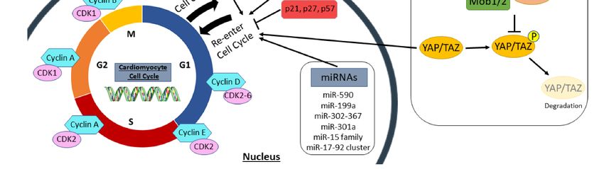
Int. J. Mol. Sci. 2021, 22, x FOR PEER REVIEW 3 of 15
Int. J. Mol. Sci. 2021, 22, 7764 3 of 16
the heart elevated cardiomyocyte DNA synthesis and increased the number of small mon-
onucleated cardiomyocytes in the heart [28].
Figure
Figure1.1.Regulators
Regulatorsof ofthe
thecardiomyocyte
cardiomyocytecell cellcycle.
cycle.Summary
Summaryof ofsignaling
signalingpathways,
pathways,cell
cellcycle
cyclegenes,
genes,and
andextracellular
extracellular
stimuli
stimulithat
that induce
induce cardiomyocyte proliferation,as
cardiomyocyte proliferation, asdiscussed
discussedininthis
thisreview.
review. This
This includes
includes cellcell cycle
cycle promotors
promotors andand inhib-
inhibitors,
itors, both the hippo and neuregulin pathways, microRNAs, and metabolic regulators, such as
both the hippo and neuregulin pathways, microRNAs, and metabolic regulators, such as hypoxia and PDK4. Cyclins hypoxia and PDK4. Cyclins
and cyclin-dependent kinases promote cell cycle activation and progression, while cyclin-dependent kinase inhibitors
and cyclin-dependent kinases promote cell cycle activation and progression, while cyclin-dependent kinase inhibitors
such as p21, p27, and p57 inhibit the cell cycle. In the hippo pathway, activation of upstream kinases (Mst1/2, Sav1, Mob1/2,
such as p21, p27, and p57 inhibit the cell cycle. In the hippo pathway, activation of upstream kinases (Mst1/2, Sav1,
and Lats1/2) phosphorylate the downstream transcription co-activators (YAP/TAZ) to promote their degradation. How-
Mob1/2,
ever, and Lats1/2)
inactivation phosphorylate
of upstream kinasesthe downstream
causes YAP/TAZ transcription co-activators
to translocate (YAP/TAZ)
to the nucleus to promote
to promote their degradation.
cell cycle re-entry in
However, inactivation
cardiomyocytes. of upstream
Neuregulin (NRG1)kinases causes
binds to YAP/TAZ
its tyrosine to translocate
receptor to the nucleus
subunits (ERBB2 to promote
and ERBB4) cell cycle
to activate re-entry in
a downstream
cardiomyocytes.
signaling pathway Neuregulin
(PI3K/AKT) (NRG1) binds to
and induce its tyrosine
myocyte receptor subunits
proliferation. (ERBB2 and ERBB4)
Several microRNAs (miRNAs) to activate a downstream
also regulate the cell
signaling
cycle pathwaynew
to influence (PI3K/AKT)
myocyte and induce Hypoxia
formation. myocyte proliferation.
and PDK4 lead Several microRNAs
to a metabolic (miRNAs)
switch also regulate the
in cardiomyocytes cellmito-
from cycle
chondrial oxidative
to influence phosphorylation
new myocyte formation. to anaerobic
Hypoxia andglycolysis
PDK4 leadfor toenergy production.
a metabolic This
switch in metabolic change
cardiomyocytes frominfluences cell
mitochondrial
cycle activation
oxidative in cardiomyocytes.
phosphorylation PDK4,glycolysis
to anaerobic pyruvate dehydrogenase kinase 4. This metabolic change influences cell cycle
for energy production.
activation in cardiomyocytes. PDK4, pyruvate dehydrogenase kinase 4.
Inhibition of cyclin-dependent kinase inhibitors (CKIs) has also been shown to reac-
tivateInhibition
the cell cycle in cardiac myocytes.
of cyclin-dependent kinaseTriple knockdown
inhibitors (CKIs) ofhasCKIs
also including
been shown p21,
to p27,
reac-
and p57the
tivate induced myocyte
cell cycle proliferation
in cardiac myocytes. with evidence
Triple of karyokinesis
knockdown and cytokinesis
of CKIs including in
p21, p27,
and p57 and
neonatal induced
adultmyocyte proliferation
cardiomyocytes [29]. with evidence
Likewise, of karyokinesis
inactivation and cytokinesis
of cyclin-dependent ki-
in neonatal
nase inhibitorand adult cardiomyocytes
2a (CDKN2a) [29]. Likewise,
or downregulation inactivation
of p21 alone inducedofcardiomyocytes
cyclin-dependent to
kinase
enter inhibitor
mitosis and2a (CDKN2a)
synthesize DNAor downregulation
[30,31]. Numerous of p21 aloneoninduced
studies cardiomyocytes
regulating various cell
to enter
cycle mitosis and
transcription synthesize
factors DNA
including [30,31].
E2F-1, E2F2, Numerous
Meis1, Tbx6,studies on regulating
and Tbx20, various
have also pro-
cell cycle
vided transcription
evidence factors including
that the proliferative E2F-1,
potential of E2F2, Meis1, Tbx6,
adult cardiac and can
myocytes Tbx20, have also
be enhanced
provided
to promoteevidence
new myocyte that the proliferative
formation potential of cardiomyocytes
from pre-existing adult cardiac myocytes
[32–37]. can be en-
Recently,
hanced to promote new myocyte formation from pre-existing cardiomyocytes
a combination treatment of cell cycle activators and inhibitors induced 15–20% of cardio- [32–37].
Recently, to
myocytes a combination
re-enter the cell treatment of cell
cycle and cyclereduced
divide, activators
scarand
sizeinhibitors
post MI, induced 15–20%
and attenuated
of cardiomyocytes
cardiac dysfunction to re-enter
[38]. Althoughthe cell
thesecycle and prove
studies divide, reduced
that scar size proliferation
cardiomyocyte post MI, and
attenuated
can be achievedcardiac dysfunction
to promote [38].regeneration
cardiac Although these studiesinprove
and repair that cardiomyocyte
cardiovascular diseases,
proliferation can be achieved to promote cardiac regeneration and repair
there is still a limitation in the number of new myocytes formed. In many of these findings, in cardiovascular
diseases, there is
cardiomyocyte still
cell a limitation
cycle activity in
wasthere-activated
number of new and myocytes
promotedformed. In many of
DNA synthesis butthese
re-
findings, cardiomyocyte cell cycle activity was re-activated and promoted
sulted in multinucleation or polyploidy without complete cytokinesis. Further study of DNA synthesis
but resulted in multinucleation or polyploidy without complete cytokinesis. Further study
of signaling pathways in the cardiomyocyte cell cycle is needed to determine how to induce
cardiomyocytes to enter the cell cycle and divide to form two daughter cells.Int. J. Mol. Sci. 2021, 22, 7764 4 of 16
Table 1. Therapeutic stimulation of endogenous cardiomyocyte proliferation and cardiac regeneration.
Pathway Molecular Factor Species Injury Completed Division Results Reference
Cyclin D1 Mouse No Injury No >40% Adult cardiomyocytes re-entered the cell cycle [20]
Cyclin D2 Mouse MI Yes Cardiomyocyte DNA synthesis up to 5 months post-MI [21]
Cyclin A2 Mouse MI Yes, in vitro Cardiac Hyperplasia develops in postnatal hearts [24]
Cell cycle promoters Cyclin A2 Rat, Pig HF, MI Yes ~18% increase in porcine ejection fraction post MI [25,26]
Cyclin B1-CDC2 Rat No Injury No * Increased number of small mononucleated myocytes [27]
CDK2 Mouse Pressure Overload No * Increased number of small less-differentiated [28]
mononuclear cardiomyocytes
Adult cardiomyocytes completed cytokinesis, expressed
p21, p27, p57 (tKD) Mouse, Rat No Injury Yes [29]
Cell cycle inhibitors neonatal genes, and resembled neonatal morphology
p21 Mouse No Injury No Induced DNA synthesis without completion of mitosis [31]
Cell cycle Increased cytokinetic and mononucleated cardiomyocytes
Meis1, Hoxb13 (dKO) Mouse MI Yes [37]
transcription factors and improved EF post MI
Combination of cell 15–20% Of adult mouse myocytes re-entered the cell cycle
4F, 2F-2i Mouse, Rat, Human MI Yes [38]
cycle regulators and completed cytokinesis
human YAP Mouse MI No Increased cardiomyocyte proliferation post MI [39]
Hippo hippo (KO) Mouse MI Yes Adult cardiomyocytes re-entered cell cycle [40]
Salv (KO, KD) Mouse HF Yes * DNA synthesis in adult myocytes up to 9 weeks post MI [41]
Neuregulin NRG1, ERBB4 Mouse, Rat MI Yes Only mononuclear cardiomyocytes completed division [42]
ERBB2 Mouse MI Yes Induced myocyte proliferation and redifferentiation [43]
human miR-590, Induced cardiomyocyte proliferation and division, and
Mouse MI Yes [44]
human miR-199a cardiac repair post MI
human miR-199a Pig IR Yes Increased myocyte proliferation at 12 days post IR [45]
miR-17-92 cluster Mouse MI Yes Promoted cardiomyocyte DNA synthesis in adult mice [46]
miRNAs
miR-19a/19b Mouse HF, MI Yes Induced cardiac regeneration up to 12 months post-MI [47]
miR-15 family (KD) Mouse IR No Increased proliferation of adult cardiomyocytes post IR [48]
miR-302-367 Mouse MI Yes Robust cardiomyocyte proliferation in the adult heart [49]
miR-301a Mouse MI Yes, in vitro Induced cell cycle re-entry of cardiomyocytes after MI [50]
Hypoxia Mouse MI Yes Increased cardiomyocyte division and cardiac repair [51]
Metabolic regulators
PDK4 (KO, KD) Mouse MI Yes Enhanced cardiomyocyte proliferation post MI [52]
miR, microRNA; KD, knockdown; tKD, triple knockdown; KO, knockout; dKO, double knockout; MI, myocardial infarction; HF, heart failure; IR, ischemia reperfusion; EF, ejection fraction; 4F represents
4 factors, i.e., CDK1, CCNB, CDK4, and CCND; 2F-2i represents 2 factors and 2 inhibitors, i.e., CDK4, CCND, MK1775 (Wee1 inhibitor), and SB431542 (TGFβ inhibitor). (*) Indicates cardiomyocyte division was
indirectly implied in the reference but needs to be confirmed by experimental evidence.Int. J. Mol. Sci. 2021, 22, 7764 5 of 16
2.2. Manipulation of Signaling Pathways to Promote New Myocyte Formation
Embryonic heart development and postnatal myocyte maturation are regulated by
multiple signaling pathways that have been modulated to stimulate cardiomyocyte pro-
liferation. One of the conserved signaling cascades involved in myocyte growth is the
hippo pathway. This pathway functions to restrict cardiomyocyte proliferation through
phosphorylation-mediated inactivation of its downstream transcription co-activators
YAP/TAZ, to cause cardiomyocytes to withdraw from the cell cycle. Therefore, inac-
tivation of hippo signaling in embryos results in enlarged hearts from overactivation of
the cardiomyocyte cell cycle [53]; conversely, embryonic deletion of YAP caused fetal
death due to cardiac hypoplasia, whereas a constitutively active form of Yap resulted in
upregulation of β-catenin, a known inducer of cardiomyocyte proliferation, and increased
myocardial mass through cardiomyocyte proliferation rather than hypertrophy [54,55].
The interaction of YAP and TEAD transcription factors in the hippo pathway is required for
cardiomyocytes to re-enter the cell cycle and divide [55], as conditional knockout of TEAD1
in murine neonates lead to complete lethality by postnatal day 9 [56]. Understanding the
role of the hippo pathway in cardiac development has led to investigations that explored
the idea that the hippo signaling cascade can be altered to promote cardiac regeneration
following injury. These studies have shown that overexpression of YAP in cardiomyocytes
attenuated myocardial fibrosis, increased cardiomyocyte cell cycle activity, and improved
cardiac function following MI in neonatal and adult mice [57]. Likewise, AAV9 delivery
of human YAP in adult mice hearts induced cardiomyocyte mitosis, improved cardiac
contractility, and enhanced mice survival after MI [39]. In contrast, heterozygous deletion
of YAP increased cardiomyocyte apoptosis and prevented myocyte hypertrophy after MI,
which led to deleterious cardiac remodeling and poor pump function [58].
Other hippo-related genes can also induce adult cardiomyocytes to re-enter the cell
cycle. Inactivation of the hippo pathway by conditional knockout of its regulatory Salv
gene in adult mice has been shown to reduce scar size, induce new myocyte formation,
and attenuate cardiac dysfunction in MI and HF disease models [40,41]. Related studies
have shown that adult myocyte overexpression of YAP alters their gene expression to re-
semble embryonic myocytes in the cell cycle. More specifically, YAP reorganizes chromatin
accessibility in TEAD and AP1 motifs to activate transcription of fetal and proliferative
genes in cardiomyocytes [59]. YAP also binds to the B-MYB subunit of the Myb-MuvB
(MMB) complex, which regulates mitotic genes, to activate transcription of cell cycle genes
in cardiomyocytes and promote their proliferation [60].
The neuregulin/ERBB2/ERBB4 pathway is also involved in cardiomyocyte prolifer-
ation. Neuregulin (NRG1) is an extracellular mitogen that binds to its tyrosine receptor
subunits ERBB2 and ERBB4 to activate downstream signaling pathways including the
PI3K/AKT pathway to induce cardiomyogenesis during fetal heart development. In adult
mammals, the neuregulin pathway can activate the cell cycle in mature cardiomyocytes to
induce proliferation. Treatment of adult rat ventricular myocytes with NRG1 promoted
mostly mononucleated cardiomyocytes to re-enter the cell cycle and complete cytokine-
sis [42]. In vivo, overexpression of ERBB4 in the postnatal heart induced DNA synthesis
in myocytes, and injection of recombinant NRG1 in control or post-MI adult mice stim-
ulated new myocyte formation by cardiomyocyte proliferation and not from progenitor
cell differentiation [42]. Similarly, overexpression of ERBB2 in all ages of mice caused
cardiac enlargement due to widespread cardiomyocyte proliferation and hypertrophy;
however, transient induction of ERBB2 in adult hearts post MI induced cardiomyocyte
dedifferentiation, proliferation, and redifferentiation to promote a more limited cardiac
regeneration that improved heart function [43]. Activations of the NRG1/ERBB2 signaling
caused a metabolic switch in border zone cardiac myocytes from oxidative phosphorylation
to glycolysis, with an upregulation in glycolysis genes and a reduction in mitochondrial
function [61]. This metabolic reprogramming induced adult murine cardiomyocytes to
re-enter the cell cycle and divide, resembling embryonic cardiac myocytes [61].Int. J. Mol. Sci. 2021, 22, 7764 6 of 16
Other signaling pathways, including PI3K/AKT/CDK7, Wnt/β-catenin, PDGFR-β,
and Notch signaling pathways can also stimulate cardiomyocytes to proliferate and induce
cardiac regeneration during heart disease or injury [62–65]. Furthermore, several studies
have discovered that multiple mitotic pathways within cardiomyocytes can crosstalk to
stimulate proliferation of pre-existing cardiomyocytes, thus, increasing myocardial mass
and promoting cardiac repair [54,66–68]. One study showed that transient overexpression
of ERBB2 activated YAP mechanotransduction signaling and an epithelial-mesenchymal
transition-like response in cardiomyocytes that induced cardiac regeneration in a HF
murine model [69]. Collectively these studies show that manipulation of several cell cycle
pathways, including hippo and neuregulin signaling, can induce cardiomyocyte mito-
sis and attenuate cardiomyocyte necrosis and cardiac dysfunction following injury. A
caution is that prolonged modulation of these pathways can lead to hypertrophic cardiomy-
opathy [43] or heart failure, where persistent inactivation of the hippo pathway causes
extensive cardiomyocyte dedifferentiation which impairs pump function and repair of the
MI injured heart [70]. Therefore, controlled, transient regulation of these pathways needs
to be developed as a strategy to replace myocytes lost to disease, and thereby improve
myocardial function following injury.
2.3. Selective Induction/Inhibition of Gene Expression can Induce Cell Cycle Re-Activation in
Cardiac Myocytes
MicroRNAs (miRNAs) regulate post-transcriptional gene expression and several
miRNAs have been linked to cardiomyocyte proliferation. High throughput functional
screening has identified 40 human miRNAs that promote cardiomyocyte growth and divi-
sion, with miR-590 and miR-199a as the major regulators of this process in neonatal and
adult rodents [71]. Following MI, therapeutic delivery of both miRNAs with an AAV9
vector improved cardiac contractility, reduced scar size, and increased cardiomyocyte
proliferation [71]. Similar results were reported when a single intracardiac injection of
synthetic human miR-590-3p and miR-199a-3p mimics promoted cardiac regeneration and
increased cardiac function in mice post MI [44]. Expression of miR-199a also benefited
ischemic hearts of a large animal model [45]. Another mitogen that was initially known
as an oncogene, is the miR-17-92 cluster, which is required for cardiomyocyte prolifer-
ation in embryonic and neonatal hearts; overexpression of this cluster in the heart can
induce cardiomyocyte DNA synthesis at all ages in mice and following cardiac injury, thus,
protecting the heart from MI-induced remodeling and dysfunction [46]. Of this cluster,
miR-19a/19b are most potent at stimulating cardiac regeneration by causing enhanced
cardiomyocyte proliferation, inhibition of apoptosis and proinflammatory immune re-
sponses, reduced fibrosis, and improved pump function up to 12 months post MI [47].
Alternatively, the miR-15 family plays a role in cardiomyocyte withdrawal from the cell
cycle and is upregulated in cardiac ventricles by postnatal day 10, with miR-195 being the
most elevated [72]; miR-195 represses several cell cycle genes including checkpoint kinase 1
(Chek1) to inhibit myocyte proliferation after birth. Thus, knockdown of the miR-15 family
increased mitosis in neonatal cardiac myocytes [72] and in adult mice which was protective
against MI-induced cardiac injury [48].
Several miRNAs have also been shown to regulate genes in signaling pathways that
influence cardiomyocyte mitosis. The miR-302-367 cluster represses signal transduction in
the hippo pathway to induce cardiomyocyte proliferation and this cluster is required for
embryonic development, as cardiac knockdown of miR-302-367 results in hypoplasia [49].
Therapeutically, transient expression of miR-302b/c, a member of the miR-302-367 cluster,
increased cardiac mass and enhanced heart function in an MI mouse model [49]. Like-
wise, other pro-proliferative miRNAs, including miR-199a-3p, activate YAP in the hippo
pathway to enhance DNA synthesis in cardiomyocytes. More specifically, miR-199a-3p
downregulates YAP inhibitory kinase, TAOK1, and the E3 ubiquitin ligase, β-TrCP, to
inhibit YAP degradation, and also represses Cofilin2 to maintain YAP nuclear localiza-
tion [73]. PTEN/PI3K/AKT is another signal transduction pathway that is activated by
miR-301a to promote cardiomyocyte re-entry into the cell cycle; miR-301a suppresses PTENInt. J. Mol. Sci. 2021, 22, 7764 7 of 16
to activate AKT and induce expression of cell cycle genes such as cyclin D1, in cardiac
myocytes [50]. Other miRNAs have been reported to promote cell cycle progression in
cardiomyocytes by targeting the TBX1/JAK2/STAT1 pathway [74], the NFkB pathway [75],
or signaling molecules such as the cell cycle inhibitors Wee1 [76] and Bim [77], among
others [78,79]. These studies suggest that MiRNAs possess the potential for replenishing
cardiomyocyte numbers in injured hearts. A cautionary note is that prolonged expression
of miRNAs can lead to cardiac arrhythmias and death from an accumulation of dedifferenti-
ated and hyper-proliferative cardiac myocytes [45,49]. Therefore, it appears that controlled,
transient expression of miRNAs is an essential component in the use of these molecules to
prevent/repair cardiac damage [49].
2.4. Changes in Cardiac Myocyte Metabolism Can Induce Cell Cycle Re-Entry
During embryonic development, the mammalian heart primarily utilizes anaerobic
metabolism to produce energy and fetal cardiomyocytes have a high proliferative capacity;
immediately after birth, the heart is exposed to blood with a much greater oxygen content
and there is a switch to aerobic metabolism to fuel the increased contractile and functional
requirements. This switch to higher oxygen levels is associated with the exit of cardiomy-
ocytes from the cell cycle and reduces their mitotic potential [80]. Neonatal exposure to
hyperoxia resulted in the production of reactive oxygen species (ROS), oxidative DNA
damage, and activation of the DNA damage response (DDR) pathway which all reduce
cardiomyocyte proliferation. Hypoxia treatment of neonates maintains postnatal prolifera-
tion of cardiac myocytes and inhibits oxidative stress [80]. These finding suggest that fetal
cardiomyocytes require hypoxia for cell cycle progression during cardiac development,
which is mediated by HIF1α expression in embryonic hearts [81]. Adult hearts may contain
a small group of hypoxic cardiomyocytes that are mononucleated and can re-enter the cell
cycle and divide [82]. The low cardiomyocyte renewal rate (Int. J. Mol. Sci. 2021, 22, 7764 8 of 16
Int. J. Mol. Sci. 2021, 22, x FOR PEER REVIEW 8 of 15
2. Systemic
Figure 2. Systemic hypoxia
hypoxia cancan induce
induce cardiomyocyte
cardiomyocyte proliferation
proliferation and
and cardiac
cardiac regeneration.
regeneration. Healthy
Healthy adult hearts
hearts have
have
normal cardiac function,
function, blood
bloodflow,
flow,and
andutilize
utilizeoxidative
oxidativephosphorylation
phosphorylation toto
generate
generateenergy. Following
energy. myocardial
Following myocardialin-
farction (MI),
infarction (MI),there
thereisisincreased
increasedcardiomyocyte
cardiomyocytehypertrophy,
hypertrophy,scar
scarformation,
formation, reduced
reduced cardiac
cardiac pump
pump function, and minimal
function, and minimal
cardiomyocyte proliferation.
cardiomyocyte proliferation. These
Theseadverse changes
adverse changescancan
progress
progressandand
leadlead
to heart failure
to heart overtime
failure withwith
overtime increased dila-
increased
tion of the myocardium, poor blood circulation, and declined cardiac function. However, a recent study in mice [51]
dilation of the myocardium, poor blood circulation, and declined cardiac function. However, a recent study in mice [51]
demonstrated that systemic hypoxemia post MI caused cardiomyocytes to undergo a metabolic switch from oxidative
demonstrated that systemic hypoxemia post MI caused cardiomyocytes to undergo a metabolic switch from oxidative
phosphorylation to anaerobic glycolysis. Hypoxia also enhanced cardiomyocyte proliferation and hypoxic myocytes re-
phosphorylation to anaerobic glycolysis.
sembled fetal cardiomyocytes. Hypoxic miceHypoxia also enhanced
had increased cardiomyocyte
angiogenesis, proliferation
reduced scar and hypoxic
size, and improved myocytes
cardiac func-
resembled fetal cardiomyocytes. Hypoxic mice had increased angiogenesis, reduced scar
tion. When the mice returned to normoxia, cardiac repair and elevated cardiac pump function was sustained. size, and improved cardiac
function. When the mice returned to normoxia, cardiac repair and elevated cardiac pump function was sustained.
2.5. Limitations in Cardiomyocyte Proliferation Techniques
Fetal mitotic cardiomyocytes utilize pyruvates in anaerobic glycolysis to generate
energy,Several
but bymethods
postnatal have
day been
7, theyemployed
switch toto studyfatty
utilize cardiomyocyte proliferation
acids in mitochondrial (Table
oxidative
2). Most commonly, cardiomyocyte cell cycle activity is measured
phosphorylation for energy and most cardiomyocytes have exited the cell cycle. Reversal by 5-ethynyl-2’-deox-
yuridine (EdU) can
of this process incorporation into new DNAproliferation,
prolong cardiomyocyte or the presence as of proteinsmice
neonatal onlyfedexpressed
a fatty
during
acid deficient diet extended the proliferative window of cardiomyocytes up to 3 B.
the cell cycle, such as Ki67, phosphorylated histone H3 (pHH3), or Aurora EdU
weeks
is incorporated
postnatally [52].into newlyfollowing
In adults formed DNA, Ki67 is expressed
MI, therapeutic deletionin the G1 to M phase
of pyruvate of the cell
dehydrogenase
cycle, pHH3 is expressed during chromosome condensation,
kinase 4 (PDK4) increased glucose oxidation which induced cardiomyocyte mitosis, and and Aurora B is expressed
during
attenuatedmitosis and during
adverse cardiac cell division
structure and [92]. Many studies
function. Likewise, have concluded thatinhibition
pharmacological new my-
ocytes are present (with cytokinesis) when these cell cycle markers
of PDK4 promoted cardiomyocyte proliferation with complete cytokinesis [52]. Together, are found. However,
other possibilities
alteration include endoreplication,
in mitochondrial metabolism to reduce endomitosis,
fatty acidand DNA damage/repair.
utilization can induce cell Most
cycle
of these markers
re-entry indicate cell
in cardiomyocytes. cycle re-entry
Confirmation of and
theseDNA synthesis,
findings wouldbut they do
enhance not reliably
confidence for
determine
translating ifthis
cardiomyocytes
novel therapy undergo
into largecell division,
animal models polyploidy,
and patients.or polynucleation. There-
Interestingly, humans
fore,
livingthese represent
at higher markers
altitudes haveof cell cycle activity
reduced mortality that maycoronary
from or may not leaddisease
heart to cell division
[89–91],
and the production of new cardiomyocytes.
suggesting that hypoxemia and modulation of mitochondrial oxidative metabolism may
offsetAthose
number of mouse
factors models
that induce have been
cardiac generated to provide more direct evidence of
diseases.
cardiomyocyte renewal. Mosaic analysis with double markers (MADM) mouse models
2.5. Limitationslabel
fluorescently in Cardiomyocyte
cardiomyocytes Proliferation
that have Techniques
completed cytokinesis. Using Cre-loxP re-
combination, daughter
Several methods cellsbeen
have are employed
single-labeled green
to study or red after cell
cardiomyocyte division [93].
proliferation (TableThis
2).
model was used cardiomyocyte
Most commonly, to validate low cell cardiomyocyte
cycle activityturnover
is measured in adults and that 0new
by 5-ethynyl-2 myocytes
-deoxyuridine
formedincorporation
(EdU) after birth or intopostnew
MI areDNA derived
or thefrom pre-existing
presence cardiomyocytes
of proteins only expressed [9].during
Rainbow,the
cell cycle,
also referredsuchtoasasKi67, phosphorylated
Brainbow mice, trackshistone
clonal H3 (pHH3),oforcardiomyocytes.
expansion Aurora B. EdU is incorpo-
Following
rated into newly
Cre-mediated formed DNA,
recombination, Ki67 myocytes
several is expressed are in the G1 to
randomly M phase
labeled withofone
theofcell
fourcycle,
flu-
pHH3
orescent is expressed
colors andduringanalysischromosome
of the hearts condensation,
at terminal and Aurora
studies B is expressed
confirms during
proliferation byInt. J. Mol. Sci. 2021, 22, 7764 9 of 16
mitosis and during cell division [92]. Many studies have concluded that new myocytes
are present (with cytokinesis) when these cell cycle markers are found. However, other
possibilities include endoreplication, endomitosis, and DNA damage/repair. Most of these
markers indicate cell cycle re-entry and DNA synthesis, but they do not reliably determine
if cardiomyocytes undergo cell division, polyploidy, or polynucleation. Therefore, these
represent markers of cell cycle activity that may or may not lead to cell division and the
production of new cardiomyocytes.
Table 2. Strategies to measure cardiomyocyte proliferation and division.
Approach Labeling Strategy Advantages Cell Cycle Phase Limitations
Labeling cardiomyocytes that EdU also labels cardiomyocytes
undergo DNA synthesis; EdU undergoing DNA damage and
EdU S repair; no evidence of whether
processing is easy, rapid, myocytes continue to mitosis
and sensitive and complete division
Does not identify if
Identifies cardiomyocytes that
Ki67 G1, S, G2 cardiomyocytes undergo
re-entered the cell cycle
cytokinesis
Unable to distinguish whether
cardiac myocytes underwent
Markers
Labeling of cardiomyocytes that cytokinesis, endoreplication, or
pHH3 G2, M
have entered Mitosis poly-nucleation; short
expression time during mitosis
so low detection
Expressed during shortest
Present between two daughter
Aurora B G2, M, cytokinesis phases of the cell cycle (M and
cells during cytokinesis
cytokinesis) so low detection
Sorts live, isolated cardiomyocytes Unable to sort isolated
cardiomyocytes from large
Molecular Beacons that are in anaphase and M, cytokinesis animal models by
telophase/cytokinesis
flow cytometry
Labeling of cardiomyocytes yellow
(GFP + RFP) when they enter the
GFP + RFP: G1-M, only Daily or frequent tamoxifen
cell cycle and upon completion of
MADM GFP and only injections can be cytotoxic for
cell division, daughter cells are
RFP: cytokinesis the animal
labeled as only green (GFP) and
only red (RFP)
Clonal expansion of labeled
cardiomyocytes identifies Indirectly measures Indirect way to measure
Rainbow
myocytes that proliferated from cytokinesis cardiomyocyte division
pre-existing myocytes
Mouse Models Identifies which stages of the cell AzG labeling unable to
cycle cardiomyocytes are present, mKO: G1, AzG: S, G2, M; distinguish whether myocytes
FUCCI by oscillations of an orange (mKO) mKO + AzG: G1/S cell are in S phase, G2 phase, or M
and green (AzG) color within cycle arrest phase; inability to
cardiomyocyte nuclei visualize cytokinesis
Inability to distinguish whether
Cardiomyocytes are labeled red
myocytes in vivo are in S phase,
when they undergo proliferation
G2 phase, M phase, or
Aurkb and division; Cytokinesis S, G2, M, cytokinesis
cytokinesis; completed
visualized by live time
cytokinesis limited to live
lapse imaging
cell imaging
Estimates the number of Indirectly measures new Accuracy of cardiomyocyte
3D Imaging Stereology cardiomyocytes in the heart using
representative thick sections myocyte numbers number is limited
Phases of the cell cycle consist of G1 (cell growth), S (DNA synthesis), G2 (cell growth and preparation for mitosis), and the mitotic phase
that includes M (mitosis, nuclear division) and cytokinesis (cytoplasmic division). EdU, 5-ethynyl-20 -deoxyuridine; pHH3, phosphorylated
histone H3; MADM, mosaic analysis with double markers; FUCCI, fluorescence ubiquitin cell cycle indicator; Aurkb, Aurora kinase B; GFP;
green fluorescent protein; RFP, red fluorescent protein; mKO, monomeric Kusabira Orange; AzG, Azami Green.
A number of mouse models have been generated to provide more direct evidence of
cardiomyocyte renewal. Mosaic analysis with double markers (MADM) mouse models
fluorescently label cardiomyocytes that have completed cytokinesis. Using Cre-loxP re-
combination, daughter cells are single-labeled green or red after cell division [93]. ThisInt. J. Mol. Sci. 2021, 22, 7764 10 of 16
model was used to validate low cardiomyocyte turnover in adults and that new myocytes
formed after birth or post MI are derived from pre-existing cardiomyocytes [9]. Rainbow,
also referred to as Brainbow mice, tracks clonal expansion of cardiomyocytes. Following
Cre-mediated recombination, several myocytes are randomly labeled with one of four
fluorescent colors and analysis of the hearts at terminal studies confirms proliferation by
single-color clusters, indicating one myocyte divided to generate adjacent myocytes. This
mouse model was used in a lineage tracing study on clonal distribution during cardiac
development [94], and corroborated increased cardiomyocyte proliferation when YAP was
overexpressed in the heart [39]. Another mouse model where cardiomyocytes express fluo-
rescence ubiquitination-based cell cycle indicators (FUCCI) specifically revealed when cells
progress from G1 to end stages of mitosis in the cell cycle. Cardiac myocytes fluorescently
express monomeric Kusabira Orange (mKO) when they are in the G1 phase and express
Azami Green (AzG) when they are in S, G2, or M phases of the cell cycle; dual labeling of
both colors indicates cell cycle arrest [95]. This allows a temporal view of cardiomyocyte
cycling through the cell cycle with cytokinesis identified by the presence of two colorless
daughter cells.
More recently, an Aurora Kinase B (Aurkb) mouse system was generated to label car-
diomyocytes red when Aurora B was expressed during mitosis and cytokinesis [96]. This
approach tracks myocytes that undergo cell proliferation and division, which eliminates
controversial problems of Aurora B staining including non-specific staining and transient
expression during mitosis. Other new methods to detect cardiomyocyte proliferation
involves using molecular beacons to target anaphase and telophase/cytokinesis mRNA in
cardiomyocytes followed by cell sorting to identify cycling myocytes that complete cell divi-
sion [97]. This technique allows isolation of a purer population of dividing cardiomyocytes
by eliminating myocytes that only undergo endoreplication or endomitosis; however, this
technique is limited to in vitro and ex vivo experiments. In addition, quantification of car-
diomyocytes by stereological practices provides measurements of cardiomyocyte number
and nucleation, which can determine the amount of new myocyte formation as compared
with control hearts [98]. These new systems should be used to confirm whether previously
reported data on cardiomyocyte renewal actually represents the generation of new cardiac
myocytes by accurately identifying cell cycle progression and completion of mitosis in
cardiac myocytes.
3. Conclusions
The human heart is believed to be a post mitotic organ, but numerous studies in ani-
mals and humans have provided evidence suggesting there can be a low rate of myocyte
renewal. The mammalian heart has a high myocyte proliferative capacity following birth,
but after several days, these cells exit the cell cycle. There are only a few cardiomyocytes
that maintain their proliferative potential throughout life which is likely responsible for
the yearly 1% cardiomyocyte renewal in humans. Following cardiac injury, embryonic
and postnatal animals can completely or partially regenerate their myocardium by pro-
liferation of pre-existing cardiomyocytes. In adults, few cardiomyocytes re-enter the cell
cycle and divide after injury, which leads to adverse cardiac remodeling and function.
Studies of embryonic cardiac development and postnatal cardiomyocytes have identified
regulators of the cardiomyocyte cell cycle that can be manipulated in adults to induce
cardiomyocyte mitosis.
Overexpression of cell cycle promoters such as cyclins and CDKs enhance cardiomy-
ocyte proliferation and prolonged expression can result in cardiac hyperplasia. These
cell cycle stimulators are reparative in MI and TAC injury models by promoting new
myocyte formation, reducing fibrosis, and attenuating cardiac pump malfunction. Like-
wise, repression of CKIs or cell cycle inhibitors induce DNA synthesis in cardiac myocytes
which can be synergistically enhanced when combined with cell cycle activators. Signal-
ing pathways involved in cell cycle progression such as the neuregulin/ERBB2/ERBB4
pathway or in cardiomyocyte cell cycle arrest including the hippo-YAP pathway haveInt. J. Mol. Sci. 2021, 22, 7764 11 of 16
been targeted to promote proliferation of pre-existing myocytes. Upregulation of YAP or
inhibition of hippo genes in neonatal and adult hearts increase cardiac mass by inducing
proliferation of mature cardiac myocytes. In disease models, modulation of this pathway
improves cardiac repair and animal survival, but persistent activation of YAP can leave
many cardiomyocytes in a dedifferentiated state which can lead to heart failure. However,
regulation of YAP by transient expression of miR-302-367 has been reported to prevent
heart failure and increased cardiac regeneration following MI. Other miRNAs have also
been reported to work independently or in conjunction with several signaling pathways to
reactivate the cardiomyocyte cell cycle and promote division, thus, improving MI outcomes.
Importantly, miRNA modulation of mitotic gene expression and manipulation of cell cycle
signaling pathways need to be tightly controlled to restore cardiomyocyte numbers after
injury without provoking tumorigenesis or accumulation of immature cardiac myocytes.
Recent studies have suggested that hypoxemia in adult mice could induce proliferation of
cardiomyocytes in healthy and post MI hearts by causing a metabolic switch in cardiomy-
ocyte mitochondrial oxidation, from oxidative phosphorylation to glycolysis. These studies
only showed modest improvement in cardiac function and cardiomyocyte proliferation,
and while myocytes may have entered the cell cycle and increased DNA synthesis, it was
not clear that they completed cytokinesis and generated new myocytes. More research
is required to discover new pathways and mechanisms that can influence cardiomyocyte
division and avoid endoreplication or endomitosis. Implementation of these practices can
increase the therapeutic potential of these treatments for enhancing cardiac regeneration
and repair following injury.
An obstacle in determination of new myocyte formation is the accurate interpretation
of studies using proliferative markers such as EdU, Ki67, pHH3, and Aurora B. Most
of these markers indicate cell cycle re-entry and DNA synthesis but only Aurora B is
present during cytokinesis [99]. However, Aurora B antibodies can have nonspecific
staining and lead to false conclusions [92]. Several new transgenic mouse models have
been established to define cardiomyocyte proliferation more clearly. MADM mice label
daughter cells of cardiac myocytes that have successfully completed cytokinesis, while
Rainbow mice analyze clonal expansion of a single-colored myocyte that divided to form
a cluster of same-colored myocytes. Aurora kinase B mice also label dividing cardiac
myocytes. Therefore, these three mouse models are available to define cardiomyocyte
division with complete cytokinesis. FUCCI mice are better suited for in vitro analysis of
cardiomyocyte proliferation through time lapse imaging, where dividing cardiomyocytes
can be visualized as they progress through the cell cycle and complete division. Methods
to isolate and quantify newly formed myocytes via molecular beacons and stereological
measurements, respectively, could also work collectively with these recently designed
mouse models to better define true cases of cardiomyocyte proliferation.
These new approaches should be implemented in future studies to develop reliable
strategies for generating new cardiac myocytes and clarifying if/when new myocytes
are generated. For example, it was originally thought that cardiomyocytes retain their
proliferative capacity up to 7 days after birth [8] but a study using FUCCI mice showed
cardiomyocyte cell cycle activity was almost complete after postnatal day 2 with most
myocytes arrested at the G1/S transition of the cell cycle [95]. In another study, it was
concluded that new cardiomyocyte formation increased four-fold following MI injury [11];
reassessment of cardiomyocyte proliferation after MI in FUCCI mice showed that cardiomy-
ocytes could indeed re-enter the cell cycle but they did not divide [95]. Therefore, these new
methods should allow more precise identification of dividing myocytes and more accurate
timelines of their cell cycle activity, which should aid in finding the optimal approach
for inducing cardiomyocyte renewal. The ideal therapy would replenish cardiomyocyte
numbers following injury, and would also promote angiogenesis, modulate inflammation,
and reduce fibrosis for complete cardiac repair and regeneration.Int. J. Mol. Sci. 2021, 22, 7764 12 of 16
Author Contributions: Conceptualization and writing—original draft preparation, J.J.; writing—
review and editing, S.M. and S.R.H.; supervision, S.R.H.; funding acquisition, S.R.H. All authors
have read and agreed to the published version of the manuscript.
Funding: This research was funded by the National Institute of Health predoctoral grant 5
F31 HL143865-03.
Conflicts of Interest: The authors declare no conflict of interest. The funders had no role in the design
of the study; in the collection, analyses, or interpretation of data; in the writing of the manuscript, or
in the decision to publish the results.
References
1. Virani, S.S.; Alonso, A.; Benjamin, E.J.; Bittencourt, M.S.; Callaway, C.W.; Carson, A.P.; Chamberlain, A.M.; Chang, A.R.; Cheng, S.;
Delling, F.N.; et al. Heart Disease and Stroke Statistics—2020 Update: A Report From the American Heart Association. Circulation
2020, 141, e139–e596. [CrossRef]
2. Sutton, M.G.; Sharpe, N. Left ventricular remodeling after Myocardial infarction: Pathophysiology and therapy. Circulation 2000,
101, 2981–2988. [CrossRef]
3. Gajarsa, J.J.; Kloner, R.A. Left ventricular remodeling in the post-infarction heart: A review of cellular, molecular mechanisms,
and therapeutic modalities. Hear. Fail. Rev. 2011, 16, 13–21. [CrossRef]
4. Prabhu, S.D.; Frangogiannis, N.G. The Biological Basis for Cardiac Repair after Myocardial infarction: From Inflammation to
Fibrosis. Circ. Res. 2016, 119, 91–112. [CrossRef]
5. Prabhu, S.D. Post-infarction ventricular remodeling: An array of molecular events. J. Mol. Cell. Cardiol. 2005, 38,
547–550. [CrossRef]
6. Antonelli, L.; Katz, M.; Bacal, F.; Makdisse, M.R.P.; Correa, A.G.; Pereira, C.; Franken, M.; Fava, A.N.; Júnior, C.V.S.; Pesaro, A.E.P.
Heart Failure with Preserved Left Ventricular Ejection Fraction in Patients with Acute Myocardial Infarction. Arq. Bras. Cardiol.
2015, 105, 145–150. [CrossRef]
7. Hausenloy, D.; Yellon, D.M. Myocardial ischemia-reperfusion injury: A neglected therapeutic target. J. Clin. Investig. 2013, 123,
92–100. [CrossRef]
8. Uygur, A.; Lee, R.T. Mechanisms of Cardiac Regeneration. Dev. Cell 2016, 36, 362–374. [CrossRef] [PubMed]
9. Ali, S.R.; Hippenmeyer, S.; Saadat, L.V.; Luo, L.; Weissman, I.L.; Ardehali, R. Existing cardiomyocytes generate cardiomyocytes at
a low rate after birth in mice. Proc. Natl. Acad. Sci. USA 2014, 111, 8850–8855. [CrossRef] [PubMed]
10. Malliaras, K.; Zhang, Y.; Seinfeld, J.; Galang, G.; Tseliou, E.; Cheng, K.; Sun, B.; Aminzadeh, M.; Marbán, E. Cardiomyocyte
proliferation and progenitor cell recruitment underlie therapeutic regeneration after myocardial infarction in the adult mouse
heart. EMBO Mol. Med. 2013, 5, 191–209. [CrossRef] [PubMed]
11. Senyo, S.; Steinhauser, M.L.; Pizzimenti, C.L.; Yang, V.K.; Cai, L.; Wang, M.; Wu, T.-D.; Guerquin-Kern, J.-L.; Lechene, C.P.; Lee,
R.T. Mammalian heart renewal by pre-existing cardiomyocytes. Nat. Cell Biol. 2013, 493, 433–436. [CrossRef]
12. Bergmann, O.; Bhardwaj, R.D.; Bernard, S.; Zdunek, S.; Barnabé-Heider, F.; Walsh, S.; Zupicich, J.; Alkass, K.; Buchholz, B.A.;
Druid, H.; et al. Evidence for Cardiomyocyte Renewal in Humans. Science 2009, 324, 98–102. [CrossRef]
13. Bergmann, O.; Zdunek, S.; Felker, A.; Salehpour, M.; Alkass, K.; Bernard, S.; Sjostrom, S.L.; Szewczykowska, M.; Jackowska, T.;
Dos Remedios, C.; et al. Dynamics of Cell Generation and Turnover in the Human Heart. Cell 2015, 161, 1566–1575. [CrossRef]
14. Zhang, Y.; Li, T.-S.; Lee, S.-T.; Wawrowsky, K.A.; Cheng, K.; Galang, G.; Malliaras, K.; Abraham, M.R.; Wang, C.; Marbán, E.
Dedifferentiation and Proliferation of Mammalian Cardiomyocytes. PLoS ONE 2010, 5, e12559. [CrossRef]
15. Zhang, Y.; López, N.G.; Li, N.; Zhang, Z.; Alver, N.; Liu, Y.; Martinson, A.M.; Mehri, A.; MacLellan, W.R. Single-cell imag-
ing and transcriptomic analyses of endogenous cardiomyocyte dedifferentiation and cycling. Cell Discov. 2019, 5, 1–15.
[CrossRef] [PubMed]
16. Wang, W.E.; Li, L.; Xia, X.; Fu, W.; Liao, Q.; Lan, C.; Yang, D.; Chen, H.; Yue, R.; Zen, C.; et al. Dedifferentiation, Prolif-
eration, and Redifferentiation of Adult Mammalian Cardiomyocytes After Ischemic Injury. Circulation 2017, 136, 834–848.
[CrossRef] [PubMed]
17. Porrello, E.R.; Mahmoud, A.I.; Simpson, E.; Hill, J.A.; Richardson, J.A.; Olson, E.N.; Sadek, H. Transient Regenerative Potential of
the Neonatal Mouse Heart. Science 2011, 331, 1078–1080. [CrossRef]
18. Poolman, R.A.; Gilchrist, R.; Brooks, G. Cell cycle profiles and expressions of p21CIP1 AND P27KIP1 during myocyte development.
Int. J. Cardiol. 1998, 67, 133–142. [CrossRef]
19. Soonpaa, M.H.; Koh, G.Y.; Pajak, L.; Jing, S.; Wang, H.; Franklin, M.T.; Kim, K.K.; Field, L.J. Cyclin D1 overexpression promotes
cardiomyocyte DNA synthesis and multinucleation in transgenic mice. J. Clin. Investig. 1997, 99, 2644–2654. [CrossRef] [PubMed]
20. Tane, S.; Kubota, M.; Okayama, H.; Ikenishi, A.; Yoshitome, S.; Iwamoto, N.; Satoh, Y.; Kusakabe, A.; Ogawa, S.; Kanai, A.; et al.
Repression of cyclin D1 expression is necessary for the maintenance of cell cycle exit in adult mammalian cardiomyocytes. J. Biol.
Chem. 2014, 289, 18033–18044. [CrossRef] [PubMed]
21. Pasumarthi, K.B.; Nakajima, H.; Hisako, O.N.; Mark, H.S.; Loren, J.F. Targeted expression of cyclin D2 results in cardiomyocyte
DNA synthesis and infarct regression in transgenic mice. Circ. Res. 2005, 96, 110–118. [CrossRef]Int. J. Mol. Sci. 2021, 22, 7764 13 of 16
22. Toischer, K.; Zhu, W.; Hünlich, M.; Mohamed, B.A.; Khadjeh, S.; Reuter, S.P.; Schäfer, K.; Ramanujam, D.; Engelhardt, S.; Field,
L.J.; et al. Cardiomyocyte proliferation prevents failure in pressure overload but not volume overload. J. Clin. Investig. 2017, 127,
4285–4296. [CrossRef]
23. Chaudhry, H.W.; Dashoush, N.H.; Tang, H.; Zhang, L.; Wang, X.; Wu, E.X.; Wolgemuth, D.J. Cyclin A2 Mediates Cardiomyocyte
Mitosis in the Postmitotic Myocardium. J. Biol. Chem. 2004, 279, 35858–35866. [CrossRef] [PubMed]
24. Cheng, R.; Asai, T.; Tang, H.; Dashoush, N.H.; Kara, R.J.; Costa, K.D.; Naka, Y.; Wu, E.X.; Wolgemuth, D.J.; Chaudhry,
H.W. Cyclin A2 Induces Cardiac Regeneration After Myocardial Infarction and Prevents Heart Failure. Circ. Res. 2007, 100,
1741–1748. [CrossRef]
25. Woo, Y.J.; Panlilio, C.M.; Cheng, R.; Liao, G.P.; Atluri, P.; Hsu, V.M.; Cohen, J.E.; Chaudhry, H.W. Therapeutic Delivery of
Cyclin A2 Induces Myocardial Regeneration and Enhances Cardiac Function in Ischemic Heart Failure. Circulation 2006, 114,
I-206. [CrossRef]
26. Shapiro, S.D.; Ranjan, A.; Kawase, Y.; Cheng, R.; Kara, R.J.; Bhattacharya, R.; Guzmán-Martínez, G.; Sanz, J.; Garcia, M.J.;
Chaudhry, H.W. Cyclin A2 Induces Cardiac Regeneration After Myocardial Infarction Through Cytokinesis of Adult Cardiomy-
ocytes. Sci. Transl. Med. 2014, 6, 224ra27. [CrossRef]
27. Bicknell, K.A.; Coxon, C.H.; Brooks, G. Forced expression of the cyclin B1-CDC2 complex induces proliferation in adult rat
cardiomyocytes. Biochem. J. 2004, 382, 411–416. [CrossRef]
28. Liao, H.-S.; Kang, P.M.; Nagashima, H.; Yamasaki, N.; Usheva, A.; Ding, B.; Lorell, B.H.; Izumo, S. Cardiac-Specific Overexpression
of Cyclin-Dependent Kinase 2 Increases Smaller Mononuclear Cardiomyocytes. Circ. Res. 2001, 88, 443–450. [CrossRef] [PubMed]
29. Di Stefano, V.; Giacca, M.; Capogrossi, M.C.; Crescenzi, M.; Martelli, F. Knockdown of Cyclin-dependent Kinase Inhibitors
Induces Cardiomyocyte Re-entry in the Cell Cycle. J. Biol. Chem. 2011, 286, 8644–8654. [CrossRef]
30. Hatzistergos, K.E.; Williams, A.R.; Dykxhoorn, D.M.; Bellio, M.A.; Yu, W.; Hare, J.M. Tumor Suppressors RB1 and CDKN2a
Cooperatively Regulate Cell-Cycle Progression and Differentiation during Cardiomyocyte Development and Repair. Circ. Res.
2019, 124, 1184–1197. [CrossRef] [PubMed]
31. Volland, C.; Schott, P.; Didié, M.; Männer, J.; Unsöld, B.; Toischer, K.; Schmidt, C.; Urlaub, H.; Nickels, K.; Knöll, R.; et al. Control
of p21Cip by BRCA1-associated protein is critical for cardiomyocyte cell cycle progression and survival. Cardiovasc. Res. 2019,
116, 592–604. [CrossRef]
32. Agah, R.; Kirshenbaum, L.A.; Abdellatif, M.; Truong, L.D.; Chakraborty, S.; Michael, L.H.; Schneider, M.D. Adenoviral delivery of
E2F-1 directs cell cycle reentry and p53-independent apoptosis in postmitotic adult myocar-dium in vivo. J. Clin. Investig. 1997,
100, 2722–2728. [CrossRef] [PubMed]
33. Ebelt, H.; Zhang, Y.; Kampke, A.; Xu, J.; Schlitt, A.; Buerke, M.; Müller-Werdan, U.; Werdan, K.; Braun, T. E2F2 expression induces
proliferation of terminally differentiated cardiomyocytes in vivo. Cardiovasc. Res. 2008, 80, 219–226. [CrossRef]
34. Mahmoud, A.I.; Kocabas, F.; Muralidhar, S.A.; Kimura, W.; Koura, A.S.; Thet, S.; Porrello, E.; Sadek, H.A. Meis1 regulates
postnatal cardiomyocyte cell cycle arrest. Nat. Cell Biol. 2013, 497, 249–253. [CrossRef]
35. Xiang, F.-L.; Guo, M.; Yutzey, K.E. Overexpression of Tbx20 in Adult Cardiomyocytes Promotes Proliferation and Improves
Cardiac Function after Myocardial Infarction. Circulation 2016, 133, 1081–1092. [CrossRef] [PubMed]
36. Haginiwa, S.; Sadahiro, T.; Kojima, H.; Isomi, M.; Tamura, F.; Kurotsu, S.; Tani, H.; Muraoka, N.; Miyake, N.; Miyake, K.; et al.
Tbx6 induces cardiomyocyte proliferation in postnatal and adult mouse hearts. Biochem. Biophys. Res. Commun. 2019, 513,
1041–1047. [CrossRef]
37. Nguyen, N.U.N.; Canseco, D.C.; Xiao, F.; Nakada, Y.; Li, S.; Lam, N.T.; Muralidhar, S.A.; Savla, J.J.; Hill, J.A.; Le, V.; et al. A
calcineurin–Hoxb13 axis regulates growth mode of mammalian cardiomyocytes. Nat. Cell Biol. 2020, 582, 271–276. [CrossRef]
38. Mohamed, T.M.; Ang, Y.-S.; Radzinsky, E.; Zhou, P.; Huang, Y.; Elfenbein, A.; Foley, A.; Magnitsky, S.; Srivastava, D. Regulation of
Cell Cycle to Stimulate Adult Cardiomyocyte Proliferation and Cardiac Regeneration. Cell 2018, 173, 104–116.e12. [CrossRef]
39. Lin, Z.; von Gise, A.; Zhou, P.; Ma, Q.; Chen, J.; Jiang, J.; Seidman, J.G.; Wang, D.-Z.; Pu, W.T. Abstract 10: Cardiac-specific
Yap Activation Improve Cardiac Function And Survival In An Experimental Murine Mi Model. Circ. Res. 2014, 115, 354–363.
[CrossRef] [PubMed]
40. Heallen, T.; Morikawa, Y.; Leach, J.; Tao, G.; Willerson, J.T.; Johnson, R.L.; Martin, J.F. Hippo signaling impedes adult heart
regeneration. Development 2013, 140, 4683–4690. [CrossRef] [PubMed]
41. Leach, J.; Heallen, T.; Zhang, M.; Rahmani, M.; Morikawa, Y.; Hill, M.; Segura, A.; Willerson, J.T.; Martin, J.F. Hippo pathway
deficiency reverses systolic heart failure after infarction. Nat. Cell Biol. 2017, 550, 260–264. [CrossRef]
42. Bersell, K.; Shima, A.; Bernhard, H.; Bernhard, K. Neuregulin1/ErbB4 signaling induces cardiomyocyte proliferation and repair
of heart injury. Cell 2009, 138, 257–270. [CrossRef] [PubMed]
43. D’Uva, G.; Aharonov, A.; Lauriola, M.; Kain, D.; Yahalom-Ronen, Y.; Carvalho, S.; Weisinger, K.; Bassat, E.; Rajchman, D.; Yifa, O.;
et al. ERBB2 triggers mammalian heart regeneration by promoting cardiomyocyte dedifferentiation and proliferation. Nat. Cell
Biol. 2015, 17, 627–638. [CrossRef]
44. Lesizza, P.; Prosdocimo, G.; Martinelli, V.; Sinagra, G.; Zacchigna, S.; Giacca, M. Single-Dose Intracardiac Injection of Pro-
Regenerative MicroRNAs Improves Cardiac Function After Myocardial Infarction. Circ. Res. 2017, 120, 1298–1304. [CrossRef]
45. Gabisonia, K.; Prosdocimo, G.; Aquaro, G.D.; Carlucci, L.; Zentilin, L.; Secco, I.; Ali, H.; Braga, L.; Gorgodze, N.; Bernini, F.;
et al. MicroRNA therapy stimulates uncontrolled cardiac repair after myocardial infarction in pigs. Nature 2019, 569, 418–422.
[CrossRef] [PubMed]You can also read