Ascophyllan Induces Activation of Natural Killer Cells in Mice In Vivo and In Vitro
←
→
Page content transcription
If your browser does not render page correctly, please read the page content below
marine drugs
Article
Ascophyllan Induces Activation of Natural Killer
Cells in Mice In Vivo and In Vitro
Wei Zhang 1 , Takasi Okimura 2 , Tatsuya Oda 3 and Jun-O Jin 1,4, *
 1   Scientific Research Center, Shanghai Public Health Clinical Center & Institutes of Biomedical Sciences,
     Shanghai Medical College, Fudan University, Shanghai 201508, China; weiwei061215@126.com
 2   Research and Development Division, Hayashikane Sangyo Co., Ltd., Shimonoseki, Yamaguchi 750-8608, Japan;
     tokimura@hayashikane.co.jp
 3   Graduate School of Fisheries Science and Environmental Studies, Nagasaki University, Nagasaki 852-8521, Japan;
     t-oda@nagasaki-u.ac.jp
 4   Department of Medical Biotechnology, Yeungnam University, Gyeongsan 38541, Korea
 *   Correspondence: jinjo@yu.ac.kr; Tel.: +82-53-810-3033; Fax.: +82-53-810-4769

 Received: 19 February 2019; Accepted: 26 March 2019; Published: 28 March 2019

 Abstract: Natural marine polysaccharides have demonstrated immune stimulatory effects in both
 mice and humans. Our previous study compared the ability of ascophyllan and fucoidan to activate
 human and mouse dendritic cells (DCs). In this study, we further examined the effect of ascophyllan
 on the activation of mouse natural killer (NK) cells in vivo and in vitro and compared it to that of
 fucoidan, a well-studied natural marine polysaccharide. Specifically, administration of ascophyllan
 to C57BL/6 mice increased the number of NK cells in the spleen when compared to the number
 in PBS-treated mice. Moreover, the number of IFN-γ-producing NK cells and expression of CD69
 were markedly upregulated by ascophyllan treatment. Ascophyllan treatment also induced IFN-γ
 production and CD69 upregulation in isolated NK cells, but did not promote cell proliferation.
 Finally, ascophyllan treatment increased the cytotoxicity of NK cells against Yac-1 cells. The effects
 of ascophyllan on NK cell activation were considerably stronger than those of fucoidan. These data
 demonstrated that ascophyllan promotes NK cell activation both in mice and in vitro, and its
 stimulatory effect on NK cells is stronger than that of fucoidan.

 Keywords: ascophyllan; fucoidan; natural killer cells; IFN-γ; cytotoxicity

1. Introduction
      Natural marine polysaccharides show various bioactivities, including anti-diabetic,
anti-inflammatory,      anti-bacterial,   anti-viral,    and immunostimulatory activities [1,2].
Moreover, plant metabolites are well conserved from algae to higher plants, indicating that
these molecules are of great biological value to these organisms [3,4]. Polysaccharide components from
algae have immunomodulatory effects in humans and mice. In particular, fucoidan, a well-studied
marine polysaccharide, can activate dendritic cells (DCs), natural killer (NK) cells, neutrophils, and T
cells [5–8]. Recently, we showed that ascophyllan purified from Ascophyllum nodosum (A. nodosum)
had immune cell activation effects that were comparable to those of fucoidan [9–11]. Ascophyllan is
a heterogeneous sulfated polysaccharide that is clearly distinguishable by its monosaccharide
composition from fucoidan, which mainly contains fucose as its sugar component [12,13].
Ascophyllan has been shown to activate mouse bone marrow-derived DCs (BMDCs) and splenic DCs,
and its effects are stronger than those of fucoidan [11]. Moreover, pro-inflammatory cytokine levels
in mouse serum are much higher following ascophyllan treatment than those following fucoidan
treatment [11]. Although the previous study examined the effects of ascophyllan on DC activation,
the stimulatory effect of ascophyllan on NK cells has not been studied or compared to that of fucoidan.

Mar. Drugs 2019, 17, 197; doi:10.3390/md17040197                              www.mdpi.com/journal/marinedrugs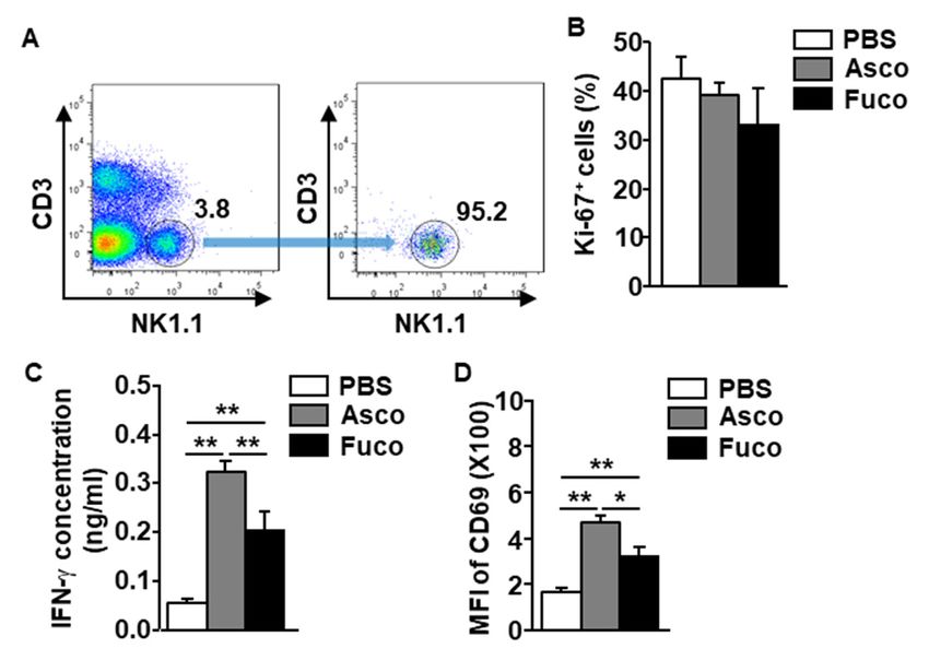
Mar. Drugs 2019, 17, 197                                                                             2 of 10

      NK cells are innate immune cells that play protective roles against bacterial and viral
infection [14,15]. Infected or damaged cells are recognized by pathogen-associated molecular patterns
or damage-associated molecular patterns, respectively, and are presented on their surface to promote
the activation of NK cells [16,17]. NK cells are the primary producers of interferon-γ (IFN-γ), which is
secreted following stimulation of the corresponding surface receptors [18,19]. IFN-γ expression by NK
cells contributes to the activation of other immune cells and subsequent pathogen elimination [18].
Moreover, regulation of target cell elimination by NK cells depends on the expression levels of major
histocompatibility complex (MHC) class I molecules [20–22]. Virus-infected cells and cancer cells
express low levels of MHC class I molecules and can be targeted for elimination by NK cells [20].
Activation of NK cells is mediated not only by pathogens and damaged cells but also by other
immune cells, such as DCs and macrophages [23–25]. Type I IFNs, which are produced by DCs,
macrophages, and other NK cells, function to activate NK cell functions, including cytotoxicity and
IFN-γ production [26,27].
      Although it was previously shown that the ability of ascophyllan to induce DC activation was
much stronger than that of fucoidan [11], the effects of ascophyllan on NK cell activation is yet to
be studied. In this study, we evaluated the effect of ascophyllan on NK cell activation in mice and
compared it with the effects of fucoidan.

2. Results and Discussion

2.1. Ascophyllan Promotes the Proliferation of NK Cells in Mice
      To evaluate the stimulatory effect of ascophyllan on NK cell proliferation, C57BL/6 mice were
intraperitoneally (i.p.) administered either ascophyllan or fucoidan (50 mg/kg, each). NK cells were
defined as NK1.1+ CD3− cells among the leukocytes (Figure 1A, B). Treatment with ascophyllan
increased the frequency of NK1.1+ CD3− cells in the spleen and blood compared to those in mice
treated with PBS at 6 h after injection (Figure 1B). In addition, the numbers of NK cells in the spleen and
blood were also significantly increased following ascophyllan treatment (Figure 1C). Proliferating NK
cells were identified by their surface expression of Ki-67, a marker of proliferating cells, detected at 6 h
after ascophyllan or fucoidan treatment. It was found that ascophyllan treatment efficiently increased
the number of Ki-67-positive cells compared to the number in the PBS-treated control mice (Figure 1D).
      Since the effect of fucoidan on NK cell activation has been well studied [5], we compared
the proliferation-inducing abilities of ascophyllan and fucoidan.               As shown in Figure 1,
ascophyllan treatment had a much greater proliferation-inducing effect in NK cells than fucoidan.
These data indicate that ascophyllan can induce NK cell proliferation and the effect is much stronger
than that of fucoidan.
control mice (Figure 1D).
Since the effect of fucoidan on NK cell activation has been well studied [5], we compared the
proliferation-inducing abilities of ascophyllan and fucoidan. As shown in Figure 1, ascophyllan
treatment had a much greater proliferation-inducing effect in NK cells than fucoidan. These data
indicate that ascophyllan can induce NK cell proliferation and the effect is much stronger than
Mar. Drugs 2019, 17, 197
that
3 of 10
of fucoidan.
Figure 1. Proliferation of natural killer (NK) cells is upregulated by ascophyllan in mice.
Either ascophyllan or fucoidan (50 mg/kg, each) was intraperitoneally (i.p.) administered to C57BL/6
mice, and NK cells were analyzed 6 h after administration. (A) The gating strategy for flow cytometry
is shown. (B) Frequencies/percentages of NK1.1+ CD3− cells in the spleen (upper panel) and blood
(lower panel) are shown. (C) Absolute numbers of NK1.1+ CD3− cells in the spleen (left panel) and
blood (right panel) are shown. (D) Expression of Ki-67 on spleen NK cells (left panel). The mean
percentage of Ki-67+ cells is shown (right panel). Data represent the average of six samples (two mice
in each group from three independent experiments). * p < 0.05, ** p < 0.01.
2.2. Ascophyllan Activates NK Cells in Mice
Our finding that ascophyllan promotes NK cell proliferation prompted us to examine the effect of
ascophyllan on activating NK cells. Either ascophyllan or fucoidan (50 mg/kg, each) was administered
i.p. to C57BL/6 mice. Six hours after administration, the spleens were harvested, and the splenocytes
were incubated in a monensin solution for an additional 4 h. The results showed that ascophyllan
treatment upregulated the intracellular production of IFN-γ in spleen NK cells (Figure 2A). In addition,
the serum concentration of IFN-γ was dramatically increased by ascophyllan treatment compared to
that induced by PBS (Figure 2B). Further, the expression of the surface marker CD69 on active NK cells
was substantially upregulated by ascophyllan (Figure 2C). Consistent with its proliferation-inducing
effects, ascophyllan also induced IFN-γ production and CD69 expression in NK cells more strongly
than fucoidan. These data suggest that ascophyllan activates spleen NK cells, and its effects are
stronger than those of fucoidan.
Fucoidan isolated from Fucus vesiculosus (F. vesiculosus) was studied in humans and mice for its
immunomodulatory effects [8,28]. Although fucoidan from F. vesiculosus showed immunostimulatory
effects on DC and NK cells, the effects of fucoidan from Macrocystis pyrifera (M. pyrifera) on DC
and NK cell activation were stronger [5]. In this study, we found that mouse NK cell activation
by ascophyllan was stronger than that by fucoidan from M. pyrifera. Based on a composition
study, fucoidan from M. pyrifera contained much higher uronic acid (UA) content than fucoidan
from F. vesiculosus [5]. Interestingly, ascophyllan also contained higher levels of UA than other
fucoidans [11,13]. Therefore, the UA content may contribute to its NK cell-activation effects. We will
examine the effects of UA on the activation of NK cells and DCs in a future study.pyrifera) on DC and NK cell activation were stronger [5]. In this study, we found that mouse NK cell
activation by ascophyllan was stronger than that by fucoidan from M. pyrifera. Based on a
composition study, fucoidan from M. pyrifera contained much higher uronic acid (UA) content than
fucoidan from F. vesiculosus [5]. Interestingly, ascophyllan also contained higher levels of UA than
other fucoidans [11,13]. Therefore, the UA content may contribute to its NK cell-activation effects. We
Mar. Drugs 2019, 17, 197 4 of 10
will examine the effects of UA on the activation of NK cells and DCs in a future study.
Figure 2. Ascophyllan activates NK cells in mice. Mice were injected with either ascophyllan
Figure50
(Asco, 2. mg/kg)
Ascophyllan activates
or fucoidan NK 50
(Fuco, cells in mice.Six
mg/kg). Mice
hourswere injected
after with
injection, either
the ascophyllan
spleens (Asco,
were harvested,
and the splenocytes were incubated in a monensin solution for 4 h. (A) Intracellular IFN-γ levelsthe
50 mg/kg) or fucoidan (Fuco, 50 mg/kg). Six hours after injection, the spleens were harvested, and in
splenocytes
spleen were(left
NK cells incubated
panel). in a monensin
Mean solution
percentage for 4 h. (A) Intracellular
of IFN-γ-producing NK cellsIFN-γ
(rightlevels in spleen
panel). NK
(B) Serum
cells (left panel).
concentration Mean percentage
of IFN-γ of IFN-γ-producing
6 h after either NK cells treatment.
ascophyllan or fucoidan (right panel).
(C)(B) Serum
CD69 concentration
expression levels
of spleen
in IFN-γ 6NK h after
cellseither ascophyllan
(left panel) or treatment.
6 h after fucoidan treatment. (C) CD69 expression
Mean fluorescence levels
intensity (MFI) ofin spleen
CD69 NK
levels
cells (left panel) 6 h after treatment. Mean fluorescence intensity (MFI) of CD69 levels
(right panel). Data represent the mean ± standard error of the mean (SEM) of six samples from three (right panel).
independent experiments, ** p < 0.01.
2.3. Ascophyllan Directly and Indirectly Activates NK Cells
In the mouse, many immune cell types are targeted by stimuli, including DCs, macrophages,
NK cells, and T cells [29–31]. These stimulated immune cells contribute to the activation of other
immune cells through cytokine production and cell-to-cell interactions [29,30]. Therefore, we next
evaluated the ability of ascophyllan to activate NK cells in mice either directly or indirectly through
other stimulated immune cells. As shown in Figure 3A, to evaluate the direct effect of ascophyllan on
NK cell activation, NK1.1+ CD3− NK cells were isolated from the leukocytes in the spleen of naïve mice
and treated with either ascophyllan or fucoidan (50 µg/mL, each). The Ki-67 staining levels on the
isolated NK cells were not increased by either ascophyllan or fucoidan (Figure 3B). However, the levels
of IFN-γ secreted into the culture medium of NK cells were dramatically increased by ascophyllan
(Figure 3C). Further, CD69 expression in isolated NK cells was also upregulated by ascophyllan
(Figure 3D). Consistent with the in vivo mouse study results, ascophyllan treatment also induced
much higher levels of IFN-γ production and CD69 expression than fucoidan. These data indicate that
ascophyllan activates NK cells directly but cannot promote the proliferation of NK cells without the
aid of other immune cells.previous studies, ascophyllan activated spleen- and lymph node (LN)-derived DCs and T cells in
mice [10]. DCs activated by ascophyllan produced high amounts of pro-inflammatory cytokines,
including IL-1, IL-12, and TNF-α [10,11]. In this study, we found that ascophyllan promoted NK cell
activation and proliferation in mice. However, the proliferation-promoting effect of ascophyllan on
isolated NK cells was diminished; therefore, the effect of ascophyllan on the induction of NK cell
Mar. Drugs 2019, 17, 197 5 of 10
proliferation may be mediated by activated DCs and cytokines.
Figure 3. Ascophyllan activates isolated NK cells. NK cells were isolated from C57BL/6 mice, and the
cells
Figurewere incubated with
3. Ascophyllan either ascophyllan
activates isolated NK(Asco, 50 µg/mL)
cells. NK or 50
cells were µg/mLfrom
isolated fucoidan (Fuco,
C57BL/6 50 µg/mL).
mice, and the
(A) Percentages of NK1.1 + CD3− cells in the splenocytes (left panel) and the purity of the NK cells
cells were incubated with either ascophyllan (Asco, 50 μg/mL) or 50 μg/mL fucoidan (Fuco, 50
(right
μg/mL). panel) are shown. of
(A) Percentages (B)NK1.1
Ki-67+CD3
expression levels
− cells in in NK cells(left
the splenocytes were measured
panel) 6 hpurity
and the after treatment.
of the NK
(C)
cellsIFN-γ
(rightconcentrations in culture
panel) are shown. (B)medium are shown.levels
Ki-67 expression (D) Surface
in NKexpression
cells wereof CD69 was measured
measured 6 h after
in NK cells. Mean ± standard error of the mean (n = 6). * p < 0.05. ** p < 0.01.
DCs and macrophages have been shown to contribute to NK cell activation in humans
and mice [32,33]. Antigen stimulation of DCs and macrophages promotes the production of
pro-inflammatory cytokines, such as IL-1, IL-12, IL-15, IL-18, TNF-α, and IFNs [34]. IL-15 promotes the
survival and differentiation of NK cells [25,35,36], and is mainly produced by activated DCs [37,38].
Moreover, IL-12 secreted by mature DCs enhances the cytotoxic activity and IFN-γ production of NK
cells [39]. In addition, IL-2 produced by activated CD4+ T cells and DCs is a potential inducer of NK
cell proliferation [23,25]. Although cytokines contribute to the activation and proliferation of NK cells,
mature DCs directly interact with NK cells to induce the activation and survival of the latter [40].
In previous studies, ascophyllan activated spleen- and lymph node (LN)-derived DCs and T cells
in mice [10]. DCs activated by ascophyllan produced high amounts of pro-inflammatory cytokines,
including IL-1, IL-12, and TNF-α [10,11]. In this study, we found that ascophyllan promoted NK cell
activation and proliferation in mice. However, the proliferation-promoting effect of ascophyllan on
isolated NK cells was diminished; therefore, the effect of ascophyllan on the induction of NK cell
proliferation may be mediated by activated DCs and cytokines.
2.4. Ascophyllan Enhances the Cytotoxic Activity of NK Cells
Next, we examined whether ascophyllan treatment enhanced the cytotoxic activity of NK cells.
C57BL/6 mice were i.p. administered either ascophyllan or fucoidan (50 mg/kg, each), and then
injected with the same concentration of either ascophyllan or fucoidan 24 h later. One day after the
last injection, NK cells were isolated from the splenocytes and co-cultured with Yac-1 cells, a mouse
lymphoma cell line [41]. The ascophyllan-treated spleen NK cells showed significantly enhanced
cytotoxic activity compared to NK cells from untreated mice (Figure 4). The cytotoxic activity of
ascophyllan-induced NK cells was stronger than that of fucoidan-treated NK cells (Figure 4). These data
demonstrate that ascophyllan enhances NK cell-mediated cytotoxicity against Yac-1 cells.by immune cells. In cancer cells and virus-infected cells, MHC class I levels are markedly lower than
the levels in healthy cells, and thus they can be targets of NK cells [44,45]. Although the MHC class I
expression levels on these cells were low, NK cells could not effectively kill these cells without being
activated [46]. In this study, we found that ascophyllan enhanced the cytotoxic activity of NK cells,
which was even stronger than the activity of fucoidan-activated NK cells. Therefore, ascophyllan may
Mar. Drugs 2019, 17, 197 6 of 10
be able to protect mice from cancer and viral infection.
Figure 4. Ascophyllan enhances the cytotoxic activity of NK cells. C57BL/6 mice were administered
Figureascophyllan
either 4. Ascophyllan enhances
(Asco, 50 mg/kg)the cytotoxic
or fucoidanactivity
(Fuco,of50NK cells. C57BL/6
mg/kg) mice were
by i.p. injection, administered
twice, separated
either ascophyllan (Asco, 50 mg/kg) or fucoidan (Fuco, 50 mg/kg) by i.p. injection,
by 24 h. Ascophyllan- or fucoidan-stimulated spleen NK cells (effector cells) were isolated twice, separated by
from
24 h. Ascophyllan-
mice 24 h after theorlast
fucoidan-stimulated spleen NKwith
injection and co-cultured cellsYac-1
(effector
cellscells) werecells)
(target isolated from
at the mice 24
indicated
h afterCytotoxicity
ratios. the last injection and co-cultured
was measured after 6 h ofwith Yac-1 cells
co-culture by the(target
lactatecells) at the indicated
dehydrogenase (LDH) ratios.
assay.
Cytotoxicity
Mean was measured
± standard error of theafter 6 h (n
mean. of =co-culture by the lactate dehydrogenase (LDH) assay. Mean ±
6) ** p < 0.01.
standard error of the mean. (n = 6) ** p < 0.01.
The most important function of NK cells is their cytotoxic activity against pathogens [42,43],
3. Materials
and and Methods
NK cell-mediated cytotoxicity is dependent on the expression of MHC class I molecules [42,43].
At steady state, cells present high levels of self-antigen on MHC class I receptors, which prevents attack
3.1.immune
by Mice cells. In cancer cells and virus-infected cells, MHC class I levels are markedly lower than
the levels in healthy
C57BL/6 cells,provided
mice were and thusbythey
thecan be targets
Shanghai of NK
Public cells Clinical
Health [44,45]. Center
Although the MHC
(SPHCC). Theclass
miceI
expression levels on these cells were low, NK cells could not effectively kill these cells
were housed under pathogen-free conditions, and the mouse room was maintained at 20–22 °C and without being
activated
50%–60%[46]. In this This
humidity. study,study
we found
was that ascophyllan
conducted enhanced
in strict the cytotoxic
accordance with theactivity of NK cells,
recommendations
which was even stronger than the activity of fucoidan-activated NK cells. Therefore,
outlined in the Guide for the Care and Use of Laboratory Animals of the SPHCC. The Committee ascophyllan may
on
be able to protect mice from cancer and viral infection.
the Ethics of Animal Experiments of SPHCC approved this study (Approval number: SYXK-2010-
0098).
3. Materials and Methods
3.2. Mice
3.1. Antibodies and Reagents
C57BL/6 mice were provided by the Shanghai Public Health Clinical Center (SPHCC). The mice
were housed under pathogen-free conditions, and the mouse room was maintained at 20–22 ◦ C and
50%–60% humidity. This study was conducted in strict accordance with the recommendations outlined
in the Guide for the Care and Use of Laboratory Animals of the SPHCC. The Committee on the Ethics
of Animal Experiments of SPHCC approved this study (Approval number: SYXK-2010-0098).
3.2. Antibodies and Reagents
Ascophyllan was purified from A. nodosum as a sulfated fucan preparation [13,47].
Briefly, in the first step, a hot water extraction was performed to obtain the total soluble
polysaccharide fraction, which contained ascophyllan, fucoidan, and trace levels of alginate.
In the second step, alginate was removed by alginate lyase digestion. The third step was
the selective precipitation of ascophyllan in the presence of NaCl and alcohol. In this step,
ascophyllan was separated from fucoidan. Finally, a composition analysis was performed to
characterize the isolated polysaccharides [45]. Fucoidan from M. pyrifera was obtained from
Sigma Aldrich (St. Louis, MO, USA). Fluorescence-conjugated Abs against IgG1, IgG2a, CD3 (17A2),
CD69 (H1.2F3), NK1.1 (PK136), Ki-67 (16A8), and IFN-γ (XMG1.2) were purchased from BioLegend
(San Diego, CA, USA).Mar. Drugs 2019, 17, 197 7 of 10
3.3. NK Cell Analysis
The spleen was cut into small fragments with curved scissors and digested in digestion buffer
(RPMI 1640 containing 2% fetal bovine serum [FBS] and collagenase IV) for 20 min at room temperature.
The digested tissue was washed with 10 ml of PBS and resuspended in 5 ml of histopaque 1077
(Sigma Aldrich). An additional 5 ml of histopaque 1077 was added to the upper layer, and 1 mL of
FBS was layered over the histopaque. The suspended cells were then centrifuged at 3000 RPM for
10 min. Splenocytes were obtained from the light density fraction (Mar. Drugs 2019, 17, 197 8 of 10
Funding: This research was funded by the National Research Foundation of Korean (grant number
NRF-2019R1C1C1003334) and the General Project of Shanghai Public Health Clinical Center (grant number
KY-GW-2018-49).
Conflicts of Interest: The authors declare no conflict of interest.
References
1. Guzman, E.A. Regulated Cell Death Signaling Pathways and Marine Natural Products That Target Them.
Mar. Drugs 2019, 17, 76. [CrossRef] [PubMed]
2. Ercolano, G.; De Cicco, P.; Ianaro, A. New Drugs from the Sea: Pro-Apoptotic Activity of Sponges and Algae
Derived Compounds. Mar. Drugs 2019, 17, 31. [CrossRef]
3. Gismondi, A.; De Rossi, S.; Canuti, L.; Novelli, S.; Di Marco, G.; Fattorini, L.; Canini, A. From Robinia
pseudoacacia L. nectar to Acacia monofloral honey: Biochemical changes and variation of biological
properties. J. Sci. Food Agric. 2018, 98, 4312–4322. [CrossRef]
4. Mimouni, V.; Ulmann, L.; Pasquet, V.; Mathieu, M.; Picot, L.; Bougaran, G.; Cadoret, J.P.; Morant-Manceau, A.;
Schoefs, B. The potential of microalgae for the production of bioactive molecules of pharmaceutical interest.
Curr. Pharm. Biotechnol. 2012, 13, 2733–2750. [CrossRef] [PubMed]
5. Zhang, W.; Oda, T.; Yu, Q.; Jin, J.O. Fucoidan from Macrocystis pyrifera has powerful immune-modulatory
effects compared to three other fucoidans. Mar. Drugs 2015, 13, 1084–1104. [CrossRef] [PubMed]
6. Kwak, J.Y. Fucoidan as a marine anticancer agent in preclinical development. Mar. Drugs 2014, 12, 851–870.
[CrossRef] [PubMed]
7. Kuznetsova, T.A.; Besednova, N.N.; Somova, L.M.; Plekhova, N.G. Fucoidan extracted from Fucus evanescens prevents
endotoxin-induced damage in a mouse model of endotoxemia. Mar. Drugs 2014, 12, 886–898. [CrossRef]
8. Jin, J.O.; Zhang, W.; Du, J.Y.; Wong, K.W.; Oda, T.; Yu, Q. Fucoidan can function as an adjuvant in vivo
to enhance dendritic cell maturation and function and promote antigen-specific T cell immune responses.
PloS ONE 2014, 9, e99396. [CrossRef]
9. Zhang, W.; Kwak, M.; Park, H.B.; Okimura, T.; Oda, T.; Lee, P.C.; Jin, J.O. Activation of Human Dendritic
Cells by Ascophyllan Purified from Ascophyllum nodosum. Mar. Drugs 2019, 17, 66. [CrossRef]
10. Zhang, W.; Okimura, T.; Xu, L.; Zhang, L.; Oda, T.; Kwak, M.; Yu, Q.; Jin, J.O. Ascophyllan functions as
an adjuvant to promote anti-cancer effect by dendritic cell activation. Oncotarget 2016, 7, 19284–19298.
[CrossRef]
11. Zhang, W.; Du, J.Y.; Jiang, Z.; Okimura, T.; Oda, T.; Yu, Q.; Jin, J.O. Ascophyllan purified from Ascophyllum
nodosum induces Th1 and Tc1 immune responses by promoting dendritic cell maturation. Mar. Drugs
2014, 12, 4148–4164. [CrossRef] [PubMed]
12. Nakano, K.; Kim, D.; Jiang, Z.; Ueno, M.; Okimura, T.; Yamaguchi, K.; Oda, T. Immunostimulatory activities
of the sulfated polysaccharide ascophyllan from Ascophyllum nodosum in in vivo and in vitro systems.
Biosci. Biotechnol. Biochem. 2012, 76, 1573–1576. [CrossRef]
13. Jiang, Z.; Okimura, T.; Yamaguchi, K.; Oda, T. The potent activity of sulfated polysaccharide, ascophyllan,
isolated from Ascophyllum nodosum to induce nitric oxide and cytokine production from mouse
macrophage RAW264.7 cells: Comparison between ascophyllan and fucoidan. Nitric oxide: Biol. Chem./ Off. J.
Nitric Oxide Soc. 2011, 25, 407–415. [CrossRef]
14. Germic, N.; Frangez, Z.; Yousefi, S.; Simon, H.U. Regulation of the innate immune system by autophagy:
Neutrophils, eosinophils, mast cells, NK cells. Cell Death Differ. 2019, 26, 703–714. [CrossRef]
15. Du, Y.; Wei, Y. Therapeutic Potential of Natural Killer Cells in Gastric Cancer. Front. Immunol. 2018, 9, 3095.
[CrossRef] [PubMed]
16. Farag, S.S.; Fehniger, T.A.; Ruggeri, L.; Velardi, A.; Caligiuri, M.A. Natural killer cell receptors: New biology
and insights into the graft-versus-leukemia effect. Blood 2002, 100, 1935–1947. [CrossRef]
17. Su, R.C.; Kung, S.K.; Gariepy, J.; Barber, B.H.; Miller, R.G. NK cells can recognize different forms of class I
MHC. J. Immunol. 1998, 161, 755–766.
18. Mah, A.Y.; Cooper, M.A. Metabolic Regulation of Natural Killer Cell IFN-gamma Production.
Crit. Rev. Immunol. 2016, 36, 131–147. [CrossRef]
19. Wang, R.; Jaw, J.J.; Stutzman, N.C.; Zou, Z.; Sun, P.D. Natural killer cell-produced IFN-gamma and TNF-alpha
induce target cell cytolysis through up-regulation of ICAM-1. J. Leukocyte Biol. 2012, 91, 299–309. [CrossRef]Mar. Drugs 2019, 17, 197 9 of 10
20. Bessoles, S.; Grandclement, C.; Alari-Pahissa, E.; Gehrig, J.; Jeevan-Raj, B.; Held, W. Adaptations of Natural
Killer Cells to Self-MHC Class I. Front. Immunol. 2014, 5, 349. [CrossRef]
21. Elliott, J.M.; Wahle, J.A.; Yokoyama, W.M. MHC class I-deficient natural killer cells acquire a licensed
phenotype after transfer into an MHC class I-sufficient environment. J. Exp. Med. 2010, 207, 2073–2079.
[CrossRef] [PubMed]
22. Lanier, L.L. Natural killer cell receptors and MHC class I interactions. Curr. Opin. Immunol. 1997, 9, 126–131.
[CrossRef]
23. Zwirner, N.W.; Domaica, C.I. Cytokine regulation of natural killer cell effector functions. BioFactors 2010, 36, 274–288.
[CrossRef] [PubMed]
24. Wu, Y.; Tian, Z.; Wei, H. Developmental and Functional Control of Natural Killer Cells by Cytokines.
Front. Immunol. 2017, 8, 930. [CrossRef] [PubMed]
25. Freeman, B.E.; Raue, H.P.; Hill, A.B.; Slifka, M.K. Cytokine-Mediated Activation of NK Cells during Viral
Infection. J. Virol 2015, 89, 7922–7931. [CrossRef]
26. Muller, L.; Aigner, P.; Stoiber, D. Type I Interferons and Natural Killer Cell Regulation in Cancer.
Front. Immunol. 2017, 8, 304. [CrossRef]
27. Madera, S.; Rapp, M.; Firth, M.A.; Beilke, J.N.; Lanier, L.L.; Sun, J.C. Type I IFN promotes NK cell expansion
during viral infection by protecting NK cells against fratricide. J. Exp. Med. 2016, 213, 225–233. [CrossRef]
[PubMed]
28. Jin, J.O.; Park, H.Y.; Xu, Q.; Park, J.I.; Zvyagintseva, T.; Stonik, V.A.; Kwak, J.Y. Ligand of scavenger receptor
class A indirectly induces maturation of human blood dendritic cells via production of tumor necrosis
factor-alpha. Blood 2009, 113, 5839–5847. [CrossRef] [PubMed]
29. Gasteiger, G.; Ataide, M.; Kastenmuller, W. Lymph node—an organ for T-cell activation and pathogen
defense. Immunol. Rev. 2016, 271, 200–220. [CrossRef]
30. Akira, S.; Uematsu, S.; Takeuchi, O. Pathogen recognition and innate immunity. Cell 2006, 124, 783–801.
[CrossRef]
31. Dubensky, T.W., Jr.; Reed, S.G. Adjuvants for cancer vaccines. Semin. Immunol. 2010, 22, 155–161. [CrossRef]
32. Lapaque, N.; Walzer, T.; Meresse, S.; Vivier, E.; Trowsdale, J. Interactions between human NK cells and
macrophages in response to Salmonella infection. J. Immunol. 2009, 182, 4339–4348. [CrossRef] [PubMed]
33. Fernandez, N.C.; Flament, C.; Crepineau, F.; Angevin, E.; Vivier, E.; Zitvogel, L. Dendritic cells (DC) promote
natural killer (NK) cell functions: Dynamics of the human DC/NK cell cross talk. Eur. Cytokine Network
2002, 13, 17–27.
34. Eastman, A.J.; Osterholzer, J.J.; Olszewski, M.A. Role of dendritic cell-pathogen interactions in the immune
response to pulmonary cryptococcal infection. Future Microbiol. 2015, 10, 1837–1857. [CrossRef]
35. Ferlazzo, G.; Pack, M.; Thomas, D.; Paludan, C.; Schmid, D.; Strowig, T.; Bougras, G.; Muller, W.A.; Moretta, L.;
Munz, C. Distinct roles of IL-12 and IL-15 in human natural killer cell activation by dendritic cells from
secondary lymphoid organs. Proc. Natl. Acad. Sci. USA. 2004, 101, 16606–16611. [CrossRef] [PubMed]
36. Huntington, N.D.; Legrand, N.; Alves, N.L.; Jaron, B.; Weijer, K.; Plet, A.; Corcuff, E.; Mortier, E.; Jacques, Y.;
Spits, H.; et al. IL-15 trans-presentation promotes human NK cell development and differentiation in vivo.
J. Exp. Med. 2009, 206, 25–34. [CrossRef] [PubMed]
37. Ruckert, R.; Brandt, K.; Bulanova, E.; Mirghomizadeh, F.; Paus, R.; Bulfone-Paus, S. Dendritic cell-derived
IL-15 controls the induction of CD8 T cell immune responses. Eur. J. Immunol. 2003, 33, 3493–3503. [CrossRef]
[PubMed]
38. Anguille, S.; Smits, E.L.; Cools, N.; Goossens, H.; Berneman, Z.N.; Van Tendeloo, V.F. Short-term cultured,
interleukin-15 differentiated dendritic cells have potent immunostimulatory properties. J. Transl. Med.
2009, 7, 109. [CrossRef]
39. Zwirner, N.W.; Ziblat, A. Regulation of NK Cell Activation and Effector Functions by the IL-12 Family of
Cytokines: The Case of IL-27. Front. Immunol. 2017, 8, 25. [CrossRef]
40. Thomas, R.; Yang, X. NK-DC Crosstalk in Immunity to Microbial Infection. J. Immunol. Res. 2016. [CrossRef]
41. Wright, S.C.; Bonavida, B. YAC-1 variant clones selected for resistance to natural killer cytotoxic factors are
also resistant to natural killer cell-mediated cytotoxicity. Proc. Natl. Acad. Sci. USA 1983, 80, 1688–1692.
[CrossRef] [PubMed]
42. Topham, N.J.; Hewitt, E.W. Natural killer cell cytotoxicity: How do they pull the trigger? Immunol. 2009, 128, 7–15.
[CrossRef]Mar. Drugs 2019, 17, 197 10 of 10
43. Smyth, M.J.; Cretney, E.; Kelly, J.M.; Westwood, J.A.; Street, S.E.; Yagita, H.; Takeda, K.; van Dommelen, S.L.;
Degli-Esposti, M.A.; Hayakawa, Y. Activation of NK cell cytotoxicity. Mol. Immunol. 2005, 42, 501–510.
[CrossRef] [PubMed]
44. Orr, M.T.; Edelmann, K.H.; Vieira, J.; Corey, L.; Raulet, D.H.; Wilson, C.B. Inhibition of MHC class I is
a virulence factor in herpes simplex virus infection of mice. PLoS Pathog. 2005, 1, e7. [CrossRef] [PubMed]
45. Seliger, B.; Ritz, U.; Ferrone, S. Molecular mechanisms of HLA class I antigen abnormalities following viral
infection and transformation. Int. J. Cancer 2006, 118, 129–138. [CrossRef]
46. He, Y.; Tian, Z. NK cell education via nonclassical MHC and non-MHC ligands. Cell. Mol. Immunol. 2017, 14, 321–330.
[CrossRef]
47. Nakayasu, S.; Soegima, R.; Yamaguchi, K.; Oda, T. Biological activities of fucose-containing polysaccharide
ascophyllan isolated from the brown alga Ascophyllum nodosum. Biosci. Biotechnol. Biochem. 2009, 73, 961–964.
[CrossRef]
48. Wang, Y.; Zhang, W.; Xu, L.; Jin, J.O. Porphyromonas gingivalis Lipopolysaccharide Induced Proliferation
and Activation of Natural Killer Cells in Vivo. Molecules 2016, 21, 1086. [CrossRef]
49. Korzeniewski, C.; Callewaert, D.M. An enzyme-release assay for natural cytotoxicity. J. Immunol. Methods
1983, 64, 313–320. [CrossRef]
© 2019 by the authors. Licensee MDPI, Basel, Switzerland. This article is an open access
article distributed under the terms and conditions of the Creative Commons Attribution
(CC BY) license (http://creativecommons.org/licenses/by/4.0/).You can also read