The Landscape of CAR-T Cell Clinical Trials against Solid Tumors-A Comprehensive Overview - OPUS 4 - KOBV
←
→
Page content transcription
If your browser does not render page correctly, please read the page content below
cancers
Review
The Landscape of CAR-T Cell Clinical Trials against
Solid Tumors—A Comprehensive Overview
Niels Schaft 1,2,3
 1   Department of Dermatology, Universtitätsklinikum Erlangen, Friedrich-Alexander-Universität,
     Erlangen-Nürnberg, Hartmannstraße 14, 91052 Erlangen, Germany; niels.schaft@uk-erlangen.de;
     Tel.: +49-9131-853-1127
 2   Comprehensive Cancer Center Erlangen European Metropolitan Area of Nuremberg (CCC ER-EMN),
     Östliche Stadtmauerstraße 30, 91054 Erlangen, Germany
 3   Deutsches Zentrum Immuntherapie (DZI), Ulmenweg 18, 91054 Erlangen, Germany

 Received: 5 August 2020; Accepted: 4 September 2020; Published: 9 September 2020

 Simple Summary: Certain immune cells, namely T cells, of cancer patients can be genetically
 manipulated to express so-called chimeric antigen receptors (CARs), which enables these cells to kill
 the tumor cells after recognition by the receptor. This therapy is very successful in the treatment of
 hematologic tumors such as lymphoma or leukemia. However, tumors growing as a solid mass are
 less susceptible to this kind of treatment. This review summarizes known data of all clinical trials
 using this therapy against solid tumors that are registered at clinicaltrials.gov.

 Abstract: CAR-T cells showed great potential in the treatment of patients with hematologic tumors.
 However, the clinical efficacy of CAR-T cells against solid tumors lags behind. To obtain a
 comprehensive overview of the landscape of CAR-T cell clinical trials against this type of cancer,
 this review summarizes all the 196 studies registered at clinicaltrials.gov. Special focus is on:
 (1) geographical distribution; (2) targeted organs, tumor entities, and antigens; (3) CAR transfer
 methods, CAR formats, and extra features introduced into the T cells; and (4) patient pretreatments,
 injection sites, and safety measurements. Finally, the few data on clinical outcome are reported.
 The last assessment of clinicaltrials.gov for the data summarized in this paper was on 4 August 2020.

 Keywords: CAR-T cell; solid tumor; clinical trial; clinicaltrials.gov; CAR format; tumor antigen;
 CAR transfer; suicide switch

1. Introduction
     T cells reprogrammed with a tumor specificity via the expression of a chimeric antigen receptor
(CAR-T cells) are increasingly used in the adoptive cellular therapy of cancer. The advantage of
transferring a CAR, in contrast to normal T-cell receptors (TCRs), is that a CAR can recognize the tumor
in an MHC-independent way. The CAR concept was originally developed by Zelig Eshhar (Weizmann
Institute of Science, Rehovot, Israel) in the late 1980s [1,2]. Most CARs are created by assembling a
tumor-antigen-binding, antibody-derived single chain Fv (scFv) and the intracellular part of the CD3ζ
chain linked in cis with one or several co-stimulatory domains [3], however many other formats exist
(see below). This modular composition allows for T-cell activation in response to antigens located on
the surface of malignant cells by binding of the scFv and subsequent signaling through the CD3ζ chain
and co-stimulatory domains [3].
     CD19-specific CAR-T cells induced impressive clinical regressions of leukemias or lymphomas
in several clinical trials [4–6]. This resulted in the approval by the FDA and EMA of Kymriah®
(Tisagenlecleucel, Basel, Switzerland), for the treatment of B-cell acute lymphoblastic leukemia (ALL),

Cancers 2020, 12, 2567; doi:10.3390/cancers12092567                           www.mdpi.com/journal/cancers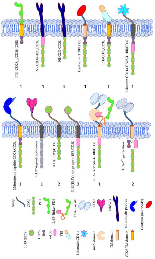
Cancers 2020, 12, 2567                                                                                         2 of 36

and Yescarta® (Axicabtagen-Ciloleucel, Santa Monica, CA, USA), for the treatment of aggressive B-cell
non-Hodgkin lymphoma [3].
      Most clinical trials focus on the elimination of such hematologic tumors; the development of CAR-T
cells against solid tumors lags behind (reviewed in [7–10]). This is due in part to the lack of suitable
tumor-specific antigens that can be targeted by CAR-T cells, causing potential on-target/off-tumor
toxicity due to the accidental killing of non-malignant bystander cells co-expressing the target
antigen [11,12].
      This review gives a comprehensive overview of all clinical trials applying CAR-T cells against
solid tumors registered at clinicaltrials.gov, irrespective of their status (i.e., withdrawn; suspended;
terminated; completed; active, but not recruiting; recruiting; not yet recruiting). The search terms
used at clinicaltrials.gov were: “CAR”, “T-body”, “designer T cells”, and “NKR-2”. Just the search
term “CAR” already resulted in 1500 found studies, from which CAR-T cell trials against hematologic
tumors, long-term follow-up, and retrospective studies, and trials not related to chimeric antigen
receptors (e.g., automotive-related studies) were filtered out. To date (last assessment: 4 August 2020),
there are 196 CAR-T cell trials against solid tumors registered.
      Looking at the geographical distribution of the registered clinical trials, it is clear that most of
these trials are performed in China (n = 99; 50.0%; Figure 1), followed by the USA (n = 85; 42.9%;
Figure 1), and only very few trials are taking place in Europe, Australia, and the rest of Asia (all together
responsible for n = 14; 7.1%; Figure 1).

      Figure 1. Schematic overview of the geographical distribution of clinical trials using CAR-T cells
      against solid tumors. (a) Number of clinical trials per country; (b) Proportional distribution of clinical
      trials per country. Data was extracted from clinicaltrials.gov.

     Data considering (1) targeted antigen, (2) targeted tumor, (3) CAR format, (4) transfer method of
the CAR into the T cells, (5) additionally introduced qualities of the CAR-T cells, (6) number of cells
applied, (7) patient pretreatment, (8) clinical outcome, (9) adverse events, and several other parameters
are summarized in the following chapters. Additional information on e.g., clinical outcome of the trials
and adverse events was gathered through literature search on pubmed.ncbi.nlm.nih.gov [13–30].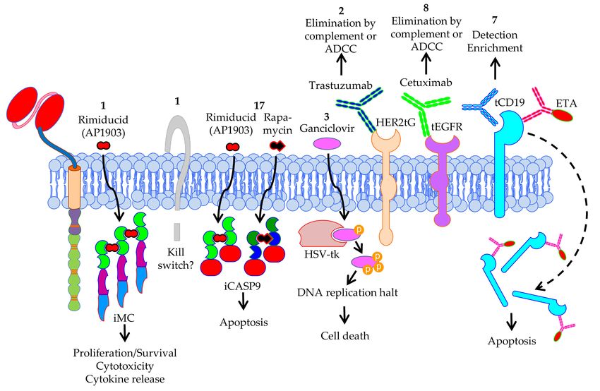
Cancers 2020, 12, 2567                                                                                       3 of 36

2. CAR-T Cell Clinical Trials against Solid Tumors—Organs, Tumor Entities, Antigens

2.1. Targeted Organs
      Many different solid tumors (see Section 2.2) are targeted in a total of 20 organs (Figure 2).
Especially the tumors in the brain/CNS, liver, pancreas, and lung are targeted in many clinical trials
(n = 45, 43, 38, and 36, respectively). This might represent the high medical need and/or the absence
of effective alternative therapies (i.e., not CAR-T therapies) for tumors in these organs. In total 51
clinical trials target several organs (Figure 2), mostly because the antigen targeted by the CAR-T cells
(see Section 2.3) is expressed on tumors in different organs (e.g., epidermal growth factor receptor
(EGFR), natural killer group 2D (NKG2D)-ligands, human epidermal growth factor receptor 2 (HER2),
mucin 1 (MUC1), and carcinoembryonic antigen (CEA)).

      Figure 2. Schematic overview of the organs targeted by CAR-T cells against solid tumors. The numbers
      indicate the number of clinical trials targeting this organ. Data was extracted from clinicaltrials.gov.
      The Motifolio Scientific Illustration Toolkit was used for the generation of this figure.

2.2. Targeted Tumor Entities
      As can be seen in Figure 3, there are 57 different tumor entities targeted by CAR-T cells registered
at clinicaltrials.gov. Nine different tumor entities were described in the brain, six in the liver, and five
in the lung (Figure 3). Unfortunately, many registered clinical trials did not exactly specify which
tumor entity was targeted. These files just indicated the organ (e.g., “brain”; not specifying which
type of tumor) (Figure 3). Furthermore, 34 registered trials just indicated “solid tumor” (Figure 3).
The four most targeted tumor entities are pancreatic cancer (n = 34), gastric cancer (n = 22), ovarian
cancer (n = 21), and colorectal cancer (n = 20) (Figure 3). This does not reflect the world-wide cancer
incidence. In 2018, the top 3 of cancer types newly diagnosed for both sexes was: (1) lung cancer
(12.3%), (2) breast cancer (12.3%), and (3) colorectal cancer (10.6%) [31–33]. This could be caused by
local difference in cancer incidence (e.g., in China, gastric cancer is the third most diagnosed cancer
after lung cancer and colorectal cancer, and even the second most common cause of cancer-related
death [34], and might therefore have a higher interest in performing clinical trials targeting this cancer
entity). Indeed, of the 22 clinical trials targeting gastric cancer, 15 were/are performed in China.
Cancers 2020, 12, 2567                                                                                           4 of 36

      Figure 3. Schematic overview of the tumor entities targeted by CAR-T cells against solid tumors
      grouped by organ. The numbers indicate the number of clinical trials targeting this tumor. Data was
      extracted from clinicaltrials.gov. The Motifolio Scientific Illustration Toolkit was used for the generation
      of parts of this figure.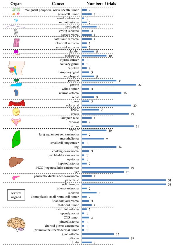
Cancers 2020, 12, 2567                                                                                 5 of 36

     At which tumor stage the CAR-T cells are applied, i.e., at an early stage (e.g., only primary tumor
present), or at a late stage with several (distant) metastases, can have an impact on the effectiveness of
CAR-T-cell therapy. However, most registered trials do not provide information on the exact treated
tumor stage. One can hypothesize that the therapy would most probably be more effective at lower
tumor burden.

2.3. Targeted Antigens
      Ideal target antigens on solid tumors unify three essential attributes: (i) uniform presence on the
surface of malignant cells reducing the risk for antigen-negative escape variants; (ii) absent expression
on non-malignant host cells precluding on-target/off-tumor activity, which harbors the potential
for severe, potentially lethal, side-effects [12]; and (iii) crucial role as an oncogenic driver in cancer
cells, which may compound antigen-shutdown due to the selective survival advantage conferred on
malignant cells. Co-expression on by-stander cells maintaining the tumor-microenvironment—such as
tumor-associated vasculature, fibroblasts, and macrophages—represents another beneficial trait.
      The registered CAR-T cell studies at clinicaltrials.gov target 44 different antigens on solid tumors
(Table 1). Table 2 shows a more detailed overview of which antigen is targeted on which tumor. Sixteen
clinical trials target several antigens at the same time [35,36], and two clinical trials do not disclose the
targeted antigen (Table 1). The top six targeted antigens that are expressed on solid tumors in many
different organs are (1) EGFR [37–41] (14 different organs), (2) NKG2D-ligands [42,43] (11 different
organs), (3) HER2 [12,44–50] (11 different organs), (4) B7-H3 (10 different organs), (5) MUC1 (9 different
organs), and (6) CEA [51–58] (9 different organs) (Table 1).
      EGFR and HER2 are members of the ErbB family of receptor tyrosine kinases (i.e., EGFR (ErbB-1),
HER2/(neu) (ErbB-2), Her 3 (ErbB-3), and Her 4 (ErbB-4). Mutations in EGFR lead to its overexpression,
which results in its constant activation and uncontrolled cell division in many different cancers (e.g.,
non-small cell lung cancer (NSCLC), colorectal cancer, pancreatic cancer, etc.) [59–61]. HER2 functions
similarly and is overexpressed mainly in breast cancer, but also in other cancer types like ovarian
cancer, glioma, and many more [62–64].
      The NKG2D-ligands MIC-A, MIC-B, and the ULBPs 1, 2, 3, 4, 5, 6 are induced-self proteins,
which are upregulated on stressed, infected, and transformed cells. These ligands can be recognized
by the NKG2D receptor expressed on NK cells, NKT cells, γ/δ T cells, and activated CD8+ αβ T
cells [65,66]. Colorectal cancers, ovarian cancers, and other cancers [67–70] express higher levels of
NKG2D-ligands and can be targeted by CAR-T cells incorporating NKG2D in the chimeric receptor.
      B7-H3 (i.e., CD276, or B7 homolog 3) is a co-stimulatory molecule for T cells and is for example
expressed on activated dendritic cells and monocytes. T cells stimulated by B7-H3 proliferate and
differentiate into cytotoxic T cells and selectively secrete IFNγ when TCR signaling and B7-H3
co-stimulation are combined [71]. It has only a limited expression on healthy tissues [72,73]. However,
B7-H3 is overexpressed on neuroblastomas, where it inhibits recognition and killing of the tumor cells
by NK cells [74]. Furthermore, CD276 is overexpressed on several other tumors—such as pancreatic
ductal adenocarcinoma (PDAC), prostate cancer, ovarian cancer, lung cancer, and clear cell renal
carcinoma—and on tumor-associated vasculature and stroma fibroblasts [73,75–85].
      Mucin 1 (MUC1) is a highly glycosylated membrane protein expressed on the surface of epithelial
cells in intestine, stomach, lung, eye, and other organs, where it inhibits pathogens from reaching the
cell membrane by binding them to oligosaccharides [86,87]. MUC1 is overexpressed on colorectal,
breast, ovarian, lung, and pancreatic cancers [88–90].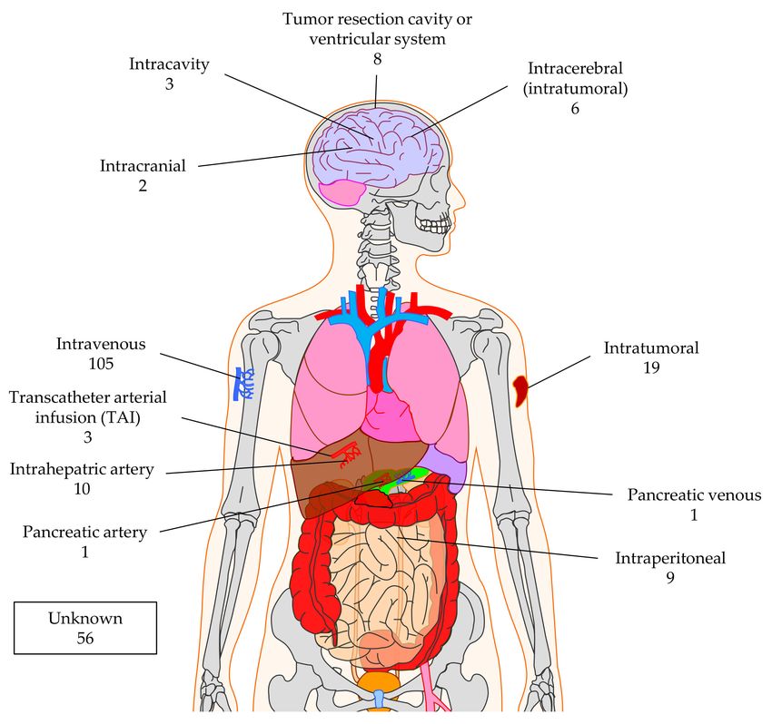
Cancers 2020, 12, 2567 6 of 36
Table 1. Summary of antigens expressed by solid tumors in different organs targeted by. CAR-T cells
in trials registered at clinicaltrials.gov.
Number of
Several
Organs
Trials
Antigen
√
AFP peptide/A2 2
√ √ √ √ √ √ √ √ √ √
B7-H3 6
√
CD20 1
√ √ √
CD44v6 2
√ √ √ √ √
CD70 2
√ √ √ √ √ √
CD133 3
CD147 √ √
2
(EMMPRIN)
√
CD171 (L1CAM) 2
√ √ √ √ √ √ √ √ √
CEA 16
claudin 18.2 √ √ √
6
(CLD18)
c-Met/hepatocyte √ √
growth factor 4
receptor
DLL3 (delta-like √
1
protein 3)
√ √ √ √ √ √ √ √ √ √ √ √ √ √
EGFR 8
EGFR family √
1
member
√
EGFRvIII 11
√
EGFR806 1
√ √ √ √ √ √ √ √
EpCAM 6
√
EphA2 2
√
ErbB2 dimers 1
√
FAP 1
FBP (folate √
3
binding protein)
√ √ √ √ √ √
GD2 24
gp100 √
1
(209-217/HLA-A2)
√ √ √ √ √ √
GPC3 (glypican-3) 18
√ √ √ √ √ √ √ √ √ √ √
HER2 17
√
ICAM1 1
√ √
IL13Rα2 6
√
Lewis Y 2
ligands of √
1
chlorotoxin
√
LMP1 (EBV) 1
√ √ √ √ √ √
mesothelin 32
√
MG7 1
√ √ √ √ √ √ √ √ √
MUC1 11
Muc1 (cleaved √
1
form)
√ √ √
MUC16ecto 1
√ √ √ √ √
TnMuc1 1
√ √ √ √ √ √
Nectin4/FAP 1
NKG2D-ligands √ √ √ √ √ √ √ √ √ √ √
(MIC-A,-B, ULBP- 6
1,-2,-3,-4,-5,-6)
√ √
PD-L1 5
√ √ √ √
PSCA 4
√ √ √ √
PSMA 11
√ √ √
ROR1 1
√ √ √ √ √
ROR2 2
√ √
VEGFR2 1
√ √ √ √ √ √ √ √ √ √ √ √ √ √
Several Ags 16
undisclosed √ √
2
antigen
Carcinoembryonic antigen (CEA, also CEACAM5) is a glycoprotein which is widely expressed
during fetal development and on some adult tissues (e.g., epithelium of the colon, stomach,
and esophagus) [91]. In normal epithelial cells of the lung and gastrointestinal tract, CEA has
an apical polarity and is facing the lumen and cannot be recognized by CAR-T cells [92]. Its function
and signaling in normal tissue are still not fully understood [93]. CEA is overexpressed in colorectal,
pancreatic, gastric, lung, and breast carcinoma where it plays a role in metastasis of the tumor [94].
In carcinomas, CEA has lost its apical polarity and is even partly shed, resulting in an increased serum
level [95]. At this stage, CEA expressed on tumor cells can be targeted by CAR-T cells [96].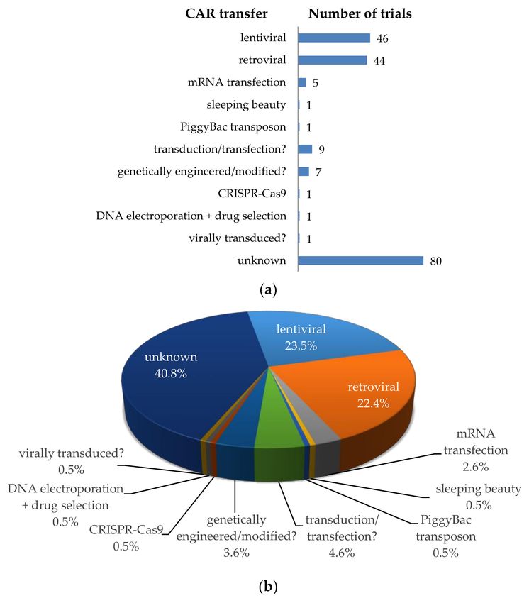
Cancers 2020, 12, 2567                                                                                                                              7 of 36

      Table 2. Summary of antigens targeted on different tumors by CAR-T cells in trials registered at
      clinicaltrials.gov.
          Organ                       Cancer Type                                                 Targeted Antigens
        brain/CNS                         brain                                                  CD133, HER2, PSMA
                                         glioma                        B7-H3, CD147, EGFR, EGFRvIII, EphA2, GD2, HER2, IL13Rα2, MUC1, CD133
                                      glioblastoma                   B7-H3, ligands of chlorotoxin, EGFRvIII, HER2, IL13Rα2, NKG2D-Ligands, PD-L1
                            primitive neuroectodermal tumor                                             B7-H3
                                choroid plexus carcinoma                                                B7-H3
                                     pineoblastoma                                                      B7-H3
                                       CNS tumor                                                B7-H3, EGFR806, HER2
                                     ependymoma                                                         B7-H3
                                    medulloblastoma                                             B7-H3, NKG2D-Ligands
       several organs               rhabdoid tumor                                                B7-H3, EGFR, GPC3
                                  Rhabdomyosarcoma                                                B7-H3, EGFR, GPC3
                           desmoplastic small round cell tumor                                       B7-H3, EGFR
                                        sarcoma                                    GD2, HER2, NKG2D-Ligands, CD133, MUC1, CD117
                                    adenocarcinoma                                                       CEA
                                                                     B7-H3, CEA, claudin 18.2, EGFR, EGFR family member, GD2, GPC3, HER2, Lewis Y,
                                      solid tumors
                                                                                 mesothelin, MUC1, MUC16ecto, TnMuc1, Nectin4, ROR2
                                                                    CD70, CD133, CEA, claudin 18.2, EGFR, EpCAM, HER2, mesothelin, MUC1, Nectin4,
         pancreas                      pancreatic
                                                                                       NKG2D-Ligands, PSCA, ROR2, EGFRvIII
                            pancreatic ductal adenocarcinoma                               claudin 18.2, mesothelin, TnMuc1
           liver                          liver                                CD133, CEA, EGFR, EpCAM, GPC3, MG7, NKG2D-Ligands
                             HCC (hepatocellular carcinoma)                AFP/HLA-A2, CD147, GPC3, MUC1, NKG2D-Ligands, c-MET, PD-L1
                                    hepatoblastoma                                                   B7-H3, EGFR
                                       hepatoma                                                         several
                                 gall bladder carcinoma                                                 EGFR
                                  cholangiocarcinoma                                             EGFR, HER2, MUC1
                                                                     CEA, EGFR, HER2, mesothelin, Lewis Y, PSCA, MUC1, PD-L1, CD80/86, MAGE-A1,
           lung                           lung
                                                                                                   MAGE-A4, GD2
                                  small cell lung cancer                                                DLL3
                                     mesothelioma                                                   FAP, mesothelin
                              lung squamous cell carcinoma                                              GPC3
                                         NSCLC                              EGFR, mesothelin, MUC1, TnMuc1, Nectin4, PD-L1, ROR1, CD80/86
       uterus/cervix                     ovarian                   CD70, CD133, CEA, EGFR, FBP, HER2, mesothelin, TnMuc1, Nectin4, NKG2D-Ligands
                                         cervical                                      mesothelin, GD2, PSMA, MUC1, mesothelin
                                     fallopian tube                                              mesothelin, TnMuc1
                                                                   CD44v6, CD70, CD133, CEA, c-MET, EpCAM, HER2, mesothelin, Muc1 (cleaved from),
           breast                        breast
                                                                                                   Nectin4, GD2
                                         TNBC                                  c-MET, mesothelin, MUC1, TnMuc1, NKG2D-Ligands, ROR1
           colon                        colorectal                                 CD133, CEA, EGFR, HER2, MUC1, NKG2D-Ligands
                                          colon                                             EpCAM, HER2, NKG2D-Ligands
          kidney                          renal                                            CD70, EGFR, VEGFR2, ROR2, AXL
                                     neuroblastoma                                         B7-H3, CD171, EGFR, GD2, PSMA
                                      wilms tumor                                                 B7-H3, EGFR, GPC3
         stomach                         gastric                   CD44v6, CEA, claudin 18.2, EGFR, EpCAM, HER2, MUC1, NKG2D-Ligands, PSCA, ROR2
          prostate                      prostate                                    CD44v6, EpCAM, NKG2D-Ligands, PSCA, PSMA
         head/neck                     esophageal                                               EpCAM, HER2, MUC1
                                    nasopharyngeal                                          EpCAM, LMP1, NKG2D-Ligands
                                        SCCHN                                                     ErbB dimers, HER2
                                     salivary gland                                                     HER2
                                     thyroid cancer                                                     ICAM1
            skin                       melanoma                            B7-H3, CD20, CD70, c-MET, GD2, gp100/HLA-A2, IL13Rα2, VEGFR2
          bladder                        bladder                                   HER2, Nectin4, NKG2D-Ligands, ROR2, PSMA, FBP
         soft tissue                synovial sarcoma                                                 B7-H3, EGFR
                                    clear cell sarcoma                                               B7-H3, EGFR
                                   soft tissue sarcoma                                        B7-H3, EGFR, GPC3, ROR2
           bone                       osteosarcoma                                                B7-H3, EGFR, GD2
                                     ewing sarcoma                                                   B7-H3, EGFR
         abdomen                       peritoneal                                              CEA, EpCAM, mesothelin
            eye                      retinoblastoma                                                  B7-H3, EGFR
                                    uveal melanoma                                                       GD2
        ovary/testis                germ cell tumor                                               B7-H3, EGFR, GPC3
     peripheral nerves   malignant peripheral nerve sheath tumor                                     B7-H3, EGFR
Cancers 2020, 12, 2567                                                                               8 of 36

      These six antigens are not perfectly tumor specific. When looking and mRNA and/or protein
expression on BioGPS (www.biogps.org [97]), The Human Protein Atlas (www.proteinatlas.org [98]),
and Expression Atlas (www.ebi.ac.uk/gxa/), all of these antigens are expressed to some extend on some
normal tissues. It is very hard to find a target antigen for CARs on solid tumors that is not expressed
on healthy tissue, and the use of CAR-T cells is a double-edged sword, because the potency of these
cells can also turn against the patient [99]. It can never be excluded that some rare but important cell
type in healthy tissue expresses the antigen. A case highlighting the lethal potential associated with
on-target/off-tumor toxicity was shared by investigators from the NCI. Shortly after infusing T cells
expressing an HER2-specific CAR to a patient with metastatic colon cancer, clinical symptoms of acute
respiratory distress syndrome were observed necessitating mechanical ventilation [12]. Unfortunately,
the patient died 5 days later [12]. The cause of death was assumed to be on-target/off-tumor toxicity
elicited by low levels of HER2 on epithelial cells in the lung. Remarkably, the CAR was derived
from the FDA-approved monoclonal antibody trastuzumab, which has been widely used without the
occurrence of severe pulmonary toxicities [100]. This justifies the addition of safety measurements
when using CAR-T cells in patients (see Section 4.3).

3. CAR-T Cell Clinical Trials against Solid Tumors—CAR Transfer Methods, CAR Formats,
Extra Features

3.1. Transfer Methods to Introduce the CAR into T Cells
     To introduce the chimeric antigen receptors into T cells, several methods can be used (Figure 4).
Most clinical trials use a viral transfer method (retroviral or lentiviral) to stably introduce the CAR.
During this procedure, a CAR encoding gene is transported by the virus into the T cell, where it is
stably integrated into the genomic DNA. The offspring of these transduced cells will all carry the CAR
gene and can express the receptor on its cell surface. Some disadvantages of viral transduction are the
random integration into the host cell’s genome, which can result in destruction or activation of some
genes (i.e., insertional mutagenesis), and the introduction of viral material/genes. This method can
cause problems in CAR-T cell treated patients. Lamers et al. described, for example, the development
of immune responses to the receptor-encoding transgene and the retroviral vector [101]. As can be seen
in Figure 4, lentiviral [102] and retroviral [103,104] transduction was mostly used for the transfer of the
CAR (i.e., n = 46; 23.5%, and n = 44; 22.4%, respectively). Unfortunately, most clinical trial registrations
do not clearly indicate which transfer method is used (i.e., transduction/transfection? n = 9, genetically
engineered/modified? n = 7, virally transduced? n = 1, or no indication at all (unknown) n = 80;
Figure 4). Some clinical trials use a non-viral gene delivery system or a transfer method integrating
the CAR gene into a specified site (i.e., sleeping beauty transposon system [103,105–107], PiggyBac
transposon system [103,107], CRISPR-Cas9 [108], or transfection of DNA or RNA [109]). The latter
two methods do not result in an integration of the CAR-encoding gene into the host cell’s genome,
which has certain advantages (highlighted in Section 4.3 for mRNA transfection).
     The time needed for the production of the CAR-T-cell product is highly variable and can even
be patient dependent. Most registered clinical trials do not provide details on the production time.
However, it is usually several weeks.

3.2. CAR Formats; the Classical and the More Exotic Models

3.2.1. CARs: The Classical Models
     Since the first CAR concept was presented by Zelig Eshhar in 1989 [1,2], several generations of
CARs were developed. The classical CAR always incorporates an antibody-based scFv, which binds to
the tumor antigen. In first generation CARs, this scFv is linked via a flexible linker and transmembrane
domain to either the intracellular signaling domain of FcεRIγ or CD3ζ [110]. In clinicaltrials.gov there
are indeed one trial registered using the first signaling domain and nine trials using CD3ζ (Figure 5).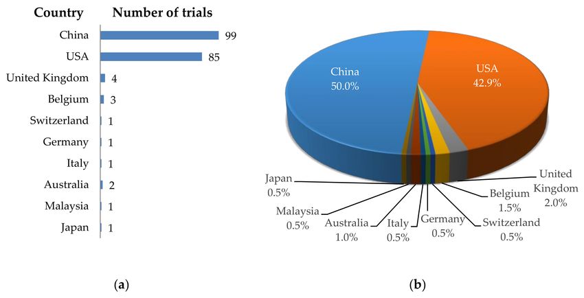
Cancers 2020, 12, 2567                                                                                             9 of 36

Most registered trials, however, use a second generation CAR [110] incorpora-ting a co-stimulatory
domain. Co-stimulation is mostly provided by CD28 or 4-1BB domains [3].

      Figure 4. Schematic overview of the methods used for CAR transfer into T cells. (a) Number of clinical
      trials using a specific transfer method; (b) Proportional distribution of clinical trials per transfer method.
      Data was extracted from clinicaltrials.gov.

      Physiologically, CD28 co-stimulation promotes the production of IL-2, -6, -10, and further
interleukins, as well as cell cycle progression, survival, differentiation, and cytolytic functions [111].
Many studies that employed CARs with a CD28 signaling domain observed potent and quick anti-tumor
effector functions. However, these were short-lived and associated with limited cell persistence in vivo
when compared to, e.g., the 4-1BB signaling domain. Notwithstanding, it was shown that human CD8+
CAR-T cells containing a CD28 co-stimulatory domain differentiate towards both a central-memory
and effector-memory type [112–119]. The transmembrane domain of CD28 used in many CARs as a
connector between extra- and intra-cellular domains is associated with improved expression of these
CARs on the surface [120,121], but might also cause tonic CAR signaling [122,123] and thereby lead to
Fas-dependent activation-induced cell death (AICD) in CAR-T cells, possibly explaining the observed
limited cell persistence [113]. Clinical trials confirmed the preclinical findings that CD28 supports
strong but short-lived anti-tumor efficacy [124,125].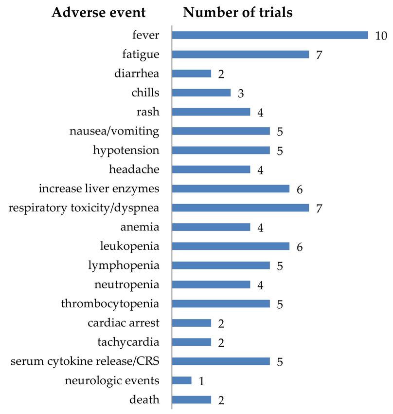
Cancers 2020, 12, 2567 10 of 36
unknown/undisclosed
scFv-4-1BB/CD3ζ
scFv-CD28/CD3ζ
scFv-FcεRIγ
scFv-CD3ζ
2 generation
or
nd
86 1 9 23 31 4
scFv-4-1BB/CD28/CD3ζ
3 generation
4 generation
scFv-CD28/4-1BB/CD3ζ
scFv-OX40/CD28/CD3ζ
4SCART
or or
th
rd
iCASP9
e.g. IL-12
e.g. IL-12
Apoptosis
5 2 2 6 5 7
OX40
CD3ζ
FcεRIγ
scFv
4-1BB
cytokine inducer
inducable caspase 9
hinge
CD28
TM domain
Figure 5. Schematic overview of the classical CAR formats used in clinical trials treating solid tumors.
The number
Figure of clinical
5. Schematic trials of
overview using a specific
the classical CAR
CAR format
formats is indicated.
used Datatreating
in clinical trials was extracted from
solid tumors.
clinicaltrials.gov. TM = transmembrane domain, iCASP9 = inducible caspase 9, 4SCART
The number of clinical trials using a specific CAR format is indicated. Data was extracted from = fourth
generation safety CAR-T cells. The Motifolio Scientific Illustration Toolkit was used for the generation
clinicaltrials.gov. TM = transmembrane domain, iCASP9 = inducible caspase 9, 4SCART = fourth
of parts of this figure.
generation safety CAR-T cells. The Motifolio Scientific Illustration Toolkit was used for the generation
of parts of this figure.
Physiological 4-1BB signaling in T cells enhances cell cycle progression and proliferation, cytokine
secretion, cytolytic potential, and inhibits clonal deletion and AICD [126,127]. CARs containing 4-1BB
as a signaling domain allowed for a more robust cell activation, as well as an increased persistence
in vivo, and 4-1BB co-stimulation promotes differentiation of CAR-T cells towards a central-memory
phenotype [4,112–118,128,129]. However, 4-1BB co-stimulated CARs showed a slower onset of
cytotoxicity,
Cancers 2020, 12,but longer durability and accumulation of CAR cells over timewww.mdpi.com/journal/cancers
x; doi: [128]. Potent anti-tumorCancers 2020, 12, 2567 11 of 36
efficacy and very long persistence of 4-1BB-containing CAR-T cells in patients were also reported in
clinical trials [4,130].
CD28 is incorporated in CARs used in 23 trials [131,132], and 4-1BB co-stimulation is used in
31 CAR-T cell trials (Figure 5). Four trials just indicate that second generation CARs are used, but do
not specify which co-stimulatory domain is included (Figure 5). In 86 registered trials, the used CAR
format is not disclosed (Figure 5).
The third generation CARs [111] used in the registered clinical trials incorporate combinations
of CD28/4-1BB, 4-1BB/CD28, or OX40/CD28 co-stimulatory domains (Figure 5) [133,134]. Six trials
mention the use of third generation CARs but do not indicate the exact co-stimulatory domains used,
or their order in the chimeric molecules (Figure 5). Fourth generation CARs (also known as “T cells
redirected for universal cytokine-mediated killing” (TRUCKs)) are in principle second generation CARs
with the extra feature that they can induce the production of e.g., cytokines in a very restricted local
fashion [135]. The effects induced depend on the cytokines that are secreted: e.g., IL-12 can activate
an innate immune response in the tumor [136], causes less susceptibility to Treg suppression [137],
and increases cytokine secretion and expansion [138,139], and IL-15 increases the anti-tumor activity of
the CAR-T cells [140]. A variant of this format is the 4SCART (fourth generation safety CAR-T) which
additionally incorporates an inducible caspase 9 as a safety measurement (will be described in detail in
Section 4.3). Five registered trials rely on the fourth generation CAR format, while seven trials use the
4SCART format (Figure 5).
3.2.2. CARs: The More Exotic Models
A group of 19 clinical trials in total use alternative binding moieties instead of a scFv directed
against an antigen expressed on the cell surface of the tumor (Figure 6).
One trial applies a so-called chimeric switch receptor (CSR), consisting of PD1 as extracellular
domain and CD28 transmembrane and intracellular signaling domain (Figure 6). As tumors often
express PD-L1 on their surface to activate the inhibitory PD1 receptor on T cells to circumvent/inhibit
an anti-tumor T-cell response, the CSR will turn the inhibitory into an activation signal induced by
CD28 [141].
Several trials target NKG2D ligands by linking NKG2D to either CD3ζ (4 trials), or 4-1BB/CD3ζ
(1 trial) (Figure 6) [43]. This strategy is very attractive, since NKG2D binds a plethora of ligands
(MIC-A, MIC-B, and the ULBPs 1, 2, 3, 4, 5, 6), that are induced-self proteins, which are upregulated on
stressed, infected, and transformed cells, as already described above, and therefore can be used for
many types of cancer.
Poseida Therapeutics, Inc., together with the City of Hope Comprehensive Cancer Center,
the Sarah Cannon Research Institute at HealthONE, and the Memorial Sloan Kettering Cancer Center,
are performing a phase 1/2 trial with CAR-T cells in which the binding moiety consists of a nanobody
(named Centyrin) specific for PSMA linked to the signaling domains of CD28 and CD3ζ (Figure 6).
The T1E-CD28/CD3ζ CAR is coupling a promiscuous ErbB ligand derived from EGF and TGFα
to a fused CD28/CD3ζ endodomain (Figure 6). This CAR can bind several ErbB2 dimers (i.e., HER2,
HER3, and EGFR) and therefore can target several tumors [142–144].
AffyImmune Therapeutics, Inc., together with the Weill Medical College of Cornell University
is clinically testing AIC100 CAR-T cells. The binding moiety of this CAR consists of the I-domain
of CD11a of LFA-1, which binds to ICAM1 on tumor cells. Intracellularly CD28, 4-1BB, and CD3ζ
domains facilitate signaling (Figure 6).
Originally used as an imaging agent to guide glioblastoma resection surgery, and to carry different
therapeutics to these tumors, a 36-amino acid long peptide of chlorotoxin, a component of scorpion
venom, was linked to the transmembrane and intracellular domains of CD28 and signaling domain
of CD3ζ to form a CAR (Figure 6). Preclinical testing showed that chlorotoxin binds to a greater
proportion of patient tumors, and cells within these tumors, while ignoring non-tumor cells in the brain
and other organs, and that this binding to its ligand is not so much influenced by tumor heterogeneityCancers 2020, 12, 2567 12 of 36
compared to other antigens such as IL13Rα2, HER2, and EGFR [145]. Therefore, the City of Hope
Medical Center, together with the National Cancer Institute is now testing this CAR in a clinical trial
against recurrent glioblastoma and recurrent malignant glioma.
Figure 6. Schematic overview of the exotic CAR formats used in clinical trials treating solid tumors.
The number of clinical trials using a specific CAR format is indicated. Data was extracted from
clinicaltrials.gov. TM = transmembrane domain, PD1 = programmed cell death protein 1, IL13 (E13Y)
= mutated IL-13 optimized to bind IL-13Rα2, TE1 = promiscuous ErbB ligand derived from EGF and
TGFα. The Motifolio Scientific Illustration Toolkit was used for the generation of parts of this figure.Cancers 2020, 12, 2567 13 of 36
CD70-CD27 interactions are important for the regulation of adaptive immunity. CD70 shows a
restricted expression on non-malignant cells, but is expressed on some solid tumors (e.g., on renal
carcinoma, pancreatic cancer, breast cancer, melanoma, and ovarian cancer) and is implicated in tumor
escape from immunosurveillance [146–150]. In a clinical trial performed by the NCI, a CAR consisting
of CD27, linked to undisclosed intracellular signaling domains is used (Figure 6).
Five clinical trials use a mutated form of IL-13, which is optimized for binding to IL-13Rα2, either
linked to the signaling domain of CD3ζ alone, or a combination of 4-BB and CD3ζ signaling domains
for the treatment of patients with glioblastoma, glioma, or melanoma (Figure 6) [151–154].
A highly complex CAR for the treatment of melanoma patients was developed by Timmune
Biotech Inc., and used in a clinical trial by the Second Affiliated Hospital of Hainan Medical University.
This so-called GPA-TriMAR binds to a peptide derived from the melanoma-associated antigen gp100,
presented in HLA-A2 through a TCR-like antibody [155,156] (Figure 6). This might increase the tumor
specificity, but nullifies the advantage of a CAR that it can bind to a native cell-surface tumor antigen,
which does not need to be processed and presented in an HLA-context and is therefore not dependent
on the HLA-type of the patient. The other two extracellular subunits are a sushi domain, which can
bind IL-15, and an IL-15-linker-PD1 construct (Figure 6). The latter two subunits are supposed to
stimulate the innate immune system. The GPA-TriMAR is linked to the intracellular signaling domains
of 4-1BB and CD3ζ [157].
Finally, two clinical trials use a TCR-like antibody (TLA) as binding moiety linked to a second
generation intracellular domain (i.e., either CD28/CD3ζ, or 4-1BB/CD3ζ) (Figure 6) [158]. Both trials
target a peptide of alphafetoprotein (AFP) presented in HLA-A2 in HCC patients. The two different
CARs were both developed by the companies Eureka Therapeutics Inc. and Aeon Therapeutics
(Shanghai, China) Co., Ltd. providing only limited published data [158].
3.3. Add-Ons; T-Cell Populations Used for Transfer or Extra Features Introduced into CAR-T Cells
3.3.1. T-Cell Populations Used for CAR-T-Cell Therapy
In many clinical trials, certain T-cell populations are used for the CAR introduction. For example,
T cells specific for VZV, EBV, adenovirus, CMV, or multivirus-specific T cells are used. The idea behind
it is that these T cells can be stimulated via their endogenous virus-specific TCR to proliferate and
therefore increase their persistence and number. Epitopes of latent viruses like EBV or CMV are
constantly presented and stimulate the CAR-T cells. Another strategy is to use virus vaccination to boost
T-cell proliferation (like VZV vaccination, or oncolytic adenovirus injected intratumorally). Some trials
used directly vaccine-specific T cells to induce this proliferation [46,159–161]. To increase persistence
of the CAR-T cells, one can alternatively use memory T cells for the transfer of the CAR [162,163].
Most trials use autologous patient T cells to introduce the CARs. However, there might be
certain situations making the use of allogeneic T cells necessary (e.g., not enough T cells can be
isolated/expanded from the patient, or CAR-T cell therapy is performed after allogeneic stem cells
transplantation). Furthermore, the use of allogeneic T cells that are genetically modified (see below)
in such a way that they are not recognized by the endogenous immune system, or can harm
healthy tissue of the patient, is very attractive, and can generate an off-the-shelf therapy for many
different patients. Alternatively, the endogenous TCR of γ/δ-T cells does not recognize peptides
presented in HLA molecules [164,165] and therefore they do not induce graft-versus-host disease
after CAR-T-cell transfer in HLA-mismatched patients [166]. This allows the use of γ/δ-T cells for the
generation of CAR-T cells from healthy donors, which are not impaired by tumor- or therapy-related
immunosuppression [167,168], and the application of CAR-T cells in a multitude of patients, irrespective
of their T-cell numbers and HLA-type. An additional positive effect is that γ/δ-T cells have an intrinsic
anti-tumor activity [169], potentiating the adoptive T-cell therapy against tumors. Moreover, the number
of CAR-γ/δ-T cells can be boosted in vivo by systemic administration of zoledronic acid [170].Cancers 2020, 12, 2567 14 of 36
3.3.2. Extra Features Introduced into CAR-T Cells
Several resistance mechanisms to CAR-T-cell therapy in solid tumors may play a role in the
observed lower effectiveness compared to CAR-T cells in hematologic malignancies [171–174].
For example, the tumor microenvironment can be hostile for CAR-T cells (e.g., unfavorable pH
or oxygen levels) or unfavorable electrolyte or cytokine concentrations, inhibiting an effective immune
response [175–177]. Additionally, the homing of CAR-T cells can be hampered in solid tumors [172].
Furthermore, solid tumors can induce inhibitory receptors on CAR-T cells like PD1 and CTLA-4,
making the CAR-T cells exhausted. The patient’s own immune cells can even attack the CAR-T
cells e.g., by antibody production [178,179]. To overcome these resistance mechanisms, several
strategies were developed, like induced expression of cytokines, expression of constitutively active
or dominant negative cytokine receptors, expression of homing receptors, prevention of anti-CAR
antibody production, or blocking of PD1/CTLA-4. All are described in more detail below.
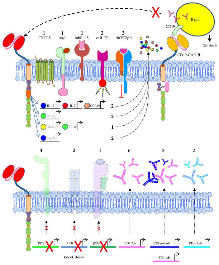
An often-used strategy to improve the effectiveness of CAR-T cells is to equip them with the
ability to secrete cytokines like IL-12, IL-15, IL-21, IL-7, or combinations thereof (Figure 7). In two
trials performed by the Second Affiliated Hospital of Guangzhou Medical University and the Sixth
Affiliated Hospital of Wenzhou Medical University in China, the used CAR-T cells produce IL-7
and CCL19 [180–183]. IL-7 is known for its positive effects on T-cell survival [184], and CCL19 is a
chemokine attracting other endogenous immune cells, like dendritic cells, B cells, and central memory
T cells [185–187] to the tumor site. IL-12, IL-15, and IL-21 are all cytokines known to stimulate immune
cells. The fact that these cytokines are produced very locally is an advantage, since some of them can
have toxic effects when applied systemically [135].
Some trials take this even a step further and use CAR-T cells co-expressing a constitutively active
IL-7 receptor [188], or a membrane-bound form of IL-15 [189] (Figure 7). Others introduce a chimeric
cytokine receptor containing the IL-4Rα ectodomain coupled to the IL-2Rβ endodomain (4αβ) resulting
in a robust expansion of CAR-T cells after IL-4 binding, a cytokine with several pathophysiologic
and therapeutic links to cancer [190] (Figure 7). TGFβ is known to have an immunosuppressive
effect, and many tumors, especially prostate cancer, secrete TGFβ and thereby promote metastasis
and neoangiogenesis and suppress T cells [191,192]. In vitro and in vivo models showed that blocking
TGFβ signaling in T cells by using a dominant-negative TGFβ receptor II (i.e., a truncated form which
lacks the intracellular domain necessary for downstream signaling) [193] resulted in an increased
ability to infiltrate, proliferate, and mediate anti-tumor responses [194]. Therefore, this dnTGFβR
was co-introduced next to a CAR specific for PSMA in T cells to treat prostate cancer patients [195]
(Figure 7).
CAR-T cells can also be engineered to express homing molecules to target these cells to specific
tissue locations. A clinical trial performed by the Sun Yat-sen University, Guangzhou, China in
collaboration with Bio-gene Technology Co., Ltd., Guangzhou, China uses CAR-T cells specific for
EGFR which were additionally transduced with the lymphoid follicle homing molecule CXCR5 [40].
Alternatively, to circumvent problems with homing of CAR-T cells to the tumor site, these cells can be
directly infused into the tumor [55,154,196,197].
In total, five clinical trials co-introduce an anti-CD19 CAR next to the tumor-antigen-specific CAR
into T cells. This extra CAR is directed against CD19 positive B cells which can produce antibodies,
and these cells will be lysed. That means that CD19-positive cells that might produce anti-CAR
antibodies, e.g., because the tumor-antigen-specific CAR is based on a murine scFv, will also be
destroyed. This will increase the persistence of the anti-tumor CAR-T cells in the patients (Figure 7).
Additionally, lymphodepleting chemotherapy will also prevent graft rejection.
The anti-tumor activity of T cells can be inhibited by various tumor-associated immunosuppressive
ligands like PD1 and CTLA-4 [198]. Several strategies are used in clinical trials to prevent inhibition of
the CAR-T cells by PD1 or CTLA-4 interactions between tumor cells and CAR-T cells. Four clinical
trials knockout the PD1 gene in the CAR-T cells (Figure 7), preventing the interaction of this molecule
with PD-L1 expressed on tumor cells. Furthermore, several studies introduce genes into CAR-TCancers 2020, 12, 2567 15 of 36
cells encoding blocking anti-PD1, anti-PD1 and anti-CTLA-4, or anti-PD-L1 antibodies, which after
secretion by the CAR-T cells result in the same prevention of inhibition by the tumor cells (Figure 7).
One study introduced a gene encoding an anti-PD1 nanobody with the same purpose (not shown).
Moreover, some trials combined CAR-T cell therapy with anti-PD1, anti-CTLA-4 antibodies ([197,199],
NCT03980288, NCT03726515, NCT01822652, NCT04003649 (see Table S1)).
Figure 7. Schematic overview of the extra features introduced into CAR-T cells used in clinical trials
treating solid tumors. The number of clinical trials using a specific feature is indicated. Data was
extracted from clinicaltrials.gov. 4αβ = chimeric cytokine receptor containing the IL-4Rα ectodomain
coupled to the IL-2Rβ endodomain, mbIL-15 = membrane-bound IL-15, caIL-7R = constitutively active
IL-7 receptor, dnTGFβR = dominant negative TGFβ receptor. The Motifolio Scientific Illustration
Toolkit was used for the generation of parts of this figure.Cancers 2020, 12, 2567 16 of 36
As already described above, the use of allogeneic CAR-T cells has certain advantages. Therefore,
several trials use T cells that are genetically modified in such a way that they are not recognized
by the endogenous immune system, or can harm healthy tissue of the patient by knocking out the
endogenous TCR and/or β2M, the latter resulting in the absence of MHC class I expression on the cell
surface (Figure 7). This approach can lead to an off-the-shelf therapy using allogeneic T cells for many
different patients.
4. CAR-T Cell Clinical Trials against Solid Tumors—Patient Pretreatments, Injection Sites, Safety
Measurements, Clinical Outcomes
4.1. Treatments of Patients before CAR-T-Cell Transfer
To provide the best conditions for the introduced CAR-T cells, it is common to perform a
lymphodepleting pretreatment of the patients. This is based on results obtained after transfer of tumor
infiltrating lymphocytes (TILs) and CD19-directed CAR-T cells. In these studies, it was shown that
lymphodepleting or conditioning chemotherapy administered prior to T-cell infusion clearly improve
persistence and efficacy of these T cells [200], for example by reducing the number of suppressive cells,
or removing competing sink cells, making IL-7 and IL-15 cytokines available for T cell expansion [201].
Different types of lymphodepleting or conditioning chemotherapies were also performed in the
CAR-T-cell clinical trials against solid tumors (Figure 8). Mostly, the classical non-myeloablative
lymphodepleting regimen with cyclophosphamide and fludarabine [202] was performed (n = 59, 28.2%;
Figure 8), but also schedules with cyclophosphamide or fludarabine as single agent are described (n = 21
and n = 2, respectively; Figure 8). Other chemotherapies include: paclitaxel + cyclophosphamide,
Temozolomide [203], or bis-1-nitrosourea + etoposide + arabinoside + cyclophosphamide (b + e + a + c)
(Figure 8). A total of 93 trials do not clearly state if preconditioning is performed (unknown, n = 75),
or what kind of preconditioning is performed (lymphodepleting pretreatment, n = 14; chemotherapy,
n = 4) (Figure 8). In the registered clinical trials that describe the timing of lymphodepletion, the interval
between lymphodepletion and CAR-T-cell application is mostly performed 3–5 days before the infusion
of CAR-T cells, for 2–4 days. Twenty-five trials explicitly mention that no lymphodepletion is executed
(Figure 8).
4.2. Injection Sites for CAR-T Cell Application
Most clinical trials apply the CAR-T cells by injecting them intravenously (n = 105; Figure 9)
counting on the correct homing of the T cells to the tumor. However, there are also other sites possible,
especially if one wants to apply the CAR-T cells very locally in the tumor or at the resection site.
For example, trials treating brain tumors use intracranial (n = 2), intracavity (n = 3), or intracerebral
(n = 6) injection, or inject into the ventricular system (n = 8) (Figure 9). Nineteen clinical trials indicated
intratumoral injection, and nine use intraperitoneal injection (Figure 9). Local treatment of liver or
pancreas cancers with CAR-T cells can be achieved by transcatheter arterial infusion (TAI; n = 3),
intrahepatic artery injection (n = 10), pancreatic artery (n = 1), or pancreatic venous (n = 1) injection
(Figure 9). A total of 56 clinical trials do not indicate the site of CAR-T-cell application (Figure 9).
4.3. Safety Measurements to Control Negative Effects of CAR-T Cells in the Patient
As already mentioned above, most antigens targeted in clinical trials with CAR-T cells against
solid tumors are not perfectly tumor specific. It can be that the antigens are expressed to some extend
on normal healthy tissues, and there an on-target/off-tumor toxicity due to the accidental killing of
non-malignant bystander cells co-expressing the target antigen can be induced by the CAR-T cells [11].
To be able to shut-off the CAR-T cells as soon as toxicity is noticed in the patient, several strategies
were developed (Figure 10). Rimiducid (AP1903) and rapamycin are molecules that are able to induce
dimerization of constructs containing inducible caspase 9, which are co-introduced with the CAR
into the T cells as a suicide switch. After dimerization, the caspase 9 induces apoptosis of the CAR-TCancers 2020, 12, 2567 17 of 36
cells and thereby the unwanted/unexpected T-cell activities are eliminated [204,205]. This kind of
suicide switch is used in 17 clinical trials (Figure 10) performed with mostly fourth generation safety
CAR-T cells (4SCART). Interestingly, rimiducid (AP1903) is also used to provide multimerization of an
inducible co-stimulatory molecule based on MyD88 and CD40 (iMC) into T cells, which allows for the
selective activation of adoptively transferred T cells in vivo resulting in enhanced anti-tumor activity
in solid tumors (Figure 10). Removal of rimiducid will switch off this co-stimulation again [206,207].
Figure 8. Schematic overview of the pretreatments used before CAR-T cells were applied in patients.
(a) Number of clinical trials using a specific pretreatment; (b) Proportional distribution of clinical trials
per pretreatment. Data was extracted from clinicaltrials.gov.
One of the oldest suicide switches used is the herpes simplex virus-thymidine kinase/ganciclovir
(HSV-tk/GCV) strategy. Mechanistically, HSV-tk phosphorylates GCV and the resulting triphosphate
form is incorporated by DNA polymerases into the DNA, leading to chain termination and cell
death [208]. HSV-tk/GCV also induces apoptosis [209]. A disadvantage of using HSV-tk/GCV is that it
can be immunogenic in immunocompetent patients causing a limited persistence of HSV-tk transduced
cells [210]. Nevertheless, three clinical trials still use the HSV-tk/GCV strategy (Figure 10) [211].
Furthermore, several trimmed molecules are used for selection and/or depletion of CAR-T cells,
like truncated HER2 (HER2tG), truncated EGFR (tEGFR), and truncated CD19 (tCD19) (Figure 10).
Trastuzumab (Herceptin® ) binds to HER2tG [212] and is used in two clinical trials for the elimination
via complement or antibody-dependent cell-mediated cytotoxicity (ADCC) of CAR-T cells in case of
on-target/off-tumor reactions (Figure 10). Cetuximab is used for the ablation of tEGFR-expressing CAR-T
cells in eight clinical trials (Figure 10) via the same mechanisms [213]. Seven clinical trials use truncated
CD19 either as selection marker for CAR-positive T cells [214], or as marker for elimination using an
anti-CD19 antibody conjugated to pseudomonas toxin (CD19-ETA’) [215] (Figure 10). One clinical trial
is using an undisclosed ‘kill switch’ as safety measurement (Figure 10).Cancers 2020, 12, 2567 18 of 36
Figure 9. Schematic overview of the injection sites used to apply CAR-T cells against solid tumors.
The numbers indicate the number of clinical trials using this injection site. Data was extracted from
clinicaltrials.gov. The Motifolio Scientific Illustration Toolkit was used for the generation of this figure.
Figure 10. Schematic overview of safety measurements used in the treatment with CAR-T cells against
solid tumors. The numbers indicate the number of clinical trials using this safety measurement.
Data was extracted from clinicaltrials.gov. The Motifolio Scientific Illustration Toolkit was used for the
generation of this figure.Cancers 2020, 12, 2567 19 of 36
A special safety measurement to circumvent prolonged autoimmunity induced by an on-target/
off-tumor reaction of the CAR is the introduction of the CAR by mRNA electroporation (n = 5, 2.6%;
Figure 4). We have previously demonstrated that transient transfection of T cells with CARs using
mRNA electroporation might be an effective and safe tool in cancer immunotherapy [121,216–220].
The electroporation procedure is based on complex physicochemical mechanisms leading to plasma
membrane perforation upon application of electric fields allowing for subsequent entry of mRNA into
the cytosol [221]. Using RNA-transfected CAR-T cells offers the advantage that the receptor expression
is temporally restricted (Figure 11), rendering potential off-target and on-target/off-tumor toxicity
transient as well. The CAR-RNA transfer strategy is especially attractive in phase 0/1 clinical trials
exploring new tumor antigens for CAR-T-cell therapy with an unknown clinical safety profile.
The mRNA transfection strategy for CARs proposed by us quite some time ago [109] has in
the meantime been applied by others in clinical trials. In patients with solid tumors c-MET was
used as a CAR-target antigen on breast cancer and melanoma [222], (NCT01837602; NCT03060356)
and mesothelin as a CAR-target antigen on mesothelioma, pancreatic cancer, and ovarian cancer
[223–225], (NCT03608618; NCT01897415; NCT01355965). RNA transfection was even explored with
non-solid tumors using CD19 and CD123 as target antigen [226], (NCT02277522; NCT02624258;
NCT02623582). The mRNA-CAR-T cells in these studies were well tolerated [222], the cells migrated
to primary and metastatic tumor sites, showed a clinical anti-tumor activity, and showed no evidence
of on-target/off-tumor toxicity against normal tissues [223]. After local application, c-MET-CAR-T cells
induced necrosis within the tumor. Importantly, some of the injected c-MET-CAR-T cells entered the
blood stream and could be monitored in the circulation for a short time [222].
CAR expression over time
RNA
Electroporation
RNA stability over
time
Figure 11. Schematic representation of introduction of a CAR by mRNA electroporation. Indicated are
the low stability of the introduced mRNA over time and the transient expression of the CAR on the
T-cell surface. The Motifolio Scientific Illustration Toolkit was used for the generation of this figure.
The clinical trials published by Beatty et al. and Maus et al., using mesothelin as antigen, showed a
cytokine release syndrome (CRS) in one mesothelioma patient resulting in adverse events (anaphylaxis,
cardiac arrest, respiratory failure, disseminated intravenous coagulation) within minutes of completing
the third infusion [223,225]. In contrast, in pancreatic cancer patients no cytokine release syndrome andCancers 2020, 12, 2567 20 of 36
no dose-limiting toxicities, but actually stable disease in two of six patients were seen [224]. When using
RNA-CAR-T cells, robust proliferation and persistence are not so important, making lymphodepletion
unnecessary, as the transient receptor expression per se necessitates repetitive injections. Unlike most
of the trials registered in clinicaltrials.gov which use virally transduced cells, which have to be applied
only once, presuming that these cells will proliferate upon tumor-antigen recognition, making repeated
applications unnecessary, RNA-transfected cells will lose CAR expression (Figure 11) and have to be
replenished from the outside to maintain cytolytic pressure on the tumor.
The possible reason for the severe adverse events in the patient described above by Maus et al. and
Beatty et al. [223,225], was that the CAR was based on a murine antibody and the adverse event was
caused by IgE antibodies specific for the scFv in the CAR (i.e., a human anti-mouse antibody (HAMA)
response), subsequently causing a CRS after an additional injection of CAR-T cells. These antibodies
were probably induced by the intermittent dosing schedule of the CAR-T cells [223,225]. After the
first two injections of RNA CAR-T cells on day 0 and day 7, the third injection was given after a
long waiting period on day 49. This is sufficient time to complete an isotype switch from IgG to IgE.
Therefore, rapid repetition of infusions seems to be best to prevent isotype switching if a HAMA
response is induced.
Although the transient expression of a CAR on the cell surface of T cells by electroporation with
mRNA can be an advantage if an on-target/off-tumor response is induced by the CAR-T cells, it can
also be a disadvantage for the applicability. It has to be carefully monitored whether the infused CAR-T
cells reach their tumor target in time before the CAR expression is too low for an effective anti-tumor
response. This might be circumvented by local infusion of these CAR-T cells at the tumor site, as it
is performed in several clinical trials (NCT01355965 [223,225], NCT01897415 [224], NCT03608618,
NCT01837602 [222]). Furthermore, the necessary repetitive application of mRNA-transfected CAR-T
cells harbors some hazards, like elaborately described above (e.g., possible isotype switch of a HAMA
response [223,225]). Additionally, this repetitive application necessitates the production and storage of
several batches of CAR-T cells, which might be cumbersome. Moreover, an anti-tumor CAR-T-cell
memory will not be induced in patients treated with mRNA-transfected CAR-T cells, which might
be a problem if the tumor is not completely eradicated and can reoccur. To draw final conclusions
on the applicability of mRNA-transfected CAR-T cells, analysis in several clinical trials is necessary.
To mitigate safety concerns, another promising strategy is the initial use of repetitive injections of
RNA-transfected CAR-T cells to probe for toxicity, and in the case of no serious side-effects, switch to
permanently transfected CAR-T cells.
4.4. Clinical Outcomes and Adverse Events of CAR-T-Cell Therapy of Solid Tumors
4.4.1. Clinical Outcomes
Of 42 clinical trials using CAR-T cells against solid tumors registered at clinicaltrials.gov the
clinical outcome could be retrieved from either clinicaltrials.gov, or through literature search on
pubmed.ncbi.nlm.nih.gov (see Table S1; also including data on number of injected cells, trial phase,
(estimated) patient number, trial status, principle investigator, and references) [25,197,199,227–247].
Some clinical outcomes were found in abstracts of ASCO meetings published in the Journal of Clinical
Oncology (Table S1). Of the 375 treated patients listed in publications reporting on clinical outcome,
13 had a complete response, 35 had a partial response, 4 had a mixed response, 121 had a stable disease,
109 had a progressive disease, 8 had no evidence of disease, 5 were not evaluable, and of 80 patients
the clinical outcome was not disclosed. This data is summarized in Table S2.
4.4.2. Adverse Events
In total, 28 clinical trials described in this review also reported on adverse events (Table S1).
The adverse events were quite diverse (Figure 12). Some adverse events were very local, and this could
be explained by looking at the tumor site (e.g., seizure when treating glioblastoma, or abdominal painYou can also read