Influence of Circadian Rhythm in the Eye: Significance of Melatonin in Glaucoma
←
→
Page content transcription
If your browser does not render page correctly, please read the page content below
biomolecules

Review
Influence of Circadian Rhythm in the Eye: Significance of
Melatonin in Glaucoma
Alejandro Martínez-Águila 1,2, * , Alba Martín-Gil 1,3 , Carlos Carpena-Torres 1,3 , Cristina Pastrana 1,3
and Gonzalo Carracedo 1,3

                                          1   Ocupharm Research Group, Biochemistry and Molecular Biology IV, Faculty of Optics and Optometry,
                                              28037 Madrid, Spain; amarting@ucm.es (A.M.-G.); ccarpena@ucm.es (C.C.-T.); crispast@ucm.es (C.P.);
                                              jgcarrac@ucm.es (G.C.)
                                          2   Research & Development Department Avizor, 28919 Leganés, Spain
                                          3   Optometry and Vision Department, Complutense University, 28037 Madrid, Spain
                                          *   Correspondence: amaguila@ucm.es; Tel.: +34-913946883

                                          Abstract: Circadian rhythm and the molecules involved in it, such as melanopsin and melatonin,
                                          play an important role in the eye to regulate the homeostasis and even to treat some ocular conditions.
                                          As a result, many ocular pathologies like dry eye, corneal wound healing, cataracts, myopia, retinal
                                          diseases, and glaucoma are affected by this cycle. This review will summarize the current scientific
                                          literature about the influence of circadian patterns on the eye, focusing on its relationship with
                                          increased intraocular pressure (IOP) fluctuations and glaucoma. Regarding treatments, two ways
                                          should be studied: the first one, to analyze if some treatments could improve their effect on the ocular
                                          disease when their posology is established in function of circadian patterns, and the second one, to
                                          evaluate new drugs to treat eye pathologies related to the circadian rhythm, as it has been stated

                                   with melatonin or its analogs, that not only could be used as the main treatment but as coadjutant,
                                          improving the circadian pattern or its antioxidant and antiangiogenic properties.
Citation: Martínez-Águila, A.;
Martín-Gil, A.; Carpena-Torres, C.;
Pastrana, C.; Carracedo, G. Influence
                                          Keywords: circadian rhythm; ocular diseases; glaucoma; melatonin; melanopsin; myopia; dry eye
of Circadian Rhythm in the Eye:
Significance of Melatonin in Glaucoma.
Biomolecules 2021, 11, 340. https://
doi.org/10.3390/biom11030340              1. Circadian Rhythm in the Eye
                                                The term circadian rhythms (circa meaning “around”, and dian meaning “day”) was
Academic Editor: Randy J. Nelson          first coined by Halberg in 1953 [1] and refers to biological cycles with periods slightly
                                          longer than 24 h (on average ~24.2 h) [2]. Body temperature, sleep duration, hormonal
Received: 28 November 2020
                                          levels, heart rate, and other physiological variables exhibit such daily oscillations [3,4].
Accepted: 20 February 2021
                                                In mammals, there are peripheral circadian clocks located in tissues like retina, heart,
Published: 24 February 2021
                                          liver, lungs, pituitary, and skeletal muscles that contain their own circadian oscillators [5,6].
                                          However, the suprachiasmatic nucleus (SCN) of the anterior hypothalamus is considered
Publisher’s Note: MDPI stays neutral
                                          to be the master circadian clock since its discovery in 1970s [7,8].
with regard to jurisdictional claims in
                                                The way to synchronize this rhythm is by external cues known as “zeitgebers” (“time
published maps and institutional affil-
                                          givers” in German). Some of these zeitgebers are temperature, feeding times, and social
iations.
                                          interactions. Nevertheless, the primary and most important cue is light, which allows us to
                                          synchronize SCN to the day–night cycle.
                                                Through the eye, light reaches the retina and is processed by retinal ganglion cells
                                          (RGCs), which drive whole visual information. However, approximately 1–2% of these
Copyright: © 2021 by the authors.
                                          cells contain a photopigment denominated melanopsin [9,10]. This small subset of RGCs
Licensee MDPI, Basel, Switzerland.
                                          received the name of intrinsic photosensitive retinal ganglion cells (ipRGCs) which are
This article is an open access article
                                          directly photosensitive [11,12]. This photic input is carried to the SCN via the retinohy-
distributed under the terms and
                                          pothalamic tract using glutamate as a neurotransmitter which also acts in gene expression,
conditions of the Creative Commons
                                          which is important in the circadian process [13]. Moreover, ipRGCs project their axons
Attribution (CC BY) license (https://
creativecommons.org/licenses/by/
                                          to the intergeniculate leaflet [14] and to the olivary pretectal nucleus of the pretectum to
4.0/).
                                          mediate pupil light reflex [15,16].

Biomolecules 2021, 11, 340. https://doi.org/10.3390/biom11030340                                      https://www.mdpi.com/journal/biomolecules
Biomolecules 2021, 11, 340 2 of 25
1.1. Melanopsin
It was in 1998 when the photopigment melanopsin was first discovered in the eye and
skin of a frog (Xenopus laevis) [17]. In mammals, however, it has been only localized in retina,
encoded in the gene OPn4. This opsin has a maximum peak of absorption at approximately
482 nm [12]. This property allows ipRGCs to act as additional photoreceptors to the
classical cones and rods, processing non-image-forming information [18].
Implications of melanopsin in the nonvisual information are supported by studies in
mice without cones and rods that still responded to light stimulus with pupillary constric-
tion [19], melatonin suppression [20], and phase shift. Additional studies in mice with the
melanopsin gene ablated only in ipRGCs showed deficits in circadian photoentrainment
and pupillary light reflex [21–23]. Furthermore, blind people with retinal damage (cones
and rods) have normal photoentrainment and pupil responses [24].
However, melanopsin is also responsible for an important process, the melatonin
synthesis suppression. Its activation by short-wavelength light (470–480 nm) [25,26] de-
creases melatonin levels both in the central nervous system (CNS) as in blood and in the
eye, following a circadian rhythm although inversed between them.
1.2. Melatonin
Melatonin (N-acetyl-5-methoxytryptamine) is the hormone responsible for regulation
of circadian and seasonal rhythms [27,28]. This molecule was first discovered and described
in the pineal gland [29], but currently it is known to be synthesized in many tissues in
the body including the eye and ocular annexes, specifically in the retina [30], iris, ciliary
body [31], crystalline lens [32,33], and lacrimal gland [34], where it regulates important pro-
cesses. As mentioned, melatonin synthesis is controlled by light–darkness cycles, increased
during the night and suppressed during the day [35], reaching a concentration peak at night
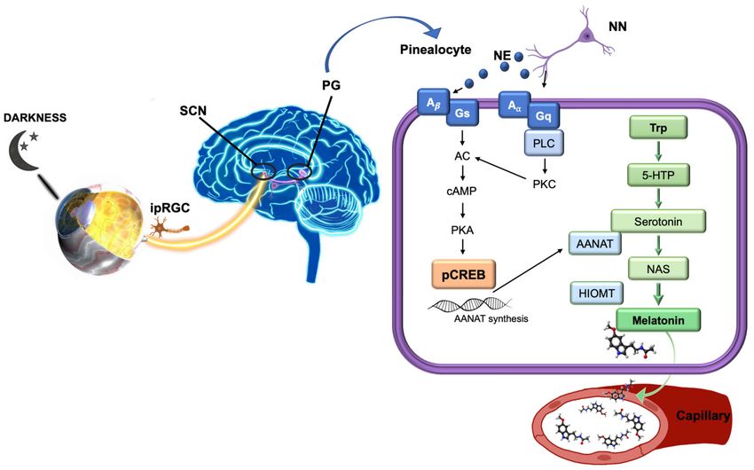
(between 02:00 to 04:00) [36]. It is initiated from tryptophan in pinealocytes [29,37], then
converted to 5-hydroxytryptophan which, in turn, is transformed into serotonin (by trypto-
phan 5-hydroxylase and aromatic amino acid decarboxylase, respectively). Following that,
serotonin is converted to N-acetylserotonin (NAT) by arylalkylamine N-acetyltransferase
(AANAT) and posteriori to melatonin by hydroxyindole-0-methyltransferase (HIOMT)
(Figure 1).
Melatonin action is mediated by cellular receptors. Two types of membrane melatonin
receptors have been characterized and cloned in mammals: MT1 and MT2 , both being
G-protein-coupled receptors with seven transmembrane domains [38,39]. A third putative
receptor called MT3 [40] was characterized as the enzyme quinone reductase 2 [40,41],
having lower affinity for melatonin when compared to MT1 and MT2.
MT1 and MT2 receptors are expressed in different parts of the brain including SCN,
cerebellum, hippocampus, substantia nigra, ventral tegmental area, in cardiovascular
system, and in almost all peripheral organs: blood vessels, adrenal glands, mammary
gland, gastrointestinal tract, liver, kidney and bladder, ovary, testis, prostate, skin, and the
immune system [42].
Focusing on the eye, receptors have been localized in different structures. On the
ocular surface, melatonin MT1 receptor has been detected in corneal epithelial cells in
different animals [43,44] and humans [45], while MT2 receptor has only been identified in
animals [43,46,47]. They have also been seen in rabbits in the iris and ciliary body [48] and
in human uveal melanocytes and human uveal melanoma cells [49]. Presence of melatonin
in aqueous humour [50] has also been confirmed as its enzymes AANAT and HIOMT [51].
Furthermore, within the lens, MT2 receptor has been identified [43] being considered as
another source of melatonin [52]. In sclera, both receptors are expressed [43,44].Biomolecules 2021, 11, 340 3 of 25
Figure 1. Diagram of melatonin synthesis in the pineal gland. (Modified and supplemented from Alkozi, 2020). Melatonin
synthesis is stimulated by signals of darkness coming from the eye through the suprachiasmatic nucleus neurons. Nore-
pinephrine may bind to α- and β-adrenergic receptors on pinealocytes activating both signaling pathways. Adenylyl cyclase
is activated to produce cAMP by both adrenergic signaling cascades. cAMP levels regulate AANAT activity, which increase
intracellular concentration of NAS converted then to melatonin by HIOMT. 5-HTP, 5-hydroxitryptophan; A, adrenergic
receptor; AANAT, arylalkylamine N-acetyltransferase; AC, adenylyl cyclase; HIOMT, hydroxyndole-O-methyltransferase;
cAMP, cyclic adenosine monophosphate; CREB, cAMP response element-binding protein; G, G protein; ipRGC, intrinsic
photosensitive retinal ganglion cells; NAS, N-acetylserotonin; NE, norepinephrine; NN, adrenergic neurons; PG, pineal
gland; PKA, protein kinase A; PKC, C kinase; PLC, phospholipase C; SCN, suprachiasmatic nucleus; and Trp, Tryptophan.
In the posterior segment, melatonin is also synthesized in the retina by photoreceptors,
and its receptors are localized in many areas of the retina, with the MT2 receptor being
more expressed than MT1 in human eyes [38]. These receptors have been detected in
plexiform layers, horizontal cells, amacrine cells, ganglion cells, and in rod photoreceptors
cells [42,43,45,53–56]. Accordingly, melatonin seems to play a role modulating dopamine
release [57], in the regulation of retinomotor movements [58], visual sensitivity [59], and
mediate regeneration of photoreceptors external discs [60].
Synthesized melatonin is released into circulation as an endocrine hormone and binds
to all receptors of target organs to regulate various physiological processes associated [55].
Melatonin levels can be measured in blood, saliva, and urine samples [61], and to keep a
normal circadian rhythm, exposure to light–dark cycles should follow a regular pattern.
Nowadays, artificial lights are present almost everywhere, especially in electronic
devices. This fact can affect our biological rhythms and is currently the subject of nu-
merous studies. Increase in exposition to lights at evening and at night can delay or
suppress normal melatonin secretion [62], through the activation of melanopsin contained
in ipRGCs [18,63–65]. Intensity and duration of the exposure, as well as light spectrum, are
considered variables in this process. Currently, there are many devices using LED light
sources with a short-wavelength emission thought to be responsible for the increase in
sleep disorders [66].
In general, circadian disruption has been linked not only to sleep disorders or jet lag
but also to different conditions such as obesity, diabetes, cardiovascular disease, cancer,
psychiatric disorders, and neurodegenerative diseases [67]. Moreover, it represents aBiomolecules 2021, 11, 340 4 of 25
common feature of Alzheimer [68,69] and Parkinson diseases [70–72], showing a strong
decrease in melatonin levels [73–75]. Additionally, some studies carried out with shift
workers, whose circadian rhythm is altered, have shown a high prevalence of some of the
aforementioned diseases [76–78].
This review will highlight the ocular pathologies such as dry eye, cataract, myopia,
retinal diseases, and glaucoma which are affected by this rhythm, focusing its relationship
with IOP fluctuations and glaucoma, and how melatonin and its analogs modify IOP.
2. Ocular Pathologies Related to Circadian Rhythm
In addition to the implications of the eye in the regulation of systemic circadian
rhythm, a large number of ocular physiological processes are also controlled by it. Any
biochemical or optical mechanisms involved in the circadian cycle could lead to developing
ocular pathologies or alterations of ocular physiology. In this section, we have reviewed
the role of circadian rhythm and melatonin (Table 1) in some ocular conditions such as dry
eye, corneal wound healing, myopia, cataract, and retinal diseases. Glaucoma will be more
deeply analyzed in the following sections.
Table 1. Summary of principal studies evaluating the effect of melatonin for the treatment of different ocular pathologies.
Ocular Pathology Reference Compound Target Results
The topical instillation of analogs of
Melatonin and
melatonin increased tears secretion: 39% with
analogs
Navarro Gil agomelatine (MT2 receptor), 28% with IIK7
(Agomelatine, IIK7, Rabbits
et al. [79] (MT2 receptor), and 20% with 5-MCA-NAT
and
(unknown receptors). Conversely, there were
Dry eye 5-MCA-NAT)
no changes with the melatonin.
The topical instillation of Ap4 A increased tear
secretion by 10%, while melatonin showed no
Hoyle et al. [80] Ap4 A + Melatonin Rabbits
effect. The synergic effect of both molecules
increased tear secretion by 34%.
The topical instillation of melatonin and IIK7
Melatonin and
improved rate of corneal healing by around
Crooke et al. analogs
Rabbits 47%, while 5-MCA-NAT showed no effect.
[47] (IIK7 and
This effect is mediated by the MT2 receptor,
5-MCA-NAT)
which is present in corneal epithelial cells.
Corneal wound
healing The topical exposition of melatonin in an ex
vivo corneal-wound model accelerated the
healing process. Notably, 60 µg/mL
Crespo-Moral Ex vivo
Melatonin melatonin accelerated the healing rate during
et al. [81] Porcine eyeball
48 h and 90 µg/mL melatonin during 72 h.
Unexpectedly, 120 µg/mL melatonin
decreased the healing rate after 96 h.
The retinal stimulation with blue light
(480 nm) reduced eye growth, which was
related to stimulate synthesis of melanopsin
Wang et al. [82] Blue light Guinea pigs
in retina and sclera, and to reduce both
synthesis of MT1 receptor and production of
Myopia melatonin in pineal gland.
The retinal melanopsin inhibition by
AA92593 intravitreal injection of an antagonist
Zheng et al. [83] (melanopsin Guinea pigs AA92593 increased both the eye growth and
antagonist) melatonin levels in the retina, these variables
being directly correlated (r = 0.74).Biomolecules 2021, 11, 340 5 of 25
Table 1. Cont.
Ocular Pathology Reference Compound Target Results
The use of melatonin in rat models reduced
lipid peroxidation and promoted both the
Different
Melatonin Rats synthesis of glutathione and antioxidative
authors [84–90]
activity of different enzymes, leading to a
reduction in cataract formation.
Cataracts
The inhibition of melanopsin present in the
lens epithelium with a yellow filter
Yellow light and
Pintor et al. [91] Rabbits (absorbance between 465–480 nm) and its
AA92593
antagonist AA92593 reduced the
concentration of ATP in the aqueous humour.
The muscular injection of melatonin reduced
Liang et al. [92] Melatonin Mice damage of photoreceptors and their apoptosis
in a retinal degeneration model.
Retinal damage The intraperitoneal injection of melatonin
reduced levels of VEGF, nitrite, and
Kaur et al. [93] Melatonin Rats
melatonin in the retina of hypoxic rats, also
reducing retinal vascular permeability.
The antioxidant effect of subcutaneous
Dieguez et al. implantation of a pellet of melatonin helped
Melatonin Mice
[94] to preserve visual function and retinal
Age-related structures in a non-exudative AMD model.
macular
In this case series study, the daily oral
degeneration
administration of 3 mg melatonin for
Yi et al. [95] Melatonin Humans 3 months improved the signs of AMD in
terms of retinal blood and retinal exudates in
more than 90% of patients.
2.1. Dry Eye
Dry eye is a multifactorial disease of the ocular surface characterized by a loss of home-
ostasis in the tear film [96]. Due to the ocular-surface health status being directly dependent
on several environmental factors [97], it is difficult to establish if the diurnal/nocturnal
variations in the ocular-surface physiology are related to these environmental factors, the
circadian rhythm, or a combination of both.
Regarding tear volume, its diurnal variations have been confirmed, which would
follow a circadian cycle [98–100]. In healthy subjects, there is a discrepancy between
studies reporting higher tear volume during the morning [98,100] and others during the
afternoon [99], but they all showed lower tear volume late in the day. On the other hand,
tear osmolarity also seems to follow a circadian pattern [99,101], despite a study of Oncel
et al. [102] showing a stable profile throughout the daytime. Niimi et al. [101] demonstrated
that tear osmolarity was drastically decreased upon awakening, quickly increased until
stabilizing its value during the first 8 h, and finally slightly increased before sleep, unlike
tear volume [98–100]. In the case of dry-eye patients, their circadian rhythm of both tear
volume and tear osmolarity could be altered compared to healthy subjects [99,100].
Supporting the idea that dry eye could be influenced by the circadian rhythm, the rela-
tionship between sleep disorders and dry-eye signs and symptoms has been proposed [103].
In mice, Li et al. [104] demonstrated that sleep deprivation induced dry eye, affecting dif-
ferent parameters such as tear secretion, ocular-surface damage, lacrimal composition,
or producing lacrimal-gland hypertrophy. In humans, different studies showed that the
prevalence of sleep disorders is higher in dry-eye patients [105–107] and how these sleep
alterations exacerbate its signs and symptoms [108].
Concerning the significance of melatonin in dry eye, it is necessary to point out that
the presence of this molecule in human tears has been recently described, following aBiomolecules 2021, 11, 340 6 of 25
circadian cycle with a higher concentration during the night [109]. In rabbits, a study
performed by Navarro Gil et al. [79] discovered the secretagogue effect of melatonin analog,
agomelatine, on tear secretion, converting them into therapeutic candidates for dry eye.
Conversely, the topical instillation of melatonin did not produce an effect on tear secretion.
Nevertheless, an earlier study by Hoyle et al. [80] showed that the combination of melatonin
with diadenosine tetraphosphate (Ap4 A), another tear secretagogue, produced a synergic
effect by increasing tear secretion.
2.2. Corneal Wound Healing
Corneal wound healing is a physiological process that involves a series of events
of cell migration, proliferation, adhesion, and differentiation. This process presents a
high interest in different ocular-surface pathologies, including dry eye [110]. It is known
that both the renewal of corneal epithelium and its regeneration is regulated by circadian
rhythm [111–113]. However, it is not clear when the maximum cell mitosis is produced,
due to the differences found amongst animal species. For example, Sasaki et al. [114]
found a higher mitotic rhythm in the corneal epithelium during the night in Japanese
quail (diurnal animal), while Xue et al. [115] found this peak during the morning in mice
(nocturnal animal). Under this context, it is reasonable to think that maximum mitotic
activity in humans would be produced during the night, but this affirmation needs to be
further tested in future studies.
The fact that corneal wound healing depends on circadian rhythm allowed the eluci-
dation of using melatonin to accelerate this process. Crooke et al. [47] studied the effect of
topical instillation of melatonin and its analogs in a corneal-wound model in rabbits. There
was an improvement of around 47% in the rate of healing with both melatonin and IIK7,
while 5-MCA-NAT showed no effect. The reverse effect of melatonin due to its combination
with luzindole (nonselective melatonin antagonist) and DH97 (MT2 receptors antagonists)
confirmed that MT2 receptor mediates the accelerating effect of melatonin. Additionally,
Crespo-Moral et al. [81] recently developed an ex vivo corneal-wound model in porcine
eyeballs that allowed us to confirm the accelerating effect of melatonin during the first 72 h
of the healing process.
2.3. Myopia
Besides myopia being a distance vision impairment when it is uncorrected, high
levels of myopia (≥6.00 diopters) produce structural ocular changes increasing the risk of
developing pathologies such as myopic maculopathy, glaucoma, or cataracts [116]. For
this reason, the control of myopia progression in the age of ocular growth is essential to
prevent this condition from becoming pathological [117].
Axial length, choroidal thickness, and different molecules involved in eye growth
have been demonstrated to be regulated by circadian rhythm. Concerning axial length,
diurnal and nocturnal animals present opposite rhythms [118,119]. In humans, axial length
is longest during the day and decreases during the night, and it seems that there are no
differences between emmetropic and myopic subjects [120,121]. In the case of the choroidal
thickness, its circadian cycle is opposed to axial length [121,122], which would confirm
that ocular growth reduces the thickness of this vascular layer. On the other hand, in vitro
studies demonstrated that the synthesis of scleral proteoglycans [123], responsible for the
structural properties of the sclera, is higher during the morning, and it is also increased
in myopic chicks [124,125]. These findings are aligned with a recent study performed in
humans by Read et al. [126], who found an increase in the anterior scleral thickness during
the night compared to its diurnal values.
In 1993, Weiss and Schaeffel [118] reported the relationship between myopia devel-
opment and circadian rhythm in chicks for the first time. They found that axial growth
cycles were altered when myopia was induced by depriving the eye of form vision, show-
ing a constant growth during day and night. Since then, many clock-related genes andBiomolecules 2021, 11, 340 7 of 25
neurotransmitters have been discovered to be involved in myopia development, including
melanopsin, melatonin, and its MT1 receptor [118,127–129].
After that, it was possible to discover that children spending more time outdoors and
exposed to increased light have not only a lower prevalence of myopia but also lower my-
opic changes in both nonmyopic and myopic children [130–136]. The effect of ambient light
in myopia control has been confirmed in animal studies, where it was found that the pres-
ence of dopamine plays an important role in reducing myopia progression [129,137–139].
It should be considered that dopamine and melatonin circadian cycles oscillate in antiphase
in the central nervous system [140], including the retina [141]. In this sense, recent studies
showed that melatonin levels in body fluids such as serum and saliva were elevated in my-
opic subjects compared to emmetropes ones [142,143], but they were reduced in urine [144].
In accordance with the above, dopamine levels were also reduced in the serum of these
myopic subjects [142].
The idea of using light to control the progression of myopia was established at
the beginning of this century. Several authors confirmed the efficacy of retinal stimu-
lation with short-wavelength light (blue) to reduce the eye growth in chicks [145–147] and
guinea pigs [82,148–150], in opposition to mid- (green) and long-wavelength (red) light.
Wang et al. [82] demonstrated that blue light (480 nm) specifically stimulates synthesis
of melanopsin in retina and sclera, reducing both expression of melatonin MT1 receptor
and concentration of melatonin in the pineal gland. These findings concur with a recent
study in guinea pigs performed by Zheng et al. [83], who studied the effect of melanopsin
inhibition by intravitreal injection of an antagonist (AA92593). This melanopsin inhibition
produced an abnormal increase in eye growth that was directly correlated to increased
melatonin levels in the retina.
2.4. Cataracts
Cataract formation is a physiological process caused by the aggregation of fiber cells
in the lens, producing a yellow opacification due to alterations in crystallins, the main
structural protein [151]. Despite cataracts being an age-related process affecting vision,
excessive light exposure also leads to oxidative damage of the lens, which accelerates its
opacification [152].
Considering that a cataract acts as a natural yellow filter that blocks blue light, it is
logical to consider its influence on circadian rhythm. On the one hand, localization of
the MT1 receptor in lens epithelial cells [43] has been suggested as being responsible for
circadian regulation of their mitosis [153,154]. Moreover, the presence of melanopsin in
human lens epithelium was recently described by Alkozi et al. [52], who found that its
specific stimulation with blue light (465–480 nm) reduces melatonin levels in the lens, while
darkness, green light (520–550 nm), and red light (625–640 nm) increase these levels. On
the other hand, concerning sleep disorders that could derive from the lens yellowing, a
meta-analysis performed by Erichsen et al. [155] concluded that cataract surgery could
help to improve them.
As mentioned above, oxidative stress plays an important role in cataract formation.
Therefore, due to the antioxidant properties of melatonin, this molecule has been presented
as a therapeutic candidate to prevent opacification. In rat models, several authors discov-
ered how the use of melatonin reduced lipid peroxidation and promoted both synthesis of
glutathione, an antioxidant, and antioxidative activity of different enzymes, leading to a
reduction of cataract formation [84–90]. Additionally, regulation of melatonin release in the
lens could open a door to new ocular therapies. In this respect, Pintor [91] found that the
inhibition of melanopsin present in the lens of rabbits with a yellow filter (absorbance at
465–480 nm) and its antagonist AA92593 reduced concentration of ATP in aqueous humour.
2.5. Retinal Diseases
The retina is not only the principal ocular structure which synthesizes melatonin, but
also some of its physiological processes are regulated by circadian rhythm. These processesBiomolecules 2021, 11, 340 8 of 25
include the regulation of photoreceptor sensitivity to light, renewal of their outer segments,
and phagocytosis of fragments of these segments by retinal pigment epithelium cells, as
well as gene expression or production of melatonin and dopamine [156]. In diurnal animals,
the synthesis of melatonin receptors of different retinal layers also follows a circadian cycle.
Melatonin receptors increase under light conditions and decrease under darkness, unlike
melatonin levels [44,56,157]. On the other hand, the levels of vascular endothelial growth
factor (VEGF) secreted by retinal pigment epithelium regulating the angiogenesis are
increased under daylight conditions [158]. Concerning the preclinical effect of melatonin,
a study by Sugawara et al. [159] found that the systemic administration of luzindole, a
nonselective melatonin receptor antagonist, protected photoreceptors from light-induced
damage in rats. Contradicting these results and in line with other authors, Liang et al. [92]
showed that the systemic administration of melatonin also reduced photoreceptor damage
and apoptosis in a mouse retinal degeneration model. This contradiction between the
beneficial and harmful effects of melatonin would manifest the complex role of circadian
rhythm in retinal neuroprotection. Nevertheless, antioxidant and antiangiogenic properties
of melatonin make it an excellent candidate for treatment of AMD and diabetic retinopathy.
Concerning the therapeutic use of melatonin in AMD, different studies confirmed its
antioxidant and antiangiogenic properties, which provide a protective effect on photorecep-
tors and retinal pigment epithelium against oxidative stress, apoptosis, and mitochondrial
DNA damage [94,160–162].
Due to diabetic retinopathy also being accompanied by retinal oxidative stress and
angiogenesis, therapeutic use of melatonin has been proposed for this disease [93,160–162].
It has been demonstrated that synthesis of melatonin in the retina is reduced in diabetic
rats [163], while, in diabetic patients with proliferative retinopathy, concentration of this
molecule in serum is also reduced during nighttime [164]. Several studies performed on
animal models demonstrated the protective effect of melatonin against retinal inflammation,
oxidative stress, angiogenesis, and apoptosis, which could help to prevent retinal damage
associated with diabetes [165,166]. Nevertheless, the clinical benefits of melatonin are still
unknown due to the lack of clinical studies.
3. Relation of Circadian Rhythm with IOP Fluctuations and Glaucoma
Glaucoma is a multifactorial optic neuropathy characterized by the damage of the
optic nerve head and irreversible loss of vision [167]. Epidemiology studies estimated that
glaucoma is the leading cause of irreversible blindness worldwide, affecting approximately
76 million individuals aged 40–80 years in 2020 [168]. Its pathophysiology is not fully
understood, but it seems that a mechanical stress and a reduction in the retinal blood flow
are related to gradual damage of the retina, causing damage of the progressive retinal
ganglion cells (RGCs), dysfunction, and death, starting from the periphery toward the
center of the retina [169].
Increased intraocular pressure (IOP) is a major risk factor in the developing of this
pathology [170–172]. For this reason, most of the pharmacological and surgical treatments
of glaucoma typically involve lowering IOP, either by decreasing the production of aqueous
humour (AH) or by increasing its outflow [173,174]. Normal IOP (~16 mmHg in humans)
is the hydrostatic pressure within the eye which is generated by the correct balance be-
tween production and drainage of aqueous humour, and it is essential to permit normal
vision [175]. The production of AH involves two consecutive processes. Firstly, a portion
of plasma from ciliary processes is filtered through the fenestrated capillaries into the
interstitial space between vessels and ciliary epithelia. Then, a portion filtered is actively
secreted into the posterior chamber by ciliary epithelial cells. In humans, these processes
are affected by age, circadian rhythm, and the presence of glaucoma [176]. The rate of AH
formation in human drops from 2.6 µL/min during daytime to 1.1 µL/min at night [177].
This rate of production decreases with age, by approximately 15–35% between ages 20 and
80 years [178]. Its rate of formation is significantly lower at night in healthy subjects and
in untreated glaucoma patients [179,180], but glaucoma patients have a higher rate of AHBiomolecules 2021, 11, 340 9 of 25
formation at night [179]. On the other hand, the majority of aqueous humour (70–90%) in
human eyes drains through the “conventional” outflow pathway in a pressure-dependent
pattern, leaving the anterior chamber through trabecular meshwork and Schlemm’s canal
in internal scleral sulcus from iridocorneal angle. A minor proportion (10–30%) of aqueous
humour drains via “non-conventional” aqueous outflow or uveoscleral pathways, which
is not pressure-dependent, through the sclera via intercellular space between ciliary mus-
cle fibers and its connective tissue [181]. Outflow facility is decreased at night in older
healthy subjects [182], reducing with age [183,184], in parallel with age-related reduction
in aqueous-humour production [185]. Nevertheless, outflow facility in ocular hypertensive
and glaucoma patients is significantly lower than in healthy subjects [179,184].
3.1. Circadian Rhythm of IOP and OPP
In light of this, it is clear why IOP is variable and influenced by circadian rhythm. IOP
fluctuates in healthy subjects within a range of 4–6 mmHg in the course of a day [186–188],
with maximum values at daybreak and minimum values at the end of afternoon [189,190].
In patients with ocular hypertension (OHT), mean described fluctuation is 6–8 mmHg, with
a mean increase of 15 mmHg during peak IOP [191,192]. While, in glaucoma patients, IOP
fluctuates in a range between 6 and 15 mmHg, with a maximum limit of up to 40 mmHg
in extreme cases [185,187]. Greater IOP fluctuation increase the odds of visual field loss
progression by 30% [193]. Most 24 h IOP monitoring studies describe higher readings
during morning, between 6:00 a.m. and 12:00 p.m. (Figure 2) [194–196]. This matches with
the aqueous-humour production and outflow pattern that significantly diminishes during
sleep [185]. However, some authors suggest that better, well-designed studies on the
importance of circadian IOP fluctuation are needed, because there is still some controversy
with respect to its basis behavior [176]. For example, Liu et al. [189] reported that a true
circadian IOP rhythm, aside from posture-related changes (sited while awake, lower than
supine while asleep), is responsible for a nocturnal IOP increase in healthy adults due to
an increased outflow resistance at night instead of a nocturnal IOP decrease as described
above. They described that IOP troughs appeared at 9:30 p.m. which is a peak at the
end of the night till before awakening (approximately at 5:30 a.m.) [197]. Moreover, in a
subsequent study, they found the average increase in IOP from day to night was higher in
younger healthy subjects (5.1 mmHg) compared to older healthy individuals (4.5 mmHg),
demonstrating that IOP is elevated at night and during supine sleep in non-glaucomatous
individuals [198]. However, in this sense, Mansouri et al. [199] caution that age differences
need to be considered when IOP curves from different studies are compared. They found
that older, healthy subjects had a mean peak IOP at around 10:20 a.m., whereas the peak
for younger healthy individuals was earlier, between 5:30 a.m. and 6:30 a.m. depending
on body posture. Furthermore, this peak also depends on the glaucomatous condition,
occurring during day, rather than at night [180] for normal-tension glaucoma patients
and primary open-angle glaucoma (POAG) eyes [200] while afternoon peaks were more
common in primary chronic-angle-closure glaucoma eyes after a laser iridotomy, as occurs
in normal eyes.Biomolecules 2021, 11, 340 10 of 25
Figure 2. Relationship between increased intraocular pressure (IOP) circadian fluctuation and serum
melatonin levels. This picture allows to compare the suggested influence of melatonin levels on IOP
circadian fluctuation during the daytime. Graphs show an adapted version of Alkozi, 2020 [186], and
Waldhauser, 1988 (above) [187], which represents the 24 h serum melatonin-level pattern in healthy
subjects, compared to an adapted version of Grippo, 2013 (below) [188], which shows the 24 h IOP
pattern of young healthy subjects. IOP starts to drop about 3 h later after serum melatonin peak
appear, which coincides with normal effect of melatonin on IOP decrease.
Additionally, other factors are noted to affect glaucoma onset and progression such
as systemic blood pressure [189] and ocular perfusion pressure (OPP) [190], which also
follow circadian patterns. In this sense, blood pressure follows a distinctive circadian
curve characterized by systolic and diastolic decline during sleep, with a trough roughly
between 2:00 a.m. and 4:00 a.m. [189,191]. This decrease is followed by a transient peak in
arterial pressure at mid-morning [192]. In this sense, Graham and Drance [193] showed
that patients with greater nocturnal blood pressure decrease were more likely to have
shown visual field loss progression despite seemingly adequate IOP control. Furthermore,
in the early manifest glaucoma trial (EMGT), Leske et al. [194] found certain vascular risk
factors were predictors of glaucoma progression being both systemic hypertension [195]
and hypotension [196] potential risk factors for glaucoma. Regarding ocular perfusion
pressure (OPP), it is defined as two-thirds of mean blood pressure minus IOP [197], and it
has been proposed by World Glaucoma Association as a risk factor in POAG [176]. In this
sense, Liu et al. described a OPP peak which occurred at night for both younger and older
subjects [198]. OPP and diurnal-to-nocturnal increase in IOP is greater in older subjects
throughout 24 h, so aging may compromise autoregulatory capacity normally present in
healthy younger individuals that ensures adequate ocular blood flow [176]. Moreover,Biomolecules 2021, 11, 340 11 of 25
in glaucoma patients, OPP is lower than control, being at its minimum around 7:00 a.m.,
just before awakening [197]. For its part, Choi et al. noted that wider circadian OPP
fluctuations in glaucoma patients were associated with excessive nocturnal blood pressure
decrease and with worse visual field indices [199], as a risk factor for glaucoma [200]. Other
authors state that circadian OPP fluctuation is the most consistent clinical risk factor for
glaucoma progression severity [201]. They found that both anatomic (retinal nerve fiber
layer thickness) and functional (visual field) outcome variables were significantly worse in
glaucoma patients with wider circadian OPP fluctuation. This suggests that an associated
glaucoma progression to eyes with defective autoregulatory mechanisms that could not
maintain consistent OPP [202].
3.2. Circadian Rhythm of Melatonin and Glaucoma
Taking all this into account, it is clear that aqueous-humour flow and IOP are influ-
enced by circadian rhythm. As has been described previously in the text, melatonin is
one of the hormones responsible for the regulation of circadian and seasonal rhythms. In
addition, it is known that melatonin is normally elevated during the night in the blood
serum and in the eye. It has been described that in young healthy subjects, the mela-
tonin serum peak is about 65 pg/mL (67.32 ± 9.18 pg/mL), and it appears around 2.30 h,
while in elder individuals (>70 years old) melatonin serum peak is lower than younger
individuals, 29.2 ± 6.1 pg/mL [203]. Nevertheless, melatonin levels may be significantly
altered in eye pathologies for instance in glaucoma [204]. Studies performed by Alkozi et al.
report that nonglaucomatous patients who exhibit IOP higher than 21 mmHg presented
concomitantly higher concentrations of melatonin [50]. While normotensive patients (IOP
below 21 mmHg) presented values of 14.6 ng/mL in aqueous humour, hypertensive pa-
tients showed melatonin concentrations of 46.6 ng/mL. These results are in agreement
with Ma et al. [204], whose previous studies measuring melatonin levels in blood in the
morning (measurements taken from 7 a.m. to 10 a.m.) found higher serum concentrations
in glaucoma patients compared with the control group. Thereby, several studies have
correlated diurnal changes in IOP and fluctuation of melatonin levels and have suggested
that some melatoninergic mechanism is involved in circadian rhythm of intraocular pres-
sure [205,206]. In this sense, studies performed on animal models [207] and more recently
on humans [208–210] suggest that melatonin or any of its analogs can significantly reduce
IOP. Furthermore, the mechanism by which melatonin produces a reduction of IOP is
described in detail in the section below.
As previously stated, it is currently unclear whether circadian issues affecting mela-
tonin secretion lowers IOP, or whether ocular hypertension, a known risk factor for glau-
coma, causes melatonin-related circadian issues. Both pathways are probably not indepen-
dent, and both may contribute to further progression of glaucoma.
As described above, glaucoma causes a significant retinal ganglion cell loss, including
intrinsically photosensitive retinal ganglion cells (ipRGCs) [211]. Drouyer et al. [212]
found that glaucoma caused an average 72% reduction of ipRGC innervations to SCN
compared with control group. In consequence, glaucoma may be the main ophthalmic
disease affecting the progression of circadian-rhythm issue. This ipRGCs loss may decrease
light perception and thus may result in abnormal diurnal variation of melatonin secretion,
furthering systemic circadian-rhythm issue. Studies performed in human glaucomatous
patients showed excess amounts of melatonin secreted during atypical time frames [204].
Therefore, the relationship between glaucoma and melatonin seems to be bidirectional, as
glaucoma inducing the death of ipRGC may affect rhythm of pineal melatonin production,
which in turn, may affect circadian system activity, as well as abnormal melatonin secreted
which could be involved in the pathogenesis of glaucoma. However, it is still unclear
which mechanism arises first.
On the other hand, despite this background, glaucoma has been reported to be asso-
ciated with a high incidence of sleep disorders [213–215], seasonal affective disorder or
depression, and anxiety [216]. Some circadian rhythm abnormalities may induce sleepBiomolecules 2021, 11, 340 12 of 25
and mood disorders as a result of loss in vision due to glaucoma progression [215,217].
Exactly how the mechanism of glaucoma may induce sleep problems is not clear, but one
possibility is that the ipRGC loss that occurs in glaucoma results in decreased light inputs
to the SCN via retino-hypothalamic tract. This may result in a decreased synchronization
of the SCN with the 24 h circadian rhythm, altering neurotransmitters in the cerebrum
by means of irregular secretions of cortisol, norepinephrine, acetylcholine, or melatonin
throughout the day, thereby resulting in sleep disorders [218].
In parallel, glaucoma patients with visual fields defects were four times as likely
to receive depression scores > 9 on the Beck depression inventory [219]. The range of
prevalence of depression in glaucoma patients was between 11.4 and 32.1% in an Aus-
tralian clinical study performed by Skalicky et al. [220]. Several studies have reported
that nocturnal disturbance of melatonin secretion may be related with the pathogene-
sis of major depressive disorders [221–223]. It may be due to melatonin being able to
inhibit adrenocorticotropic hormone-stimulated cortisol production [224]; therefore, an
abnormally reduction in melatonin secretion may be related with the hypercortisolemia
described as one of the most prevalence signs in depressive individuals [225]. However,
further studies are necessary to fully characterize the relationship between glaucoma,
circadian rhythms, and mood disorders.
4. Melatonin and Analogs: Its Function on IOP
Melatonin has been described since 1984 as a natural compound with hypotensive
effect [226]. Its action in IOP is mediated through MT1 , MT2 , and the putative MT3
melatonin receptor, located in the ciliary body, which leads to a decrease in chloride efflux
from non-pigmented epithelial cells (NPE) through an increase in cAMP [227]. Lowering
this efflux causes a decrease in aqueous-humour production and finally, a reduction of IOP.
A significant number of melatonin analogs have also shown hypotensive efficacy
in New Zealand white rabbits: N-acetyltryptamine, 6-chloromelatonin, 2-iodomelatonin,
2-phenylmelatonin, IIK7, agomelatine [207], and 5-MCA-NAT [228]. Between them, 5-
MCA-NAT was the most efficacious one, reaching almost 50% IOP reduction and lasting
for more than 8 h [228]. Furthermore, this reduction is not fully reverted to basal values
and could last for up to 96 h [229].
Another melatonin analogue with a good lowering effect is agomelatine. This com-
pound is a potent agonist of the MT1 and MT2 melatonin receptors [230] and a nonselective
inhibitor of the 5-HT2C serotonin receptor [231]. This compound is approved for the treat-
ment of major depression [232] but when applied topically in New Zealand white rabbits,
it reduces 20% IOP [207].
Studies with melatonin analogs have also been done on hypertensive conditions. The
Trendelenburg position [233] consists of placing the animal with the head down at different
angles to the horizontal causing a status venous that increases IOP from the first minute and
for at least 20 min, reaching 160% (5.2 mmHg more) [207]. In such a position, melatonin,
agomelatine, and 5-MCA-NAT could increase their efficacy by 50% more with respect to
normotensive conditions [207], showing the higher IOP, and obtaining better results.
Looking for a more complex animal model with fully developed glaucoma, DBA/2J
mice were selected. They spontaneously develop pseudo-exfoliative glaucoma [234] in-
creasing their IOP from 6 months reaching the glaucoma stage at 9 months with marked
alteration in electroretinogram [235]. Experiments using this model have also proven that
melatonin and 5-MCA-NAT were able to reduce IOP by a half more in glaucomatous
mice than in control mice [236], as occurred in hypertensive conditions. Furthermore, a
long-term application of 5-MCA-NAT for 3 months could partially counteract the rise in
IOP observed in nontreated glaucomatous mice [236]. This treatment not only produced
an acute IOP reduction but also a long-term effect with a final difference of 18% between
both groups.
As previously stated, agomelatine was approved for major depression in 2009, and
after our group performed experiments to confirm its effect on IOP, an interesting studyBiomolecules 2021, 11, 340 13 of 25
was performed with hypertensive psychiatric patients. In it, agomelatine was used to treat
their condition, but IOP was also evaluated, showing a reduction of roughly 30% after 15
and 30 days of treatment [208].
However, not only has agomelatine been used in humans, but also melatonin in
different studies and conditions, showing promising results. Samples was the first one
who used melatonin on normotensive humans to test its hypotensive effect, showing
that with only 500 µg of melatonin, IOP could decrease [237]. Years later, Ismail et al.
demonstrated that 10 mg melatonin before cataract surgery not only reduced IOP from 17.9
to 13.8 mmHg but also produced anxiolytic effect, leading to better surgical conditions and
outcomes [209]. Recently, a nutritional supplement based on 1 mg melatonin has also been
developed especially for glaucoma patients, showing that melatonin not only reduced IOP
in an acute effect but could also reduce IOP in the morning during the 3 days tested [210].
It has to be pointed out that combined therapy for glaucoma control is a common
method when monotherapy is no longer effective [238]. Consequently, this combination of
melatonin with other hypotensive compounds could not only be beneficial in terms of IOP
reduction, but also for its antioxidant and antiangiogenic properties [93–95,160–162,239] as
mentioned in point 2.5 retinal diseases. Furthermore, there are some studies which show
promising results about a possible slow down vision worsening over time in glaucoma.
Park et al. show that RGC survival was greater in retinas treated with melatonin 2 weeks
after ischemia-reperfusion [240], and Belforte et al. demonstrate that melatonin prevented
and reversed the effect of ocular hypertension on retinal function and diminished the
vulnerability of retinal ganglion cells during a 6 week period [206].
Although melatonin receptor activation is the main cause of IOP reduction [228],
there are some results that cannot explain IOP reduction patterns, such as the increase in
5-MCA-NAT effect with corynanthine, an α1 antagonist [227], or when there is both hyper-
tension and increased melatonin levels [50]. In those cases, functional units constituted by
melatonin receptors in complex with other proteins must be considered.
It has been reported that MT1 and MT2 receptors may form heteromers in a heterolo-
gous expression system [241,242]. Furthermore, MT2 receptor may interact with serotonin
5-HT2C [243], being of particular interest due to the discover of agomelatine, a potent
agonist of the MT1 and MT2 melatonin receptors [230] and a non-selective inhibitor of the
5-HT2C serotonin receptor [231]. Recently, interactions of MT1 and MT2 receptors with α1
antagonist have been shown to increase melatonin hypotensive effect to almost 50% and
lasting for 6 h [244].
Apart from the aforementioned interactions with heteromers, there are key results
from melatonin and 5.MCA-NAT regarding regulation of gene expressions that impact
on their interaction with other hypotensive compounds. In this way, both compounds
reduce the expression of carbonic anhydrase 2 (CAII) and 12 (CAXII) in in vitro assays [229].
Moreover, in vivo results confirm this theory. A single application of 5-MCA-NAT could
raise dorzolamide (a carbonic anhydrase inhibitor) IOP reduction applied for 3 days,
increasing its efficacy by a half up to 45% [229].
A similar behavior has been obtained with adrenoceptors. On the one hand, melatonin
and 5-MCA-NAT were able to significantly reduce the expression of the β2 -adrenergic
receptor and up-regulate the α2A receptor. On the other hand, if a single dose of melatonin
or 5-MCA-NAT was applied, and then daily treated with either timolol (nonselective
β-adrenergic receptor antagonist) or brimonidine (selective α2A -adrenergic agonist), the
hypotensive effect of the adrenoceptor-acting drugs was increased especially for the later,
reaching almost 80% reduction [245].
Finally, the effect of melatonin and its analogs on different animal models or patients
has been summarized in Table 2.Biomolecules 2021, 11, 340 14 of 25
Table 2. Summary of principal studies evaluating the effect of melatonin and analogs for glaucoma treatment.
Compound Animal Model/ Patients Results Reference
New Zealand white rabbits
22.0% reduction Pintor et al. [228]
(NZWR)
Hypertensive NZWR 43.6% reduction Martinez-Águila et al. [207]
Control (C57BL/6J) and 19.4 normotensive vs 32.6%
Melatonin Martinez-Águila et al. [236]
glaucomatous mice (DBA/2J) glaucomatous mice
17% reduction with 0.5 mg;
From 17.9 to 13.8 mmHg (23%) Samples et al. [237]; Ismail
Normotensive patients with 10 mg; et al. [209] and Carracedo et al.
From 15.7 to 14.7 mmHg (7%) [210]
with 1 mg.
New Zealand white rabbits
20.8% reduction Martinez-Águila et al. [207]
(NZWR)
Agomelatine Hypertensive NZWR 68.8% reduction Martinez-Águila et al. [207]
From 23.4 to 14.3 mmHg (34%)
Glaucomatous patients Pescosolido et al. [208]
with 25 mg
New Zealand white rabbits
42.5% reduction Pintor et al. [228]
(NZWR)
Hypertensive NZWR 85.6% reduction Martinez-Águila et al. [207]
20.7% normotensive vs 29.3%
5-MCA-NAT Control (C57BL/6J) and glaucomatous mice.
Martinez-Águila et al. [236]
glaucomatous mice (DBA/2J) 13% reduction with 3 months
treatment vs placebo
Glaucomatous monkeys
7.0 mmHg (19%) reduction Serle et al. [246]
(Macaca fascicularis)
N-Acetyltryptamine 6.0%,
6-chloromelatonin 9.0%,
New Zealand white rabbits
Other melatonin analogs 1-Iodomelatonin 9.0% and Pintor et al. [228]
(NZWR)
2-phenylmelatonin 8.7%
reduction
From 16.6 to 9.0 mmHg (46%)
Melatonin + Prazosin Glaucomatous mice (DBA/2J) Hanan et al. [244]
reduction
5-MCA-NAT increase
New Zealand white rabbits dorzolamide effect by 45% vs
5-MCA-NAT + Anhydrases Crooke et al. [229]
(NZWR) 32% dorzolamide only for 3
days.
Melatonin increase
brimonidine effect by 29.3% vs
40.6% brimonidine only and
Melatonin + adrenergic New Zealand white rabbits 5-MCA-NAT 39.1%, for 4 days.
Crooke et al. [245]
compounds (NZWR) Melatonin increase timolol
effect by 39.8% vs 25.8%
timolol only and 5-MCA-NAT
42.6% only one day.
5. Summary
This review summarizes the current scientific literature on the influence of circadian
patterns on the eye. The circadian rhythm is involved in many processes in different parts
of the eye. From the ocular surface, the most anterior structure of the eye, to the retina, the
most posterior, the circadian rhythm, and the molecules are involved in playing an impor-
tant role to regulate the homeostasis and even to treat some conditions. For instance, the
influence of sleep disorders on dry-eye symptoms and signs has been demonstrated [103].Biomolecules 2021, 11, 340 15 of 25
Melatonin analogues were found to be a potential treatment of dry eye, improving the
symptomatology and mainly signs like tear volume or tear composition [79,109]. In the
case of melatonin, no effect has been found directly over the dry-eye symptoms, more than
its capacity to improve corneal wound healing [47]. However, its role in sleep-disorder
regulation could be used as coadjutant treatment of ocular surface diseases due to the im-
provement in sleep disorders. In the posterior pole of the eye, the effect of circadian rhythm
is not clear but melatonin properties such as antioxidant and antiangiogenic effects could be
important to enhance other main treatments for AMD or other retinal diseases [94,160–162].
Nowadays, the most significant protagonist of circadian rhythm on the eye is about
the refractive development role and the IOP regulation. Probably one of the most important
concerns for ophthalmology is the myopia incidence increasing in recent decades on a
global basis. Some Asian countries present incidences of over 90% in children between 10
to 13 years old. Myopia is already widely recognized as a significant public health issue,
causing visual loss and a risk factor for a range of other serious ocular conditions [247].
Recent studies showed that molecules involved in the circadian rhythm, and even the own
circadian rhythm, play an important role in the axial growth of the eye [248]. For instance,
some studies have found that the endogenous circadian rhythm in sclera modifies the scleral
proteoglycan synthesis underlying the oscillations in the eye length. In addition, Weis and
Schaeffel [118] found that eye elongation is higher during the day than at night, and Nickla
et al. [125] described less axial growth in myopic defocus if the light, as treatment, is applied
in the afternoon rather than the morning. Moreover, potential treatments for inhibiting
melatonin and enhancing dopamine have been proposed [249]. It is very interesting to
observe that classical treatment, such as atropine to control myopia progression, which
seemed to have its effect over the accommodation to reduce the axial growth of the eye,
really is controlled by a regulator of G-protein signaling (RGS). Concretely, this role is
performed by RGS2, a feedback inhibitor of melatonin production in the pineal gland [250],
being down-regulated in the sclera by atropine and modifying the scleral collagen layer
thickening, hindering the axial growth [251]. Regarding IOP, it seems clear that it is
regulated in part by circadian rhythm because aqueous-humour formation, and its outflow
through the trabecular meshwork are different depending on the day or night. Moreover,
for glaucoma, other factors affect glaucoma onset and progression such as systemic blood
pressure [189] and ocular perfusion pressure [190], both following circadian patterns.
However, these IOP changes during the day do not only affect glaucoma but probably eye
growth as well. Some studies have found that changes in IOP are related to modifications
in scleral proteoglycan synthesis, and therefore, it is possible that IOP rhythm influence
the eye size and the refraction development [125]. On the other hand, melatonin and its
analogues have been described as potential drugs for glaucoma treatment, decreasing
the IOP.
6. Conclusions
The diagnosis of certain eye conditions could be improved if an all-day monitoring of
some parameters were made. It is currently possible to analyze the IOP using a specific
contact-lens sensor able to capture the IOP for 24 h [252]. This type of measurement
could help to get better diagnosis and therefore to prescribe the best treatment for the
patient. Regarding treatments, two ways should be studied: the first one, to analyze if
some treatments could improve their effect on the ocular disease if their posology could be
established in function of circadian patterns. For instance, is it better to instill atropine for
myopia control progression or a hypotensive drug for glaucoma in the morning or at night?
The second one, to evaluate new drugs to treat eye pathologies related to the circadian
rhythm, as melatonin or its analogues, not only as the main treatment but as coadjutant,
improving the circadian pattern or for its antioxidant and antiangiogenic properties.
Author Contributions: All authors have participated in writing—review and editing. All authors
have read and agreed to the published version of the manuscript.Biomolecules 2021, 11, 340 16 of 25
Funding: This research was funded by Ministerio Economía y Competitividad, grant number
SAF2016-77084-R.
Acknowledgments: In memoriam of Suso Pintor who built our group and worked continuously on
the relation of melatonin and IOP.
Conflicts of Interest: The authors declare no conflict of interest.
References
1. Martinez-O’Ferrall, J.A. Circadian rhythms. J. Occup. Med. 1968, 10, 305–315. [CrossRef]
2. Emens, J.S.; Burgess, H.J. Effect of Light and Melatonin and Other Melatonin Receptor Agonists on Human Circadian Physiology.
Sleep Med. Clin. 2015, 10, 435–453. [CrossRef] [PubMed]
3. Fukuhara, C.; Aguzzi, J.; Bullock, N.; Tosini, G. Effect of long-term exposure to constant dim light on the circadian system of rats.
Neurosignals 2005, 14, 117–125. [CrossRef] [PubMed]
4. Vandewalle, G.; Middleton, B.; Rajaratnam, S.M.; Stone, B.M.; Thorleifsdottir, B.; Arendt, J.; Dijk, D.J. Robust circadian rhythm in
heart rate and its variability: Influence of exogenous melatonin and photoperiod. J. Sleep Res. 2007, 16, 148–155. [CrossRef]
5. Yamazaki, S.; Numano, R.; Abe, M.; Hida, A.; Takahashi, R.; Ueda, M.; Block, G.D.; Sakaki, Y.; Menaker, M.; Tei, H. Resetting
central and peripheral circadian oscillators in transgenic rats. Science 2000, 288, 682–685. [CrossRef] [PubMed]
6. Yoo, S.H.; Yamazaki, S.; Lowrey, P.L.; Shimomura, K.; Ko, C.H.; Buhr, E.D.; Siepka, S.M.; Hong, H.K.; Oh, W.J.; Yoo, O.J.; et al.
PERIOD2::LUCIFERASE real-time reporting of circadian dynamics reveals persistent circadian oscillations in mouse peripheral
tissues. Proc. Natl. Acad. Sci. USA 2004, 101, 5339–5346. [CrossRef]
7. Stephan, F.K.; Zucker, I. Circadian rhythms in drinking behavior and locomotor activity of rats are eliminated by hypothalamic
lesions. Proc. Natl. Acad. Sci. USA 1972, 69, 1583–1586. [CrossRef]
8. Moore, R.Y.; Eichler, V.B. Loss of a circadian adrenal corticosterone rhythm following suprachiasmatic lesions in the rat. Brain Res.
1972, 42, 201–206. [CrossRef]
9. Schmidt, T.M.; Chen, S.K.; Hattar, S. Intrinsically photosensitive retinal ganglion cells: Many subtypes, diverse functions. Trends
Neurosci. 2011, 34, 572–580. [CrossRef] [PubMed]
10. Provencio, I.; Rodriguez, I.R.; Jiang, G.; Hayes, W.P.; Moreira, E.F.; Rollag, M.D. A novel human opsin in the inner retina. J.
Neurosci. 2000, 20, 600–605. [CrossRef] [PubMed]
11. Hattar, S.; Liao, H.W.; Takao, M.; Berson, D.M.; Yau, K.W. Melanopsin-containing retinal ganglion cells: Architecture, projections,
and intrinsic photosensitivity. Science 2002, 295, 1065–1070. [CrossRef]
12. Berson, D.M.; Dunn, F.A.; Takao, M. Phototransduction by retinal ganglion cells that set the circadian clock. Science 2002, 295,
1070–1073. [CrossRef] [PubMed]
13. Ebling, F.J. The role of glutamate in the photic regulation of the suprachiasmatic nucleus. Prog. Neurobiol. 1996, 50, 109–132.
[CrossRef]
14. Pickard, G.E. Bifurcating axons of retinal ganglion cells terminate in the hypothalamic suprachiasmatic nucleus and the inter-
geniculate leaflet of the thalamus. Neurosci. Lett. 1985, 55, 211–217. [CrossRef]
15. Hattar, S.; Kumar, M.; Park, A.; Tong, P.; Tung, J.; Yau, K.W.; Berson, D.M. Central projections of melanopsin-expressing retinal
ganglion cells in the mouse. J. Comp. Neurol. 2006, 497, 326–349. [CrossRef]
16. McDougal, D.H.; Gamlin, P.D. The influence of intrinsically-photosensitive retinal ganglion cells on the spectral sensitivity and
response dynamics of the human pupillary light reflex. Vision Res. 2010, 50, 72–87. [CrossRef] [PubMed]
17. Provencio, I.; Jiang, G.; De Grip, W.J.; Hayes, W.P.; Rollag, M.D. Melanopsin: An opsin in melanophores, brain, and eye. Proc.
Natl. Acad. Sci. USA 1998, 95, 340–345. [CrossRef]
18. Panda, S.; Provencio, I.; Tu, D.C.; Pires, S.S.; Rollag, M.D.; Castrucci, A.M.; Pletcher, M.T.; Sato, T.K.; Wiltshire, T.;
Andahazy, M.; et al. Melanopsin is required for non-image-forming photic responses in blind mice. Science 2003, 301, 525–527.
[CrossRef] [PubMed]
19. Lucas, R.J.; Douglas, R.H.; Foster, R.G. Characterization of an ocular photopigment capable of driving pupillary constriction in
mice. Nat. Neurosci. 2001, 4, 621–626. [CrossRef] [PubMed]
20. Lucas, R.J.; Foster, R.G. Neither functional rod photoreceptors nor rod or cone outer segments are required for the photic inhibition
of pineal melatonin. Endocrinology 1999, 140, 1520–1524. [CrossRef] [PubMed]
21. Göz, D.; Studholme, K.; Lappi, D.A.; Rollag, M.D.; Provencio, I.; Morin, L.P. Targeted destruction of photosensitive retinal
ganglion cells with a saporin conjugate alters the effects of light on mouse circadian rhythms. PLoS ONE 2008, 3, e3153. [CrossRef]
[PubMed]
22. Hatori, M.; Le, H.; Vollmers, C.; Keding, S.R.; Tanaka, N.; Buch, T.; Waisman, A.; Schmedt, C.; Jegla, T.; Panda, S. Inducible
ablation of melanopsin-expressing retinal ganglion cells reveals their central role in non-image forming visual responses. PLoS
ONE 2008, 3, e2451. [CrossRef]
23. Güler, A.D.; Ecker, J.L.; Lall, G.S.; Haq, S.; Altimus, C.M.; Liao, H.W.; Barnard, A.R.; Cahill, H.; Badea, T.C.; Zhao, H.; et al.
Melanopsin cells are the principal conduits for rod-cone input to non-image-forming vision. Nature 2008, 453, 102–105. [CrossRef]
[PubMed]You can also read