The Role of Purinergic Receptors in the Circadian System - MDPI
←
→
Page content transcription
If your browser does not render page correctly, please read the page content below
International Journal of
            Molecular Sciences

Review
The Role of Purinergic Receptors in the
Circadian System
Amira A.H. Ali , Gayaneh Avanes Avakian and Charlotte Von Gall *
 Institute of Anatomy II, Medical Faculty, Heinrich-Heine-University, Moorenstrasse 5, 40225 Düsseldorf,
 Germany; amira.ali@med.uni-duesseldorf.de (A.A.H.A.); GayanehAvakian@aol.com (G.A.A.)
 * Correspondence: charlotte.vongall@med.uni-duesseldorf.de; Tel.: +49-(0)211-81-15044;
   Fax: +49-(0)211-81-15046

 Received: 23 March 2020; Accepted: 6 May 2020; Published: 12 May 2020

 Abstract: The circadian system is an internal time-keeping system that synchronizes the behavior
 and physiology of an organism to the 24 h solar day. The master circadian clock, the suprachiasmatic
 nucleus (SCN), resides in the hypothalamus. It receives information about the environmental
 light/dark conditions through the eyes and orchestrates peripheral oscillators. Purinergic signaling is
 mediated by extracellular purines and pyrimidines that bind to purinergic receptors and regulate
 multiple body functions. In this review, we highlight the interaction between the circadian system and
 purinergic signaling to provide a better understanding of rhythmic body functions under physiological
 and pathological conditions.

 Keywords: purinergic receptors; circadian clock; SCN; urinary bladder

1. Introduction
     Purinergic signaling has been implicated in multiple brain functions such as learning and memory,
locomotor and feeding behavior, and sleep (reviewed in [1]), as well as body functions such as
gastrointestinal and cardiac functions or micturition (reviewed in [2,3]). Importantly, all of these brain
and body functions show time-of-day-dependent variations controlled by the circadian system. Thus,
in this review, we will summarize the knowledge on purinergic signaling within the mammalian
circadian system.

2. The Circadian System and the Molecular Clockwork
     Life on Earth has evolved under the influence of rhythmic changes in the environment. Thus,
living organisms have developed internal circadian clocks, which allow anticipating these rhythmic
changes and adapting their behavior and physiology accordingly. Circadian clocks continue to oscillate
with a period length close to the solar day of approximately 24 h, even in the absence of a rhythmic
light–dark cycle. Molecular clockwork ensures the precision. Under natural conditions, the phase
and period of circadian clocks are entrained to the environmental time. In addition to persistence and
resetting, a true circadian oscillator is temperature compensated. Circadian clocks consist of three major
components: a central circadian oscillator, input pathways to allow entrainment, and output pathways
that orchestrate circadian rhythms in behavior and physiology [4]. The circadian clock is found in
various species. Cyanobacteria are the simplest organisms and the only prokaryotes known to have
a robust circadian clock [5]. The cyanobacterial molecular clockwork is based on post-translational
modification, and its main component KaiC is an autokinase, autophosphatase, and ATPase whose daily
rhythms of phosphorylation and ATPase activity are key features of the timekeeping mechanism [5].
In eukaryotes, the molecular clock is based on autoregulatory transcription–translation feedback loops
(TTFL) of so-called clock genes. The Nobel Prize in Physiology or Medicine 2017 was awarded jointly to

Int. J. Mol. Sci. 2020, 21, 3423; doi:10.3390/ijms21103423                          www.mdpi.com/journal/ijms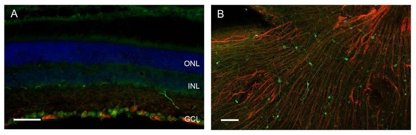
Int. J. Mol. Sci. 2020, 21, 3423                                                                               2 of 17

Jeffrey C. Hall, Michael Rosbash, and Michael W. Young for their discoveries of molecular mechanisms
controlling the circadian rhythm using Drosophila as a model organism. The major principle of the
Drosophila molecular clockwork is conserved also in mammals. Here, the positive components are the
transcription factors Clock and Bmal1 that heterodimerize and bind to E-box elements. The negative
components are the periods (Per1 and Per2) and cryptochromes (Cry1 and Cry2) that form together with
casein kinases, which regulate Per phosphorylation and turnover [6], a negative regulatory complex
that inhibits Clock/Bmal1-dependent transcription [7]. Importantly, E-box elements are present in the
regulatory regions, promoters of the genes encoding for the Pers and Crys, as well as in other genes
encoding for key regulators in cell function; therefore, they represent the so-called clock-controlled
genes (Ccg; Figure 1). Thus, in addition to a time-keeping mechanism, the molecular clockwork drives
rhythmic cell function.

      Figure 1. The mammalian suprachiasmatic nucleus (SCN) molecular clockwork. Light is received
      by the eye and transmitted by the retinal ganglion cells, forming the retino-hypothalamic tract,
      to the suprachiasmatic nucleus (SCN). Light/dark information is encoded by the release of the
      neurotransmitters glutamate (Glu) and pituitary adenylate cyclase-activating peptide (PACAP).
      Glutamate and PACP signal transduction lead to the activation of the transcription factor CREB,
      which modulates the intrinsic SCN molecular clockwork. The molecular clockwork consists of the
      transcriptional/translational feedback loops of so-called clock genes. The transcription factors Clock (C)
      and Bmal1 (B) bind to E-box elements in the regulatory region of the negative transcription regulators,
      the periods (P) and cryptochromes (Cr), as well as of clock-controlled genes (Ccg). The P and Cr
      accumulate, become phosphorylated by casein kinases (ε/δ), and form a complex that negatively
      interferes with C and B activity. Eventually, Cr and P transcription is downregulated, the complex is
      degraded, and a new cycle starts. Each cycle takes approximately 24 h (circadian). Accordingly, the
      Ccg are rhythmically expressed and encode for neuropeptides that transmit the time information to
      other parts of the hypothalamus regulating the autonomic nervous system or the endocrine system.

     In mammals, the circadian system comprises the retina (light input), the hypothalamic
suprachiasmatic nucleus (SCN; central circadian oscillator), and subordinate/peripheral oscillators in
the brain and the body (Figure 2) [4]. Molecular clockwork is not only present in the SCN but also in
subordinate oscillators in the brain and in the periphery. The SCN is directly entrained by light via
input from retinal ganglion cells (RGCs). The axons of RGCs convey light/dark information to the SCN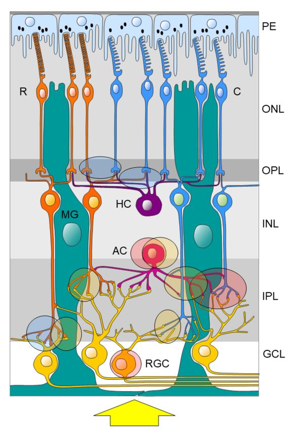
Int. J. Mol. Sci. 2020, 21, 3423                                                                            3 of 17

by the release of glutamate and pituitary adenylate cyclase-activating peptide (PACAP) ([8], reviewed
in [9]). Subsequent signal transduction pathways including phosphorylation of the transcription factor
CREB [10–12] and activation of Per expression leads to an adjustment of the molecular SCN clockwork
(Figure 1). Moreover, the SCN provides rhythmic signals for the temporal synchronization of various
peripheral organs and systems within the body. Important rhythmic signaling molecules within the
circadian system are the “hormone of darkness” melatonin, which is released from the pineal gland and
the retina (reviewed by [13,14]), and the “stress hormone” corticosterone, which is released from the
adrenal gland [15,16]. In addition, the autonomic nervous system is under the control of the circadian
system and controls rhythmic organ function [17]. This internal temporal synchronization is essential
for mental and global health [18].

      Figure 2. The mammalian circadian system. Light is received by the eye (input) and transmitted to the
      central circadian oscillator in the suprachiasmatic nucleus. The central circadian oscillator provides
      rhythmic output signals via the autonomous nervous system and the endocrine system to peripheral
      circadian oscillators in other parts of the brain and the body such as the liver (left) and the urinary
      bladder (right).

3. Purinergic Signaling
     The purinergic signaling pathway is mediated by extracellular purines (adenosine, ADP, and ATP)
and pyrimidines (UDP and UTP) through binding to purinergic receptors. Purinergic receptors are
classified into P1 purinoreceptors that are stimulated by adenosine and P2 purinoreceptors, which are
stimulated by variety of nucleotides. P1 receptors include A1, A2A, A2B, and A3 receptor subtypes,
whereas the P2 receptors are further subdivided into G-protein-coupled receptors (P2Y) and ion
channel ligand-gated receptors (P2X). P2Y receptors involve P2Y1, P2Y2, P2Y4, P2Y6, P2Y11, P2Y12,
P2Y13, and P2Y14, while P2X are subclassified into P2X1–P2X7 subtypes [19,20].
     Purinergic receptors can be found in almost every mammalian tissue and are essential for a
wide variety of body functions. In the mammalian central nervous system, activation of purinergic
receptors is involved in cellular differentiation, neurotransmission, and ion transport through ion
channels [21,22], which are the gatekeepers of neuronal excitability. Interestingly, circadian changes in
the gating properties of ion channels control cellular signaling mechanisms that regulate circadian
gene expression and cell function [23].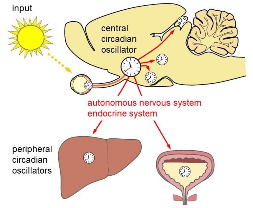
Int. J. Mol. Sci. 2020, 21, 3423                                                                          4 of 17

     However, the role of purinergic signaling within the mammalian circadian system remains largely
unknown. Therefore, in this review, we focus on purinergic signaling in components of the circadian
system including the retina, the SCN, and a peripheral oscillator (e.g., the urinary bladder).

4. Purinergic Signaling within the Retina
     The mammalian retina does not only process light to generate an image of the environment
but also measures environmental irradiance. The latter function is essential for light entrainment of
circadian rhythms.
     Light entrainment of circadian rhythms does not require functional rods and cones [24]. In
2002, a new subset of retinal ganglion cells (RGCs) was identified to be intrinsically photosensitive
(ipRGCs) [25,26]. These ipRGCs express the photopigment melanopsin [25] (Figure 3). The ipRGCs do
not only mediate light entrainment of the circadian system but also other non-image-forming visual
functions such as the pupillary light reflex [27]. The ipRGCs project directly to the SCN and other
non-image-forming brain regions such as the olivary pretectal nucleus, which controls the pupillary
light reflex, the ventral sub-paraventricular zone, and the sleep active ventrolateral preoptic nucleus
as well as image-forming brain regions such as the lateral geniculate nucleus [28,29]. The ipRGCs
represent a very small subset (1%) of the RGCs [30] and comprises at least five subtypes (M1–M5) with
differences in morphology, physiological properties, and projections (reviewed in [31]). In the retina,
they form a functional gap junction-coupled unit with non-photosensitive cells [32]. Moreover, ipRGCs
receive input from rods and cones that modulate their light response [33] and might account for the
residual light entrainment in melanopsin-deficient mice [34,35]. Both the retinal pigment epithelium
(PE) and Müller glia play roles in melanopsin chromophore regeneration [36].

      Figure 3. Representative melanopsin immunoreaction (green) in retinal ganglion cells (A) in a coronal
      section, scale bar 50 µm, and (B) in a whole-mount mouse retina preparation, scale bar 100 µm.
      Beta-tubulin-immunoreactive axons are shown in red, cell nuclei (DAPI) are shown in blue. ONL, outer
      nuclear layer; INL, inner nuclear layer; GCL, ganglion cell layer.

     The retina has its own circadian clock with a robust circadian rhythm in clock gene expression
and a rhythmic release of melatonin independent of the SCN [37,38]. The retinal circadian clock
and the hormone melatonin play important roles in the regulation of retinal development [39]
and function [40–42]. Specifically, melatonin modulates the amplitude of the a- and b-waves of
the electroretinogram (ERG), indicating a role of the hormone in regulating circadian changes in
retinal function. The daily and circadian rhythms in the ERG response are mediated through the
G-protein-coupled MT1 and MT2 receptors [43–45] and are PKC-dependent [45]. In addition, melatonin
has receptor-independent functions including detoxification of reactive oxygen species (ROS) and
other reactive molecules [13], which support the integrity of the mitochondria as well as cell function
and survival [46,47]. Increased ROS production is involved in retinal pathologies such as age-related
macular degeneration, glaucoma, and retinopathy [13]. Thus, melatonin is a potential preventive and
therapeutic agent in the treatment of these diseases [13].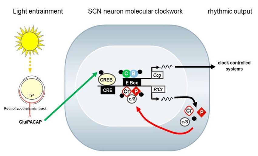
Int. J. Mol. Sci. 2020, 21, 3423                                                                    5 of 17

     Interestingly, release and content of retinal adenosine are higher at night as compared to day,
suggesting regulation by a circadian clock [48]. This darkness-evoked increase in the level of
extracellular adenosine results primarily from an increase in the conversion of extracellular ATP into
adenosine [48]. In addition, light leads to a decrease in extracellular adenosine levels presumably
because of decreased ATP release [48]. Possible sources of extracellular ATP in the retina include
both neurons as well as Müller glia [49]. Activity of ecto-ATPase could be demonstrated in the IPL
surrounding Müller glia processes and rod bipolar cell terminals [50]. Ectonucleotidases hydrolyze
ATP to ADP and ultimately to adenosine [51] and, therefore, play a pivotal role in purinergic signal
transmission as they control their availability at purinergic P2 receptors [52]. In various prosencephalic
brain regions, ectonucleotidases show a time-of-day-dependent expression pattern, which is modulated
by melatonin [53], and is mediated by MT1 and MT2 receptors [54] suggesting the melatoninergic
signaling as an interface between the purinergic system and the circadian system in general [54].
However, little is known about the role of melatonin on the purinergic system in the retina.
     Purines can contribute to retinal neurotransmission and/or neuromodulation (reviewed in [55]).
At the mRNA level, ionotropic P2X receptors [56–62] and metabotropic P2Y receptors [58,59,63] are
expressed on RGCs, bipolar cells, and Müller glia. Using immunohistochemistry, the P2X and P2Y
receptor subtypes could be detected in all layers and cell types of the retina. Further characterization
of the P2X receptors by colocalization studies using immunofluorescence and/or electron microscopy
revealed that P2X receptors are segregated to specific circuits within the retina [55]. The P2X2 receptor
could be detected on a subpopulation of GABAergic (but not dopaminergic) amacrine cells, on large
RGCs (type 1 alpha), and within the IPL associated with cone bipolar cell axon terminals [64]. The P2X3
receptor is also present on GABAergic amacrine cells but within the INL associated with both the rod
and cone bipolar cell terminals [50]. The P2X7 receptor could be detected in the outer plexiform layer
(OPL) at the rod and cone photoreceptor terminals and the horizontal cell processes as well as in the
inner plexiform layer (IPL) associated with rod bipolar cell axon terminals [65] (Figure 4).
     Functionally, P2X7 signaling modulates the a-wave of the ERG [65] and RGC response to a
light stimulus [66], consistent with its putative role in modulating photoreceptor and RGC function.
Moreover, ATP induces apoptosis of photoreceptors presumably by P2X7 signaling [67]. In the absence
of extracellular ATP, P2X7 receptors expressed on macrophages can act as scavenger receptors that play
an important role in the innate immune system [68]. There is increasing evidence that P2X7 receptor
function contributes to retinal pathologies such as glaucoma [69–71], inherited retinal degenerations,
as well as inflammatory changes associated with AMD [72] and diabetic retinopathy (reviewed in [68]).
However, little is known about the interaction of the circadian clock and/or melatonin and the purinergic
system in retinal physiology and pathology.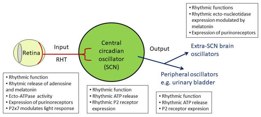
Int. J. Mol. Sci. 2020, 21, 3423                                                                             6 of 17

      Figure 4. Potential function of P2X2, P2X3, and P2X7 receptors in the retina based on subcellular
      localization. P2X2 receptor activation (red-shaded form) might modulate amacrine cell (AC) as well as
      retinal ganglion cell (RGC) function and specifically the cone (C) signaling pathway. P2X3 receptor
      activation (yellow-shaded form) might modulate amacrine (AC) cell function and both the rod (R)
      and cone (C) signaling pathways. P2X7 receptor activation (blue-shaded form) might modulate the
      photoreceptor as well as horizontal cell function (HC) and specifically the rod (R) signaling pathway.
      The yellow arrow indicates the light direction. GCL; ganglion cell layer, INL; inner nuclear layer, IPL;
      inner plexiform layer, ONL; outer nuclear layer, OPL; outer plexiform layer, MG; Müller glia, PE;
      pigment epithelium. Subcellular localization of P2 receptors is based on previous findings [50,64,65].

5. Purinergic Signaling within the SCN
     The SCN is the central circadian oscillator that coordinates circadian rhythms throughout the body
(Figures 1 and 2). SCN neurons show circadian rhythms in electrical (reviewed in [73]) and metabolic
(reviewed in [74]) activity as well as neurotransmitter release (reviewed in [73]). At the cellular level,
the molecular clockwork in the SCN is self-sustained. However, intercellular network properties are
essential for a coherent and strong circadian rhythmicity (reviewed in [75]). SCN neurons are primarily
GABAergic [76] but corelease of a variety of neuropeptides [77]. Two major functionally different
subdivisions of the SCN are the VIPergic ventrolateral core region and the AVPergic dorsomedial shell
region [78] (Figure 5).
Int. J. Mol. Sci. 2020, 21, 3423 7 of 17
Figure 5. Representative immunoreaction of neuropeptides in the SCN. (A) VIP immunoreaction is
present in perikarya of the ventrolateral core region (arrows) and a dense neuropil throughout the entire
SCN. (B) AVP immunoreaction is present in perikarya of the dorsomedial shell region (arrowheads).
Scale bar, 100 µm.
The core region receives direct input from the retinal ganglion cells, while the shell region sends
rhythmic signals to other brain regions to synchronize the other brain and peripheral clocks. However,
the core region is essential also for rhythmic behavior [79]. Rhythmic clock gene expression in the
core region depends upon the light/dark cycle, while those in the shell region persist even in constant
darkness [80]. VIP and GABA are crucial for coupling within the SCN network, while VIP and AVP
are important for coupling the SCN with other brain regions (reviewed by [81]). There is increasing
evidence that communication between astrocytes and neurons contributes largely to circadian rhythm
generation [82,83]. However, mice with a targeted deletion of the glial gap-junction proteins connexin30
and connexin43 show a relatively mild circadian phenotype [84].
ATP plays a significant role in astrocyte–astrocyte as well as astrocyte–neuron intercellular
communication [85,86]. Many brain regions exhibit a circadian rhythm in ATP content that is negatively
correlated with electrical and metabolic activity [87]. Among these, the amplitude of the ATP rhythm
is the largest in the SCN [88]. Moreover, in the SCN the rhythm of extracellular ATP accumulation
persists in vitro [89] and is dependent on clock gene expression and inositol triphosphate signaling [90].
Thus, extracellular ATP might play an important role in intercellular communication within the SCN.
Moreover, ectonucleotidases show a circadian rhythm in various brain regions [54]. They rapidly
hydrolyze extracellular ATP to its metabolites: ADP, AMP, and adenosine [91]. This process terminates
P2 activation and prevents receptor desensitization [92]. However, ADP is also an agonist for the
Gi-coupled P2Y12 receptor and the Gq-coupled P2Y1 receptors [93]. Thus, both extracellular ATP
content and ectonucleotidase activity determine P2 receptor agonist availability. Various P2X and P2Y
transcripts [94,95] and proteins [94,96] are expressed in the SCN. P2X7 and P2Y receptors contribute to
the ATP-stimulated increase of intracellular calcium levels in SCN cells [94]. Moreover, P2X2 receptor
signaling modulates GABAergic inhibitory synaptic transmission in the SCN [94]. These data suggest
a role of ATP and purinergic receptor signaling in SCN synaptic transmission. Rhythmic receptor
activation can be achieved by rhythmic availability of the agonist and/or of the receptor. Therefore,
we have previously analyzed time-of-day-dependent expression of P2 receptors in the SCN [97].
At the mRNA level, P2X2, P2X3, P2X4, P2X5, P2X7, P2Y1, P2Y4, P2Y6, P2Y12, and P2Y14 show a
time-of-day-dependent variation with lowest levels at the end of the dark phase and highest levels at
the early dark phase [97]. At the protein level, P2X1, P2X3, and P2X4 as well as P2Y2, P2Y6, P2Y12, and
P2Y14 show a time-of-day-dependent oscillation (Table 1). P2X4 has the strongest expression in the
SCN (Figure 6). P2X3 is increased during the late light phase in the core region, and P2X1, P2X3, as well
as P2X4 have the highest levels during the dark phase in both SCN subregions (Table 1). In contrast,
P2Y12 is increased during the late dark phase in the core region, and P2Y2, P2Y6, P2Y12, as well as
P2Y14 have highest levels during the early and mid-light phase in both SCN subregions (Table 1).
These data show a temporal and spatial redistribution of P2 receptor subtypes in the SCN and suggest
a potential role of P2 signaling in light entrainment and coupling between the SCN subdivisions. P2XInt. J. Mol. Sci. 2020, 21, 3423 8 of 17
receptors are more abundant during darkness and are, thus, in phase with the highest levels of their
dominant agonist, ATP [89]. In contrast, P2Y receptors are more abundant during the light phase and
are, thus, in antiphase with the ATP peak. This is consistent with P2Y receptor activation by other
agonists besides ATP such as ADP or pyrimidines. However, little is known about the rhythmicity of
extracellular ADP of other purines or pyrimidines in the SCN.
Table 1. Time-of-day-dependent expression of P2 receptors in the SCN obtained from mice at different
zeitgeber times (ZTs). ZT00 is defined as lights on. White and black bars indicate light and darkness,
respectively. P2 receptors with a time-of-day-dependent variation in expression are marked with
an asterisk, and peak levels are indicated with bold symbols. The intensity of immunoreaction was
categorized arbitrarily: + = low, ++ = moderate, +++ = high. Data are based on [97].
Zeitgeber Time (ZT) 02 06 10 14 18 22
Light/dark
subregion core shell core shell core shell core shell core shell core shell
P2X1 * + + + + + + + + + + ++ ++
P2X2 + + + + + + + + + + + +
P2X3 * + + + + ++ + ++ ++ ++ ++ ++ ++
P2X4 * ++ ++ ++ ++ ++ ++ +++ +++ +++ +++ +++ +++
P2X5 ++ ++ ++ ++ ++ ++ ++ ++ ++ ++ ++ ++
P2X6 ++ ++ ++ ++ ++ ++ ++ ++ ++ ++ ++ ++
P2X7 ++ ++ ++ ++ ++ ++ ++ ++ ++ ++ ++ ++
P2Y1 + + + + + + + + + + + +
P2Y2 * ++ ++ ++ ++ + + + + + + + +
P2Y4 + + + + + + + + + + + +
P2Y6 * + + ++ ++ + + + + + + + +
P2Y11 + + + + + + + + + + + +
P2Y12 * ++ ++ + + + + + + + + ++ +
P2Y13 ++ ++ ++ ++ ++ ++ ++ ++ ++ ++ ++ ++
P2Y14 * ++ + ++ ++ + + + + + + + +
Figure 6. Representative P2X4 immunoreaction in the SCN. Brain sections obtained from mice at two
different time points during the 12 h light/12 h dark cycle. Zeitgeber time (ZT02) is two hours after
lights on, ZT22 is two hours before lights on. Scale bar, 100 µm.
6. Purinergic Signaling within a Peripheral Oscillator, the Urinary Bladder
As mentioned above, the circadian system controls rhythmic behavior and physiology to ensure
optimal performance. Behavior and physiology are tightly locked to the sleep–wake cycle. Diurnal
animals are not only physically and mentally more active during the day but also consume and
metabolize most of their food and water during this time, whereas nocturnal animals do so during the
night. Thus, for any study of the behavior or physiological function, the subjective time of day of the
experimental animal should be taken into consideration. In this context, it is important to emphasize
that laboratory rodents such as mice and rats are nocturnal and, thus, are not in the same phase as the
diurnal human. However, in both diurnal and nocturnal animals, the SCN provides endocrine and
neuronal rhythmic signals for temporal synchronization within the body. The SCN directly targets
the hypothalamic center for hormonal and autonomic control, the paraventricular nucleus (PVN) [98].
The PVN contains 1) neuroendocrine neurons controlling the pituitary hormone secretion and 2)Int. J. Mol. Sci. 2020, 21, 3423 9 of 17
preautonomic neurons. The preautonomic PVN neurons balance sympathetic and parasympathetic
drive to the organs by projections to the intermediolateral column of the spinal cord and the dorsal
motor nucleus of the vagus, respectively [17]. Purines act as cotransmitters with acetylcholine in
parasympathetic nerves acting on P2X receptors in many different organs [3]. However, little is known
about the rhythmic release of ATP and rhythmic activation of purinergic receptors in the periphery.
Interestingly, purinergic signaling in the urinary bladder has been known since the 1970s (reviewed
in [2,99]). On the other hand, urinary bladder function is highly rhythmic [100]. Therefore, this section
focuses on the current knowledge about rhythmic purinergic signaling in this organ.
The muscles controlling micturition are innervated by autonomic and somatic nerves. During the
urine storage phase, sympathetic stimulation prevails, and the internal urethral sphincter is tonically
contracted while the detrusor muscle is relaxed. At increasing bladder volume, the firing rate of sensory
fibers from the bladder increases, initiating the voiding reflex and causing a conscious sensation of
urinary urge. During micturition, parasympathetic stimulation causes the detrusor muscle to contract
and the internal urethral sphincter to relax, while somatic innervation causes the external urethral
sphincter to relax. After voiding, the storage phase restarts [101,102].
Urine production and voiding occur predominantly during the active phase, whereas during
the inactive phase, kidney function is decreased and the storage capacity of the urinary bladder is
increased [103]. Disruption of this temporal regulation, for example, in elderly people or in patients
with neurodegenerative diseases with nocturia, affects sleep quality as well as general quality of
life and ultimately increases morbidity and mortality [71–74]. Therefore, a better understanding of
rhythmic function of the urinary bladder is highly relevant.
In mice, the rhythm of functional bladder capacity is dependent on functional molecular
clockwork [104]. Bladder smooth muscle cells have an internal molecular clockwork that drives
rhythmic expression of connexin43 (Cx43), a gap junction protein [104]. Surprisingly, mice expressing a
reduced level of Cx43 have a larger functional bladder capacity [104]. Thus, Cx43 negatively regulates
capacity contributing to rhythmic bladder function. Consistent with its regulatory function, Cx43 is
expressed in human and mouse urothelia lining the bladder lumen [105]. Furthermore, the mouse
urothelia contains a molecular clockwork and displays a rhythmic Cx43 expression with highest levels
during the active phase [105].
Importantly, ATP concentration in the bladder lumen undergoes daily variations, peaking in
phase with Cx43. Moreover, Cx43 controls rhythmic as well as mechanically induced ATP release
from urothelial cells, presumably by forming ATP-releasing hemichannels [105]. Thus, a rhythmic
molecular clockwork in the urothelia might control Cx43 hemichannel function providing rhythmic
ATP release. This time-of-day-dependent change in ATP release from the urothelia might shape
homeostatic regulation of bladder function.
Homeostatically, increased hydrostatic pressure as well as mechanosensory stimulation, such
as distension, evokes ATP release from the urothelia [106,107]. The urothelia itself expresses P2X
receptors, which modulate the apical membrane composition of umbrella cells [108–113]. Suburothelial
sensory nerve fibers express P2X and P2Y receptors, which respond to ATP release from the urothelial
cells during bladder distension, mediating the voiding reflex and nociception [114–116]. Moreover,
ATP acts as an excitatory cotransmitter with acetylcholine in parasympathetic nerves on P2X receptors
and mediates the contractile response of the detrusor muscle [112,117–123]. As mentioned above,
rhythmic parasympathetic innervation is controlled by the circadian system. Thus, ATP release from
the parasympathetic nerve endings and consequently P2 receptor function in the detrusor muscle
might undergo time-of-day-dependent changes.
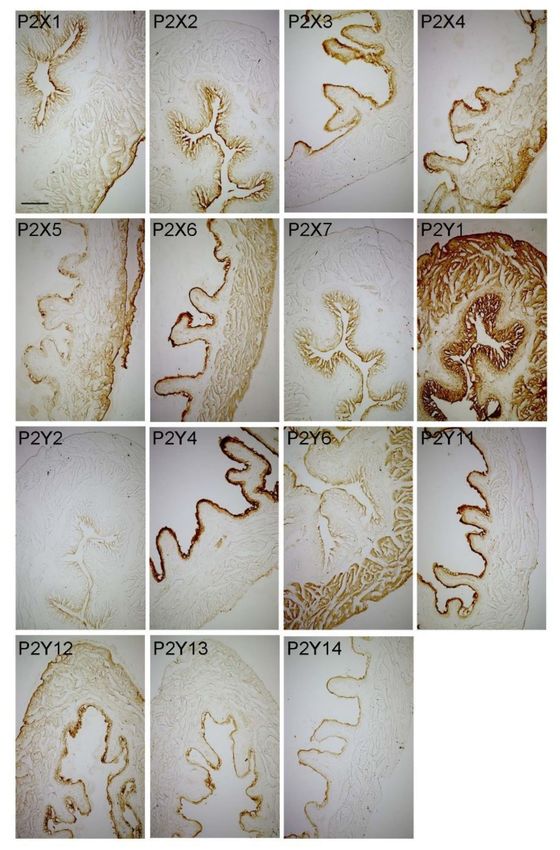
We performed a mapping of P2X and P2Y receptors (Figure 7, Table 2) in the urinary bladders
of mice sacrificed during the early day/inactive phase when storage capacity is increasing. In the
urothelium, we found a more or less intense immunoreaction of all P2X and P2Y receptors, suggesting
a potential role of all receptors in mediating autocrine/paracrine regulation of urothelial function by
purines and pyrimidines (unpublished data). In the suburothelial layer, a substantial P2X1-, P2X2-,Int. J. Mol. Sci. 2020, 21, 3423 10 of 17
P2X7-, and P2Y1 immunoreaction was present, suggesting a major role of these receptors in mediating
mechanosensation. In the detrusor muscle, a substantial immunoreaction was observed for P2X6,
P2Y1, and P2Y6 (Table 2). The P2Y6 receptor is selectively activated by pyrimidines, and the contractile
effect of UDP on vascular smooth muscle cells is lost in P2Y6-deficient mice [124]. This mapping of P2
receptor subtypes in the bladder provides a structural basis for further functional, pathological, and
pharmacological analyses.
Figure 7. Representative microphotographs showing immunoreaction of P2X1-7 receptors and P2Y1-4,
6, and 11-14 in the mouse urinary bladder. Scale bar, 200 µm.
Importantly, under pathological conditions rhythmic bladder function is disrupted, and, as
mentioned above, dysregulation of rhythmic bladder function affects the sleep–wake cycle. ATP
release [123,125] as well as expression of P2X and/or P2Y receptors are changed in various bladder
diseases [126,127]. Importantly, purinergic signaling has a high therapeutic potential [128]. Thus,
timed application of receptor-selective purinergic drugs might be a successful therapeutic strategy for
interrupting the vicious cycle of chronodisruption and bladder dysfunction.Int. J. Mol. Sci. 2020, 21, 3423 11 of 17
Table 2. Expression of P2 receptors in the urinary bladder wall subregions. The intensity of
immunoreaction was categorized arbitrarily: -, absent; (+), very weak; +, low; ++, moderate; +++,
high. Immunohistochemistry was performed as described previously [97].
P2 Receptors Urothelium Sub-Urothelium Detrusor Muscle
P2X1 ++ ++ (+)
P2X2 ++ ++ -
P2X3 ++ - -
P2X4 +++ - +
P2X5 ++ - +
P2X6 +++ - ++
P2X7 ++ ++ -
P2Y1 +++ +++ +++
P2Y2 + + -
P2Y4 +++ - -
P2Y6 + - ++
P2Y11 +++ - -
P2Y12 ++ - +
P2Y13 + - -
P2Y14 + - -
7. Summary
In this review, we gathered current knowledge on purinergic signaling in the major components
of the mammalian circadian system: the retina (input), the SCN (central oscillator), and the urinary
bladder as an example of a peripheral oscillator (Figure 8). For the urinary bladder, we discussed
the interaction of chronodisruption and organ dysfunction. However, further detailed analyses of
spatiotemporal distribution of agonists and receptors are needed to better understand purinergic
signaling in health and disease and to fully exploit its high therapeutic potential.
Figure 8. Summary of purinergic signaling and circadian rhythm in the components of the mammalian
circadian system. RHT: retinohypothalamic tract, SCN: suprachiasmatic nucleus.
Author Contributions: Conceptualization, C.v.G., A.A.H.A.; Visualization, A.A.H.A.; G.A.A. and C.v.G.;
Writing—original draft, C.v.G., A.A.H.A.; Writing—review & editing, A.A.H.A., G.A.A. and C.v.G. All authors
have read and agreed to the published version of the manuscript.
Acknowledgments: This Study is supported by Heinrich Heine University, Düsseldorf, Germany. We thank
Christine Opfermann-Rüngeler for excellent graphic design.
Conflicts of Interest: The authors declare no conflicts of interest.Int. J. Mol. Sci. 2020, 21, 3423 12 of 17
References
1. Burnstock, G. Introduction to purinergic signalling in the brain. Adv. Exp. Med. Biol. 2020, 1202, 1–12.
[PubMed]
2. Burnstock, G. Purinergic signalling in the urinary tract in health and disease. Purinergic Signal 2014, 10,
103–155. [CrossRef] [PubMed]
3. Burnstock, G. Purinergic signalling: From discovery to current developments. Exp. Physiol. 2014, 99, 16–34.
[CrossRef] [PubMed]
4. Rainbow, R.S.M. The suprachiasmatic nucleus and the circadian timekeeping system of the body. In
Neuroscience in the 21st Century; Pfaff, D.W., Ed.; Springer: New York, NY, USA, 2013.
5. Cohen, S.E.; Golden, S.S. Circadian rhythms in cyanobacteria. Microbiol. Mol. Biol. Rev. 2015, 79, 373–385.
[CrossRef]
6. Etchegaray, J.P.; Yu, E.A.; Indic, P.; Dallmann, R.; Weaver, D.R. Casein kinase 1 delta (CK1delta) regulates
period length of the mouse suprachiasmatic circadian clock in vitro. PLoS ONE 2010, 5, e10303. [CrossRef]
7. Reppert, S.M.; Weaver, D.R. Coordination of circadian timing in mammals. Nature 2002, 418, 935–941.
[CrossRef]
8. Chen, D.; Buchanan, G.F.; Ding, J.M.; Hannibal, J.; Gillette, M.U. Pituitary adenylyl cyclase-activating peptide:
A pivotal modulator of glutamatergic regulation of the suprachiasmatic circadian clock. Proc. Natl. Acad. Sci.
USA 1999, 96, 13468–13473. [CrossRef]
9. Hannibal, J. Neurotransmitters of the retino-hypothalamic tract. Cell Tissue Res. 2002, 309, 73–88. [CrossRef]
10. Gau, D.; Lemberger, T.; Von Gall, C.; Kretz, O.; Le Minh, N.; Gass, P.; Schmid, W.; Schibler, U.; Korf, H.W.;
Schütz, G. Phosphorylation of CREB Ser142 regulates light-induced phase shifts of the circadian clock.
Neuron 2002, 34, 245–252. [CrossRef]
11. Ding, J.M.; Faiman, L.E.; Hurst, W.J.; Kuriashkina, L.R.; Gillette, M.U. Resetting the biological clock:
Mediation of nocturnal CREB phosphorylation via light, glutamate, and nitric oxide. J. Neurosci. 1997, 17,
667–675. [CrossRef]
12. Ginty, D.D.; Kornhauser, J.M.; Thompson, M.A.; Bading, H.; Mayo, K.E.; Takahashi, J.S.; Greenberg, M.E.
Regulation of creb phosphorylation in the suprachiasmatic nucleus by light and a circadian clock. Science
1993, 260, 238–241. [CrossRef] [PubMed]
13. Blasiak, J.; Reiter, R.J.; Kaarniranta, K. Melatonin in retinal physiology and pathology: The case of age-related
macular degeneration. Oxid. Med. Cell. Longev. 2016. [CrossRef] [PubMed]
14. Korf, H.W.; Von Gall, C. Mice, melatonin and the circadian system. Mol. Cell. Endocrinol. 2006, 252, 57–68.
[CrossRef] [PubMed]
15. Balsalobre, A.; Brown, S.A.; Marcacci, L.; Tronche, F.; Kellendonk, C.; Reichardt, H.M.; Schütz, G.; Schibler, U.
Resetting of circadian time in peripheral tissues by glucocorticoid signaling. Science 2000, 289, 2344–2347.
[CrossRef] [PubMed]
16. Leliavski, A.; Dumbell, R.; Ott, V.; Oster, H. Adrenal clocks and the role of adrenal hormones in the regulation
of circadian physiology. J. Biol. Rhythms 2015, 30, 20–34. [CrossRef] [PubMed]
17. Buijs, R.M.; Escobar, C.; Swaab, D.F. The circadian system and the balance of the autonomic nervous system.
Handb. Clin. Neurol. 2013, 117, 173–191. [PubMed]
18. Rijo-Ferreira, F.; Takahashi, J.S. Genomics of circadian rhythms in health and disease. Genome Med. 2019, 11,
82. [CrossRef]
19. Tang, Y.; Illes, P. Regulation of adult neural progenitor cell functions by purinergic signaling. Glia 2017, 65,
213–230. [CrossRef]
20. Ralevic, V.; Burnstock, G. Receptors for purines and pyrimidines. Pharmacol. Rev. 1998, 50, 413–492.
21. Peterson, T.S.; Camden, J.M.; Wang, Y.; Seye, C.I.; Wood, W.G.; Sun, G.Y.; Erb, L.; Petris, M.J.; Weisman, G.A.
P2Y2 nucleotide receptor-mediated responses in brain cells. Mol. Neurobiol. 2010, 41, 356–366. [CrossRef]
22. Kaebisch, C.; Schipper, D.; Babczyk, P.; Tobiasch, E. The role of purinergic receptors in stem cell differentiation.
Comput. Struct. Biotechnol. J. 2015, 13, 75–84. [CrossRef] [PubMed]
23. Ko, G.Y.; Shi, L.; Ko, M.L. Circadian regulation of ion channels and their functions. J. Neurochem. 2009, 110,
1150–1169. [CrossRef] [PubMed]Int. J. Mol. Sci. 2020, 21, 3423 13 of 17
24. Freedman, M.S.; Lucas, R.J.; Soni, B.; Von Schantz, M.; Muñoz, M.; David-Gray, Z.; Foster, R. Regulation of
mammalian circadian behavior by non-rod, non-cone, ocular photoreceptors. Science 1999, 284, 502–504.
[CrossRef] [PubMed]
25. Hattar, S.; Liao, H.W.; Takao, M.; Berson, D.M.; Yau, K.W. Melanopsin-containing retinal ganglion cells:
Architecture, projections, and intrinsic photosensitivity. Science 2002, 295, 1065–1070. [CrossRef]
26. Berson, D.M.; Dunn, F.A.; Takao, M. Phototransduction by retinal ganglion cells that set the circadian clock.
Science 2002, 295, 1070–1073. [CrossRef]
27. Lucas, R.J.; Hattar, S.; Takao, M.; Berson, D.M.; Foster, R.G.; Yau, K.W. Diminished pupillary light reflex at
high irradiances in melanopsin-knockout mice. Science 2003, 299, 245–247. [CrossRef]
28. Gooley, J.J.; Lu, J.; Fischer, D.; Saper, C.B. A broad role for melanopsin in nonvisual photoreception. J.
Neurosci. 2003, 23, 7093–7106. [CrossRef]
29. Hattar, S.; Kumar, M.; Park, A.; Tong, P.; Tung, J.; Yau, K.W.; Berson, D.M. Central projections of
melanopsin-expressing retinal ganglion cells in the mouse. J. Comp. Neurol. 2006, 497, 326–349. [CrossRef]
30. Berson, D.M. Strange vision: Ganglion cells as circadian photoreceptors. Trends Neurosci. 2003, 26, 314–320.
[CrossRef]
31. Schmidt, T.M.; Chen, S.K.; Hattar, S. Intrinsically photosensitive retinal ganglion cells: Many subtypes,
diverse functions. Trends Neurosci. 2011, 34, 572–580. [CrossRef]
32. Sekaran, S.; Foster, R.G.; Lucas, R.J.; Hankins, M.W. Calcium imaging reveals a network of intrinsically
light-sensitive inner-retinal neurons. Curr. Biol. 2003, 13, 1290–1298. [CrossRef]
33. Dacey, D.M.; Liao, H.W.; Peterson, B.B.; Robinson, F.R.; Smith, V.C.; Pokorny, J.; Yau, K.W.; Gamlin, P.D.
Melanopsin-expressing ganglion cells in primate retina signal colour and irradiance and project to the LGN.
Nature 2005, 433, 749–754. [CrossRef] [PubMed]
34. Panda, S.; Sato, T.K.; Castrucci, A.M.; Rollag, M.D.; DeGrip, W.J.; Hogenesch, J.B.; Provencio, I.; Kay, S.A.
Melanopsin (Opn4) requirement for normal light-induced circadian phase shifting. Science 2002, 298,
2213–2216. [CrossRef] [PubMed]
35. Ruby, N.F.; Brennan, T.J.; Xie, X.; Cao, V.; Franken, P.; Heller, H.C.; O’Hara, B.F. Role of melanopsin in
circadian responses to light. Science 2002, 298, 2211–2213. [CrossRef]
36. Zhao, X.; Pack, W.; Khan, N.W.; Wong, K.Y. Prolonged inner retinal photoreception depends on the visual
retinoid cycle. J. Neurosci. 2016, 36, 4209–4217. [CrossRef]
37. Tosini, G.; Menaker, M. Circadian rhythms in cultured mammalian retina. Science 1996, 272, 419–421.
[CrossRef]
38. Tosini, G.; Menaker, M. The clock in the mouse retina: Melatonin synthesis and photoreceptor degeneration.
Brain Res. 1998, 789, 221–228. [CrossRef]
39. Baba, K.; Piano, I.; Lyuboslavsky, P.; Chrenek, M.A.; Sellers, J.T.; Zhang, S.; Gargini, C.; He, L.; Tosini, G.;
Iuvone, P.M. Removal of clock gene Bmal1 from the retina affects retinal development and accelerates cone
photoreceptor degeneration during aging. Proc. Natl. Acad. Sci. USA 2018, 115, 13099–13104. [CrossRef]
40. Storch, K.F.; Paz, C.; Signorovitch, J.; Raviola, E.; Pawlyk, B.; Li, T.; Weitz, C.J. Intrinsic circadian clock of
the mammalian retina: Importance for retinal processing of visual information. Cell 2007, 130, 730–741.
[CrossRef]
41. Cameron, M.A.; Barnard, A.R.; Hut, R.A.; Bonnefont, X.; Van der Horst, G.T.; Hankins, M.W.; Lucas, R.J.
Electroretinography of wild-type and Cry mutant mice reveals circadian tuning of photopic and mesopic
retinal responses. J. Biol. Rhythms 2008, 23, 489–501. [CrossRef]
42. Tosini, G.; Baba, K.; Hwang, C.K.; Iuvone, P.M. Melatonin: An underappreciated player in retinal physiology
and pathophysiology. Exp. Eye Res. 2012, 103, 82–89. [CrossRef] [PubMed]
43. Baba, K.; Pozdeyev, N.; Mazzoni, F.; Contreras-Alcantara, S.; Liu, C.M.; Kasamatsu, M.; Martinez-Merlos, T.;
Strettoi, E.; Iuvone, P.M.; Tosini, G. Melatonin modulates visual function and cell viability in the mouse retina
via the MT1 melatonin receptor. Proc. Natl. Acad. Sci. USA 2009, 106, 15043–15048. [CrossRef] [PubMed]
44. Sengupta, A.; Baba, K.; Mazzoni, F.; Pozdeyev, N.V.; Strettoi, E.; Iuvone, P.M.; Tosini, G. Localization of
melatonin receptor 1 in mouse retina and its role in the circadian regulation of the electroretinogram and
dopamine levels. PLoS ONE 2011, 6. [CrossRef] [PubMed]
45. Baba, K.; Benleulmi-Chaachoua, A.; Journe, A.S.; Kamal, M.; Guillaume, J.L.; Dussaud, S.; Gbahou, F.;
Yettou, K.; Liu, C.M.; Contreras-Alcantara, S.; et al. Heteromeric MT1/MT2 melatonin receptors modulate
photoreceptor function. Sci. Signal. 2013, 6. [CrossRef]Int. J. Mol. Sci. 2020, 21, 3423 14 of 17
46. Leon, J.; Acuna-Castroviejo, D.; Sainz, R.M.; Mayo, J.C.; Tan, D.X.; Reiter, R.J. Melatonin and mitochondrial
function. Life Sci. 2004, 75, 765–790. [CrossRef]
47. Xu, G.Q.; Zhao, J.; Liu, H.Y.; Wang, J.; Lu, W.F. Melatonin inhibits apoptosis and oxidative stress of mouse
leydig cells via a SIRT1-dependent mechanism. Molecules 2019, 24. [CrossRef]
48. Ribelayga, C.; Mangel, S.C. A circadian clock and light/dark adaptation differentially regulate adenosine in
the mammalian retina. J. Neurosci. 2005, 25, 215–222. [CrossRef]
49. Newman, E.A. A purinergic dialogue between glia and neurons in the retina. Novartis Found. Symp. 2006,
276, 193–202.
50. Puthussery, T.; Fletcher, E.L. Neuronal expression of P2X3 purinoceptors in the rat retina. Neuroscience 2007,
146, 403–414. [CrossRef]
51. Zimmermann, H. Biochemistry, localization and functional roles of ecto-nucleotidases in the nervous system.
Prog. Neurobiol. 1996, 49, 589–618. [CrossRef]
52. Zimmermann, H.; Zebisch, M.; Strater, N. Cellular function and molecular structure of ecto-nucleotidases.
Purinerg. Signal 2012, 8, 437–502.
53. Homola, M.; Pfeffer, M.; Fischer, C.; Zimmermann, H.; Robson, S.C.; Korf, H.W. Expression of
ectonucleotidases in the prosencephalon of melatonin-proficient C3H and melatonin-deficient C57Bl mice:
Spatial distribution and time-dependent changes. Cell Tissue Res. 2015, 362, 163–176. [CrossRef] [PubMed]
54. Homola, M.; Pfeffer, M.; Robson, S.C.; Fischer, C.; Zimmermann, H.; Korf, H.W. Melatonin receptor deficiency
decreases and temporally shifts ecto-5’-nucleotidase mRNA levels in mouse prosencephalon. Cell Tissue Res.
2016, 365, 147–156. [CrossRef] [PubMed]
55. Sanderson, J.; Dartt, D.A.; Trinkaus-Randall, V.; Pintor, J.; Civan, M.M.; Delamere, N.A.; Fletcher, E.L.; Salt, T.E.;
Grosche, A.; Mitchell, C.H. Purines in the eye: Recent evidence for the physiological and pathological role of
purines in the RPE, retinal neurons, astrocytes, Muller cells, lens, trabecular meshwork, cornea and lacrimal
gland. Exp. Eye Res. 2014, 127, 270–279. [CrossRef] [PubMed]
56. Brandle, U.; Guenther, E.; Irrle, C.; Wheeler-Schilling, T.H. Gene expression of the P2X receptors in the rat
retina. Mol. Brain Res. 1998, 59, 269–272. [CrossRef]
57. Brandle, U.; Kohler, K.; Wheeler-Schilling, T.H. Expression of the P2X7-receptor subunit in neurons of the rat
retina. Mol. Brain Res. 1998, 62, 106–109. [CrossRef]
58. Fries, J.E.; Wheeler-Schilling, T.H.; Guenther, E.; Kohler, K. Expression of P2Y1, P2Y2, P2Y4, and P2Y6
receptor subtypes in the rat retina. Invest. Ophthalmol. Vis. Sci. 2004, 45, 3410–3417. [CrossRef]
59. Fries, J.E.; Wheeler-Schilling, T.H.; Kohler, K.; Guenther, E. Distribution of metabotropic P2Y receptors in the
rat retina: A single-cell RT-PCR study. Mol. Brain Res. 2004, 130, 1–6. [CrossRef]
60. Jabs, R.; Guenther, E.; Marquordt, K.; Wheeler-Schilling, T.H. Evidence for P2X(3), P2X(4), P2X(5) but not for
P2X(7) containing purinergic receptors in Muller cells of the rat retina. Mol. Brain Res. 2000, 76, 205–210.
[CrossRef]
61. Wheeler-Schilling, T.H.; Marquordt, K.; Kohler, K.; Guenther, E.; Jabs, R. Identification of purinergic receptors
in retinal ganglion cells. Mol. Brain Res. 2001, 92, 177–180. [CrossRef]
62. Wheeler-Schilling, T.H.; Marquordt, K.; Kohler, K.; Jabs, R.; Guenther, E. Expression of purinergic receptors
in bipolar cells of the rat retina. Mol. Brain Res. 2000, 76, 415–418. [CrossRef]
63. Fries, J.E.; Goczalik, I.M.; Wheeler-Schilling, T.H.; Kohler, K.; Guenther, E.; Wolf, S.; Wiedemann, P.;
Bringmann, A.; Reichenbach, A.; Francke, M. Identification of P2Y receptor subtypes in human muller glial
cells by physiology, single cell RT-PCR, and immunohistochemistry. Invest. Ophthalmol. Vis. Sci. 2005, 46,
3000–3007. [CrossRef] [PubMed]
64. Puthussery, T.; Fletcher, E.L. P2X2 receptors on ganglion and amacrine cells in cone pathways of the rat
retina. J. Comp. Neurol. 2006, 496, 595–609. [CrossRef] [PubMed]
65. Puthussery, T.; Fletcher, E.L. Synaptic localization of P2X7 receptors in the rat retina. J. Comp. Neurol. 2004,
472, 13–23. [CrossRef] [PubMed]
66. Chavda, S.; Luthert, P.J.; Salt, T.E. Light-evoked retinal ganglion cell synaptic responses and microglial
morphology are modulated by P2X7 receptor activation and bacterial lipopolysaccharide (LPS). Invest.
Ophthalmol. Vis. Sci. 2014, 55.
67. Puthussery, T.; Fletcher, E. Extracellular ATP induces retinal photoreceptor apoptosis through activation of
purinoceptors in rodents. J. Comp. Neurol. 2009, 513, 430–440. [CrossRef]Int. J. Mol. Sci. 2020, 21, 3423 15 of 17
68. Fletcher, E.L.; Wang, A.Y.; Jobling, A.I.; Rutar, M.V.; Greferath, U.; Gu, B.; Vessey, K.A. Targeting P2X7
receptors as a means for treating retinal disease. Drug Discov. Today 2019, 24, 1598–1605. [CrossRef]
69. Resta, V.; Novelli, E.; Vozzi, G.; Scarpa, C.; Caleo, M.; Ahluwalia, A.; Solini, A.; Santini, E.; Parisi, V.; Di
Virgilio, F. Acute retinal ganglion cell injury caused by intraocular pressure spikes is mediated by endogenous
extracellular ATP. Eur. J. Neurosci. 2007, 25, 2741–2754. [CrossRef]
70. Reigada, D.; Lu, W.; Zhang, M.; Mitchell, C.H. Elevated pressure triggers a physiological release of Atp from
the retina: Possible role for pannexin hemichannels. Neuroscience 2008, 157, 396–404. [CrossRef]
71. Dutot, M.; Olivier, E.; Wakx, A.; Rat, P. The Role of the P2X7 Receptor in Ocular Stresses: A Potential
Therapeutic Target. Vision 2017, 1, 14. [CrossRef]
72. Vessey, K.A.; Gu, B.J.; Jobling, A.I.; Phipps, J.A.; Greferath, U.; Tran, M.X.; Dixon, M.A.; Baird, P.N.;
Guymer, R.H.; Wiley, J.S.; et al. Loss of function of P2X7 receptor scavenger activity in aging mice a novel
model for investigating the early pathogenesis of age-related macular degeneration. Am. J. Clin. Pathol. 2017,
187, 1670–1685. [CrossRef] [PubMed]
73. Gillette, M.U. SCN electrophysiology in vitro: Rhythmic activity and endogenous clock properties. In
Suprachiasmatic Nucleus: The Mind’s Clock; Reppert, S.M.K., Moore, C.D., Eds.; Oxford University Press Inc:
New York, NY, USA, 1991.
74. Schwartz, W.J. SCN metabolic activity in vivo. In Suprachiasmatic Nucleus: The Mind’s Clock; Reppert, D.C.M.,
Klein, S.M., Editor, R.Y., Eds.; Oxford University Press Inc: New York, NY, USA, 1991.
75. Welsh, D.K.; Takahashi, J.S.; Kay, S.A. Suprachiasmatic nucleus: Cell autonomy and network properties.
Annu. Rev. Physiol. 2010, 72, 551–577. [CrossRef] [PubMed]
76. Moore, R.Y.; Speh, J.C. GABA is the principal neurotransmitter of the circadian system. Neurosci. Lett. 1993,
150, 112–116. [CrossRef]
77. Maywood, E.S.; Chesham, J.E.; O’Brien, J.A.; Hastings, M.H. A diversity of paracrine signals sustains
molecular circadian cycling in suprachiasmatic nucleus circuits. Proc. Natl. Acad. Sci. USA 2011, 108,
14306–14311. [CrossRef] [PubMed]
78. Moore, R.Y.; Speh, J.C.; Leak, R.K. Suprachiasmatic nucleus organization. Cell Tissue Res. 2002, 309, 89–98.
[CrossRef] [PubMed]
79. Yan, L.; Karatsoreos, I.; Lesauter, J.; Welsh, D.K.; Kay, S.; Foley, D.; Silver, R. Exploring spatiotemporal
organization of SCN circuits. Cold Spring Harb. Symp. Quant. Biol. 2007, 72, 527–541. [CrossRef]
80. Hamada, T.; LeSauter, J.; Venuti, J.M.; Silver, R. Expression of period genes: Rhythmic and nonrhythmic
compartments of the suprachiasmatic nucleus pacemaker. J. Neurosci. 2001, 21, 7742–7750. [CrossRef]
81. Bittman, E.L. Circadian rhythms: Understanding the SCN connectome. Curr. Biol. 2016, 26, R840–R843.
[CrossRef]
82. Brancaccio, M.; Edwards, M.D.; Patton, A.P.; Smyllie, N.J.; Chesham, J.E.; Maywood, E.S.; Hastings, M.H.
Cell-autonomous clock of astrocytes drives circadian behavior in mammals. Science 2019, 363, 187–192.
[CrossRef]
83. Brancaccio, M.; Patton, A.P.; Chesham, J.E.; Maywood, E.S.; Hastings, M.H. Astrocytes control circadian
timekeeping in the suprachiasmatic nucleus via glutamatergic signaling. Neuron 2017, 93, 1420–1435.
[CrossRef]
84. Ali, A.A.H.; Stahr, A.; Ingenwerth, M.; Theis, M.; Steinhauser, C.; Von Gall, C. Connexin30 and Connexin43
show a time-of-day dependent expression in the mouse suprachiasmatic nucleus and modulate rhythmic
locomotor activity in the context of chronodisruption. Cell Commun. Signal 2019, 17, 61. [CrossRef] [PubMed]
85. Halassa, M.M.; Haydon, P.G. Integrated brain circuits: Astrocytic networks modulate neuronal activity and
behavior. Annu. Rev. Physiol. 2010, 72, 335–355. [CrossRef] [PubMed]
86. Haydon, P.G. GLIA: Listening and talking to the synapse. Nat. Rev. Neurosci. 2001, 2, 185–193. [CrossRef]
87. Yamazaki, S.; Ishida, Y.; Inouye, S. Circadian rhythms of adenosine triphosphate contents in the
suprachiasmatic nucleus, anterior hypothalamic area and caudate putamen of the rat–negative correlation
with electrical activity. Brain Res. 1994, 664, 237–240. [CrossRef]
88. Veitenhansl, M.; Stegner, K.; Hierl, F.X.; Dieterle, C.; Feldmeier, H.; Gutt, B.; Landgraf, R.; Garrow, A.P.;
Vileikyte, L.; Findlow, A.; et al. Diabetologia. In Proceedings of the 40(th) EASD Annual Meeting of
the European Association for the Study of Diabetes, Munich, Germany, 5–9 September 2004; Volume 47,
pp. A1–A464.Int. J. Mol. Sci. 2020, 21, 3423 16 of 17
89. Womac, A.D.; Burkeen, J.F.; Neuendorff, N.; Earnest, D.J.; Zoran, M.J. Circadian rhythms of extracellular ATP
accumulation in suprachiasmatic nucleus cells and cultured astrocytes. Eur. J. Neurosci. 2009, 30, 869–876.
[CrossRef] [PubMed]
90. Marpegan, L.; Swanstrom, A.E.; Chung, K.; Simon, T.; Haydon, P.G.; Khan, S.K.; Liu, A.C.; Herzog, E.D.;
Beaule, C. Circadian regulation of ATP release in astrocytes. J. Neurosci. 2011, 31, 8342–8350. [CrossRef]
91. Yegutkin, G.G. Nucleotide- and nucleoside-converting ectoenzymes: Important modulators of purinergic
signalling cascade. Biochim. Biophys. Acta 2008, 1783, 673–694. [CrossRef]
92. Junger, W.G. Immune cell regulation by autocrine purinergic signalling. Nat. Rev. Immunol. 2011, 11, 201–212.
[CrossRef]
93. Daniel, J.L.; Dangelmaier, C.; Jin, J.; Kim, Y.B.; Kunapuli, S.P. Role of intracellular signaling events in
ADP-induced platelet aggregation. Thromb. Haemost. 1999, 82, 1322–1326. [CrossRef]
94. Bhattacharya, A.; Vavra, V.; Svobodova, I.; Bendova, Z.; Vereb, G.; Zemkova, H. Potentiation of inhibitory
synaptic transmission by extracellular ATP in rat suprachiasmatic nuclei. J. Neurosci. 2013, 33, 8035–8044.
[CrossRef]
95. Collo, G.; North, R.A.; Kawashima, E.; Merlo-Pich, E.; Neidhart, S.; Surprenant, A.; Buell, G. Cloning OF P2X5
and P2X6 receptors and the distribution and properties of an extended family of ATP-gated ion channels. J.
Neurosci. 1996, 16, 2495–2507. [CrossRef] [PubMed]
96. Xiang, Z.; He, C.; Burnstock, G. P2X5 receptors are expressed on neurons containing arginine vasopressin
and nitric oxide synthase in the rat hypothalamus. Brain Res. 2006, 1099, 56–63. [CrossRef] [PubMed]
97. Lommen, J.; Stahr, A.; Ingenwerth, M.; Ali, A.A.H.; Von Gall, C. Time-of-day-dependent expression of
purinergic receptors in mouse suprachiasmatic nucleus. Cell Tissue Res. 2017, 369, 579–590. [CrossRef]
[PubMed]
98. Buijs, R.M.; La Fleur, S.E.; Wortel, J.; Van Heyningen, C.; Zuiddam, L.; Mettenleiter, T.C.; Kalsbeek, A.;
Nagai, K.; Niijima, A. The suprachiasmatic nucleus balances sympathetic and parasympathetic output to
peripheral organs through separate preautonomic neurons. J. Comp. Neurol. 2003, 464, 36–48. [CrossRef]
[PubMed]
99. Burnstock, G. Purinergic nerves. Pharmacol. Rev. 1972, 24, 509–581. [PubMed]
100. Noh, J.Y.; Han, D.H.; Yoon, J.A.; Kim, M.H.; Kim, S.E.; Ko, I.G.; Kim, K.H.; Kim, C.J.; Cho, S. Circadian
rhythms in urinary functions: Possible roles of circadian clocks? Int. Neurourol. J. 2011, 15, 64–73. [CrossRef]
101. Fowler, C.J.; Griffiths, D.; De Groat, W.C. The neural control of micturition. Nat. Rev. Neurosci. 2008, 9,
453–466. [CrossRef]
102. Hill, W.G. Control of urinary drainage and voiding. Clin. J. Am. Soc. Nephrol. 2015, 10, 480–492. [CrossRef]
103. Van Hoeck, K.; Bael, A.; Lax, H.; Hirche, H.; Van Gool, J.D. Circadian variation of voided volume in normal
school-age children. Eur. J. Pediatr. 2007, 166, 579–584. [CrossRef]
104. Negoro, H.; Kanematsu, A.; Doi, M.; Suadicani, S.O.; Matsuo, M.; Imamura, M.; Okinami, T.; Nishikawa, N.;
Oura, T.; Matsui, S.; et al. Involvement of urinary bladder Connexin43 and the circadian clock in coordination
of diurnal micturition rhythm. Nat. Commun. 2012, 3, 809. [CrossRef]
105. Sengiku, A.; Ueda, M.; Kono, J.; Sano, T.; Nishikawa, N.; Kunisue, S.; Tsujihana, K.; Liou, L.S.; Kanematsu, A.;
Shimba, S.; et al. Circadian coordination of ATP release in the urothelium via connexin43 hemichannels. Sci.
Rep. 2018, 8, 1996. [CrossRef]
106. Wang, E.C.; Lee, J.M.; Ruiz, W.G.; Balestreire, E.M.; Von Bodungen, M.; Barrick, S.; Cockayne, D.A.;
Birder, L.A.; Apodaca, G. ATP and purinergic receptor-dependent membrane traffic in bladder umbrella
cells. J. Clin. Invest. 2005, 115, 2412–2422. [CrossRef] [PubMed]
107. Ferguson, D.R.; Kennedy, I.; Burton, T.J. ATP is released from rabbit urinary bladder epithelial cells by
hydrostatic pressure changes–a possible sensory mechanism? J. Physiol. 1997, 505, 503–511. [CrossRef]
[PubMed]
108. Lee, H.Y.; Bardini, M.; Burnstock, G. P2X receptor immunoreactivity in the male genital organs of the rat.
Cell Tissue Res. 2000, 300, 321–330. [CrossRef] [PubMed]
109. Burnstock, G.; Dumsday, B.; Smythe, A. Atropine resistant excitation of the urinary bladder: The possibility
of transmission via nerves releasing a purine nucleotide. Br. J. Pharmacol. 1972, 44, 451–461. [CrossRef]
110. Tempest, H.V.; Dixon, A.K.; Turner, W.H.; Elneil, S.; Sellers, L.A.; Ferguson, D.R. P2X and P2X receptor
expression in human bladder urothelium and changes in interstitial cystitis. BJU Int. 2004, 93, 1344–1348.
[CrossRef]Int. J. Mol. Sci. 2020, 21, 3423 17 of 17
111. Svennersten, K.; Hallen-Grufman, K.; De Verdier, P.J.; Wiklund, N.P.; Poljakovic, M. Localization of P2X
receptor subtypes 2, 3 and 7 in human urinary bladder. BMC Urol. 2015, 15, 81. [CrossRef]
112. Elneil, S.; Skepper, J.N.; Kidd, E.J.; Williamson, J.G.; Ferguson, D.R. Distribution of P2X(1) and P2X(3)
receptors in the rat and human urinary bladder. Pharmacology 2001, 63, 120–128. [CrossRef]
113. Sun, Y.; Keay, S.; Lehrfeld, T.J.; Chai, T.C. Changes in adenosine triphosphate-stimulated ATP release suggest
association between cytokine and purinergic signaling in bladder urothelial cells. Urology 2009, 74, 1163–1168.
[CrossRef]
114. Burnstock, G. Release of vasoactive substances from endothelial cells by shear stress and purinergic
mechanosensory transduction. J. Anat. 1999, 194, 335–342. [CrossRef]
115. Rong, W.; Spyer, K.M.; Burnstock, G. Activation and sensitisation of low and high threshold afferent fibres
mediated by P2X receptors in the mouse urinary bladder. J. Physiol. 2002, 541, 591–600. [CrossRef] [PubMed]
116. Cockayne, D.A.; Hamilton, S.G.; Zhu, Q.M.; Dunn, P.M.; Zhong, Y.; Novakovic, S.; Malmberg, A.B.; Cain, G.;
Berson, A.; Kassotakis, L.; et al. Urinary bladder hyporeflexia and reduced pain-related behaviour in
P2X3-deficient mice. Nature 2000, 407, 1011–1015. [CrossRef] [PubMed]
117. Vial, C.; Evans, R.J. P2X receptor expression in mouse urinary bladder and the requirement of P2X(1) receptors
for functional P2X receptor responses in the mouse urinary bladder smooth muscle. Br. J. Pharmacol. 2000,
131, 1489–1495. [CrossRef] [PubMed]
118. Birder, L.A.; Ruan, H.Z.; Chopra, B.; Xiang, Z.; Barrick, S.; Buffington, C.A.; Roppolo, J.R.; Ford, A.P.; De
Groat, W.C.; Burnstock, G. Alterations in P2X and P2Y purinergic receptor expression in urinary bladder
from normal cats and cats with interstitial cystitis. Am. J. Renal Physiol. 2004, 287, F1084–F1091. [CrossRef]
119. Lee, H.Y.; Bardini, M.; Burnstock, G. Distribution of P2X receptors in the urinary bladder and the ureter of
the rat. J. Urol. 2000, 163, 2002–2007. [CrossRef]
120. Vulchanova, L.; Arvidsson, U.; Riedl, M.; Wang, J.; Buell, G.; Surprenant, A.; North, R.A.; Elde, R. Differential
distribution of two ATP-gated channels (P2X receptors) determined by immunocytochemistry. Proc. Natl.
Acad. Sci. USA 1996, 93, 8063–8067. [CrossRef]
121. Burnstock, G.; Kennedy, C. Is there a basis for distinguishing two types of P2-purinoceptor? Gen. Pharmacol.
1985, 16, 433–440. [CrossRef]
122. McMurray, G.; Dass, N.; Brading, A.F. Purinoceptor subtypes mediating contraction and relaxation of
marmoset urinary bladder smooth muscle. Br. J. Pharmacol. 1998, 123, 1579–1586. [CrossRef]
123. Heppner, T.J.; Werner, M.E.; Nausch, B.; Vial, C.; Evans, R.J.; Nelson, M.T. Nerve-evoked purinergic signalling
suppresses action potentials, Ca2+ flashes and contractility evoked by muscarinic receptor activation in
mouse urinary bladder smooth muscle. J. Physiol. 2009, 587, 5275–5288. [CrossRef]
124. Bar, I.; Guns, P.J.; Metallo, J.; Cammarata, D.; Wilkin, F.; Boeynams, J.M.; Bult, H.; Robaye, B. Knockout mice
reveal a role for P2Y6 receptor in macrophages, endothelial cells, and vascular smooth muscle cells. Mol.
Pharmacol. 2008, 74, 777–784. [CrossRef]
125. Burnstock, G. Purinergic signalling: Pathophysiology and therapeutic potential. Keio J. Med. 2013, 62, 63–73.
[CrossRef] [PubMed]
126. Birder, L.A.; Barrick, S.R.; Roppolo, J.R.; Kanai, A.J.; De Groat, W.C.; Kiss, S.; Buffington, C.A. Feline interstitial
cystitis results in mechanical hypersensitivity and altered ATP release from bladder urothelium. Am. J.
Physiol. Renal Physiol. 2003, 285, F423–F429. [CrossRef] [PubMed]
127. Sun, Y.; Keay, S.; De Deyne, P.G.; Chai, T.C. Augmented stretch activated adenosine triphosphate release
from bladder uroepithelial cells in patients with interstitial cystitis. J. Urol. 2001, 166, 1951–1956. [CrossRef]
128. Burnstock, G. The therapeutic potential of purinergic signalling. Biochem. Pharmacol. 2018, 151, 157–165.
[CrossRef] [PubMed]
© 2020 by the authors. Licensee MDPI, Basel, Switzerland. This article is an open access
article distributed under the terms and conditions of the Creative Commons Attribution
(CC BY) license (http://creativecommons.org/licenses/by/4.0/).You can also read