A Contemporary View of Respiratory Syncytial Virus (RSV) Biology and Strain-Specific Differences - MDPI
←
→
Page content transcription
If your browser does not render page correctly, please read the page content below
pathogens
Review
A Contemporary View of Respiratory Syncytial Virus
(RSV) Biology and Strain-Specific Differences
Mansi C. Pandya, Sean M. Callahan, Kyryll G. Savchenko and Christopher C. Stobart *
 Department of Biological Sciences, Butler University, Indianapolis, IN 46208, USA;
 mpandya@butler.edu (M.C.P.); smcallah@butler.edu (S.M.C.); ksavchen@butler.edu (K.G.S.)
 * Correspondence: cstobart@butler.edu

 Received: 28 March 2019; Accepted: 4 May 2019; Published: 21 May 2019

 Abstract: Respiratory syncytial virus (RSV) is a human respiratory pathogen which remains a leading
 viral cause of hospitalizations and mortality among infants in their first year of life. Here, we review
 the biology of RSV, the primary laboratory isolates or strains which have been used to best characterize
 the virus since its discovery in 1956, and discuss the implications for genetic and functional variations
 between the established laboratory strains and the recently identified clinical isolates.

 Keywords: respiratory syncytial virus; RSV; strain-specific differences; viral evolution

1. An Overview of RSV Biology

1.1. RSV Disease and Vaccines
     Respiratory syncytial virus (RSV) is a human respiratory virus that is responsible for the majority
of acute lower respiratory tract infections in infants worldwide [1]. In addition, it continues to
be commonly associated with lower respiratory infections and mortality among the elderly and in
individuals with compromised immune states [2–6]. In 2015, an estimated 33.1 million cases of acute
lower respiratory infections that occurred worldwide were associated with RSV [7]. Of those cases
identified, approximately 10% were associated with hospitalization and 0.4% resulted in mortality.
Although RSV infections occur regularly in healthy infants with no predisposing factors, conditions
associated with enhanced susceptibility for RSV disease include premature birth, lack of initial infection,
lack of or limited levels of maternal antibodies for protection against RSV, the presence of certain
genetic predisposing traits, and/or the presence of cardiopulmonary disease [8–13]. Despite a high
global burden of disease being attributable to RSV, there remains no commercially-available vaccines
for RSV.
     The World Health Organization (WHO) has identified RSV as a primary target for future vaccine
development and has begun to establish standards for comparison. While many existing vaccines
target a single primary population, the development of vaccines for RSV will likely need to target
three distinct populations: Young infants (within the first three to six months of life), older infants
and toddlers, and the elderly [2]. Numerous vaccine approaches are currently being explored to
address one or more of these target populations, including live-attenuated, chimeric, vector-based,
particle-based, subunit, and monoclonal antibody-based vaccine platforms [2]. The continued study of
existing and developing strains of RSV will be essential to the future development of RSV vaccines, in
order to understand the evolution of the virus and any potential changes in antigenicity that may arise.

1.2. RSV Genome
    RSV belongs to the order Mononegavirales and family Pneumoviridae. It is an enveloped virus with
a non-segmented, negative-sense RNA genome (-ssRNA) that is approximately 15.2 kb in length [13].

Pathogens 2019, 8, 67; doi:10.3390/pathogens8020067                         www.mdpi.com/journal/pathogens
Pathogens 2019, 8, x FOR PEER REVIEW 2 of 15
Pathogens 2019, 8, 67 2 of 15
RSV belongs to the order Mononegavirales and family Pneumoviridae. It is an enveloped virus
with a non-segmented, negative-sense RNA genome (-ssRNA) that is approximately 15.2 kb in
The genomic
length organization
[13]. The genomicof RSV consists of
organization of 10
RSVgenes which
consists collectively
of 10 genes which encode 11 proteins
collectively encode (Figure
11 1).
RSV forms filamentous
proteins enveloped
(Figure 1). RSV formsvirions that are
filamentous primarily
enveloped covered
virions in the
that are attachment
primarily covered(G)in and
the fusion
attachment (G)
(F) glycoproteins, withanda fusion
lesser(F) glycoproteins,
amount with a lesser
of integrated amount of
pentameric integrated
small pentameric(SH)
hydrophobic smallproteins
hydrophobic (SH) proteins within the envelope structure [13,14]. RSV F and G (see below) are
within the envelope structure [13,14]. RSV F and G (see below) are responsible for mediating viral
responsible for mediating viral attachment and entry into cell hosts. Directly beneath the envelope,
attachment and entry into cell hosts. Directly beneath the envelope, the viral matrix (M) protein
the viral matrix (M) protein plays a key role in the assembly and stability of virion structures [13,15].
plays a Within
key role theinvirion
the assembly and stability(N),
are the nucleoprotein of virion structures
phosphoprotein (P),[13,15]. Within the virion
and RNA-dependent RNA are the
nucleoprotein
polymerase(N),(L),
phosphoprotein
which protect the (P), and
viral RNA-dependent
genome RNA polymerase
and mediate replication, along with(L), which protect the
a transcription
processivity
viral genome factor (M2-1)
and mediate [13]. The two
replication, most
along common
with reverse genetics
a transcription systems, which
processivity factoreach employ
(M2-1) [13]. The
two most common reverse genetics systems, which each employ the A2 strain geneticwith
the A2 strain genetic background, necessitate co-expression of these four viral proteins a
background,
full-length antigenomic construct for infectious clone assembly [15,16]. In addition to the
necessitate co-expression of these four viral proteins with a full-length antigenomic construct for
aforementioned structural genes and replicative machinery, RSV also produces two nonstructural
infectious clone(NS1
proteins assembly [15,16].
and NS2), In addition
which have beentoshown
the aforementioned
to suppress innate structural
immunegenes and and
signaling replicative
machinery, RSV also produces two
antagonize apoptotic pathways [17–20].nonstructural proteins (NS1 and NS2), which have been shown to
suppress innate immune signaling and antagonize apoptotic pathways [17–20].
M2-2
M2-1
Glycoprotein (G)
Fusion (F) Protein Small Hydrophobic (SH) Protein
Polymerase (L)
Phosphoprotein (P)
Nucleoprotein (N) + -ssRNA Genome Matrix (M) Protein
Figure 1. The genomic organization and virion structure of respiratory syncytial virus (RSV) strain A2
are shown with each of the 10 genes color-coded based on their relative functions: Immunomodulatory
(light blue), envelope
Figure structure
1. The genomic (green), surface
organization structures
and virion structurefor
of attachment and entry
respiratory syncytial (red),
virus and
(RSV) replication
strain
and genomic
A2 arestability
shown (dark
with blue).
each ofA the
size 10
scale is provided
genes withbased
color-coded 1 kb gradations for evaluation
on their relative functions: of gene
Immunomodulatory
size. NS1/NS2, (lightprotein
nonstructural blue), envelope
1/2; N, structure (green),P,
nucleocapsid; surface structures for M,
phosphoprotein; attachment
matrix;andSH, small
entry (red), and replication and genomic stability (dark blue). A size scale is provided with 1 kb
hydrophobic glycoprotein; G, attachment glycoprotein; F, fusion glycoprotein; L, large polymerase
gradations for evaluation of gene size. NS1/NS2, nonstructural protein 1/2; N, nucleocapsid; P,
protein (RdRP).
phosphoprotein; M, matrix; SH, small hydrophobic glycoprotein; G, attachment glycoprotein; F,
fusion glycoprotein; L, large polymerase protein (RdRP).
1.3. RSV Antigenicity and Infectivity
RSV1.3.GRSV
andAntigenicity and Infectivity
F are arguably the most well-studied proteins of the virus due to their critical roles in
mediating attachment and fusion, respectively, as well as being responsible for inducing the majority
of neutralizing antibodies in vivo [21–24]. RSV G is produced in membrane-bound and secreted
forms during infection [25–28]. The membrane-bound form of RSV G is heavily glycosylated and
exhibits two highly variable mucin-like regions and a central conserved region [29–32]. Overall, RSV
G exhibits the most variable RSV gene sequence and has been used in the characterization of virus
evolution and to establish genetic variants of circulating RSV [33]. While several host proteins have
been identified as possible targets of RSV attachment in a wide range of cells, including heparin sulfate,
surfactant protein A (SP-A), the fractalkine receptor (CX3CR1), and annexin II, it remains unclear
what the primary host target for RSV G-mediated attachment is during infection in the human airwayPathogens 2019, 8, 67 3 of 15
epithelium [34–39]. This initial interaction between RSV G and the host cell facilitates and likely aids in
the engagement of RSV F to drive fusion between the viral envelope and host membrane [40]. During
fusion, RSV F undergoes a dynamic conformational change from a metastable pre-fusion trimer to a
stable post-fusion state [14,33,41,42]. While several host receptors for F attachment have been proposed,
including nucleolin and the epidermal growth factor receptor (EGFR), the possible portals of RSV entry
and the potential receptors for host fusion remain unclear [43–45]. Studies evaluating neutralizing
antibodies of RSV F have identified several neutralizing sites, with the most potent neutralizing
antibodies being associated with the binding of the prefusion conformational state of RSV F [24,41,46].
Subsequently, most current vaccine design efforts are focused on optimizing RSV F as a platform for
inducing immunologic protection [2,47].
1.4. RSV Genetic Diversity
Traditionally, RSV is classified into two distinct groups or subtypes, RSV-A and RSV-B, which
diverged approximately 350 years ago and are based on antigenic and sequence-based variations
predominately associated with RSV G [48–51]. RSV exhibits seasonality with multiple genotypes, often
in co-circulation with a dominance shift between RSV-A and RSV-B types every one to two years [52].
Within each of these two groups, several genotypes have been identified and described [49,50,53,54].
Recently, several unique genetic modifications in RSV G have been identified, which include a
72-nucleotide duplication (referred to as the ON genotype) associated with RSV-A types and a
60-nucleotide duplication (referred to as the BA genotype) associated with RSV-B types [53,55–57]. In
the cases of each of these two new genotypes, they have rapidly become the predominant forms found
in circulation worldwide and appear to increase in vitro viral fitness and attachment [58]. Furthermore,
variations in RSV-A and RSV-B over time appear to correlate with the induction of anti-G monoclonal
antibodies that recognize the primary epitopes, thus indicating immune-driven RSV evolution [59].
2. Common Laboratory Strains of RSV
Since its initial isolation from a chimpanzee in 1956 during an outbreak of coryza in a colony of
animals and subsequent discovery in humans shortly after, most research on RSV has centered on the
use of a limited number of historical isolates [60–62]. These prototypic “laboratory strains”, while
playing critical roles for identifying the key functions of viral proteins and the general virology of RSV
infection, have shown in limited studies to exhibit subtle to significant differences in cytopathology,
antigenicity, and pathogenicity, both in vitro and in vivo, when compared to other laboratory strains
and clinical isolates. In this section, we will review the origins, general biology, and use of several
common prototypic laboratory strains.
2.1. RSV Long
The RSV Long strain was first isolated by Robert Chanock from a child with bronchopneumonia
in 1956 and was subsequently passaged 11 to 13 times in HEp-2 cells [62]. As the “first” prototypic
strain, the Long strain was used extensively in the 1960s and 1970s to characterize the initial physical
properties of the virus, antigenic variations between RSV isolates, pathophysiology, and epidemiology
of RSV disease [60,63–70]. The RSV Long strain has also played an important role in the establishment
of early animal models of RSV infection in vivo, including mice, ferrets, and cotton rats [71–73].
Although existing platforms using the genetic background of A2 had been available, in 2014, the first
reverse genetics platform based on the Long genetic background was developed [74].
An early study in 1963 that compared the antigenic responses to strains Long and CH-18537
(described below) was pivotal in suggesting that antigenically distinct strains of RSV exist [66]. Later
studies would classify these strains into subtypes (groups) A and B, respectively, due in large part to
the antigenic differences in the attachment glycoprotein G [48,75]. Along with strain A2 (described
below), the Long strain continues to be recognized as a prototypic group (or subtype) A virus and is
regularly used in comparative studies of antigenicity and neutralization [76,77].Pathogens 2019, 8, 67 4 of 15
2.2. RSV A2
RSV strain A2 was first isolated in 1961 from the lower respiratory tract of an infant in Melbourne,
Australia [78]. Since its initial isolation, RSV A2 has been established as the prototypic A strain for the
study of RSV and remains the most common platform for the development of RSV live-attenuated
vaccine candidates.
Although not recapitulating human disease, numerous studies have employed the A2 strain to
elucidate and better characterize the immune responses to RSV in both mice and cotton rats [63,79–81].
In several common cell culture lines for the propagation and study of RSV (HEp-2, Vero, A549 cells) and
in airway epithelial cultures, RSV A2 is generally associated with higher replication kinetics and more
cytopathic effects earlier in infection when compared to other “laboratory” or clinical isolates [42,81–83].
It has been hypothesized that this may be due to a long history of passage-based adaptation in these cell
lines [82]. The flexibility and ease by which A2 is able to replicate efficiently in a variety of cell platforms
has also led many to believe that the virus utilizes a wide array of different receptors and attachment
factors, including heparin sulfate and nucleolin [38,82]. While no significant morphological differences
in virion structure or organization were observed between A2 and a clinical isolate (A/TN/12/11-19),
studies have reported that RSV A2 exhibits less thermal stability and potentially less pre-fusion F
availability when compared to strain A2-line19F, further implicating that appreciable differences in
stability and antigenicity may exist at a molecular level, even between similar strains [14,42,84].
A key factor in the use of A2 in RSV research has been the establishment of two separate reverse
genetics platforms based on the A2 genetic background. In 1995, the first reverse genetics system for
RSV was developed through the co-transfection of a plasmid encoding the antigenome, along with
supplementary plasmids expressing N, P, M2-1, and L proteins [16]. While this initial system was
not very efficient, it did yield a platform to recover mutant infectious viruses and opened the door to
the development of some of the first recombinant live-attenuated vaccines based on the A2 genetic
background. In 2012, a similar reverse genetics platform was developed which utilized a bacterial
artificial chromosome (BAC) for expression of the antigenome and was associated with more efficient
virus recovery [15]. While the 1995 system was initially based on the A2 genetic background, the
2012 system was initially engineered to generate an A2-line19F virus. Studies comparing A2 and
A2-line19F showed that the F protein of strain Line19 (see discussion below) was associated with more
pathogenesis in mice when compared to strain A2 [83].
In summary, A2 continues to be a prototypic RSV strain and has been used extensively as a reverse
genetics platform for the development of the majority of live-attenuated vaccine candidates to date.
The strain is a key system for the study of RSV structure and has played a key role in elucidating
immune responses in animal models to RSV infection [79].
2.3. RSV Line 19 and A2-line19F
Line 19 is an RSV subtype A virus that was first isolated from an infant with respiratory illness
at the University of Michigan Hospital in 1967 and initially expanded in WI-38 cells [85]. However,
subsequent studies of genomic differences between the Line 19 and Long strains have suggested that
Line 19 may be a Long strain, mouse-adapted through serial intracranial inoculations in suckling mice
generated in the same laboratory [73,79]. The pathogenesis of Line 19 infection in mice is distinct
and different from other common strains such as A2 and Long. Infections in mice with strain Line
19 are biased towards a more TH 2-type antiviral response, with increased goblet cell expansion and
elevated IL-13 and MUC5AC levels when compared to the TH 1 responses associated with A2 and
Long [86,87]. Compared to strain A2, Line 19 is also associated with lower overall titers in both HEp-2
cells and in mice in vivo [86]. A follow-up study using a recombinant A2-line19F virus (A2 expressing
the F protein of Line 19) showed that the fusion protein of strain Line 19 was genetically unique and
responsible for the differential immunopathology observed between the strains [83]. Regardless of
its origin, strain Line 19 and a related strain, A2-line19F, have been established as important mouse
models of pulmonary pathophysiology and immunologic responses.Pathogens 2019, 8, 67 5 of 15
2.4. RSV CH-18537
CH-18537 is a RSV subtype B virus that was first isolated from a throat swab of a child with
upper respiratory disease in 1962. The virus initially expanded in Wistar 26 cells [66]. Early studies
in the 1960s established that CH-18537 differed in antigenicity from the other contemporary isolates
at the time [66]. Since these initial studies, CH-18537 has been established as a common prototype
RSV-B strain for genetic studies and continues to be used for evaluating antigenicity and genetic
diversity [75,88–92].
2.5. RSV Memphis-37
The Memphis-37 strain is an RSV subtype A virus isolated as a nasal aspirate in 2001 from
a 4-month-old African American male child with bronchiolitis [93]. The initial isolate was plaque
purified and expanded as a GMP-lot in FDA-approved Vero cells for use in human clinical trials.
Memphis-37 has been used to study RSV pathogenesis, the dynamics of immunologic responses to
infection in healthy immunocompetent adults, and as a platform for testing inhibitors, vaccines, and
therapeutics for RSV infection [94–97]. Intranasal inoculation of neonatal lambs with Memphis-37 has
been shown to better reflect the pathogenesis and immune responses seen in humans when compared
to other animal models [98]. Memphis-37 remains a primary model to study RSV pathogenesis and
immunologic responses in humans.
3. Contemporary RSV Strains, Pathogenesis, and Viral Evolution
3.1. The Genetics and Evolution of RSV Clinical Isolates
Contemporary RSV clinical isolates are initially classified into either RSV-A or RSV-B subgroups,
then often further classified into one of a rapidly growing number of genotypes [49,50,53,56,99].
Different genotypes are known to co-circulate during RSV seasons and genotypic dominance can vary
based on the year and location [99,100]. Epidemiological studies often reveal genetic (and sometimes
phenotypic) variance between isolates of the same subgroup and genotype [101]. The continual
emergence of new strains and the identification of new genotypes highlights the ongoing evolution of
the virus worldwide.
In the last 20 years, there have been two new emergent genotypes which have each taken
precedence: ON1 and BA. The BA genotype emerged in samples obtained in Buenos Aires in 1999.
These viruses exhibited a 60-nucleotide duplication within a hypervariable region in the G gene of
RSV-B and within a few years became the predominant form of RSV-B detected worldwide [50,102–105].
Analysis of BA strains in vitro has shown that the G duplication impacts both viral attachment and
replication, providing a fitness advantage over viruses lacking the duplication [58]. In parallel to
the emergence of the G duplication in RSV-B strains, the more recent emergence and predominance
of the RSV-A ON1 genotype over NA1 (its precursor lineage lacking the G duplication) has been
well-noted and described [55,106,107]. During the RSV season of 2010–2011 in Ontario, Canada, a
novel RSV genotype (ON1) containing a 72-nucleotide duplication in the C-terminal region of RSV
G was identified, which resulted in up to seven potential additional O-glycosylation sites within the
protein. Similar to the BA genotype, the ON1 genotype quickly become common worldwide among
RSV-A isolates [56,108,109]. Several recent groups have started describing mutations in RSV G which
result in premature stop codons and deletions of the ON1 and BA genotypes [110]. These studies may
indicate a growing level of immunity within some populations.
Analysis of the RSV F and G of recent isolates from 2015–2017 in the United States has shown not
only that changes in RSV G were continuing to occur, but also changes in known antigenic sites in RSV
F [109]. Recent analysis of isolates in Kenya has shown that adaptive amino acid changes are also being
detected in other viral genes, including the viral polymerase (L) and M2-1 proteins [111]. A recent
study from Lebanon has identified two additional RSV-A genotype variants (LBA1 and LBA2), but
has also indicated a selection of variants with increased resistance to palivizumab, the only availablePathogens 2019, 8, 67 6 of 15
prophylaxis option worldwide for RSV [112]. While nearly all studies evaluating recent clinical isolates
have focused on genetic variations (rather than phenotypic differences), these viruses are known to
differ appreciably in genetic sequence from current laboratory strains. Several studies have been
performed to determine the relative mutation rates of RSV strains and have shown rates ranging from
10-3 to 10-4 nucleotide substitutions/site/year, depending upon the location and strain [49,50,113,114].
These data strongly suggest that evolutionary pressures are continuing to drive RSV evolution and
potentially
Pathogens 2019,push modern
8, x FOR circulating RSV strains further from the common laboratory strains isolated
PEER REVIEW 6 of 15
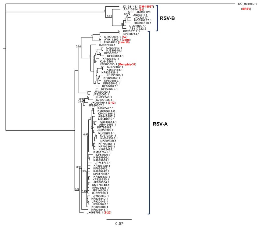
during 1950s and 1960s. We performed a phylogenetic analysis of RSV G gene sequences, comparing
commonSimilar
protein. laboratory strains
to the and contemporary
BA genotype, clinical isolates
the ON1 genotype (Figure
quickly 2). common
become From our worldwide
analysis, and those
among
performed by others, laboratory strains tend to cluster out and separate from contemporary
RSV-A isolates [56,108,109]. Several recent groups have started describing mutations in RSV G clinical
isolates,
which indicating
result that the viruses
in premature have likely
stop codons evolved not
and deletions only
of the in genetic
ON1 and BA space, but potentially
genotypes also
[110]. These
in phenotype
studies [49,50,53,57].
may indicate a growing level of immunity within some populations.
Figure 2.
Figure Phylogenetic analysis
2. Phylogenetic analysisof
of an
an alignment
alignmentof of the
the RSV
RSV GG gene
gene sequences
sequences of of common
common laboratory
laboratory
strains (identified in red)
strains red) and
and contemporary
contemporaryclinical
clinicalRSV-A
RSV-Aand
andRSV-B
RSV-Bisolates.
isolates.AABayesian
Bayesianinference
inferenceof
phylogenetic
of phylogeneticrelationships between
relationships RSV G
between nucleotide
RSV sequences
G nucleotide is shown.
sequences is The phylogenetic
shown. outgroup
The phylogenetic
was bovinewas
outgroup RSV bovine
(BRSV). RSV
Numbers on branches
(BRSV). Numbers are on
estimates for PPs
branches are(posterior
estimatesprobabilities) from the
for PPs (posterior
Bayesian inference (only numbers higher than 0.8 are shown). Common laboratory
probabilities) from the Bayesian inference (only numbers higher than 0.8 are shown). Common RSV-A strains A2,
Long, LineRSV-A
laboratory 19, and Memphis
strains (which
A2, Long, are19,
Line described in Section
and Memphis (which2), are
as well as strains
described A2001/2-20,
in Section and
2), as well
A2001/3-12
as (which have
strains A2001/2-20, been
and more recently
A2001/3-12 (whichisolated
have and
beenused
moretorecently
study RSV pathogenesis)
isolated and usedareto shown
study
for phylogenetic
RSV pathogenesis) comparison.
are shownCommon laboratory
for phylogenetic RSV-B strains
comparison. CH-18537
Common and B1 are
laboratory also strains
RSV-B shown
for comparison.
CH-18537 and B1 are also shown for comparison.
Analysis of the RSV F and G of recent isolates from 2015–2017 in the United States has shown
not only that changes in RSV G were continuing to occur, but also changes in known antigenic sites
in RSV F [109]. Recent analysis of isolates in Kenya has shown that adaptive amino acid changes are
also being detected in other viral genes, including the viral polymerase (L) and M2-1 proteins [111].
A recent study from Lebanon has identified two additional RSV-A genotype variants (LBA1 andPathogens 2019, 8, 67 7 of 15
3.2. Disease, Pathogenesis, and Cytopathology among Clinical Isolates
Numerous recent studies have surveyed clinical isolates obtained from sites around the world.
From these multiyear studies, both the RSV-A and RSV-B strains co-circulate, with the dominance of
strains varying over time [115–117]. However, RSV-A infections appear more frequent and appear
to be associated with higher transmissibility than RSV-B [118–120]. The persistence of both of these
distinct groups may explain the ability of previously infected individuals to remain susceptible to RSV
infections [121]. While several studies have evaluated the disease potential of obtained RSV clinical
isolates, the relationships and extent of variation, with regards to genetic sequences, genotypes, and
clinical severities, remain unclear [122]. Confounding these issues, several multiyear studies have
indicated significant phenotypic differences among the surveyed clinical isolates, while other studies
have reported that no significant differences were observed [115–117,122–125]. Furthermore, very few
studies have begun to evaluate the replication and pathophysiology of clinical isolates. The ambiguity
in available clinical data, along with a lack of replication and pathophysiology data highlights an
extensive need for understanding the current biology of circulating strains of RSV.
The limited studies that have begun to evaluate clinical strain differences, both in vitro and in vivo,
have highlighted significant differences among strains, even within the same genotype grouping.
In vitro models of infection have historically relied upon infections in continuous cell lines including
HEp-2, A549, BEAS-2B, and Vero cells, however recent studies have demonstrated that these cell
lines fail to recapitulate the native architecture of the airway epithelium and may bias the entry,
spread, and infectivity of RSV [126–129]. A study using primary pediatric bronchial epithelial cells
(PBECs) investigated whether the replication and cytopathology of prototypic laboratory strain A2
was representative of a panel of four recent clinical isolates. In this study, Villenave et al. demonstrated
that A2 exhibits markedly different cytopathology, viral titers, and cytokine secretion than the clinical
isolates surveyed [82]. Hypersecretion of mucus is a hallmark of severe neonatal RSV infections and
is associated with higher disease potential in the lower respiratory tract [130]. In another study by
Stokes et al., an analysis of a panel of six clinical isolates demonstrated significant differences among
strains in disease severity, lung IL-13 cytokine production, and mucus production during infections in
mice [81]. As previously described, studies have shown that the G duplications associated with the
ON1 and BA genotypes may confer increased fitness, both in vitro and in vivo, over those lacking these
modifications, yet none of the prototypic laboratory strains express these important genetic variations.
Collectively, these studies appear to suggest that conventional laboratory strains of RSV may not best
represent the biology and pathology of clinical isolates circulating today.
4. Summary and Future Implications for Vaccine Design
As reviewed here, advances in our understanding of the biology of RSV continue to be driven
through the extensive study of a limited number of prototypic laboratory strains. These studies have
made great progress towards establishing the basis for current vaccine design efforts. However, while
distinct differences have been observed (Table 1), far less is known on whether these “laboratory”
models of RSV and contemporary clinical isolates differ significantly in infectivity, replication, or
cytopathology. Extensive efforts are currently underway, by a multitude of groups, to develop effective
vaccines for RSV. It remains unclear how well these candidate platforms will perform in providing
broadly neutralizing coverage to circulating clinical isolates today and the near future. More effort
should be afforded to the study of more recent isolates and to identify how these viruses fundamentally
differ from the laboratory strains that have been characterized in the past. Understanding the extent
of variations in genetics and phenotypes among RSV strains will inform better design of future
vaccine constructs while also providing a more detailed picture of the landscape of RSV disease and
pathogenesis in human populations today.Pathogens 2019, 8, 67 8 of 15
Table 1. Comparative summary of laboratory and clinical isolates reviewed. The genetic type and
common characteristics and applications are provided for each strain (or group).
Virus Strain Type and Designation Characteristics and Applications
• Isolated in 1956
RSV-A (Laboratory
Long • Primarily used today in studies of antigenicity
Strain)
• Prototypic RSV-A model
• Isolated in 1961
• Most well-studied strain in use today; a prototypic RSV-A model
RSV-A (Laboratory • Higher replication kinetics in vitro compared to other strains
A2
Strain) • Mild cytopathology in animal models compared to other strains
• Most commonly employed reverse genetics system
• Most common strain used in live-attenuated vaccine preparations
• Isolated in 1967 (A2-line19F synthesized in 2009)
• Primarily used in pathogenesis and immunology studies
Line RSV-A (Laboratory
19/A2-line19F Strain) • Lower viral load, but more severe pathophysiology in
animal models
• Exhibits enhanced thermal stability compared to other strains
• Isolated in 1962
RSV-B (Laboratory
CH 18537 • Primarily used in antigenicity studies
Strain)
• Prototypic RSV-B model
• Isolated in 2001
RSV-A (Laboratory
Memphis-37 • Produced as a GMP lot for clinical studies
Strain)
• Primarily used in human pathogenesis and challenge studies
• Vary significantly in genetic diversity and differ from conventional
laboratory strains
• Most studies to date have focused on genetic variations
RSV-A and RSV-B and epidemiology
Clinical Isolates
Isolates • Many circulating types today exhibit a G protein duplication (ON1
and BA genotypes)
• Limited pathogenesis studies suggest variations in cytopathology
and pathogenesis between isolates
Author Contributions: M.C.P., S.M.C., and C.C.S. were directly involved in the writing and editing of this review.
K.G.S. conceptualized and performed the phylogenetic analysis as well as provided editing and writing support.
All four authors have read this manuscript and approve it for submission.
Acknowledgments: We acknowledge Sean Berthrong for his guidance and helpful discussions pertaining to
the organization and direction of this review. We also acknowledge Butler University Department of Biological
Sciences and the Holcomb Awards Committee for their funding and logistical support of our research.
Conflicts of Interest: The authors of this manuscript declare no conflicts of interest.
References
1. Nair, H.; Simoes, E.A.; Rudan, I.; Gessner, B.D.; Azziz-Baumgartner, E.; Zhang, J.S.F.; Feikin, D.R.;
A Mackenzie, G.; Moiïsi, J.C.; Roca, A.; et al. Global and regional burden of hospital admissions for
severe acute lower respiratory infections in young children in 2010: A systematic analysis. Lancet 2013, 381,
1380–1390. [CrossRef]
2. Mazur, N.; Higgins, D.; Nunes, M.C.; Melero, J.A.; Langedijk, A.C.; Horsley, N.; Buchholz, U.J.; Openshaw, P.J.;
McLellan, J.S.; Englund, J.A.; et al. The respiratory syncytial virus vaccine landscape: Lessons from the
graveyard and promising candidates. Lancet Infect Dis. 2018, 18, e295–e311. [CrossRef]
3. Thompson, W.W.; Shay, D.K.; Weintraub, E.; Brammer, L.; Cox, N.; Anderson, L.J.; Fukuda, K. Mortality
Associated with Influenza and Respiratory Syncytial Virus in the United States. JAMA 2003, 289, 179–186.
[CrossRef]
4. Dowell, S.F.; Anderson, L.J.; Gary, H.E.; Erdman, D.D.; Plouffe, J.F.; File, T.M.; Marston, B.J.; Breiman, R.F.
Respiratory Syncytial Virus Is an Important Cause of Community-Acquired Lower Respiratory Infection
among Hospitalized Adults. J. Infect. Dis. 1996, 174, 456–462. [CrossRef]Pathogens 2019, 8, 67 9 of 15
5. Walsh, E.E.; Peterson, D.R.; Falsey, A.R. Is Clinical Recognition of Respiratory Syncytial Virus Infection
in Hospitalized Elderly and High-Risk Adults Possible? J. Infect. Dis. 2007, 195, 1046–1051. [CrossRef]
[PubMed]
6. Falsey, A.R.; Hennessey, P.A.; Formica, M.A.; Cox, C.; Walsh, E.E. Respiratory Syncytial Virus Infection in
Elderly and High-Risk Adults. N. Engl. J. Med. 2005, 352, 1749–1759. [CrossRef]
7. Shi, T.; McAllister, D.A.; O’Brien, K.L.; Simões, E.A.F.; Madhi, S.A.; Gessner, B.D.; Polack, F.P.; Balsells, E.;
Acacio, S.; Aguayo, C.; et al. Global, regional, and national disease burden estimates of acute lower respiratory
infections due to respiratory syncytial virus in young children in 2015: A systematic review and modelling
study. Lancet 2017, 390, 946–958. [CrossRef]
8. Miyairi, I.; DeVincenzo, J.P. Human Genetic Factors and Respiratory Syncytial Virus Disease Severity. Clin.
Microbiol. Rev. 2008, 21, 686–703. [CrossRef]
9. McIntosh, K.; Kurachek, S.C.; Cairns, L.M.; Burns, J.C.; Goodspeed, B. Treatment of respiratory viral infection
in an immunodeficient infant with ribavirin aerosol. Am. J. Dis. Child. 1984, 138, 305–308. [CrossRef]
10. Macdonald, N.E.; Hall, C.B.; Suffin, S.C.; Alexson, C.; Harris, P.J.; Manning, J.A. Respiratory Syncytial Viral
Infection in Infants with Congenital Heart Disease. N. Engl. J. Med. 1982, 307, 397–400. [CrossRef] [PubMed]
11. Cunningham, C.K.; McMillan, J.A.; Gross, S.J. Rehospitalization for respiratory illness in infants of less than
32 weeks’ gestation. PEDIATRICS 1991, 88, 527–532. [PubMed]
12. Berkovich, S. Acute respiratory illness in the premature nursery associated with respiratory syncytial virus
infections. Pediatrics 1964, 34, 753–760. [PubMed]
13. Collins, P.L.; Melero, J.A. Progress in understanding and controlling respiratory syncytial virus: Still crazy
after all these years. Virus Res. 2011, 162, 80–99. [CrossRef]
14. Ke, Z.; Dillard, R.S.; Chirkova, T.; Leon, F.; Stobart, C.C.; Hampton, C.M.; Strauss, J.D.; Rajan, D.; Rostad, C.A.;
Taylor, J.V.; et al. The Morphology and Assembly of Respiratory Syncytial Virus Revealed by Cryo-Electron
Tomography. Viruses 2018, 10, 446. [CrossRef] [PubMed]
15. Hotard, A.L.; Shaikh, F.Y.; Lee, S.; Yan, D.; Teng, M.N.; Plemper, R.K.; Crowe, J.E.; Moore, M.L.; Lopez-Ona, A.
A Stabilized Respiratory Syncytial Virus Reverse Genetics System Amenable to Recombination Mediated
Mutagenesis. Virology 2012, 434, 129–136. [CrossRef]
16. Collins, P.L.; Hill, M.G.; Camargo, E.; Grosfeld, H.; Chanock, R.M.; Murphy, B.R. Production of infectious
human respiratory syncytial virus from cloned cDNA confirms an essential role for the transcription
elongation factor from the 5’ proximal open reading frame of the M2 mRNA in gene expression and provides
a capability for vaccine development. Proc. Natl. Acad. Sci. USA 1995, 92, 11563–11567.
17. Bitko, V.; Shulyayeva, O.; Mazumder, B.; Musiyenko, A.; Ramaswamy, M.; Look, D.C.; Barik, S.
Nonstructural proteins of respiratory syncytial virus suppress premature apoptosis by an NF-κB-dependent,
interferon-independent mechanism and facilitate virus growth. J. Virol. 2007, 81, 1786–1795. [CrossRef]
18. Swedan, S.; Musiyenko, A.; Barik, S. Respiratory Syncytial Virus Nonstructural Proteins Decrease Levels of
Multiple Members of the Cellular Interferon Pathways. J. Virol. 2009, 83, 9682–9693. [CrossRef]
19. Spann, K.M.; Tran, K.C.; Collins, P.L. Effects of Nonstructural Proteins NS1 and NS2 of Human Respiratory
Syncytial Virus on Interferon Regulatory Factor 3, NF-κB, and Proinflammatory Cytokines. J. Virol. 2005, 79,
5353–5362. [CrossRef]
20. Lo, M.S.; Brazas, R.M.; Holtzman, M.J. Respiratory Syncytial Virus Nonstructural Proteins NS1 and NS2
Mediate Inhibition of Stat2 Expression and α/β Interferon Responsiveness. J. Virol. 2005, 79, 9315–9319.
[CrossRef] [PubMed]
21. Teng, M.N.; Whitehead, S.S.; Collins, P.L. Contribution of the Respiratory Syncytial Virus G Glycoprotein
and Its Secreted and Membrane-Bound Forms to Virus Replication In Vitro and In Vivo. Virology 2001, 289,
283–296. [CrossRef]
22. Sastre, P.; Melero, J.A.; Garcia-Barreno, B.; Palomo, C. Comparison of affinity chromatography and adsorption
to vaccinia virus recombinant infected cells for depletion of antibodies directed against respiratory syncytial
virus glycoproteins present in a human immunoglobulin preparation. J. Med. Virol. 2005, 76, 248–255.
[CrossRef]
23. Cortjens, B.; Yasuda, E.; Yu, X.; Wagner, K.; Claassen, Y.B.; Bakker, A.Q.; Van Woensel, J.B.M.; Beaumont, T.
Broadly Reactive Anti-Respiratory Syncytial Virus G Antibodies from Exposed Individuals Effectively Inhibit
Infection of Primary Airway Epithelial Cells. J. Virol. 2017, 91, e02357-16. [CrossRef]Pathogens 2019, 8, 67 10 of 15
24. Ngwuta, J.O.; Chen, M.; Modjarrad, K.; Joyce, M.G.; Kanekiyo, M.; Kumar, A.; Yassine, H.M.; Moin, S.M.;
Killikelly, A.M.; Chuang, G.-Y.; et al. Prefusion F–specific antibodies determine the magnitude of RSV
neutralizing activity in human sera. Sci. Transl. Med. 2015, 7, 309ra162. [CrossRef]
25. Levine, S.; Klaiber-Franco, R.; Paradiso, P.R. Demonstration that Glycoprotein G Is the Attachment Protein of
Respiratory Syncytial Virus. J. Virol. 1987, 68, 2521–2524. [CrossRef] [PubMed]
26. Hendricks, D.A.; Baradaran, K.; McIntosh, K.; Patterson, J.L. Appearance of a Soluble Form of the G Protein
of Respiratory Syncytial Virus in Fluids of Infected Cells. J. Virol. 1987, 68, 1705–1714. [CrossRef] [PubMed]
27. Roberts, S.R.; Lichtenstein, D.; Ball, L.A.; Wertz, G.W. The membrane-associated and secreted forms of the
respiratory syncytial virus attachment glycoprotein G are synthesized from alternative initiation codons. J.
Virol. 1994, 68, 4538–4546. [PubMed]
28. Kwilas, S.; Liesman, R.M.; Zhang, L.; Walsh, E.; Pickles, R.J.; Peeples, M.E. Respiratory Syncytial Virus
Grown in Vero Cells Contains a Truncated Attachment Protein That Alters Its Infectivity and Dependence on
Glycosaminoglycans. J. Virol. 2009, 83, 10710–10718. [CrossRef]
29. Melero, J.A.; Mas, V.; McLellan, J.S. Structural, antigenic and immunogenic features of respiratory syncytial
virus glycoproteins relevant for vaccine development. Vaccine 2017, 35, 461–468. [CrossRef]
30. Johnson, P.R.; Spriggs, M.K.; Olmsted, R.A.; Collins, P.L. The G glycoprotein of human respiratory syncytial
viruses of subgroups A and B: Extensive sequence divergence between antigenically related proteins. Proc.
Natl. Acad. Sci. USA 1987, 84, 5625–5629. [CrossRef]
31. Collins, P.L.; Mottet, G. Oligomerization and post-translational processing of glycoprotein G of human
respiratory syncytial virus: Altered O-glycosylation in the presence of brefeldin A. J. Virol. 1992, 73, 849–863.
[CrossRef] [PubMed]
32. Satake, M.; Coligan, J.E.; Elango, N.; Norrby, E.; Venkatesan, S. Respiratory syncytial virus envelope
glycoprotein (G) has a novel structure. Nucleic Acids Res. 1985, 13, 7795–7812. [CrossRef] [PubMed]
33. McLellan, J.S.; Ray, W.C.; Peeples, M.E. Structure and function of respiratory syncytial virus surface
glycoproteins. Curr. Top Microbiol. Immunol. 2013, 372, 83–104. [CrossRef] [PubMed]
34. Tripp, R.A.; Jones, L.P.; Haynes, L.M.; Zheng, H.; Murphy, P.M.; Anderson, L.J. CX3C chemokine mimicry by
respiratory syncytial virus G glycoprotein. Nat. Immunol. 2001, 2, 732–738. [CrossRef]
35. Hickling, T.P.; Malhotra, R.; Bright, H.; McDowell, W.; Blair, E.D.; Sim, R.B. Lung Surfactant Protein A
Provides a Route of Entry for Respiratory Syncytial Virus into Host Cells. Viral. Immunol. 2000, 13, 125–135.
[CrossRef] [PubMed]
36. Barr, F.E.; Pedigo, H.; Johnson, T.R.; Shepherd, V.L. Surfactant Protein-A Enhances Uptake of Respiratory
Syncytial Virus by Monocytes and U937 Macrophages. Am. J. Respir. Cell Mol. Boil. 2000, 23, 586–592.
[CrossRef] [PubMed]
37. Malhotra, R.; Ward, M.; Bright, H.; Priest, R.; Foster, M.R.; Hurle, M.; Blair, E.; Bird, M. Isolation and
characterisation of potential respiratory syncytial virus receptor(s) on epithelial cells. Microbes Infect. 2003, 5,
123–133. [CrossRef]
38. Feldman, S.A.; Audet, S.; Beeler, J.A. The Fusion Glycoprotein of Human Respiratory Syncytial Virus
Facilitates Virus Attachment and Infectivity via an Interaction with Cellular Heparan Sulfate. J. Virol. 2000,
74, 6442–6447. [CrossRef] [PubMed]
39. Chirkova, T.; Stobart, C.C.; Hartert, T.V.; Lin, S.; Gaston, K.A.; Anderson, L.J.; Oomens, A.G.P.;
Boyoglu-Barnum, S.; Moore, M.L.; Ziady, A.G.; et al. CX3CR1 is an important surface molecule for
respiratory syncytial virus infection in human airway epithelial cells. J. Gen. Virol. 2015, 96, 2543–2556.
[CrossRef]
40. Meng, J.; Hotard, A.L.; Currier, M.G.; Lee, S.; Stobart, C.C.; Moore, M.L. Respiratory Syncytial Virus
Attachment Glycoprotein Contribution to Infection Depends on the Specific Fusion Protein. J. Virol. 2016, 90,
245–253. [CrossRef]
41. McLellan, J.S.; Chen, M.; Leung, S.; Graepel, K.W.; Du, X.; Yang, Y.; Zhou, T.; Baxa, U.; Yasuda, E.; Beaumont, T.;
et al. Structure of RSV Fusion Glycoprotein Trimer Bound to a Prefusion-Specific Neutralizing Antibody.
Science 2013, 340, 1113–1117. [CrossRef] [PubMed]
42. Stobart, C.C.; Rostad, C.A.; Ke, Z.; Dillard, R.S.; Hampton, C.M.; Strauss, J.D.; Yi, H.; Hotard, A.L.; Meng, J.;
Pickles, R.J.; et al. A live RSV vaccine with engineered thermostability is immunogenic in cotton rats despite
high attenuation. Nat. Commun. 2016, 7, 13916. [CrossRef]Pathogens 2019, 8, 67 11 of 15
43. Tayyari, F.; Marchant, D.; Moraes, T.J.; Duan, W.; Mastrangelo, P.; Hegele, R.G. Identification of nucleolin as a
cellular receptor for human respiratory syncytial virus. Nat. Med. 2011, 17, 1132–1135. [CrossRef] [PubMed]
44. Villenave, R.; Nguyen, M.T.; Hammonds, J.; Sakamoto, K.; Lee, S.; Meng, J.; Currier, M.G.; Stobart, C.C.;
Hotard, A.L.; Pretto, C.D.; et al. EGFR Interacts with the Fusion Protein of Respiratory Syncytial Virus Strain
2-20 and Mediates Infection and Mucin Expression. PLOS Pathog. 2016, 12, e1005622.
45. Battles, M.B.; McLellan, J.S. Respiratory syncytial virus entry and how to block it. Nat. Rev. Microbiol. 2019,
17, 233–245. [CrossRef] [PubMed]
46. McLellan, J.S.; Chen, M.; Joyce, M.G.; Sastry, M.; Stewart-Jones, G.B.E.; Yang, Y.; Zhang, B.; Chen, L.;
Srivatsan, S.; Zheng, A.; et al. Structure-Based Design of a Fusion Glycoprotein Vaccine for Respiratory
Syncytial Virus. Science 2013, 342, 592–598. [CrossRef]
47. Karron, R.A.; Buchholz, U.J.; Collins, P.L. Live-Attenuated Respiratory Syncytial Virus Vaccines; Springer Nature:
Berlin, Germany, 2013; Volume 372, pp. 259–284.
48. Mufson, M.A.; Orvell, C.; Rafnar, B.; Norrby, E. Two Distinct Subtypes of Human Respiratory Syncytial Virus.
J. Virol. 1985, 66, 2111–2124. [CrossRef]
49. Zlateva, K.T.; Lemey, P.; Vandamme, A.-M.; Van Ranst, M. Molecular Evolution and Circulation Patterns of
Human Respiratory Syncytial Virus Subgroup A: Positively Selected Sites in the Attachment G Glycoprotein.
J. Virol. 2004, 78, 4675–4683. [CrossRef]
50. Zlateva, K.T.; Lemey, P.; Moës, E.; Vandamme, A.-M.; Van Ranst, M. Genetic Variability and Molecular
Evolution of the Human Respiratory Syncytial Virus Subgroup B Attachment G Protein. J. Virol. 2005, 79,
9157–9167. [CrossRef]
51. Sande, C.J.; Mutunga, M.N.; Medley, G.F.; Cane, P.A.; Nokes, D.J. Group- and genotype-specific neutralizing
antibody responses against respiratory syncytial virus in infants and young children with severe pneumonia.
J. Infect. Dis. 2013, 207, 489–492. [CrossRef] [PubMed]
52. Waris, M. Pattern of Respiratory Syncytial Virus Epidemics in Finland: Two-Year Cycles with Alternating
Prevalence of Groups A and B. J. Infect. Dis. 1991, 163, 464–469. [CrossRef] [PubMed]
53. Trento, A.; Abrego, L.; Rodríguez-Fernández, R.; González-Sánchez, M.I.; González-Martínez, F.; Delfraro, A.;
Pascale, J.M.; Arbiza, J.; Melero, J.A. Conservation of G-Protein Epitopes in Respiratory Syncytial Virus
(Group A) Despite Broad Genetic Diversity: Is Antibody Selection Involved in Virus Evolution? J. Virol. 2015,
89, 7776–7785. [CrossRef] [PubMed]
54. Peret, T.C.; Golub, J.A.; Anderson, L.J.; Hall, C.B.; Schnabel, K.C. Circulation patterns of genetically distinct
group A and B strains of human respiratory syncytial virus in a community. J. Virol. 1998, 79, 2221–2229.
[CrossRef] [PubMed]
55. Agoti, C.N.; Otieno, J.R.; Gitahi, C.W.; Cane, P.A.; Nokes, D.J. Rapid Spread and Diversification of Respiratory
Syncytial Virus Genotype ON1, Kenya. Emerg. Infect. Dis. 2014, 20, 950–959. [CrossRef] [PubMed]
56. Eshaghi, A.; Duvvuri, V.R.; Lai, R.; Nadarajah, J.T.; Li, A.; Patel, S.N.; Low, D.E.; Gubbay, J.B. Genetic
Variability of Human Respiratory Syncytial Virus A Strains Circulating in Ontario: A Novel Genotype with a
72 Nucleotide G Gene Duplication. PLoS ONE 2012, 7, e32807. [CrossRef] [PubMed]
57. Trento, A.; Galiano, M.; Videla, C.; Carballal, G.; García-Barreno, B.; Melero, J.A.; Palomo, C. Major changes
in the G protein of human respiratory syncytial virus isolates introduced by a duplication of 60 nucleotides.
J. Virol. 2003, 84, 3115–3120. [CrossRef] [PubMed]
58. Hotard, A.L.; Laikhter, E.; Brooks, K.; Hartert, T.V.; Moore, M.L.; Lopez-Ona, A. Functional Analysis of the
60-Nucleotide Duplication in the Respiratory Syncytial Virus Buenos Aires Strain Attachment Glycoprotein.
J. Virol. 2015, 89, 8258–8266. [CrossRef]
59. Botosso, V.F.; Zanotto, P.M.D.A.; Ueda, M.; Arruda, E.; Gilio, A.E.; Vieira, S.E.; Stewien, K.E.; Peret, T.C.T.;
Jamal, L.F.; Pardini, M.I.D.M.C.; et al. Positive Selection Results in Frequent Reversible Amino Acid
Replacements in the G Protein Gene of Human Respiratory Syncytial Virus. PLOS Pathog. 2009, 5, e1000254.
[CrossRef]
60. Chanock, R.; Finberg, L. Recovery from infants with respiratory illness of a virus related to chimpanzee
coryza agent (CCA). II. Epidemiologic aspects of infection in infants and young children. Am. J. Hyg. 1957,
66, 291–300.
61. Blount, R.E.; Morris, J.A.; Savage, R.E. Recovery of cytopathogenic agent from chimpanzees with coryza.
Proc. Soc. Exp. Boil. Med. 1956, 92, 544–549.Pathogens 2019, 8, 67 12 of 15
62. Chanock, R.; Finberg, L. Recovery from Infants with Respiratory Illness of A Virus Related To Chimpanzee
Coryza Agent (Cca). Am. J. Epidemiol. 1957, 66, 281–290. [CrossRef]
63. Sullender, W.M. Respiratory Syncytial Virus Genetic and Antigenic Diversity. Clin. Microbiol. Rev. 2000, 13,
1–15. [CrossRef] [PubMed]
64. Johnson, K.M.; Chanock, R.M.; Rifkind, D.; Kravetz, H.M.; Knight, V. Respiratory syncytial virus. IV.
Correlation of virus shedding, serologic response, and illness in adult volunteers. JAMA 1961, 176, 663–667.
65. Coates, H.V.; Forsyth, B.R.; Chanock, R.M. Biophysical Studies of Respiratory Syncytial Virus I. Density of
Respiratory Syncytial Virus and Associated Complement-Fixing Antigens in a Cesium Chloride Density
Gradient. J. Bacteriol. 1966, 91, 1263–1269.
66. Coates, H.V.; Kendrick, L.; Chanock, R.M. Antigenic Differences between Two Strains of Respiratory Syncytial
Virus. Exp. Boil. Med. 1963, 112, 958–964. [CrossRef]
67. Suto, T.; Yano, N.; Ikeda, M.; Miyamoto, M.; Takai, S.; Shigeta, S.; Hinuma, Y.; Ishida, N. Respiratory Syncytial
Virus Infection and Its Serologic Epidemiology. Am. J. Epidemiol. 1965, 82, 211–224. [CrossRef] [PubMed]
68. Doggett, J.E.; Taylor-Robinson, D. Serological studies with respiratory syncytial virus. Arch. Virol. 1965, 15,
601–608. [CrossRef]
69. Wulff, H.; Kidd, P.; Wenner, H.A. Respiratory Syncytial Virus: Observations on Antigenic Heterogeneity.
Exp. Boil. Med. 1964, 115, 240–243. [CrossRef]
70. Norrby, E.; Marusyk, H.; Örvell, C. Morphogenesis of Respiratory Syncytial Virus in a Green Monkey Kidney
Cell Line (Vero). J. Virol. 1970, 6, 237–242.
71. Prince, G.A.; Jenson, A.B.; Horswood, R.L.; Camargo, E.; Chanock, R.M. The pathogenesis of respiratory
syncytial virus infection in cotton rats. Am. J. Pathol. 1978, 93, 771–791.
72. Prince, G.A.; Porter, D.D. The pathogenesis of respiratory syncytial virus infection in infant ferrets. Am. J.
Pathol. 1976, 82, 339–352. [PubMed]
73. Cavallaro, J.J.; Maassab, H.F. Adaptation of Respiratory Syncytial (RS) Virus to Brain of Suckling Mice. Exp.
Boil. Med. 1966, 121, 37–41. [CrossRef]
74. Hu, B.; Jiang, J.; Zhan, J.; Li, G.; Jiang, Y.; Guan, X.; Chen, Y.; Fang, Z. Development of a reverse genetics
system for respiratory syncytial virus long strain and an immunogenicity study of the recombinant virus.
Virol. J. 2014, 11, 142. [CrossRef]
75. Anderson, L.J.; Hierholzer, J.C.; Tsou, C.; Hendry, R.M.; Fernie, B.F.; Stone, Y.; McIntosh, K. Antigenic
Characterization of Respiratory Syncytial Virus Strains with Monoclonal Antibodies. J. Infect. Dis. 1985, 151,
626–633. [CrossRef]
76. Rossey, I.; Sedeyn, K.; Wrapp, D.; Kanekiyo, M.; Chen, M.; Mas, V.; Spitaels, J.; Schepens, B.; Saelens, X.;
Gilman, M.S.A.; et al. Potent single-domain antibodies that arrest respiratory syncytial virus fusion protein
in its prefusion state. Nat. Commun. 2017, 8, 14158. [CrossRef]
77. Jones, H.G.; Ritschel, T.; Pascual, G.; Brakenhoff, J.P.J.; Keogh, E.; Furmanova-Hollenstein, P.; Lanckacker, E.;
Wadia, J.S.; Gilman, M.S.A.; Williamson, R.A.; et al. Structural basis for recognition of the central conserved
region of RSV G by neutralizing human antibodies. PLOS Pathog. 2018, 14, e1006935. [CrossRef]
78. Lewis, F.A.; Rae, M.L.; Lehmann, N.I.; Ferris, A.A. A syncytial virus associated with epidemic disease of the
lower respiratory tract in infants and young children. Med. J. Aust. 1961, 48, 932–933.
79. Woolums, A.R.; Lee, S.; Moore, M.L. Animal Models of Respiratory Syncytial Virus Pathogenesis and Vaccine
Development: Opportunities and Future Directions. RSV 2011. Available online: https://www.intechopen.
com/download/pdf/24392 (accessed on 5 May 2019).
80. Moore, M.L.; Stokes, K.L.; Hartert, T.V. The impact of viral genotype on pathogenesis and disease severity:
respiratory syncytial virus and human rhinoviruses. Curr. Opin. Immunol. 2013, 25, 761–768. [CrossRef]
[PubMed]
81. Stokes, K.L.; Chi, M.H.; Sakamoto, K.; Newcomb, D.C.; Currier, M.G.; Huckabee, M.M.; Lee, S.;
Goleniewska, K.; Pretto, C.; Williams, J.V.; et al. Differential Pathogenesis of Respiratory Syncytial Virus
Clinical Isolates in BALB/c Mice. J. Virol. 2011, 85, 5782–5793. [CrossRef] [PubMed]
82. Villenave, R.; O’Donoghue, D.; Thavagnanam, S.; Touzelet, O.; Skibinski, G.; Heaney, L.G.; McKaigue, J.P.;
Coyle, P.V.; Shields, M.D.; Power, U.F. Differential cytopathogenesis of respiratory syncytial virus prototypic
and clinical isolates in primary pediatric bronchial epithelial cells. Virol. J. 2011, 8, 43. [CrossRef]Pathogens 2019, 8, 67 13 of 15
83. Moore, M.L.; Chi, M.H.; Luongo, C.; Lukacs, N.W.; Polosukhin, V.V.; Huckabee, M.M.; Newcomb, D.C.;
Buchholz, U.J.; Crowe, J.E.; Goleniewska, K.; et al. A Chimeric A2 Strain of Respiratory Syncytial Virus
(RSV) with the Fusion Protein of RSV Strain Line 19 Exhibits Enhanced Viral Load, Mucus, and Airway
Dysfunction. J. Virol. 2009, 83, 4185–4194. [CrossRef]
84. Rostad, C.A.; Stobart, C.C.; Todd, S.O.; Molina, S.A.; Lee, S.; Blanco, J.C.G.; Moore, M.L. Enhancing the
Thermostability and Immunogenicity of a Respiratory Syncytial Virus (RSV) Live-Attenuated Vaccine by
Incorporating Unique RSV Line19F Protein Residues. J. Virol. 2018, 92, e01568-17. [CrossRef] [PubMed]
85. Herlocher, M.; Ewasyshyn, M.; Sambhara, S.; Gharaee-Kermani, M.; Cho, D.; Lai, J.; Klein, M.; Maassab, H.
Immunological properties of plaque purified strains of live attenuated respiratory syncytial virus (RSV) for
human vaccine. Vaccine 1999, 17, 172–181. [CrossRef]
86. Lukacs, N.W.; Moore, M.L.; Rudd, B.D.; Berlin, A.A.; Collins, R.D.; Olson, S.J.; Ho, S.B.; Peebles, R.S.
Differential Immune Responses and Pulmonary Pathophysiology Are Induced by Two Different Strains of
Respiratory Syncytial Virus. Am. J. Pathol. 2006, 169, 977–986. [CrossRef]
87. Moore, M.L.; Peebles, R.S. Respiratory syncytial virus disease mechanisms implicated by human, animal
model, and in vitro data facilitate vaccine strategies and new therapeutics. Pharmacol. Ther. 2006, 112,
405–424. [CrossRef] [PubMed]
88. Belshe, R.B.; Anderson, E.L.; Walsh, E.E. Immunogenicity of Purified F Glycoprotein of Respiratory Syncytial
Virus: Clinical and Immune Responses to Subsequent Natural Infection in Children. J. Infect. Dis. 1993, 168,
1024–1029. [CrossRef]
89. Ghildyal, R.; Hogg, G.; Mills, J.; Meanger, J. Detection and subgrouping of respiratory syncytial virus directly
from nasopharyngeal aspirates. Clin. Microbiol. Infect. 1997, 3, 120–123. [CrossRef]
90. Storch, G.A.; Park, C.S. Monoclonal antibodies demonstrate heterogeneity in the G glycoprotein of prototype
strains and clinical isolates of respiratory syncytial virus. J. Med Virol. 1987, 22, 345–356. [CrossRef]
91. Lim, C.S.; Kumarasinghe, G.; Chow, V.T.K. Sequence and phylogenetic analysis of SH, G, and F genes and
proteins of Human respiratory syncytial virus isolates from Singapore. Acta Virol. 2003, 47, 97–104.
92. Walsh, E.E.; Brandriss, M.W.; Schlesinger, J.J. Immunological Differences between the Envelope Glycoproteins
of Two Strains of Human Respiratory Syncytial Virus. J. Virol. 1987, 68, 2169–2176. [CrossRef]
93. Kim, Y.-I.; DeVincenzo, J.P.; Jones, B.G.; Rudraraju, R.; Harrison, L.; Meyers, R.; Cehelsky, J.; Álvarez, R.;
Hurwitz, J.L. Respiratory Syncytial Virus Human Experimental Infection Model: Provenance, Production,
and Sequence of Low-Passaged Memphis-37 Challenge Virus. PLoS ONE 2014, 9, 113100. [CrossRef]
[PubMed]
94. DeVincenzo, J.P.; Wilkinson, T.; Vaishnaw, A.; Cehelsky, J.; Meyers, R.; Nochur, S.; Harrison, L.; Meeking, P.;
Mann, A.; Moane, E.; et al. Viral Load Drives Disease in Humans Experimentally Infected with Respiratory
Syncytial Virus. Am. J. Respir. Crit. Care Med. 2010, 182, 1305–1314. [CrossRef] [PubMed]
95. Bagga, B.; Harrison, L.; Roddam, P.; DeVincenzo, J. Unrecognized prolonged viral replication in the
pathogenesis of human RSV infection. J. Clin. Virol. 2018, 106, 1–6. [CrossRef]
96. DeVincenzo, J.; Lambkin-Williams, R.; Wilkinson, T.; Cehelsky, J.; Nochur, S.; Walsh, E.; Meyers, R.; Gollob, J.;
Vaishnaw, A. A randomized, double-blind, placebo-controlled study of an RNAi-based therapy directed
against respiratory syncytial virus. Proc. Natl. Acad. Sci. USA 2010, 107, 8800–8805. [CrossRef] [PubMed]
97. Eyles, J.E.; Johnson, J.E.; Megati, S.; Roopchand, V.; Cockle, P.J.; Weeratna, R.; Makinen, S.; Brown, T.P.;
Lang, S.; Witko, S.E.; et al. Nonreplicating Vaccines Can Protect African Green Monkeys from the Memphis
37 Strain of Respiratory Syncytial Virus. J. Infect. Dis. 2013, 208, 319–329. [CrossRef]
98. Larios Mora, A.; Detalle, L.; Van Geelen, A.; Davis, M.S.; Stohr, T.; Gallup, J.M.; Ackermann, M.R. Kinetics of
Respiratory Syncytial Virus (RSV) Memphis Strain 37 (M37) Infection in the Respiratory Tract of Newborn
Lambs as an RSV Infection Model for Human Infants. PLoS ONE 2015, 10, e0143580. [CrossRef]
99. Cui, G.; Zhu, R.; Qian, Y.; Deng, J.; Zhao, L.; Sun, Y.; Wang, F. Genetic Variation in Attachment Glycoprotein
Genes of Human Respiratory Syncytial Virus Subgroups A and B in Children in Recent Five Consecutive
Years. PLoS ONE 2013, 8, e75020. [CrossRef] [PubMed]
100. Pretorius, M.A.; Van Niekerk, S.; Tempia, S.; Moyes, J.; Cohen, C.; Madhi, S.A.; Venter, M. Replacement and
Positive Evolution of Subtype A and B Respiratory Syncytial Virus G-Protein Genotypes From 1997–2012 in
South Africa. J. Infect. Dis. 2013, 208, 227–237. [CrossRef]
101. Mufson, M.A.; Belshe, R.B.; Örvell, C.; Norrby, E. Respiratory Syncytial Virus Epidemics: Variable Dominance
of Subgroups A and B Strains Among Children, 1981–1986. J. Infect. Dis. 1988, 157, 143–148. [CrossRef]You can also read