Complete Genomic Sequence of Human Coronavirus OC43: Molecular Clock Analysis Suggests a Relatively Recent Zoonotic Coronavirus Transmission Event
←
→
Page content transcription
If your browser does not render page correctly, please read the page content below
JOURNAL OF VIROLOGY, Feb. 2005, p. 1595–1604                                                                                    Vol. 79, No. 3
0022-538X/05/$08.00⫹0 doi:10.1128/JVI.79.3.1595–1604.2005
Copyright © 2005, American Society for Microbiology. All Rights Reserved.

Complete Genomic Sequence of Human Coronavirus OC43: Molecular
  Clock Analysis Suggests a Relatively Recent Zoonotic Coronavirus
                         Transmission Event
           Leen Vijgen, Els Keyaerts, Elien Moës, Inge Thoelen, Elke Wollants, Philippe Lemey,
                             Anne-Mieke Vandamme, and Marc Van Ranst*
         Laboratory of Clinical and Epidemiological Virology, Department of Microbiology and Immunology, Rega Institute
                                   for Medical Research, University of Leuven, Leuven, Belgium
                                            Received 14 June 2004/Accepted 16 September 2004

                                                                                                                                                 Downloaded from http://jvi.asm.org/ on March 10, 2021 by guest
            Coronaviruses are enveloped, positive-stranded RNA viruses with a genome of approximately 30 kb. Based
          on genetic similarities, coronaviruses are classified into three groups. Two group 2 coronaviruses, human
          coronavirus OC43 (HCoV-OC43) and bovine coronavirus (BCoV), show remarkable antigenic and genetic
          similarities. In this study, we report the first complete genome sequence (30,738 nucleotides) of the prototype
          HCoV-OC43 strain (ATCC VR759). Complete genome and open reading frame (ORF) analyses were performed
          in comparison to the BCoV genome. In the region between the spike and membrane protein genes, a 290-
          nucleotide deletion is present, corresponding to the absence of BCoV ORFs ns4.9 and ns4.8. Nucleotide and
          amino acid similarity percentages were determined for the major HCoV-OC43 ORFs and for those of other
          group 2 coronaviruses. The highest degree of similarity is demonstrated between HCoV-OC43 and BCoV in all
          ORFs with the exception of the E gene. Molecular clock analysis of the spike gene sequences of BCoV and
          HCoV-OC43 suggests a relatively recent zoonotic transmission event and dates their most recent common
          ancestor to around 1890. An evolutionary rate in the order of 4 ⴛ 10ⴚ4 nucleotide changes per site per year was
          estimated. This is the first animal-human zoonotic pair of coronaviruses that can be analyzed in order to gain
          insights into the processes of adaptation of a nonhuman coronavirus to a human host, which is important for
          understanding the interspecies transmission events that led to the origin of the severe acute respiratory
          syndrome outbreak.

   Coronaviruses are large (120- to 160-nm), roughly spherical                (CCoV), feline infectious peritonitis virus (FIPV), human
particles with a linear, nonsegmented, capped, and polyade-                   coronavirus 229E (HCoV-229E), and the recently identified
nylated positive-sense single-stranded RNA genome that is                     human coronavirus NL63 (HCoV-NL63). Group 2 contains
encapsidated in a helical nucleocapsid. The envelope is derived               the murine hepatitis virus (MHV), bovine coronavirus
from intracellular membranes and contains a characteristic                    (BCoV), human coronavirus OC43 (HCoV-OC43), rat sialo-
crown of widely spaced club-shaped spikes that are 12 to 24 nm                dacryoadenitis virus (SDAV), porcine hemagglutinating en-
long. The genus Coronavirus (International Committee on the                   cephalomyelitis virus (PHEV), canine respiratory coronavirus
Taxonomy of Viruses database [ICTVdb], virus code                             (CRCoV), and equine coronavirus (ECoV). Group 3 contains
03.019.0.1) belongs to the family Coronaviridae in the order                  the avian infectious bronchitis virus (IBV) and turkey corona-
Nidovirales (7, 8).                                                           virus (TCoV). The SARS coronavirus (SARS-CoV) is not as-
   Before the 2002-to-2003 severe acute respiratory syndrome                  signed to any of these groups but is most closely related to
(SARS) epidemic, coronaviruses were somewhat neglected in                     group 2 coronaviruses (21, 54).
human medicine, but they have always been of considerable                        HCoV-OC43 (ICTVdb code 19.0.1.0.006) and HCoV-229E
importance in animal health. Coronaviruses infect a variety of                (ICTVdb code 19.0.1.0.005) were isolated in 1967 from volun-
livestock, poultry, and companion animals, in whom they can                   teers at the Common Cold Unit in Salisbury, United Kingdom.
cause serious and often fatal respiratory, enteric, cardiovascu-              HCoV-OC43 was initially propagated on ciliated human em-
lar, and neurologic diseases (25). Most of our understanding                  bryonic tracheal and nasal organ cultures (42). HCoV-OC43
about the molecular pathogenic properties of coronaviruses                    and HCoV-229E are responsible for 10 to 30% of all common
has been achieved by the veterinary virology community.                       colds, and infections occur mainly during the winter and early
   The coronaviruses are classified into three groups based on                spring (38). The incubation period is 2 to 4 days. During the
genetic and serological relationships (19). Group 1 contains                  2002-to-2003 winter season, a new human coronavirus, HCoV-
the porcine epidemic diarrhea virus (PEDV), porcine trans-                    NL63, was isolated from a 7-month-old child suffering from
missible gastroenteritis virus (TGEV), canine coronavirus                     bronchiolitis and conjunctivitis in The Netherlands (61). Seven
                                                                              additional HCoV-NL63-infected individuals, both infants and
                                                                              adults, were identified, indicating that HCoV-NL63 can be
  * Corresponding author. Mailing address: Laboratory of Clinical             considered an important new etiologic agent in respiratory
and Epidemiological Virology, Department of Microbiology and Im-
munology, Rega Institute for Medical Research, University of Leuven,
                                                                              tract infections. Coronaviruses infect all age groups, and rein-
Minderbroedersstraat 10, BE-3000 Leuven, Belgium. Phone: 32-16-               fections are common. The infection can be subclinical and is
347908. Fax: 32-16-347900. E-mail: marc.vanranst@uz.kuleuven.ac.be.           usually mild, but there have been reports of more-severe lower

                                                                       1595
1596 VIJGEN ET AL. J. VIROL.
respiratory tract involvement in infants and elderly people (17, report the complete HCoV-OC43 sequence (30,738 bases) and
60). Human coronaviruses can induce a demyelinating disease the comparative characterization and evolutionary relationship
in rodents and can infect primary cultures of human astrocytes of the BCoV–HCoV-OC43 pair. This is the first animal-human
and microglia. A possible etiological role for HCoV-OC43 and zoonotic pair of coronaviruses that can be analyzed in order to
HCoV-229E in multiple sclerosis is being debated (4, 13, 15). gain insights into the processes of adaptation of a nonhuman
The coronavirus genomes are the largest of the known RNA coronavirus to a human host.
viruses (27 to 31.5 kb) and are polycistronic, generating a
MATERIALS AND METHODS
nested set of subgenomic RNAs with common 5⬘ and 3⬘ se-
Preparation of HCoV-OC43 RNA. An HCoV-OC43 strain (VR759) was ob-
quences (35). The 5⬘ two-thirds of the genome consists of two tained from the American Type Culture Collection (ATCC). The ATCC VR759
large replicase open reading frames (ORFs), ORF1a and strain originated from a volunteer with a common cold-like illness at the Com-
ORF1b. The ORF1a polyprotein (pp1a) can be extended with mon Cold Unit in Salisbury, United Kingdom (42). HCoV-OC43 was propagated
ORF1b-encoded sequences via a ⫺1 ribosomal frameshift at a in a human rhabdomyosarcoma (RD) cell line, obtained from the European
Collection of Cell Cultures (ECACC). The supernatant was harvested after 7
conserved slippery site (6), generating the ⬎7,000-amino-acid days of incubation at 33°C, and RNA was isolated by using the QIAamp viral
polyprotein pp1ab, which includes the putative RNA-depen- RNA kit (QIAGEN, Westburg, The Netherlands). A real-time quantitative re-
Downloaded from http://jvi.asm.org/ on March 10, 2021 by guest
dent RNA polymerase (RdRp) and RNA helicase (HEL) ac- verse transcription PCR (RT-PCR) (Taqman; Perkin-Elmer Applied Biosys-
tivity (20, 39). The polyproteins pp1a and pp1ab are autocata- tems, Foster City, Calif.) was developed to determine the number of RNA copies
present in the supernatant.
lytically processed by two or three different viral proteases
Sequencing of the HCoV-OC43 genome. To determine the HCoV-OC43
encoded by ORF1a: one or two papain-like proteases (PLP1 genomic sequence, a set of overlapping RT-PCR products (average size, 1.5 kb)
and PLP2) and a 3C-like protease (3CLpro) (39, 67, 68). Other encompassing the entire genome was generated. For both RT-PCR and sequenc-
putative domains presumably associated with a 3⬘-to-5⬘ exonu- ing, oligonucleotide primers were designed in regions that were conserved be-
clease (ExoN) activity, a poly(U)-specific endo-RNase (Xen- tween the BCoV and MHV genomes. The forward PCR primer in the 5⬘-
terminal sequence (OC43F1 [5⬘-GATTGTGAGCGATTTGC-3⬘]) was based on
doU) activity, and a 2⬘-O-methyltransferase (2⬘-O-MT) activity the HCoV-OC43 5⬘ untranslated region partial sequence (H. Y. Wu, J. S. Guy,
are predicted in pp1ab (27, 54). The 3⬘ end of a coronavirus D. Yoo, R. Vlasak, and D. A. Brian, unpublished data; GenBank accession
genome includes several structural and accessory protein number AF523847). To generate RT-PCR products containing the exact 3⬘-
genes: an envelope-associated hemagglutinin esterase (HE) terminal sequence, we used oligonucleotide OC43R74 (5⬘-TTTTTTTTTTGTG
ATTCTTCCA-3⬘) based on the conserved 3⬘-end sequence of all known group
glycoprotein gene, present only in group 2 coronaviruses; a
2 coronaviruses. By using 150 sequencing primers, sequencing in both directions
spike (S) glycoprotein gene; an envelope (E) protein gene; a was performed on an ABI Prism 3100 genetic analyzer (Perkin-Elmer Applied
matrix (M) glycoprotein gene; a nucleocapsid (N) phospho- Biosystems) using the BigDye terminator cycle sequencing kit (version 3.1).
protein gene; and several ORFs that encode putative nonstruc- Chromatogram sequencing files were inspected with Chromas 2.2 (Technely-
tural (ns) proteins (35). sium, Helensvale, Australia), and contigs were prepared by using SeqMan II
(DNASTAR, Madison, Wis.).
Coronaviruses are well equipped to adapt rapidly to chang- DNA and protein sequence analyses. ORF analysis was performed by using the
ing ecological niches by the high mutation rate of their RNA NCBI ORF finder (http://www.ncbi.nlm.nih.gov/gorf/gorf.html). Potential 3C-
genome (about 10⫺4 nucleotide substitution/site/year) and like protease cleavage sites were identified by using the NetCorona 1.0 server
high recombination frequencies (51). Many animal coronavi- (30). DNA and protein similarity searches were performed using the NCBI
WWW-BLAST (basic local alignment search tool) server on the GenBank DNA
ruses cause long-term or persistent enzootic infections. Long
database, release 118.0 (2). Pairwise nucleotide and protein sequence alignments
periods of coronavirus infection combined with a high muta- were performed by using FASTA algorithms in the ALIGN program on the
tion and recombination rate increase the probability that a GENESTREAM network server (http://www2.igh.cnrs.fr) at the Institut de Gé-
virus mutant with an extended host range might arise. nétique Humaine in Montpellier, France (47). Maizel-Lenk dot matrix plots
The current emergence of the SARS-CoV is an example of were calculated using the pairwise FLAG 1.0 (fast local alignment for gigabases)
algorithm at the server of the Biomedical Engineering Center of the Industrial
a crossing of the animal-human species barrier. It is likely that Technology Research Institute in Hsinchu City, Taiwan (http://bioinformatics
the SARS-CoV was enzootic in an unknown animal or bird .itri.org.tw/prflag/prflag.php). Multiple sequence alignments were prepared by
species before suddenly emerging as a virulent virus for hu- using CLUSTALW (58) and CLUSTALX, version 1.82 (59) and were manually
mans. Chinese scientists found that six masked palm civets edited in GENEDOC (46). Phylogenetic analyses were conducted by using
MEGA, version 2.1 (34).
(Paguma larvata) and a racoon dog (Nyctereutes procyonoides)
Evolutionary rate analyses and timing of the most recent common ancestor.
for sale in an exotic food market in Shenzhen, in the Guang- The relationship between isolation date and genetic divergence was investigated
dong province in Southern China, were harboring a virus very using a linear regression, based on a maximum-likelihood tree, as implemented
similar to the SARS-CoV (1). Thirteen percent of the civet in the Path-O-Gen software, kindly provided by Andrew Rambaut (University of
merchants tested at markets in Guangdong also had SARS Oxford, Oxford, United Kingdom). Evolutionary rates and divergence times
were estimated by using maximum likelihood in the TipDate software package,
antibodies. Sequence analysis showed that the animal version version 1.2 (49), and Bayesian inference in BEAST, version 1.03 (kindly made
of the SARS-CoV contained an extra stretch of 29 bases (22). available by A. J. Drummond and A. Rambaut, University of Oxford; http:
It is still not clear whether the civets were a reservoir for the //evolve.zoo.ox.ac.uk/beast/). The molecular clock hypothesis was tested by using
virus or were infected by another species. the likelihood ratio test.
Nucleotide sequence accession number. The nucleotide sequence data reported
HCoV-OC43 and BCoV (ICTVdb code 03.019.0.01.002)
in this paper were deposited in GenBank under accession number AY391777 by
show remarkable antigenic and genetic similarities (23, 29, 36, using the National Center for Biotechnology Information (NCBI; Bethesda,
44, 52, 63, 65). They both have hemagglutinating activity by Md.) BankIt v3.0 submission tool (http://www3.ncbi.nlm.nih.gov/BankIt/).
attaching to the N-acetyl-9-O-acetylneuraminic acid moiety on
red blood cells (33). BCoV causes severe diarrhea in newborn RESULTS
calves. The complete nucleotide sequences of different BCoV HCoV-OC43 complete genomic sequence. We report here
strains are known, but only fragments of the HCoV-OC43 the complete nucleotide sequence of the prototype HCoV-
genome had been determined previously. In this paper, we OC43 strain (VR759), isolated in 1967 from an adult withVOL. 79, 2005 GENOMIC CHARACTERIZATION OF HUMAN CORONAVIRUS OC43 1597
Downloaded from http://jvi.asm.org/ on March 10, 2021 by guest
FIG. 1. Linear representation of the ORFs of the group 2 coronaviruses and SARS-CoV. Nucleotide insertions (open arrowheads) and
deletions (solid arrowheads) in the HCoV-OC43 genome compared to BCoV are shown.
common cold-like symptoms (42). The HCoV-OC43 genome ORF organization of HCoV-OC43. The HCoV-OC43 ge-
encompasses 30,738 nucleotides [excluding the 3⬘ poly(A) tail] nome contains 11 major ORFs flanked by 5⬘ and 3⬘ untrans-
and was deposited in the GenBank database under accession lated regions of 211 and 288 nucleotides, respectively. A linear
number AY391777. The HCoV-OC43 genome has a GC-con- representation of the major ORFs of HCoV-OC43, other
tent of 36.9%. group 2 coronaviruses, and SARS-CoV is given in Fig. 1. Table1598 VIJGEN ET AL. J. VIROL.
TABLE 1. Positions of the major ORFs of HCoV-OC43 (ATCC VR759) and BCoV (Mebus strain)
Nucleotide position No. of No. of
ORF
Virus Start ORF First ATG Stop codon bases amino acids
ORF1a HCoV-OC43 205 211 13362 13,158 4,385
BCoV 205 211 13362 13,158 4,385
ORF1b HCoV-OC43 13332 13566 21497 8,166 2,721
BCoV 13332 13566 21494 8,163 2,720
ns2 HCoV-OC43 21498 21507 22343 846 281
BCoV 21495 21504 22340 846 281
HE HCoV-OC43 22334 22355 23629 1,296 431
BCoV 22331 22352 23626 1,296 431
Downloaded from http://jvi.asm.org/ on March 10, 2021 by guest
S HCoV-OC43 23623 23644 27729 4,107 1,368
BCoV 23620 23641 27732 4,113 1,370
ns4.9 HCoV-OC43 NAa NA NA NA NA
BCoV 27811 27889 28026 216 71
ns12.9 HCoV-OC43 27802 27817 28146 345 114
BCoV 28095 28110 28439 345 114
E HCoV-OC43 28133 28133 28387 255 84
BCoV 28426 28426 28680 255 84
M HCoV-OC43 28381 28402 29094 714 237
BCoV 28674 28695 29387 714 237
N HCoV-OC43 29095 29104 30450 1,356 451
BCoV 29388 29397 30743 1,356 451
Ia HCoV-OC43 29102 29165 29347 246 81
Ib HCoV-OC43 29348 29441 29788 441 146
I BCoV 29395 29458 30081 687 228
a
NA, not applicable.
1 shows a comparison of the positions of the major ORFs of as ExoN (aa 5901 to 6421), XendoU (aa 6422 to 6796), and a
HCoV-OC43 and BCoV strain Mebus. 2⬘-O-MT domain (aa 6797 to 7095) (Fig. 2) (27, 54). The
The first two-thirds of the genome consists of two large amino-terminal part of the polyprotein is predicted to be
replicase ORFs, ORF1a and ORF1b. ORF1a is 4,383 codons cleaved by the papain-like proteases (68), while the carboxy-
long and overlaps with ORF1b, which consists of 2,721 codons. terminal part is putatively processed by the main coronavirus
The coronavirus replicase polyprotein of 7,095 amino acids protease, 3CLpro (67).
(aa) is synthesized by a ⫺1 ribosomal frameshift at a conserved The 3⬘-proximal part of the HCoV-OC43 genome contains
slippery site (UUUAAAC, nucleotides 13335 to 13341). In this several ORFs, which encode a variety of structural and acces-
polyprotein (GenBank accession number AAR01012) numer- sory proteins. Downstream of ORF1b, a nonstructural protein
ous putative functional domains are predicted: PLP1 and PLP2 gene (ns2) of 837 nucleotides, which is a group 2-specific gene,
(aa 852 to 2750), 3CLpro (aa 3247 to 3549), RdRp (aa 4370 to is present. Although these group 2-specific genes are not es-
5297), HEL (aa 5298 to 5900) (20, 39), and several putative sential for viral growth, recent work has shown that deletion of
nidovirus homologs of cellular RNA-processing enzymes, such MHV ns2 leads to a significant attenuation of the virus when
FIG. 2. Overview of the putative domain organization and potential proteolytic cleavage sites of the HCoV-OC43 replicase polyprotein pp1ab.
Cleavage sites that are predicted to be processed by 3C-like protease are indicated by black arrowheads, while potential papain-like protease
cleavage sites are indicated by white arrowheads. The following predicted domains are shown: papain-like proteases 1 and 2 (PLP1 and PLP2),
X domain (X), putative transmembrane domains 1, 2, and 3 (TM1, TM2, and TM3), 3C-like protease (3CL), growth factor-like domain (GFL),
RdRp, metal ion-binding domain (MB), HEL, ATPase, putative 3⬘-to-5⬘ exonuclease (ExoN), putative poly(U)-specific endo-RNase (XendoU),
and a putative S-adenosylmethionine-dependent ribose 2⬘-O-methyltransferase (MT).VOL. 79, 2005 GENOMIC CHARACTERIZATION OF HUMAN CORONAVIRUS OC43 1599
TABLE 2. Nucleotide and amino acid similarities of the major HCoV-OC43 (ATCC VR759) ORFs with the ORFs of BCoV, CRCoV,
PHEV, ECoV, MHV, and SDAV
HCoV-OC43 % Nucleotide (amino acid) similarity
ORF BCoV CRCoV PHEV ECoV MHV SDAV
a
ORF1a 97.4 (97.0) NA NA NA 69.3 (65.9) NA
ORF1b 97.8 (98.6) NA NA NA 82.7 (87.1) NA
ns2 95.1 (95.0) NA NA NA 59.0 (59.7) 60.3 (51.5)
HE 96.7 (95.3) NA 89.8 (88.7) 73.7 (72.9) 60.8 (58.1)b 64.2 (59.5)
S 93.5 (91.4) 92.8 (89.9) 81.7 (81.0) 79.3 (79.2) 66.5 (62.9) 67.0 (64.4)
ns12.9 96.1 (93.6) NA 89.7 (86.2) 88.8 (81.7) 55.0 (47.4) 59.8 (51.4)
E 98.0 (96.4) NA 99.6 (98.8) 96.1 (91.7) 72.5 (65.9) 69.2 (66.7)
M 94.8 (94.3) NA 92.1 (93.0) 91.2 (88.7) 77.9 (83.5) 77.5 (82.3)
N 96.8 (96.4) NA 94.3 (94.0) NA 71.8 (69.6) 72.0 (69.9)
Ia/Ibc 97.1 (NDd) NA 95.2 (ND) NA 72.3 (ND) 71.4 (ND)
a
NA, not applicable; the corresponding sequence is not available in the GenBank database.
Downloaded from http://jvi.asm.org/ on March 10, 2021 by guest
b
The 5⬘ end of the MHV HE ORF is missing due to a frameshift mutation or sequencing error in NC_001846.
c
The OC43 (ATCC VR759 strain) internal ORF (I) coding region contains a stop codon at position 29345, resulting in two potential coding regions of 60 aa (Ia)
and 115 aa (Ib). This stop codon is not present in BCoV, which has the capacity to code for a 207-aa protein. This stop codon is also absent in PHEV, MHV, and SADV.
The percentage of nucleotide similarity is calculated for the continuous Ia/Ib region.
d
ND, not done.
inoculated into mice (12). The HE gene, another group 2-spe- (EToV; accession number X52374), belonging to the genus
cific gene, consists of 1,275 nucleotides and encodes a protein Torovirus in the family Coronaviridae. Three phylogenetic clus-
of 424 aa. The S gene is located immediately downstream of ters, corresponding to the three coronavirus groups, can be
the HE gene and has 4,086 nucleotides. The S protein, which demonstrated. The SARS-CoV forms a separate branch, al-
consists of 1,361 aa, plays an important role in the attachment though there is strong support for monophyly of SARS-CoV
of the virus to cell surface receptors and induces the fusion of with the group 2 coronaviruses, such as HCoV-OC43 and
the viral and cellular membranes (5, 55). In the genomic region BCoV.
between the S gene and the membrane glycoprotein (M) gene, Based on the nucleotide sequence coding for the spike pro-
two ORFs can be identified: the ns12.9 gene encodes a putative tein, a maximum-likelihood phylogenetic tree was constructed
nonstructural protein of 12.9 kDa and is 330 nucleotides long for HCoV-OC43 and several BCoV strains for which the date
(43), while the E gene, 255 nucleotides long, codes for the E of isolation was known (Table 3; Fig. 5). HEC4408, a corona-
protein of approximately 9.5 kDa (43). At the 3⬘ end of the virus isolated in 1988 from a child with acute diarrhea, was also
HCoV-OC43 genome, four major ORFs are present. The M included in the analysis and has actually been shown to be a
gene is 693 nucleotides long and encodes a polypeptide of 230 BCoV (66). The time to the most recent common ancestor
aa with a molecular size of approximately 26 kDa (24). The (TMRCA) of HCoV-OC43 and BCoV was dated by three
membrane glycoprotein is anchored in the viral membrane methods (Fig. 6). Linear regression of root-to-tip divergence
with only a short amino-terminal domain exposed to the exte- versus sampling time situates the TMRCA of HCoV-OC43
rior of the viral envelope. The nucleocapsid protein (N) gene, and BCoV in 1891. The maximum-likelihood estimate for
consisting of 1,347 nucleotides, lies at the 3⬘ end of the genome TMRCA is 1873, with a 95% confidence interval of 1815 to
and encodes a 448-aa protein, which is associated with the 1899. The Bayesian coalescent approach dates TMRCA
RNA genome to form the nucleocapsid inside the viral enve- around 1890 (95% highest posterior density interval, 1859 to
lope. In the 5⬘ part of the HCoV-OC43 N region, two small 1912). This estimate was highly consistent under different de-
internal (I) ORFs can be identified (Ia and Ib). In BCoV, this mographic models, including an exponential-growth model,
region is uninterrupted and contains a single I gene which has which resulted in a TMRCA around 1893 (95% confidence
the capacity to code for a 207-aa protein (37). interval, 1866 to 1918). The evolutionary rate of BCoV was
HCoV-OC43 sequence similarity to other group 2 coronavi- also calculated by these three methods (Table 4). A maximum-
ruses. The sequence similarity among HCoV-OC43, BCoV, likelihood evolutionary rate of 4.3 ⫻ 10⫺4 substitutions per site
CRCoV, PHEV, ECoV, MHV, and SDAV was investigated by per year was estimated (95% confidence interval, 2.7 ⫻ 10⫺4 to
pairwise alignments of the corresponding ORFs and their pro- 6.0 ⫻ 10⫺4). A likelihood ratio test indicated that the molec-
teins (Table 2). HCoV-OC43 showed the highest percentage of ular clock hypothesis could not be rejected (P ⫽ 0.10).
similarity to BCoV in all ORFs except for the HCoV-OC43 E
gene, which showed 99.6% identity on the nucleotide level and
DISCUSSION
98.8% identity on the protein level to the PHEV E gene.
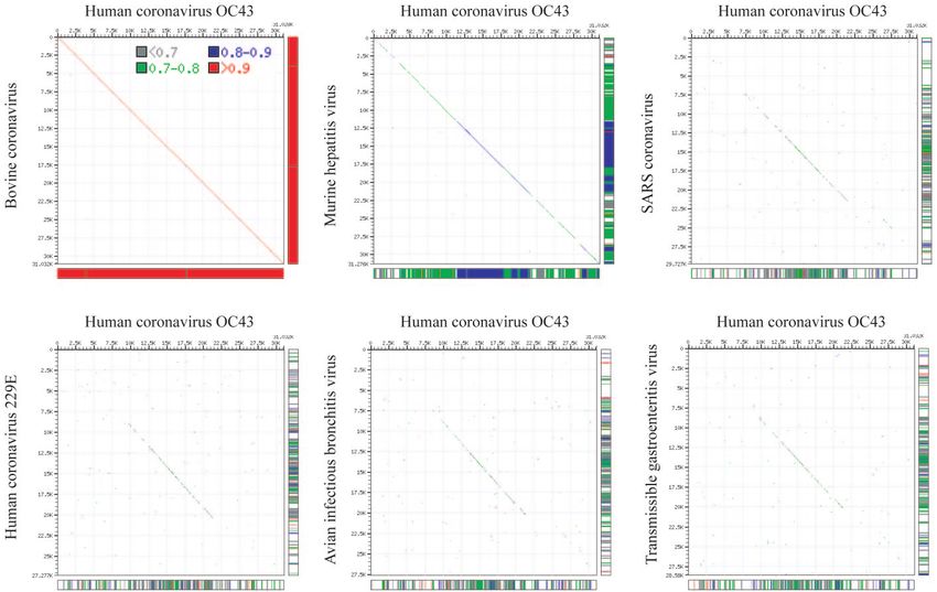
Maizel-Lenk dot matrix plots illustrate the similarity between We report in this paper the first complete genome sequence
HCoV-OC43 and BCoV (Fig. 3). (30,738 nucleotides) of the prototype HCoV-OC43 strain
Phylogenetic analysis. A neighbor-joining phylogenetic tree (VR759). Until now, only partial sequence fragments of the
of HCoV-OC43 and 11 other coronaviruses was constructed structural protein genes of HCoV-OC43 were available in
based on an alignment of ORF1b replicase amino acid se- GenBank, leaving the greater 5⬘ part of the genome to be
quences (Fig. 4). As an outgroup, we used the equine torovirus determined. The recent discovery of a new human coronavirus,1600
FIG. 3. Maizel-Lenk dot matrix plots: the complete genome sequence of HCoV-OC43 is compared to the complete genome sequences of BCoV, MHV, SARS-CoV, HCoV-229E, IBV, and TGEV,
respectively. Sequence identities are indicated by a dot.
Downloaded from http://jvi.asm.org/ on March 10, 2021 by guestVOL. 79, 2005 GENOMIC CHARACTERIZATION OF HUMAN CORONAVIRUS OC43 1601
FIG. 4. Phylogenetic analysis of the coronavirus ORF1b replicase
Downloaded from http://jvi.asm.org/ on March 10, 2021 by guest
amino acid sequences. The HCoV-OC43 ORF1b protein (GenBank
accession number AY391777) was compared to other coronaviruses
and to an equine torovirus as an outgroup. Group 1, HCoV-229E
(accession number AF304460), HCoV-NL63 (AY567487), PEDV FIG. 5. Maximum-likelihood phylogenetic tree of spike gene nu-
strain CV777 (AF353511), and TGEV strain Purdue (AJ271965). cleotide sequences of HCoV-OC43 and several BCoV strains for
Group 2, BCoV strain Mebus (U00735), MHV type 2 (MHV-2; which the date of isolation was known.
AF201929), MHV strain Penn 97-1 (AF208066), and MHV-A59
(X51939). Group 3, IBV strain Beaudette (M95169), IBV strain LX4
(AY338732), IBV strain BJ (AY319651). SARS-CoV strain Frank- SARS-CoV, necessitates a better understanding of the
furt-1 (AY291315) is not classified in any of these groups but is most
genomic structure and evolution of other known coronaviruses
closely related to group 2 coronaviruses. Outgroup, equine Berne
torovirus (EToV; X52374). Regions that were poorly conserved in the in order to gain insights in how this new type could have
manually edited multiple protein sequence alignment were deleted emerged.
from the alignment. All columns containing gaps were removed. The The prototype HCoV-OC43 strain (ATCC VR759) is a lab-
resulting alignment included 2,083 characters (1,122 being parsimony oratory strain that, since its isolation in 1967, has been pas-
informative) and contained the meld of the following HCoV-OC43
fragments: 13686–13721, 13737–13793, 13797–13820, 13857–13889, saged 7 times in human embryonic tracheal organ culture,
13869–13994, 14013–14090, 14127–14174, 14247–14390, 14397–14594, followed by 15 passages in suckling mouse brain cells and an
14598–14756, 14766–14855, 14859–15230, 15243–15443, 15480–15674, unknown number of passages in human rectal tumor HRT-18
15684–15719, 15729–15764, 15786–15854, 15864–15989, 16023–16358, cells and/or Vero cells. During the passage history, it is likely
16374–16715, 16719–16898, 16902–17093, 17115–17258, 17268–17336,
that a number of mutations have accumulated. It would be
17340–17363, 17379–17501, 17535–17561, 17568–17825, 17925–18008,
18018–18032, 18069–18101, 18207–18440, 18450–18527, 18531–18563, interesting to analyze the complete nucleotide sequence of
18576–18602, 18612–18929, 18942–19010, 19,026–19139, 19143–19259, contemporary HCoV-OC43 strains that are free from in vitro
19284–19466, 19479–19625, 19686–19793, 20289–20318, 20370–20603, expansion mutations.
20625–20708, 20718–20762, 20769–20885, 20907–21008, 21045–21125, Nucleotide and amino acid similarity percentages were de-
21135–21233, 21252–21296, 21309–21431, and 21438–21476. The fre-
quencies of occurrence of particular bifurcations (percentage of 10,000 termined for the major HCoV-OC43 ORFs and those of other
bootstrap replicate calculations) are indicated at the nodes. group 2 coronaviruses (BCoV, CRCoV, PHEV, ECoV, MHV,
and SDAV). For all HCoV-OC43 ORFs, the highest similarity
demonstrated was that to the corresponding BCoV ORFs,
TABLE 3. Date and area of isolation of bovine and human coronaviruses used to calculate TMRCA
Isolation GenBank
Strain Isolation area Reference
date accession no.
BCoV-LY138 1965 Utah AF058942 64
BCoV Mebus 1972 Quebec, Canada U00735 18
BCoV Quebec 1972 Quebec, Canada AF220295 32
BCoV-BECS 1979 France D00731 64
BCoV-COBAAA 1989 Giessen, Germany M80844 66
BCoV-BCQ7373 1992 Quebec, Canada AF239306 18
BCoV-BCQ1523 1994 Quebec, Canada AF239307 18
BCoV-OK05143 1996 Kansas AF058944 18
BCoV-LSU94 1994 Louisiana AF058943 9
BCoV-BCO44175 1997 Ontario, Canada AF239309 18
BCoV-OntarioBCO43277 1997 Ontario, Canada AH010241 18
BCoV-BCQ3994 1998 Quebec, Canada AF339836 18
BCoV-ENT 1998 Texas AF391541 10, 57
BCoV-LUN 1998 Texas AF391542 57
HEC4408 1988 Laubach-Wetterfeld, Germany L07748 William Herbst, personal
communication
HCoV-OC43 (VR759) 1967 Salisbury, United Kingdom AY391777 421602 VIJGEN ET AL. J. VIROL.
determinants for BCoV, HCoV-OC43, and PHEV have been
demonstrated (62, 63). A phylogenetic tree was also con-
structed for the spike gene of HCoV-OC43 and several BCoV
isolates for which the date of isolation could be traced. Differ-
ent molecular clock calculations situate the most recent com-
mon ancestor of HCoV-OC43 and the different BCoV isolates
around 1890. We suggest that around 1890, BCoV might have
jumped the species barrier and became able to infect humans,
resulting in the emergence of a new type of human coronavirus
(HCoV-OC43), a scenario similar to the origin of the SARS
outbreak. Indisputable evidence for the bovine-to-human di-
rection of the interspecies transmission event, instead of a
human-to-bovine direction, is not available. However, we con-
sider the occurrence of a 290-nucleotide deletion (correspond-
FIG. 6. Results of the evolutionary rate analysis. Line a, linear
Downloaded from http://jvi.asm.org/ on March 10, 2021 by guest
regression of root-to-tip divergence (y axis) versus sampling time (x ing to the absence of BCoV ns4.9 and ns4.8) in HCoV-OC43
axis). The point at which the regression line crosses the time axis relative to the BCoV genome to be a potential supporting
indicates the TMRCA (1891). Line b, maximum-likelihood estimate argument, as this additional sequence fragment in BCoV is
(1873) with 95% confidence intervals (1815 to 1899) for the TMRCA. also present in MHV and SDAV. Consequently, we assume
Curve c, marginal posterior probability (right y axis) for the TMRCA
that a deletion from BCoV to HCoV-OC43 rather than an
obtained by using the Bayesian coalescent approach. The vertical bars
in the distribution represent the 95% highest posterior density interval. insertion in the opposite direction took place during evolution,
Dates of isolation of HCoV-OC43 and BCoV strains are indicated by and thus, we hypothesize that the interspecies transmission
grey dots. event occurred from bovines to humans.
Nevertheless, it is possible that two other group 2 coronavi-
ruses, CRCoV and PHEV, might have played a role in the
except for the HCoV-OC43 E gene, which showed 99.6% iden- emergence of HCoV-OC43. CRCoV appears to be very closely
tity on the nucleotide level and 98.8% identity on the amino related to BCoV and HCoV-OC43 (16), and for the HCoV-
acid level with the PHEV E gene. Based on the high similarity OC43 E gene, the highest percentage of similarity was found
between HCoV-OC43 and PHEV in E, and between HCoV- with the PHEV E gene, suggesting a possible recombination
OC43 and BCoV in all the other major ORFs, some hypoth- event. To elucidate the evolutionary relationship of HCoV-
eses concerning the origin of HCoV-OC43 can be put forward. OC43 and BCoV with CRCoV and PHEV, complete genome
Adaptation of BCoV to a human host and a recombination sequence data of CRCoV and PHEV would be required. Mo-
event between BCoV and PHEV leading to a new type of lecular dating has frequently been used to investigate the ori-
coronavirus with a different species specificity could both have gin of viral epidemics (31, 40, 48). The reliability of such an
been responsible for the emergence of a new human corona- analysis is dependent on the validity of the molecular clock
virus. hypothesis, which assumes that the evolutionary rate is roughly
Phylogenetic analysis of coronavirus ORF1b replicase pro- constant in the lineages of a phylogenetic tree. Although this
tein sequences confirms the presence of three coronavirus assumption is frequently violated for viral sequence data (28),
group clusters and a separate branch for SARS-CoV, which a molecular clock test indicated that this hypothesis could not
seems to be most closely related to group 2 coronaviruses (21, be rejected for the coronavirus data set investigated here.
54). HCoV-OC43 and BCoV cluster together, demonstrating In the second half of the nineteenth century, a highly infec-
the close relationship between the two viruses. There is in fact tious respiratory disease with a high mortality rate affected
more divergence between the different MHV strains or be- cattle herds around the world (11, 41). The same disease, or a
tween the different IBV strains than between HCoV-OC43 and similar disease, is now known as contagious bovine pleuro-
BCoV. The close relationship between HCoV-OC43 and pneumonia (CBPP) and is caused by Mycoplasma mycoides
BCoV on the genetic level has also been shown to correspond mycoides. In the nineteenth century, the clinical symptoms of
to a close antigenic relationship: by using monoclonal antibod- CBPP would have been difficult to distinguish from those of
ies directed against the BCoV S protein, common antigenic BCoV pneumonia, and it can be hypothesized that the bovine
respiratory disease in the second half of the nineteenth century
might have been similar to the coronavirus-associated shipping
TABLE 4. Evolutionary rate estimations of the BCoV–HCoV- fever disease (56). Most industrialized countries mounted mas-
OC43 pair sive culling operations in the period between 1870 and 1890
(11) and were able to eradicate the disease by the beginning of
95%
Method
Substitutions/site/yr
Confidence the twentieth century. During the slaughtering of CBPP-af-
(10⫺4)
interval (10⫺4) fected herds, there was ample opportunity for the culling per-
Linear regression 5.0
sonnel to come into contact with bovine respiratory secretions.
Maximum likelihood 4.3 2.7–6.0 These respiratory secretions could have contained BCoV, ei-
Bayesian inference ther as the causal agent or as a coinfecting agent.
1st codon position 4.7 3.1–6.5 Interestingly, around the period in which the BCoV inter-
2nd codon position 3.6 2.2–4.8 species transmission would probably have taken place, a hu-
3rd codon position 7.8 5.4–10.4
man epidemic ascribed to influenza was spreading around theVOL. 79, 2005 GENOMIC CHARACTERIZATION OF HUMAN CORONAVIRUS OC43 1603
world. The 1889–1890 pandemic probably originated in Cen- 6. Brierley, I., M. E. Boursnell, M. M. Binns, B. Bilimoria, V. C. Blok, T. D.
Brown, and S. C. Inglis. 1987. An efficient ribosomal frame-shifting signal in
tral Asia (3) and was characterized by malaise, fever, and the polymerase-encoding region of the coronavirus IBV. EMBO J. 6:3779–
pronounced central nervous system symptoms (53). A signifi- 3785.
cant increase in case fatality with increasing age was observed. 7. Büchen-Osmond, C. 2003. ICTVdB: the Universal Virus Database of the
International Committee on Taxonomy of Viruses, version 3, 03.019.0.1
Absolute evidence that an influenza virus was the causative (Coronaviridae). Biomedical Informatics Core, Northeast Biodefense Cen-
agent of this epidemic was never obtained, due to the lack of ter, Columbia University, New York, N.Y.
tissue samples from that period. However, postepidemic anal- 8. Cavanagh, D. 1997. Nidovirales: a new order comprising Coronaviridae and
Arteriviridae. Arch. Virol. 142:629–633.
ysis in 1957 of the influenza antibody pattern in sera of people 9. Chouljenko, V. N., K. G. Kousoulas, X. Q. Lin, and J. Storz. 1998. Nucleo-
who were 50 to 100 years old indicated that H2N2 influenza tide and predicted amino acid sequences of all genes encoded by the 3⬘
genomic portion (9.5 kb) of respiratory bovine coronaviruses and compari-
antibodies might have originated from the 1889–1890 pan- sons among respiratory and enteric coronaviruses. Virus Genes 17:33–42.
demic (45). However, it is tempting to speculate about an 10. Chouljenko, V. N., X. Q. Lin, J. Storz, K. G. Kousoulas, and A. E. Gorbale-
alternative hypothesis, that the 1889–1890 pandemic may have nya. 2001. Comparison of genomic and predicted amino acid sequences of
respiratory and enteric bovine coronaviruses isolated from the same animal
been the result of interspecies transmission of bovine corona- with fatal shipping pneumonia. J. Gen. Virol. 82:2927–2933.
viruses to humans, resulting in the subsequent emergence of 11. Crookshank, E. M. 1897. Infectious pleuro-pneumonia, p. 239–248. In E. M.
Downloaded from http://jvi.asm.org/ on March 10, 2021 by guest
HCoV-OC43. The dating of the most recent common ancestor Crookshank (ed.), A textbook of bacteriology including the etiology and
prevention of infective diseases. W. B. Saunders, Philadelphia, Pa.
of BCoV and HCoV-OC43 to around 1890 is one argument. 12. de Haan, C. A., P. S. Masters, X. Shen, S. Weiss, and P. J. Rottier. 2002. The
Another argument is the fact that central nervous system group-specific murine coronavirus genes are not essential, but their deletion,
by reverse genetics, is attenuating in the natural host. Virology 296:177–189.
symptoms were more pronounced during the 1889–1890 epi-
13. Dessau, R. B., G. Lisby, and J. L. Frederiksen. 2001. Coronaviruses in brain
demic than in other influenza outbreaks. It has been shown tissue from patients with multiple sclerosis. Acta Neuropathol. (Berlin) 101:
that HCoV-OC43 has neurotropism and can be neuroinvasive 601–604.
14. Domingo, E., and J. J. Holland. 1988. High error rates, population equilib-
(4). rium and evolution of RNA replication systems, p. 3–36. In E. Domingo, J. J.
Maximum-likelihood phylogenetic analysis of the spike gene Holland, and P. Ahlquist (ed.), RNA genetics, vol. 3. CRC Press, Boca
of HCoV-OC43 and several BCoV strains for which the date of Raton, Fla.
15. Edwards, J. A., F. Denis, and P. J. Talbot. 2000. Activation of glial cells by
isolation is known indicates that these strains evolved accord- human coronavirus OC43 infection. J. Neuroimmunol. 108:73–81.
ing to a molecular clock. An evolutionary rate on the order of 16. Erles, K., C. Toomey, H. W. Brooks, and J. Brownlie. 2003. Detection of a
4 ⫻ 10⫺4 nucleotide change per site per year was estimated, group 2 coronavirus in dogs with canine infectious respiratory disease. Vi-
rology 310:216–223.
and this rate was highly consistent across the different methods 17. Gagneur, A., J. Sizun, S. Vallet, M. C. Legr, B. Picard, and P. J. Talbot. 2002.
used. This rate falls within the range reported for other RNA Coronavirus-related nosocomial viral respiratory infections in a neonatal and
paediatric intensive care unit: a prospective study. J. Hosp. Infect. 51:59–64.
viruses, including SARS-CoV (14, 50, 51). 18. Gelinas, A. M., M. Boutin, A. M. Sasseville, and S. Dea. 2001. Bovine
This study provides evidence for viral promiscuity, a phe- coronaviruses associated with enteric and respiratory diseases in Canadian
nomenon that has already been reported for several animal dairy cattle display different reactivities to anti-HE monoclonal antibodies
and distinct amino acid changes in their HE, S and ns4.9 protein. Virus Res.
coronaviruses, including BCoV, for which the potential to in- 76:43–57.
fect other species, including humans, has already been de- 19. Gonzalez, J. M., P. Gomez-Puertas, D. Cavanagh, A. E. Gorbalenya, and L.
scribed (26, 66). The isolation of the SARS-CoV from masked Enjuanes. 2003. A comparative sequence analysis to revise the current tax-
onomy of the family Coronaviridae. Arch. Virol. 148:2207–2235.
palm civets and raccoon dogs indicates that this new type of 20. Gorbalenya, A. E., E. V. Koonin, A. P. Donchenko, and V. M. Blinov. 1989.
coronavirus was also enzootic in an animal species before sud- Coronavirus genome: prediction of putative functional domains in the non-
structural polyprotein by comparative amino acid sequence analysis. Nucleic
denly emerging as a virulent virus for humans. The character- Acids Res. 17:4847–4861.
ization of the BCoV–HCoV-OC43 pair presented in this study 21. Gorbalenya, A. E., E. J. Snijder, and W. J. Spaan. 2004. Severe acute
provides insights into the process of adaptation of a nonhuman respiratory syndrome coronavirus phylogeny: toward consensus. J. Virol.
78:7863–7866.
coronavirus to a human host, which is important for under- 22. Guan, Y., B. J. Zheng, Y. Q. He, X. L. Liu, Z. X. Zhuang, C. L. Cheung, S. W.
standing the interspecies transmission events that led to the Luo, P. H. Li, L. J. Zhang, Y. J. Guan, K. M. Butt, K. L. Wong, K. W. Chan,
origin of the SARS outbreak. W. Lim, K. F. Shortridge, K. Y. Yuen, J. S. M. Peiris, and L. L. M. Poon.
2003. Isolation and characterisation of viruses related to the SARS corona-
virus from animals in Southern China. Science 302:276–278.
ACKNOWLEDGMENTS 23. Hogue, B. G., B. King, and D. A. Brian. 1984. Antigenic relationships among
proteins of bovine coronavirus, human respiratory coronavirus OC43, and
We thank all our colleagues at the Laboratory of Clinical and Epi- mouse hepatitis coronavirus A59. J. Virol. 51:384–388.
demiological Virology, Department of Microbiology and Immunology, 24. Hogue, B. G., and D. A. Brian. 1986. Structural proteins of human respira-
Rega Institute for Medical Research, University of Leuven, Leuven, tory coronavirus OC43. Virus Res. 5:131–144.
Belgium, for helpful comments and discussions. 25. Holmes, K. V., and M. M. Lai. 1996. Coronaviridae: the viruses and their
This work was supported by a fellowship from the Flemish Fonds replication, p. 1075–1093. In B. N. Fields, D. M. Knipe, and P. M. Howley
voor Wetenschappelijk Onderzoek (FWO) to L.V. and by FWO grant (ed.), Fields virology, 3rd ed. Raven Press, New York, N.Y.
26. Ismail, M. M., K. O. Cho, L. A. Ward, L. J. Saif, and Y. M. Saif. 2001.
G.0288.01. Experimental bovine coronavirus in turkey poults and young chickens. Avian
Dis. 45:157–163.
REFERENCES 27. Ivanov, K. A., V. Thiel, J. C. Dobbe, Y. van der Meer, E. J. Snijder, and J.
1. Abbott, A. 2003. Pet theory comes to the fore in fight against SARS. Nature Ziebuhr. 2004. Multiple enzymatic activities associated with severe acute
423:576. respiratory syndrome coronavirus helicase. J. Virol. 78:5619–5632.
2. Altschul, S. F., W. Gish, W. Miller, E. W. Myers, and D. J. Lipman. 1990. 28. Jenkins, G. M., A. Rambaut, O. G. Pybus, and E. C. Holmes. 2002. Rates of
Basic local alignment search tool. J. Mol. Biol. 215:403–410. molecular evolution in RNA viruses: a quantitative phylogenetic analysis. J.
3. Anonymous. 1958. Influenza 1889 and 1957. Lancet i:833–835. Mol. Evol. 54:156–165.
4. Arbour, N., R. Day, J. Newcombe, and P. J. Talbot. 2000. Neuroinvasion by 29. Kamahora, T., L. H. Soe, and M. M. Lai. 1989. Sequence analysis of nu-
human respiratory coronaviruses. J. Virol. 74:8913–8921. cleocapsid gene and leader RNA of human coronavirus OC43. Virus Res.
5. Bosch, B. J., R. van der Zee, C. A. de Haan, and P. J. Rottier. 2003. The 12:1–9.
coronavirus spike protein is a class I virus fusion protein: structural and 30. Kiemer, L., O. Lund, S. Brunak, and N. Blom. 2004. Coronavirus 3CL-pro
functional characterization of the fusion core complex. J. Virol. 77:8801– proteinase cleavage sites: possible relevance to SARS virus pathology. BMC
8811. Bioinformatics 5:72.1604 VIJGEN ET AL. J. VIROL.
31. Korber, B., M. Muldoon, J. Theiler, F. Gao, R. Gupta, A. Lapedes, B. H. juanes. 1992. Genetic evolution and tropism of transmissible gastroenteritis
Hahn, S. Wolinsky, and T. Bhattacharya. 2000. Timing the ancestor of the coronaviruses. Virology 190:92–105.
HIV-1 pandemic strains. Science 288:1789–1796. 52. Sasseville, A. M., M. Boutin, A. M. Gelinas, and S. Dea. 2002. Sequence of
32. Kourtesis, A. B., A. M. Gelinas, and S. Dea. 2001. Genomic and antigenic the 3⬘-terminal end (8.1 kb) of the genome of porcine haemagglutinating
variations of the HE glycoprotein of bovine coronaviruses associated with encephalomyelitis virus: comparison with other haemagglutinating corona-
neonatal calf diarrhea and winter dysentery. Arch. Virol. 146:1219–1230. viruses. J. Gen. Virol. 83:2411–2416.
33. Krempl, C., B. Schultze, and G. Herrler. 1995. Analysis of cellular receptors 53. Sisley, R. 1891. The epidemic of 1889-1890. Bokhara. St. Petersburgh. Ber-
for human coronavirus OC43. Adv. Exp. Med. Biol. 380:371–374. lin, p. 47–53. In R. Sisley (ed.), Epidemic influenza: notes on its origin and
34. Kumar, S., K. Tamura, I. B. Jakobsen, and M. Nei. 2001. MEGA2: molec- method of spread. Longmans, Green, and Co., London, United Kingdom.
ular evolutionary genetics analysis software. Bioinformatics 17:1244–1245. 54. Snijder, E. J., P. J. Bredenbeek, J. C. Dobbe, V. Thiel, J. Ziebuhr, L. L. M.
35. Lai, M. M., and D. Cavanagh. 1997. The molecular biology of coronaviruses. Poon, Y. Guan, M. Rozanov, W. J. M. Spaan, and A. E. Gorbalenya. 2003.
Adv. Virus Res. 48:1–100. Unique and conserved features of genome and proteonome of SARS-coro-
36. Lapps, W., and D. A. Brian. 1985. Oligonucleotide fingerprints of antigeni- navirus, an early split-off from the coronavirus group 2 lineage. J. Mol. Biol.
cally related bovine coronavirus and human coronavirus OC43. Arch. Virol. 331:991–1004.
86:101–108. 55. Spaan, W., D. Cavanagh, and M. C. Horzinek. 1988. Coronaviruses: struc-
37. Lapps, W., B. G. Hogue, and D. A. Brian. 1987. Sequence analysis of the ture and genome expression. J. Gen. Virol. 69:2939–2952.
bovine coronavirus nucleocapsid and matrix protein gene. Virology 157:47– 56. Storz, J., L. Stine, A. Liem, and G. A. Anderson. 1996. Coronavirus isolation
57. from nasal swab samples of cattle with signs of respiratory tract disease after
38. Larson, H. E., S. E. Reed, and D. A. J. Tyrell. 1980. Isolation of rhinoviruses shipping. J. Am. Vet. Med. Assoc. 208:1452–1456.
Downloaded from http://jvi.asm.org/ on March 10, 2021 by guest
and coronaviruses from 38 colds in adults. J. Med. Virol. 5:221–229. 57. Storz, J., X. Lin, C. W. Purdy, V. N. Chouljenko, K. G. Kousoulas, F. M.
39. Lee, H. J., C. K. Shieh, A. E. Gorbalenya, E. V. Koonin, N. La Monica, J. Enright, W. C. Gilmore, R. E. Briggs, and R. W. Loan. 2000. Coronavirus
Tuler, A. Bagdzhadzhyan, and M. M. Lai. 1991. The complete sequence (22 and Pasteurella infections in bovine shipping fever pneumonia and Evans’
kilobases) of murine coronavirus gene 1 encoding the putative proteases and criteria for causation. J. Clin. Microbiol. 38:3291–3298.
RNA polymerase. Virology 180:567–582. 58. Thompson, J. D., D. G. Higgins, and T. J. Gibson. 1994. CLUSTAL W:
40. Lemey, P., O. G. Pybus, B. Wang, N. K. Saksena, M. Salemi, and A. M. improving the sensitivity of progressive multiple sequence alignment through
Vandamme. 2003. Tracing the origin and history of the HIV-2 epidemic. sequence weighting, position-specific gap penalties and weight matrix choice.
Proc. Natl. Acad. Sci. USA 100:6588–6592. Nucleic Acids Res. 22:4673–4680.
41. McEachran, D. 1879. Extract report of the Minister of Agriculture for the
59. Thompson, J. D., T. J. Gibson, F. Plewniak, F. Jeanmougin, and D. G.
Dominion of Canada for 1878: report of special investigation into existence
Higgins. 1997. The CLUSTAL_X Windows interface: flexible strategies for
of cattle disease in the United States, p. 206–209. In Thomas Walley (ed.),
multiple sequence alignment aided by quality analysis tools. Nucleic Acids
The four bovine scourges: pleuro-pneumonia, foot-and-mouth disease, cattle
Res. 25:4876–4878.
plague, tubercle (scrofula) with an appendix on the inspection of live animals
60. Vabret, A., T. Mourez, S. Gouarin, J. Petitjean, and F. Freymuth. 2003. An
and meat. MacLachlan and Stewart, Edinburgh, United Kingdom.
outbreak of coronavirus OC43 respiratory infection in Normandy, France.
42. McIntosh, K., W. B. Becker, and R. M. Chanock. 1967. Growth in suckling
Clin. Infect. Dis. 36:985–989.
mouse brain of “IBV-like” viruses from patients with upper respiratory tract
disease. Proc. Natl. Acad. Sci. USA 58:2268–2273. 61. van der Hoek, L., K. Pyrc, M. F. Jebbink, W. Vermeulen-Oost, R. J. M.
43. Mounir, S., and P. J. Talbot. 1993. Human coronavirus OC43 RNA 4 lacks Berkhout, K. C. Wolthers, P. M. E. Wertheim-van Dillen, J. Kaandorp, J.
two open reading frames located downstream of the S gene of bovine coro- Spaargaren, and B. Berkhout. 2004. Identification of a new human corona-
navirus. Virology 192:355–360. virus. Nat. Med. 10:368–373.
44. Mounir, S., P. Labonte, and P. J. Talbot. 1993. Characterization of the 62. Vautherot, J. F., and J. Laporte. 1983. Utilization of monoclonal antibodies
nonstructural and spike proteins of the human respiratory coronavirus for antigenic characterization of coronaviruses. Ann. Rech. Vet. 14:437–444.
OC43: comparison with bovine enteric coronavirus. Adv. Exp. Med. Biol. 63. Vieler, E., T. Schlapp, C. Anders, and W. Herbst. 1995. Genomic relationship
342:61–67. of porcine hemagglutinating encephalomyelitis virus to bovine coronavirus
45. Mulder, J., and N. Masurel. 1958. Pre-epidemic antibody against 1957 strain and human coronavirus OC43 as studied by the use of bovine coronavirus S
of Asiatic influenza in serum of older people living in The Netherlands. gene-specific probes. Arch. Virol. 140:1215–1223.
Lancet i:810–814. 64. Zhang, X., K. G. Kousoulas, and J. Storz. 1991. Comparison of the nucle-
46. Nicholas, K. B., H. B. Nicholas, and D. W. Deerfield. 1997. GeneDoc: otide and deduced amino acid sequences of the S genes specified by virulent
analysis and visualization of genetic variation. EMBnet News 4:14. and avirulent strains of bovine coronaviruses. Virology 183:397–404.
47. Pearson, W. R., T. Wood, Z. Zhang, and W. Miller. 1997. Comparison of 65. Zhang, X., K. G. Kousoulas, and J. Storz. 1992. The hemagglutinin/esterase
DNA sequences with protein sequences. Genomics 46:24–36. gene of human coronavirus strain OC43: phylogenetic relationships to bo-
48. Pybus, O. G., A. J. Drummond, T. Nakano, B. H. Robertson, and A. Ram- vine and murine coronaviruses and influenza C virus. Virology 186:318–323.
baut. 2003. The epidemiology and iatrogenic transmission of hepatitis C 66. Zhang, X. M., W. Herbst, K. G. Kousoulas, and J. Storz. 1994. Biological and
virus in Egypt: a Bayesian coalescent approach. Mol. Biol. Evol. 20:381–387. genetic characterization of a hemagglutinating coronavirus isolated from a
49. Rambaut, A. 2000. Estimating the rate of molecular evolution: incorporating diarrhoeic child. J. Med. Virol. 44:152–161.
noncontemporaneous sequences into maximum likelihood phylogenies. 67. Ziebuhr, J., E. J. Snijder, and A. E. Gorbalenya. 2000. Virus-encoded pro-
Bioinformatics 16:395–399. teinases and proteolytic processing in the Nidovirales. J. Gen. Virol. 81:853–
50. Salemi, M., W. M. Fitch, M. Ciccozzi, M. J. Ruiz-Alvarez, G. Rezza, and 879.
M. J. Lewis. 2004. Severe acute respiratory syndrome coronavirus sequence 68. Ziebuhr, J., V. Thiel, and A. E. Gorbalenya. 2001. The autocatalytic release
characteristics and evolutionary rate estimate from maximum likelihood of a putative RNA virus transcription factor from its polyprotein precursor
analysis. J. Virol. 78:1602–1603. involves two paralogous papain-like proteases that cleave the same peptide
51. Sanchez, C. M., F. Gebauer, C. Sune, A. Mendez, J. Dopazo, and L. En- bond. J. Biol. Chem. 276:33220–33232.You can also read