Glucose availability but not changes in pancreatic hormones sensitizes hepatic AMPK activity during nutritional transition in rodents
←
→
Page content transcription
If your browser does not render page correctly, please read the page content below
JBC Papers in Press. Published on March 17, 2020 as Manuscript RA119.010244
      The latest version is at https://www.jbc.org/cgi/doi/10.1074/jbc.RA119.010244

    Glucose availability but not changes in pancreatic hormones sensitizes hepatic
               AMPK activity during nutritional transition in rodents

        Camille Huet1, Nadia Boudaba1, Bruno Guigas2, Benoit Viollet1, and Marc Foretz1*

From the 1Université de Paris, Institut Cochin, CNRS, INSERM, F-75014 Paris, France and
2
 Department of Parasitology, Leiden University Medical Center, Leiden, Netherlands.

                Running title: Hepatic AMPK regulation during nutritional transition

*To whom correspondence should be addressed: Marc Foretz, Institut Cochin, Département
d’Endocrinologie Métabolisme et Diabète, 24, rue du Faubourg Saint-Jacques, 75014 Paris, France.
Phone: 33.1.44.41.24.38; Fax: 33.1.44.41.24.21; email: marc.foretz@inserm.fr

Keywords: AMP‐activated kinase (AMPK), glucose, insulin, glucagon, nutritional transition,
metabolic regulation, energy homeostasis, nutrient sensing, metformin, liver, hepatocyte

                                                                                                          Downloaded from http://www.jbc.org/ by guest on October 30, 2020
Abstract                                              charge. Our results suggest that nutritional
                                                      changes (i.e. glucose availability), rather than
    The cellular energy sensor AMP-activated          the related hormonal changes (i.e. the
protein kinase (AMPK) is a metabolic                  glucagon:insulin ratio), sensitize AMPK
regulator that mediates adaptation to                 activation to the energetic stress induced by the
nutritional variations in order to maintain a         dietary transition during fasting. This effect is
proper energy balance in cells. We show here          critical for preserving the cellular energy state
that suckling-weaning and fasting-refeeding           in the liver.
transitions in rodents are associated with
changes in AMPK activation and the cellular
energy state in the liver. These nutritional          Introduction
transitions were characterized by a metabolic
switch from lipid to glucose utilization,                 AMP-activated protein kinase (AMPK) is a
orchestrated by modifications in glucose levels       major energy sensor that regulates cellular and
and the glucagon:insulin ratio in the                 whole-body energy homeostasis (1). It is
bloodstream. We therefore investigated the            widely accepted that AMPK integrates
respective roles of glucose and pancreatic            nutritional and hormonal signals to maintain
hormones on AMPK activation in mouse                  the cellular energy balance and execute
primary hepatocytes. We found that glucose            appropriate     metabolic     functions    (e.g.,
starvation transiently activates AMPK,                inhibition of ATP-consuming pathways and
whereas changes in glucagon and insulin levels        promotion of ATP-generating pathways) in
had no impact on AMPK. Challenge of                   response     to   nutritional     environmental
hepatocytes with metformin-induced metabolic          challenges. AMPK is activated in response to a
stress strengthened both AMPK activation and          variety of metabolic stresses or hormonal
cellular energy depletion limited-glucose             changes that typically change the cellular
conditions, whereas neither glucagon nor              AMP/ATP and ADP/ATP ratios caused by
insulin altered AMPK activation. Although             increasing ATP consumption or reducing ATP
both insulin and glucagon induced AMPKα               production, such as that observed following
phosphorylation at its Ser-485/491 residue,           starvation, exercise, hypoxia, ischemia, or
they did not affect its activity. Finally, the        inhibition    of    mitochondrial      oxidative
decrease in cellular ATP levels in response to        phosphorylation.
an energy stress was additionally exacerbated             AMPK is a heterotrimeric complex
under fasting conditions and by AMPK                  consisting of a catalytic α subunit and two
deficiency in hepatocytes, revealing metabolic        regulatory subunits, β and γ. Each subunit has
inflexibility and emphasizing the importance          at least two isoforms. The α-subunit contains
of AMPK for maintaining hepatic energy                the kinase domain, which is normally active

                                                  1
only when a critical residue, Thr172, is                  residue     and     3-hydroxy-3-methylglutaryl
phosphorylated within the activation loop (2).            (HMG) coenzyme A (CoA) reductase
The upstream kinases that phosphorylate this              (HMGCR) at Ser871 residue (8-12). The
site have been identified as the tumor                    transition from the fasting to refed state is
suppressor liver kinase B1 (LKB1) and                     associated with modifications in hepatic lipid
Ca2+/calmodulin-activated        protein     kinase       metabolism (i.e., increased fatty-acid synthesis
(CaMKK2). While Thr172 residue represents                 and decreased fatty-acid oxidation) that appear
the major AMPK phosphorylation and                        to coincide with changes in the activation and
activation      site     in     the      α-subunit,       phosphorylation of the AMPK α-subunit at
phosphorylation of some Ser/Thr residues                  Thr172 and ACC at Ser79 (13-16). This
within the ST loop by PKA, Akt, and GSK3,                 observation raises the possibility that the
associated       with       reduced      α-Thr172         modulation of AMPK activity may contribute
phosphorylation, has been reported to inhibit             to the shift of lipid metabolism in the liver
AMPK activity (1,3). The β-subunit acts as a              from catabolism to anabolism. However, the
scaffold to link the three subunits and contains          specific cues that mediate such changes in
a myristoylation site that is important for the           AMPK signaling are still poorly understood.
subcellular localization and activation of                The hepatic metabolic adaptations that occur
AMPK (4-6). The γ-subunit contains four                   during fasting/refeeding are primarily triggered
tandem repeats of the cystathionine β-synthase            by changes in the glucagon/insulin ratio.

                                                                                                              Downloaded from http://www.jbc.org/ by guest on October 30, 2020
(CBS) motif, which provides binding sites for             During fasting, plasma glucagon levels are
the regulatory nucleotides, AMP, ADP, and                 high and plasma insulin and glucose levels are
ATP.                                                      low. By contrast, refeeding increases plasma
    Binding of AMP or ADP activates AMPK                  glucose and insulin concentrations. Hence,
by various mechanisms that are all inhibited by           changes in glucose availability and/or the level
ATP. They include the promotion of AMPK α-                of pancreatic hormones may directly modulate
subunit Thr172 phosphorylation by the                     hepatic AMPK activity during this metabolic
upstream kinase LKB1 and inhibition of α-                 transition. AMPK activity during fasting and
Thr172       dephosphorylation       by     protein       refeeding may thus be regulated by glucagon
phosphatases. In addition, binding of AMP, but            or insulin-stimulated changes in kinase
not ADP, causes allosteric activation of up to            phosphorylation,     respectively     (15,17,18).
10-fold. Activation of AMPK can also occur                Consistent with this possibility, the AMPK α-
independently of AMP/ADP binding through                  subunit is phosphorylated at multiple sites,
α-Thr172 phosphorylation by CaMKK2 in                     including α1-Ser485/α2-Ser491, by the
response to increased intracellular Ca2+ levels.          insulin-activated protein kinase Akt, inhibiting
An       additional       AMP/ADP-independent             subsequent phosphorylation of α-Thr172 by
mechanism is engaged upon glucose removal                 upstream kinases (19,20).
by the formation of an axin/LKB1/AMPK                        Here, we provide evidence that hepatic
complex at the surface of lysosomes, leading              AMPK activity is insensitive to changes in
to the phosphorylation and activation of a                insulin and glucagon levels but is instead
compartmentalized pool of AMPK. The                       sensitive to variations in glucose availability.
activation of distinct subcellular pools of               Such regulation is central to defining the
AMPK may play an important role in the                    threshold of AMPK activation during
phosphorylation of specific downstream                    metabolic/energy stress in the liver.
targets. Indeed, a recent study reported that the
intensity of stress stimulation triggers                  Results
differential AMPK activation in the lysosomal,
cytosolic, and mitochondrial fractions to target          Nutritional transition is associated with
specific metabolic pathways, depending on the             changes in AMPK activation and the energy
metabolic status of the cell (7).                         state in the liver
    In the liver, AMPK plays a crucial role in               The      suckling-weaning    transition    is
the regulation of lipid partitioning between              accompanied by marked changes in metabolic
oxidative and biosynthetic pathways through               pathways in the liver (i.e., a metabolic switch
the phosphorylation and inactivation of its               from lipid to glucose utilization with a
well-established        targets,       acetyl-CoA         decrease in lipid oxidation and an increase in
carboxylase (ACC) 1/2 at Ser79/Ser212                     glycolysis and lipogenesis) (21). During the

                                                      2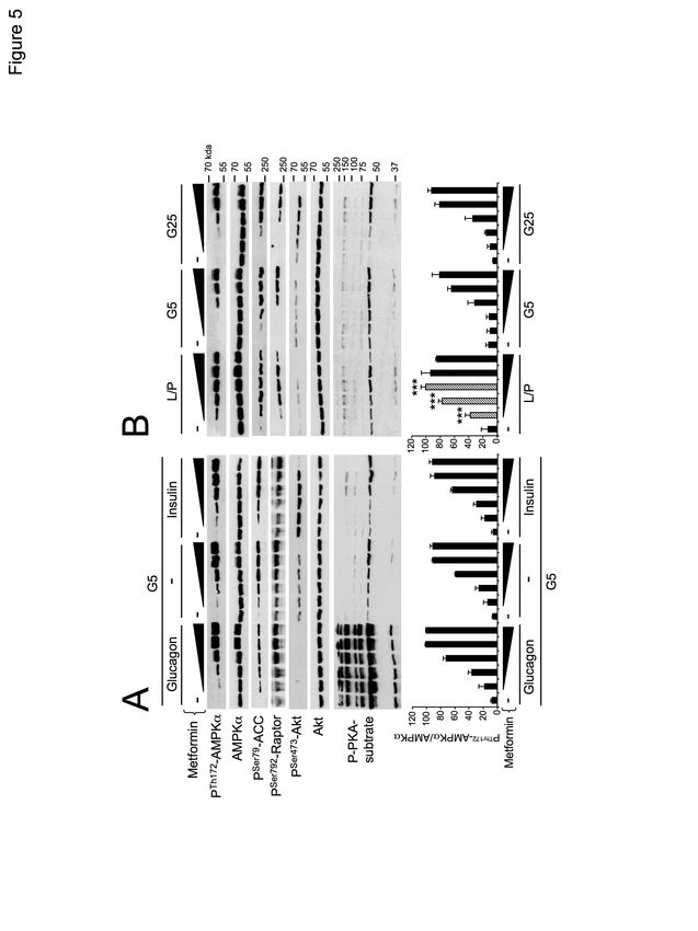
suckling     period,     the    plasma    insulin       deficient liver (Fig. 3A and 3B). In agreement
concentration is low and that of glucagon high          with the inhibitory action of metformin on
because of the ingestion of milk, which is a            mitochondrial complex 1 activity (22,23), we
high-fat, low-carbohydrate food. The transition         found that metformin treatment led to a
from a milk diet to a high-carbohydrate diet at         reduction in respiration in control hepatocytes,
weaning leads to an increase in blood glucose           which was accentuated in AMPK-deficient
and plasma insulin levels (Fig. 1A). Weaning            hepatocytes (Fig. 3C). These results
from maternal milk was associated with a                demonstrate the protective role of AMPK in
decrease in the phosphorylation of AMPK at α-           maintaining hepatic energy homeostasis in
Thr172 and that of its substrate ACC at Ser79           response to a metabolic challenge induced by a
in the liver, whereas the abundance of the              reduction in cellular energy charge.
AMPK and ACC proteins was unchanged (Fig.
1B). In this context, increased phosphorylation         Metabolic stress-induced AMPK activation is
of Akt at Ser473 reflects activation of the             strengthened in hepatocytes incubated under
insulin-signaling      pathway      (Fig.   1B).        simulated fasting conditions
Conversely, AMPK signaling was highly                       Given the modulation of AMPK activity
active in the liver of suckling rats, as                during       the      fasting/refeeding      and
characterized by an increase in the                     suckling/weaning transitions, we hypothesized
phosphorylation of AMPK-α-Thr172 and                    that the regulation of hepatic AMPK is driven

                                                                                                            Downloaded from http://www.jbc.org/ by guest on October 30, 2020
ACC-Ser79 (Fig. 1B).                                    by changes in glucose availability and/or
    As expected, the transition from fasting to         glucagon or insulin-stimulated changes in the
refeeding was associated with an increase in            kinase phosphorylation status (15,17,18). We
blood glucose and insulin levels and a decrease         thus treated mouse primary hepatocytes with
in blood glucagon levels (Fig. 1C). In the liver        the AMPK activators, metformin, AICAR, or
of starved mice, phosphorylation of AMPK-α-             A-769662, under various nutritional and
Thr172 and ACC-Ser79 was markedly higher                hormonal conditions mimicking the fasting or
than that of refed mice (Fig. 1D), in agreement         fed states to identify the nature of the stimuli
with previous studies (13-16). Conversely, the          that modulate hepatic AMPK activity.
increase in blood insulin levels in refed mice              Under low glucose/basal conditions (5 mM
induced the phosphorylation of Akt at Ser473            glucose),      metformin        induced       the
residue (Fig. 1D). During fasting, changes in           phosphorylation of AMPK at α-Thr172 and
AMPK activation were associated with a lower            that of its downstream targets ACC at Ser79
cellular energy state, as revealed by the               and Raptor at Ser792 in concentration-
decrease and increase of hepatic ATP and ADP            dependent manner (Fig. 4). Incubation of
concentrations, respectively, resulting in a            hepatocytes with high glucose concentrations
significant increase in the ADP/ATP ratio (Fig.         (25 mM glucose) and insulin, to mimic
2). Thus, hepatic AMPK activation induced by            feeding, increased the phosphorylation of Akt
fasting is associated with a decrease in the            at Ser473 but did not alter metformin-induced
cellular energy state in the liver.                     AMPK phosphorylation relative to that of the
                                                        basal condition (Fig. 4). In contrast, culturing
AMPK deficiency exacerbates cellular energy             the hepatocytes in a medium that simulated
depletion in response to metabolic stress in            fasting conditions, which contained glucagon
the liver                                               and in which glucose was replaced with lactate
    AMPK plays a crucial role in maintaining            and pyruvate, robustly enhanced metformin-
energy homeostasis during periods of                    induced          AMPK            phosphorylation
metabolic stress. We therefore hypothesized             concomitantly with an increase in the PKA
that AMPK deficiency in the liver may alter             substrate phosphorylation pattern (Fig. 4).
sensitivity to an energy stress. We tested this
hypothesis by treating hepatic AMPK-deficient           Metabolic stress-induced energy depletion is
mice with metformin, a mitochondrial                    worsened in AMPK-deficient hepatocytes
respiratory chain inhibitor (22,23). Metformin          incubated under simulated fasting conditions
treatment induced a marked increase in the                 We next assessed the effect of changes in
ADP/ATP ratio in the livers of control                  glucose concentrations and pancreatic hormone
animals. Importantly, the increase in the               levels in culture medium on metformin-
ADP/ATP ratio was greater in AMPK-                      induced energy depletion in both control and

                                                    3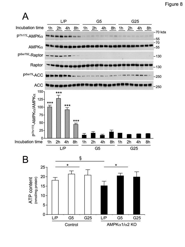
AMPK-deficient hepatocytes. In the absence of results with A-769662, a direct small molecule
metformin, the cellular energy charge was AMPK activator (Fig. S1). Furthermore,
similar between control and AMPK-deficient AMPK and ACC phosphorylation induced by
hepatocytes and was not altered after an 8-h the cell-permeable AMPK activator AICAR
incubation under conditions mimicking fasting was unaltered by treatment with the pancreatic
(glucose-free medium containing lactate and hormones glucagon or insulin (Fig. 7A). In
pyruvate plus glucagon) or refeeding (25 mM contrast, a comparison of the action of AICAR
glucose plus insulin), indicating that these at various glucose concentrations showed
conditions are insufficient to alter the cellular AMPK phosphorylation to be stimulated to a
energy state (Fig. 5A and 5B). Metformin greater extent in glucose-free medium
treatment strongly correlated with a marked containing lactate and pyruvate plus glucagon
decrease in ATP and energy charge in both than medium containing only 5 mM glucose or
control and AMPK-deficient hepatocytes. As 25 mM glucose plus insulin (Fig. 7B). In
observed in the liver (Fig. 3A and 3B), the summary, AMPK activation in hepatocytes is
decrease in ATP levels and energy charge in enhanced by the scarcity of glucose, whereas
response to metformin was greater in AMPK- changes in insulin or glucagon concentrations
deficient hepatocytes than control hepatocytes do not affect its activity.
(Fig. 5A and 5B). Metformin-induced energy
depletion was also greater when hepatocytes Switching to a glucose-free medium
Downloaded from http://www.jbc.org/ by guest on October 30, 2020
were incubated under fasting-like than containing lactate and pyruvate transiently
refeeding-like conditions, and this effect was activates AMPK in hepatocytes
greater in AMPK-deficient hepatocytes than In the previous experiments (Fig. 4, 6, 7
control hepatocytes. Thus, the higher and S1), we observed no changes in AMPK-α-
activation of AMPK in response to an energy Thr172 phosphorylation after the incubation of
stress in hepatocytes incubated under fasting- hepatocytes for 8 h with various glucose
like conditions coincides with a greater cellular concentrations in the absence of activators
energy deficiency. Moreover, the aggravation (metformin, AICAR or A-769662), This
of energy depletion observed in AMPK- observation is consistent with the lack of an
deficient hepatocytes incubated under fasting effect on the cellular energy state (Fig. 5). We
conditions is consistent with the role of AMPK tested whether changes in glucose availability
in regulating the cellular energy balance to could activate AMPK at early time points.
restore cellular ATP levels to normal values. Incubation of hepatocytes with a glucose-free
medium containing lactate and pyruvate
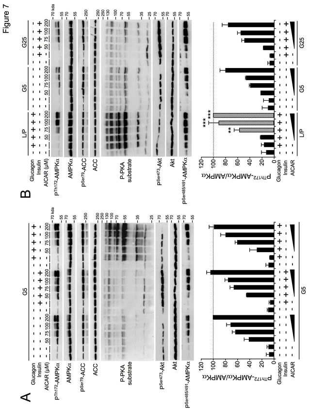
Glucose availability, but not pancreatic transiently activated AMPK signaling within 2
hormones, sensitizes AMPK activation during h (Fig. 8A). Indeed, phosphorylation of α-
metabolic stress in hepatocytes Thr172-AMPK and its downstream targets
To dissociate the impact of glucagon and ACC and Raptor were maximal at 2 h and
insulin signaling from that of glucose nearly returned to basal levels after 8 h (Fig.
availability on AMPK activity, we separately 8A). In contrast, incubation in a medium
examined their respective effects on containing 5 or 25 mM glucose did not modify
metformin-induced AMPK activation in AMPK signaling (Fig. 8A). Moreover, AMPK
hepatocytes. In the presence of 5 mM glucose, activation induced by the absence of glucose in
treatment with glucagon or insulin induced the culture medium correlated with a low but
sustained phosphorylation of PKA-substrates significant decrease in intracellular ATP levels
or Akt-Ser473, respectively, but they had no at 2 h, which was amplified in AMPK-
effect on the pattern of change of AMPK deficient hepatocytes (Fig. 8B). These results
phosphorylation at α-Thr172 or that of its indicate that the lack of glucose induces
downstream targets (ACC at Ser79 and Raptor transient activation of AMPK, which acts to
at Ser792) induced by metformin (Fig. 6A). adapt hepatocyte metabolism and maintain
Conversely, the incubation of hepatocytes in cellular energy levels.
glucose-free medium containing lactate and We next assessed AMPK activation in
pyruvate led to greater metformin-induced response to various activators at early time
AMPK phosphorylation than that in points in hepatocytes incubated with various
hepatocytes incubated with 5 or 25 mM levels of glucose. Activation of AMPK by
glucose for 8 h (Fig. 6B). We obtained similar metformin (Fig. 9) or AICAR (Fig. S2A) was
4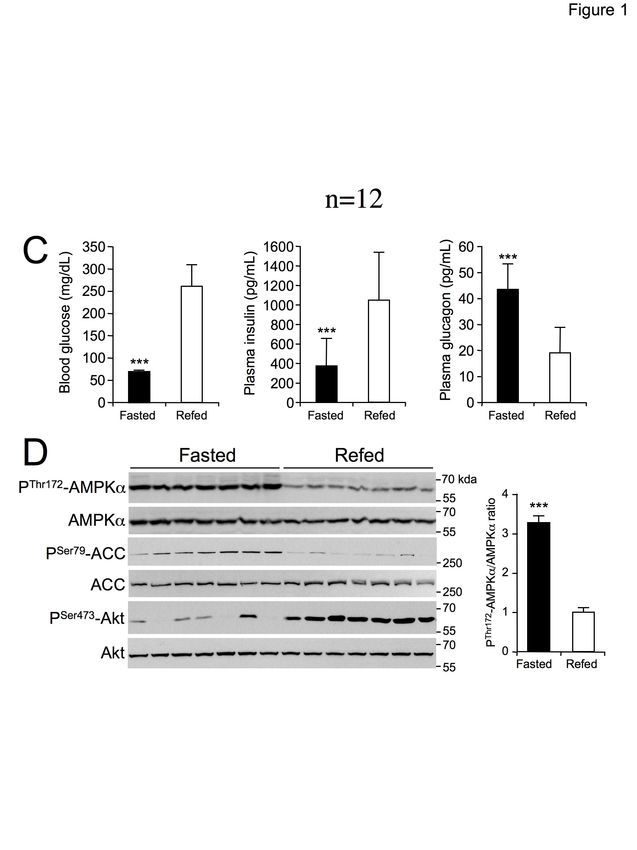
enhanced when hepatocytes were incubated in             display normal hepatic glucose and lipid
glucose-free medium containing lactate and              homeostasis and are not prone to insulin
pyruvate or with low glucose concentrations (5          resistance, suggesting that the decrease in
mM glucose). Similarly, incubation of                   AMPK activity associated with insulin
hepatocytes with the direct small molecule              resistance may be a consequence, rather than a
activator A-769662 induced more pronounced              cause, of changes in hepatic metabolism (10).
ACC and Raptor phosphorylation in glucose-                  By contrast, we and others have shown that
free medium containing lactate and pyruvate             reversible and physiological variation of
(Fig. S2B). Unexpectedly, A-769662-mediated             hepatic AMPK activity occurs during the
AMPK phosphorylation was only induced in                fasting-refeeding      and     suckling-weaning
the absence of glucose (Fig. S2B).                      transitions (13-16) (Fig. 1). These changes in
                                                        AMPK activity may account for the shift in
Phosphorylation of AMPKα on the                         hepatic lipid metabolism from catabolism to
Ser485/491 residue does not alter its activity          anabolism. However, the catabolic and/or
in hepatocytes                                          anabolic     stimuli    responsible    for   the
   Insulin and agents that elevate cellular             physiological modulation of AMPK activity
cAMP have been reported to inhibit AMPK                 during fasting are still poorly understood. It
activity through the phosphorylation of                 has been hypothesized that acute changes in
AMPKα at Ser485/491 by Akt and PKA,                     hepatic AMPK activity are due to fluctuations

                                                                                                            Downloaded from http://www.jbc.org/ by guest on October 30, 2020
respectively (20,24). We found that both                in plasma levels of insulin and the counter
insulin and glucagon weakly induced AMPK                regulatory action of glucagon (34). Indeed,
phosphorylation at α-Ser485/491 in primary              conditions associated with increased glucagon
hepatocytes. In contrast, AICAR induced                 activate AMPK, possibly through modulation
massive phosphorylation at this site, likely            of the hepatic energy charge (increase in the
resulting from autophosphorylation, as                  AMP/ATP ratio) and PKA-induced activation
previously described (24). However, AMPK                of LKB1 (18,35). Conversely, insulin has been
activity was not attenuated by increased α-             reported to decrease AMPK activity through
Ser485/491 phosphorylation, as shown by                 the phosphorylation of AMPKα at α1-
maintenance of the phosphorylation of its               Ser485/α2-Ser491, with a concomitant loss of
downstream target ACC (Fig. 7A and 7B),                 both AMPKα-Thr172 and ACC-Ser79
indicating the absence of an inhibitory effect of       phosphorylation (17,36).
Ser485/491 phosphorylation on AMPK                          The role of the phosphorylation at
activation in primary hepatocytes.                      Ser485/491 is not well understood and it is still
                                                        unclear     whether      this    phosphorylation
Discussion                                              contributes to enzyme regulation and AMPK
                                                        activity. Interestingly, mutation of the Ser485
    Over the past decade, the fuel-sensing              residue to mimic phosphorylation by
enzyme AMPK has attracted much attention                introduction of an aspartate residue in the
because of the associations drawn between the           AMPKα1 subunit is not sufficient to inhibit
wide range of its metabolic downstream                  AMPK activation by liver purified AMPK
targets, including fatty-acid synthesis and             kinase (37). Although phosphorylation at
oxidation, mitochondrial function, oxidative            Ser485/491 has been shown to correlate with
stress, inflammation, and autophagy and the             the inhibition of AMPK activity in a variety of
alteration of these pathways by insulin                 tissues,     we      show      that    increased
resistance and metabolic syndrome-associated            phosphorylation at this site is proportional to
disorders (25). Although there is no clear              the increase in the phosphorylation of the
evidence that polymorphisms in genes                    downstream AMPK target ACC-Ser79 in
encoding AMPK subunits influence the                    primary hepatocytes (Fig. 7). Similarly,
occurrence of metabolic syndrome (26-28), a             HepG2 cells treated with troglitazone showed
sustained decrease in AMPK activity has been            an increase in phosphorylation of AMPKα at
found in the liver, skeletal muscle, and adipose        both Thr172 and Ser485 residues, associated
tissue from obese or hyperglycemic rodents              with      an     increase     in     ACC-Ser79
and humans in association with insulin                  phosphorylation (38). This is also reminiscent
resistance (29-33). Nevertheless, we have               of the effect of acute renal ischemia causing
shown that liver-specific AMPK deficient mice           simultaneous phosphorylation of AMPKα-

                                                    5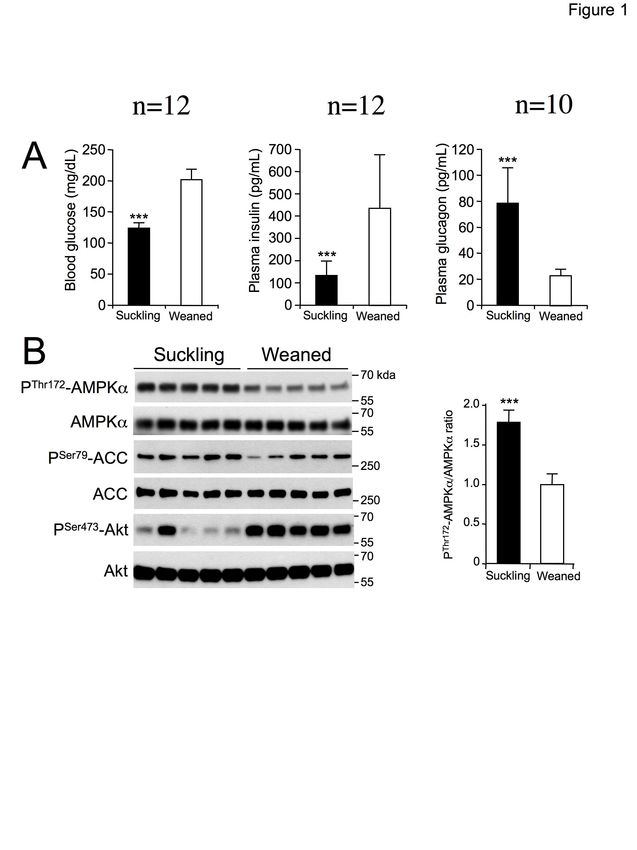
Thr172 and AMPKα-Ser485 in the kidney                   the cellular energy charge. Of note, transient
(39). Furthermore, despite that glucagon and            AMPK activation induced by glucose
insulin pretreatments induce phosphorylation            starvation was low compared to drug-induced
at α-Ser485/491, this is not sufficient to reduce       AMPK activation (Fig. 9 and Fig. S2),
the phosphorylation of the activation loop α-           suggesting that substitution of glucose by
Thr172 in response to AICAR (Fig. 7). It is             lactate and pyruvate in medium had a
likely that the increase in Ser485/491                  relatively low impact on cellular energy levels.
phosphorylation              reflects          an           Furthermore, consistent with its role as an
autophosphorylation event, concomitant with             energy sensor acting to restore energy
the increase in AMPK activation, as previously          homeostasis, primary hepatocytes incubated in
demonstrated (24). It was suggested that                the absence of glucose (but incubated with
phosphorylation of AMPK at this specific site           lactate and pyruvate) exhibited enhanced
may represent a regulatory mechanism to                 activation of AMPK and amplified energy
prevent over-stimulation of AMPK, in addition           depletion in response to metformin-induced
to potential cross-talk with inhibitory signaling       energy stress relative to that of hepatocytes
pathways (24). Thus, further studies will be            incubated with glucose (5 or 25 mM) (Fig. 4,
required to better understand the physiological         5, 6 and 9). Such an enhanced response to
impact of such phosphorylation on hepatic               energy stress observed in the context of low
metabolism.                                             glucose availability may be attributable to

                                                                                                           Downloaded from http://www.jbc.org/ by guest on October 30, 2020
    We and others have shown that AMPK                  lower ATP generation due to an overall
activation in the liver during fasting results          decrease in the glycolytic flux.
from an increase in AMP/ATP and ADP/ATP                     Unexpectedly, we showed that the
ratios (40,41) (Fig. 2). However, the nature of         stimulation of AMPK signaling by the small
the stimuli altering the AMP/ATP and                    molecule A-769662 was enhanced by low
ADP/ATP ratios and subsequent AMPK                      glucose levels and that AMPKα-Thr172
activity in the liver is somewhat unclear. Our          phosphorylation was transiently induced under
results suggest that nutritional changes (i.e.,         conditions of glucose starvation (Fig. S1 and
glucose availability) rather than related               S2). These effects appear to be paradoxical
hormonal changes (i.e., glucagon/ insulin ratio)        since A-769662 causes activation of AMPK
likely underly the sensitization of AMPK to             independently of α-Thr172 phosphorylation
energetic stress induced by the dietary                 via an allosteric mechanism and without
transition that takes place during fasting.             compromising the cellular AMP/ATP ratio
    Recent studies suggest that starvation-             (43-45). Nevertheless, we have previously
induced AMPKα-Thr172 phosphorylation in                 shown that AMP-induced phosphorylation of
the liver requires the formation of a ternary           AMPK is enhanced by A-769662 (10,44).
complex between axin, LKB1, and AMPK                    Thus, the AMPK-α-Thr172 phosphorylation
(16). Interestingly, AMP binding to AMPK has            observed with A-769662 in glucose-free
been shown to enhance its binding to the axin-          medium can be interpreted as a synergic effect
LKB1 complex and thus promote axin-                     resulting        from          AMPK-α-Thr172
dependent AMPKα-Thr172 phosphorylation                  phosphorylation caused by a transient increase
(7,16). Also, it has been reported that AMPK            in cellular AMP levels in response to glucose
can sense glucose starvation independently of           starvation and A-769662-binding to the AMPK
changes in adenine nucleotide concentrations            complex.
through the formation of a lysosomal complex                ATP depletion was more pronounced in the
(42). Although we were unable to detect                 livers of hepatic AMPK-deficient mice treated
activation of AMPK in primary mouse                     with metformin than in those of control mice
hepatocytes incubated in glucose-free medium            (Fig. 3). Similarly, ATP levels in primary
containing lactate and pyruvate for 8 h, we             AMPK-deficient hepatocytes were much lower
observed transient AMPK activation within 2             than in control hepatocytes following
h, associated with a decrease in cellular ATP           incubation with metformin (Fig. 5), as
levels (Fig. 8). Such transient activation of           previously reported (44). The depletion of
AMPK observed in hepatocytes after switching            hepatic ATP observed during fasting has also
from glucose to lactate and pyruvate (Fig. 8)           been shown to be amplified in the livers of
can be seen as a counter signal to adapt the            AMPK-deficient mice (41). This exacerbated
metabolism to glucose starvation and maintain           decrease in ATP levels in response to an

                                                    6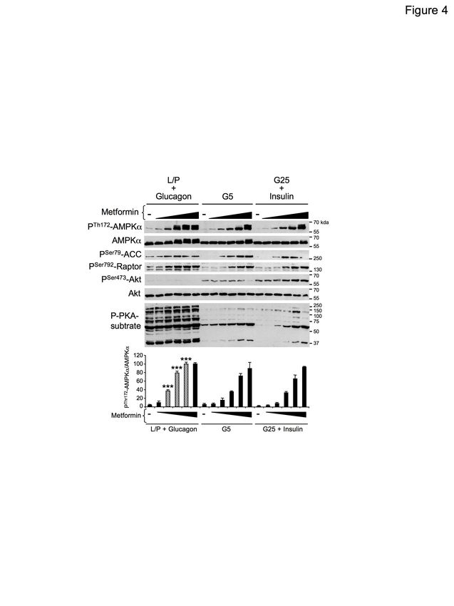
energy stress reveals metabolic inflexibility in         energy state in the liver by promoting a
AMPK-deficient hepatocytes and emphasizes                metabolic switch from the utilization of
the importance of AMPK in the maintenance                glucose to that of other substrates, notably,
of the hepatic energy charge through the                 fatty acids, to supply energy needs.
control of adaptive mitochondrial function (46-
49). Of note, in the absence of energy stress,           Experimental procedures
ATP levels in the livers and primary
hepatocytes from AMPK-deficient mice are no              Reagents and antibodies
different than those of the control counterparts,           Metformin (#D5035) and glucagon were
reinforcing the notion that hepatic AMPK is              purchased from Sigma. AICAR was purchased
activated only during times of energy stress to          from Toronto Research Chemicals. A-769662
maintain the energy balance. These results               was kindly provided by Dr. Anudharan
demonstrate that AMPK activation is crucial              Balendran (Astra Zeneca). Human insulin
for maintaining energy homeostasis in the liver          (Actrapid) was purchased from Novo Nordisk.
during the metabolic transition that occurs              Primary antibodies directed against total
during fasting.                                          AMPKα (#2532), AMPKα phosphorylated at
    AMPK has a high therapeutic potential for            Thr172 (#2531), phospho-AMPKα1(Ser485)/
the management of dysregulated metabolism in             AMPKα2(Ser491) (#4185), total acetyl-CoA
the liver. Notably, pharmacological AMPK                 carboxylase      (ACC)     (#3676),     ACC

                                                                                                             Downloaded from http://www.jbc.org/ by guest on October 30, 2020
activation has shown beneficial effects in the           phosphorylated at Ser79 (#3661), total Raptor
treatment of liver steatosis (10,43,50,51). We           (#2280), Raptor phosphorylated at Ser792
have shown that drug-induced AMPK                        (#2083),     total   Akt     (#9272),    Akt
activation      decreases       hepatic      lipid       phosphorylated at Ser473 (#4058), and
accumulation, both by inhibiting lipid                   phospho-PKA substrate (#9624) were all
synthesis and by stimulating fatty-acid                  purchased from Cell Signaling Technology.
oxidation (10). Thus, the modulation of hepatic          HRP-conjugated secondary antibodies were
AMPK activity by the nutritional state may               purchased from Calbiochem. All other
have implications in future clinical practice.           materials unless otherwise indicated were
Indeed, our findings predict that hepatic                purchased from Sigma.
AMPK activation in response to the
administration of an AMPK-activating drug                Animals
may be enhanced during fasting. On the other                Animal studies were approved by the Paris
hand, postprandial delivery of an AMPK-                  Descartes University ethics committee (no.
activating compound may counteract the                   CEEA34.BV.157.12) and performed under
lowing of hepatic AMPK activity due to the               French authorization to experiment on
massive influx of glucose into liver after the           vertebrates (no.75-886) in accordance with the
ingestion of a carbohydrate-rich meal. In both           European guidelines. C57BL/6J mice were
conditions, the nutritional state may influence          obtained from Harlan France. Liver-specific
the downstream effects of AMPK. In the                   double knockout of AMPKα1 and AMPKα2
fasting state, AMPK-induced fatty-acid                   catalytic subunits was achieved by crossing
oxidation will be boosted. In the fed state, lipid       AMPKα1lox/lox mice with AMPKα2lox/lox mice
synthesis from glucose will be inhibited by              and then crossing the progeny with Alfp-Cre
drug-induced AMPK activation. Consequently,              transgenic mice to generate AMPKα1lox/lox,
the delivery of future AMPK activating                   α2lox/lox (control) and AMPKα1lox/lox, α2lox/lox-
therapies will need to consider the nutritional          Alfp-Cre (liver AMPKα1/α2 KO) mice (10).
state and diet composition (low- or high-                All mice were maintained in a barrier facility
carbohydrate) to adapt the dosage.                       under a 12/12-h light/dark cycle with free
    In summary, our studies support the notion           access to water and standard mouse diet (in
that reversible AMPK activation observed in              terms of energy: 65% carbohydrate, 11% fat,
the liver during nutritional transition (typically       24% protein).
during fasting) results in a decrease in cellular
energy charge, which is modulated by glucose             Suckling and weaned rats
availability rather changes in pancreatic                   Litters of 13-day-old Wistar rats with their
hormone levels. In this context, AMPK                    mother were obtained from Janvier France.
activation is critical to preserve the cellular          Litters were housed in individual cages under a

                                                     7
12/12-h light/dark cycle in a temperature-             modified version of the collagenase method as
controlled environment with free access to             described previously (44). The cells were
water and standard diet. When the pups were            plated in M199 medium with Glutamax
18 and 19 days of age, the mothers were fed            supplemented with 100 U/ml penicillin, 100
separately from their offspring from 9:00 to           µg/ml streptomycin, 10% (v/v) FBS, 500 nM
12:00 a.m. and 4:00 to 7:00 p.m. to avoid early        dexamethasone        (Sigma),      100      nM
weaning of the pups. Twenty-day-old suckling           triiodothyronine (Sigma), and 10 nM insulin
pups were separated from their mother for 3 h          (Actrapid, Novo Nordisk) at a density of 4 x
to allow gastric emptying. Then, rats were             105 cells/well in six-well plates or 1 x 10⁶
force-weaned by gavage with 5 g/kg glucose or          cells/60-mm-diameter cell culture plate. After
replaced with their mother. After 3 h, rats were       attachment (3 to 4 h), hepatocytes were
sacrificed by decapitation. Blood was quickly          maintained in M199 medium with antibiotics
collected, and the liver was immediately               and 100 nM dexamethasone for 16 h. The cells
removed and frozen in liquid nitrogen in < 25          were then stimulated with the respective
s. Livers were stored at -80°C until analysis.         compounds or hormones for the times
After sacrifice, the stomachs of suckling pups         indicated in the figure legends in glucose-free
were checked to ensure that they were filled           DMEM medium supplemented with 100 nM
with milk.                                             dexamethasone and lactate/pyruvate (10:1
                                                       mM) or glucose (5 or 25 mM).

                                                                                                          Downloaded from http://www.jbc.org/ by guest on October 30, 2020
Fasting and refeeding experiments
    For the fasting-refeeding experiments, mice        Hepatocyte oxygen consumption assay
were fasted for 24 h or fasted for 24 h and then           Primary mouse hepatocytes were plated
refed a high-carbohydrate diet (70%                    onto collagen I-coated Seahorse 96-well plates
carbohydrate in terms of total kcal with 64%           at a density of 10,000 cells/well. After 4 h,
sucrose in terms of weight, Harlan TD.08247)           primary hepatocytes were cultured for 16 h in
for 3 h. At the end of the refeeding period,           M199 medium containing antibiotics and 100
mice were sacrificed by cervical dislocation           nM dexamethasone. Hepatocytes were then
and the liver immediately removed and frozen           switched to glucose-free DMEM supplemented
in liquid nitrogen in < 25 s. Livers were stored       with lactate/pyruvate (10:1 mM) and 100 nM
at -80°C until analysis.                               dexamethasone 1 h prior to measuring
                                                       respiration. The oxygen consumption rate
Blood glucose and plasma pancreatic                    (OCR,      mitochondrial    respiration)  was
hormone measurement                                    monitored using the Seahorse Bioscience
   Blood    glucose     concentrations    were         XF96 Extra Cellular Flux Analyzer in real
determined from blood isolated from the tail           time. The OCR was acquired under basal
vein with a glucometer (Roche Diagnostics).            conditions and 15, 30, and 45 min after
Blood was collected into heparin-containing            injection with 1 mM metformin. Results were
tubes and centrifuged to obtain plasma. Plasma         normalized to total protein/well after
insulin and glucagon levels were determined            completion of the assay and are expressed as
using mouse or rat ELISA kits (Crystal Chem).          pmoles O2 consumed per µg of protein per
                                                       minute.
In vivo metformin treatment
    Ten-week-old      control      and     liver       Measurement        of    adenine     nucleotide
AMPKα1/α2 KO mice in a fed state were                  concentrations
injected intraperitoneally with saline or 200              Adenine nucleotide concentrations were
mg/kg metformin to induce a hepatic energy             determined in cell extracts prepared from
stress. Mice were sacrificed by cervical               cultured hepatocytes or liver samples using an
dislocation 1 h after metformin administration         enzymatic method (44). Primary hepatocytes
and the liver extracted and frozen in liquid           were treated as described in the figure legends,
nitrogen in < 25 s. Livers were stored at -80°C        the culture medium removed, and cells on 60-
until adenine nucleotide analysis.                     mm-diameter cell culture plates (1 x 10⁶
                                                       cells/plate) scraped into 200 µl 6% (v/v) ice-
Mouse primary hepatocytes                              cold HClO4 in < 5 s. For the liver, mice were
   Mouse primary hepatocytes were isolated             treated as described in the figure legends. At
from 10-12-week-old male mice using a                  the end of treatment, mice were sacrificed by

                                                   8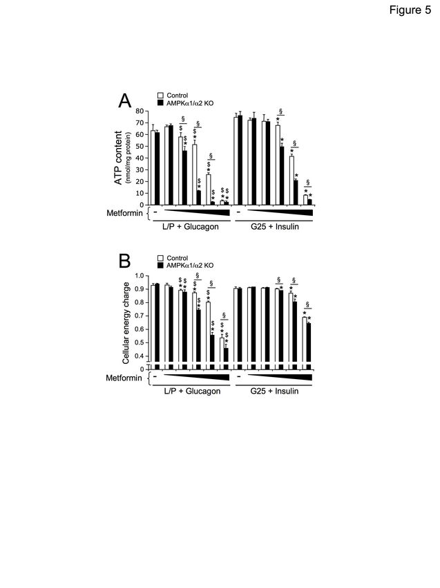
cervical dislocation and the livers extracted            ice-cold lysis buffer with a ball-bearing
and frozen in liquid nitrogen in < 25 s. Two             homogenizer (Retsch). The lysates and
hundred milligrams of liver were homogenized             homogenates were centrifuged for 10 min at
in 1 ml 6% (v/v) ice-cold HClO4. Cell extracts           10,000 x g at 4°C and the supernatants
were centrifuged at 10,000 g for 10 min at 4°C.          removed for determination of total protein
The acid supernatant was neutralized and used            content with a BCA protein assay kit (Thermo
for spectrophotometric determination of                  Fisher Scientific). Fifty micrograms of protein
adenine nucleotides. Standard curves for ATP,            from the supernatant was separated on 10%
ADP, and AMP were constructed with 25, 50,               SDS-PAGE        gels     and    transferred    to
75, 100, 125, and 150 µM of each nucleotide.             nitrocellulose membranes. The membranes
Determination of the adenine nucleotides                 were blocked for 30 min at 37°C with Tris-
presented in the Fig. 8B was performed by                buffered saline supplemented with 0.05%
high-performance liquid chromatography as                NP40      and     5%      nonfat    dry     milk.
described previously (52). Adenine nucleotide            Immunoblotting was performed with the
levels are expressed in µmol/g of liver weight           antibodies indicated in the figure legends,
or nmol/mg of protein. The energy charge was             following standard procedures, and the signals
calculated using the following equation:                 detected by chemiluminescence reagents
[ATP+ADP/2]/[ATP+ADP+AMP],               where           (Thermo). Total and phosphorylated AMPK,
AMP, ADP, and ATP are the respective tissue              ACC, Raptor, and Akt were probed using

                                                                                                             Downloaded from http://www.jbc.org/ by guest on October 30, 2020
concentrations (53).                                     separate membranes. X-ray films were scanned
                                                         and band intensities were quantified by Image
Western-blot analysis                                    J (NIH) densitometry analysis.
    After the incubation times indicated in the
figure legends, cultured hepatocytes were lysed          Statistical analysis
in ice-cold lysis buffer containing 50 mM Tris,              Results are expressed as the means ± SD.
pH 7.4, 1% Triton X-100, 150 mM NaCl, 1                  Comparisons between groups were made by
mM EDTA, 1 mM EGTA, 10% glycerol, 50                     unpaired two-tailed Student’s t-tests or one-
mM NaF, 5 mM sodium pyrophosphate, 1 mM                  way      ANOVA,        in   conjunction  with
Na3VO4, 25 mM sodium-β-glycerophosphate,                 Bonferroni’s post hoc test for multiple
1 mM DTT, 0.5 mM PMSF, and protease                      comparisons,       when    appropriate, using
inhibitors (Complete Protease Inhibitor                  GraphPad Prism 5.0 (GraphPad Software Inc.).
Cocktail; Roche). Lysates were sonicated on              Differences between groups were considered
ice for 15 seconds to shear DNA and reduce               statistically significant when P < 0.05.
viscosity. Pieces of liver were homogenized in

Data Availability Statement: All data presented and discussed are contained within the manuscript.

The abbreviations used are: AMPK, AMP-activated protein kinase; ACC, acetyl-CoA carboxylase;
AICAR, 5-aminoimidazole-4-carboxamide ribonucleotide; L/P, glucose-free medium containing 10
mM lactate and 1 mM pyruvate; G5, medium containing 5 mM glucose; G25, medium containing 25
mM glucose.

Financial support: This work was supported by grants from Inserm, the CNRS, the Université Paris
Descartes, the Région Ile-de-France (CORDDIM), and the Société Francophone du Diabète (SFD).
N.B. is a recipient of a doctoral fellowship from the French Government (Ministère de la Recherche et
des Enseignements Supérieurs).

Author contributions: CH and NB performed experiments. BG performed the adenine nucleotide
measurement by HPLC and edited the manuscript. BV interpreted the data and wrote the manuscript.
MF conceived, designed, and performed experiments, interpreted the data, wrote the manuscript, and
directed this study.

Conflict of interest: The authors declare no conflict of interest associated with this study.

                                                    9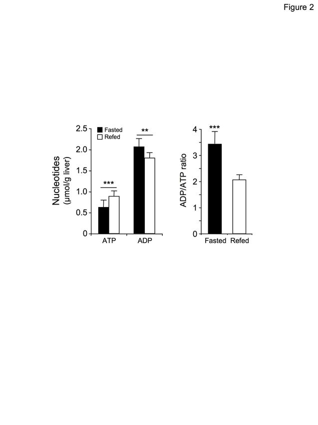
References

1.     Hardie, D. G. (2014) AMPK--sensing energy while talking to other signaling pathways. Cell Metab 20,
       939-952
2.     Willows, R., Sanders, M. J., Xiao, B., Patel, B. R., Martin, S. R., Read, J., Wilson, J. R., Hubbard, J.,
       Gamblin, S. J., and Carling, D. (2017) Phosphorylation of AMPK by upstream kinases is required for
       activity in mammalian cells. Biochem J 474, 3059-3073
3.     Viollet, B., Horman, S., Leclerc, J., Lantier, L., Foretz, M., Billaud, M., Giri, S., and Andreelli, F.
       (2010) AMPK inhibition in health and disease. Crit Rev Biochem Mol Biol 45, 276-295
4.     Oakhill, J. S., Chen, Z. P., Scott, J. W., Steel, R., Castelli, L. A., Ling, N., Macaulay, S. L., and Kemp,
       B. E. (2010) beta-Subunit myristoylation is the gatekeeper for initiating metabolic stress sensing by
       AMP-activated protein kinase (AMPK). Proc Natl Acad Sci U S A 107, 19237-19241
5.     Liang, J., Xu, Z. X., Ding, Z., Lu, Y., Yu, Q., Werle, K. D., Zhou, G., Park, Y. Y., Peng, G., Gambello,
       M. J., and Mills, G. B. (2015) Myristoylation confers noncanonical AMPK functions in autophagy
       selectivity and mitochondrial surveillance. Nat Commun 6, 7926
6.     Warden, S. M., Richardson, C., O'Donnell, J., Jr., Stapleton, D., Kemp, B. E., and Witters, L. A. (2001)
       Post-translational modifications of the beta-1 subunit of AMP-activated protein kinase affect enzyme
       activity and cellular localization. Biochem J 354, 275-283
7.     Zong, Y., Zhang, C. S., Li, M., Wang, W., Wang, Z., Hawley, S. A., Ma, T., Feng, J. W., Tian, X., Qi,
       Q., Wu, Y. Q., Zhang, C., Ye, Z., Lin, S. Y., Piao, H. L., Hardie, D. G., and Lin, S. C. (2019)
       Hierarchical activation of compartmentalized pools of AMPK depends on severity of nutrient or energy

                                                                                                                     Downloaded from http://www.jbc.org/ by guest on October 30, 2020
       stress. Cell Res 29, 460-473
8.     Foretz, M., Ancellin, N., Andreelli, F., Saintillan, Y., Grondin, P., Kahn, A., Thorens, B., Vaulont, S.,
       and Viollet, B. (2005) Short-term overexpression of a constitutively active form of AMP-activated
       protein kinase in the liver leads to mild hypoglycemia and fatty liver. Diabetes 54, 1331-1339
9.     Foretz, M., Even, P. C., and Viollet, B. (2018) AMPK Activation Reduces Hepatic Lipid Content by
       Increasing Fat Oxidation In Vivo. Int J Mol Sci 19
10.    Boudaba, N., Marion, A., Huet, C., Pierre, R., Viollet, B., and Foretz, M. (2018) AMPK Re-Activation
       Suppresses Hepatic Steatosis but its Downregulation Does Not Promote Fatty Liver Development.
       EBioMedicine 28, 194-209
11.    Fullerton, M. D., Galic, S., Marcinko, K., Sikkema, S., Pulinilkunnil, T., Chen, Z. P., O'Neill, H. M.,
       Ford, R. J., Palanivel, R., O'Brien, M., Hardie, D. G., Macaulay, S. L., Schertzer, J. D., Dyck, J. R., van
       Denderen, B. J., Kemp, B. E., and Steinberg, G. R. (2013) Single phosphorylation sites in Acc1 and
       Acc2 regulate lipid homeostasis and the insulin-sensitizing effects of metformin. Nat Med 19, 1649-
       1654
12.    Loh, K., Tam, S., Murray-Segal, L., Huynh, K., Meikle, P. J., Scott, J. W., van Denderen, B., Chen, Z.,
       Steel, R., LeBlond, N. D., Burkovsky, L. A., O'Dwyer, C., Nunes, J. R. C., Steinberg, G. R., Fullerton,
       M. D., Galic, S., and Kemp, B. E. (2019) Inhibition of Adenosine Monophosphate-Activated Protein
       Kinase-3-Hydroxy-3-Methylglutaryl Coenzyme A Reductase Signaling Leads to Hypercholesterolemia
       and Promotes Hepatic Steatosis and Insulin Resistance. Hepatol Commun 3, 84-98
13.    Assifi, M. M., Suchankova, G., Constant, S., Prentki, M., Saha, A. K., and Ruderman, N. B. (2005)
       AMP-activated protein kinase and coordination of hepatic fatty acid metabolism of
       starved/carbohydrate-refed rats. Am J Physiol Endocrinol Metab 289, E794-800
14.    Suchankova, G., Tekle, M., Saha, A. K., Ruderman, N. B., Clarke, S. D., and Gettys, T. W. (2005)
       Dietary polyunsaturated fatty acids enhance hepatic AMP-activated protein kinase activity in rats.
       Biochem Biophys Res Commun 326, 851-858
15.    Munday, M. R., Milic, M. R., Takhar, S., Holness, M. J., and Sugden, M. C. (1991) The short-term
       regulation of hepatic acetyl-CoA carboxylase during starvation and re-feeding in the rat. Biochem J 280
       ( Pt 3), 733-737
16.    Zhang, Y. L., Guo, H., Zhang, C. S., Lin, S. Y., Yin, Z., Peng, Y., Luo, H., Shi, Y., Lian, G., Zhang, C.,
       Li, M., Ye, Z., Ye, J., Han, J., Li, P., Wu, J. W., and Lin, S. C. (2013) AMP as a low-energy charge
       signal autonomously initiates assembly of AXIN-AMPK-LKB1 complex for AMPK activation. Cell
       Metab 18, 546-555
17.    Witters, L. A., and Kemp, B. E. (1992) Insulin activation of acetyl-CoA carboxylase accompanied by
       inhibition of the 5'-AMP-activated protein kinase. J Biol Chem 267, 2864-2867
18.    Kimball, S. R., Siegfried, B. A., and Jefferson, L. S. (2004) Glucagon represses signaling through the
       mammalian target of rapamycin in rat liver by activating AMP-activated protein kinase. J Biol Chem
       279, 54103-54109

                                                      10
19. Hawley, S. A., Ross, F. A., Gowans, G. J., Tibarewal, P., Leslie, N. R., and Hardie, D. G. (2014)
Phosphorylation by Akt within the ST loop of AMPK-alpha1 down-regulates its activation in tumour
cells. Biochem J 459, 275-287
20. Horman, S., Vertommen, D., Heath, R., Neumann, D., Mouton, V., Woods, A., Schlattner, U.,
Wallimann, T., Carling, D., Hue, L., and Rider, M. H. (2006) Insulin antagonizes ischemia-induced
Thr172 phosphorylation of AMP-activated protein kinase alpha-subunits in heart via hierarchical
phosphorylation of Ser485/491. J Biol Chem 281, 5335-5340
21. Pillay, D., and Bailey, E. (1982) Lipogenesis at the suckling-weaning transition in liver and brown
adipose tissue of the rat. Biochim Biophys Acta 713, 663-669
22. El-Mir, M. Y., Nogueira, V., Fontaine, E., Averet, N., Rigoulet, M., and Leverve, X. (2000)
Dimethylbiguanide inhibits cell respiration via an indirect effect targeted on the respiratory chain
complex I. J Biol Chem 275, 223-228
23. Owen, M. R., Doran, E., and Halestrap, A. P. (2000) Evidence that metformin exerts its anti-diabetic
effects through inhibition of complex 1 of the mitochondrial respiratory chain. Biochem J 348 Pt 3,
607-614
24. Hurley, R. L., Barre, L. K., Wood, S. D., Anderson, K. A., Kemp, B. E., Means, A. R., and Witters, L.
A. (2006) Regulation of AMP-activated protein kinase by multisite phosphorylation in response to
agents that elevate cellular cAMP. J Biol Chem 281, 36662-36672
25. Ruderman, N. B., Carling, D., Prentki, M., and Cacicedo, J. M. (2013) AMPK, insulin resistance, and
the metabolic syndrome. J Clin Invest 123, 2764-2772
26. Sun, M. W., Lee, J. Y., de Bakker, P. I., Burtt, N. P., Almgren, P., Rastam, L., Tuomi, T., Gaudet, D.,
Downloaded from http://www.jbc.org/ by guest on October 30, 2020
Daly, M. J., Hirschhorn, J. N., Altshuler, D., Groop, L., and Florez, J. C. (2006) Haplotype structures
and large-scale association testing of the 5' AMP-activated protein kinase genes PRKAA2, PRKAB1,
and PRKAB2 [corrected] with type 2 diabetes. Diabetes 55, 849-855
27. Horikoshi, M., Hara, K., Ohashi, J., Miyake, K., Tokunaga, K., Ito, C., Kasuga, M., Nagai, R., and
Kadowaki, T. (2006) A polymorphism in the AMPKalpha2 subunit gene is associated with insulin
resistance and type 2 diabetes in the Japanese population. Diabetes 55, 919-923
28. Keshavarz, P., Inoue, H., Nakamura, N., Yoshikawa, T., Tanahashi, T., and Itakura, M. (2008) Single
nucleotide polymorphisms in genes encoding LKB1 (STK11), TORC2 (CRTC2) and AMPK alpha2-
subunit (PRKAA2) and risk of type 2 diabetes. Mol Genet Metab 93, 200-209
29. Kraegen, E. W., Saha, A. K., Preston, E., Wilks, D., Hoy, A. J., Cooney, G. J., and Ruderman, N. B.
(2006) Increased malonyl-CoA and diacylglycerol content and reduced AMPK activity accompany
insulin resistance induced by glucose infusion in muscle and liver of rats. Am J Physiol Endocrinol
Metab 290, E471-479
30. Ha, S. K., Kim, J., and Chae, C. (2011) Role of AMP-activated protein kinase and adiponectin during
development of hepatic steatosis in high-fat diet-induced obesity in rats. J Comp Pathol 145, 88-94
31. Yu, X., McCorkle, S., Wang, M., Lee, Y., Li, J., Saha, A. K., Unger, R. H., and Ruderman, N. B.
(2004) Leptinomimetic effects of the AMP kinase activator AICAR in leptin-resistant rats: prevention
of diabetes and ectopic lipid deposition. Diabetologia 47, 2012-2021
32. Muse, E. D., Obici, S., Bhanot, S., Monia, B. P., McKay, R. A., Rajala, M. W., Scherer, P. E., and
Rossetti, L. (2004) Role of resistin in diet-induced hepatic insulin resistance. J Clin Invest 114, 232-239
33. Gauthier, M. S., O'Brien, E. L., Bigornia, S., Mott, M., Cacicedo, J. M., Xu, X. J., Gokce, N., Apovian,
C., and Ruderman, N. (2011) Decreased AMP-activated protein kinase activity is associated with
increased inflammation in visceral adipose tissue and with whole-body insulin resistance in morbidly
obese humans. Biochem Biophys Res Commun 404, 382-387
34. Hasenour, C. M., Berglund, E. D., and Wasserman, D. H. (2013) Emerging role of AMP-activated
protein kinase in endocrine control of metabolism in the liver. Mol Cell Endocrinol 366, 152-162
35. Berglund, E. D., Kang, L., Lee-Young, R. S., Hasenour, C. M., Lustig, D. G., Lynes, S. E., Donahue, E.
P., Swift, L. L., Charron, M. J., and Wasserman, D. H. (2010) Glucagon and lipid interactions in the
regulation of hepatic AMPK signaling and expression of PPARalpha and FGF21 transcripts in vivo. Am
J Physiol Endocrinol Metab 299, E607-614
36. Mankouri, J., Tedbury, P. R., Gretton, S., Hughes, M. E., Griffin, S. D., Dallas, M. L., Green, K. A.,
Hardie, D. G., Peers, C., and Harris, M. (2010) Enhanced hepatitis C virus genome replication and lipid
accumulation mediated by inhibition of AMP-activated protein kinase. Proc Natl Acad Sci U S A 107,
11549-11554
37. Woods, A., Vertommen, D., Neumann, D., Turk, R., Bayliss, J., Schlattner, U., Wallimann, T., Carling,
D., and Rider, M. H. (2003) Identification of phosphorylation sites in AMP-activated protein kinase
(AMPK) for upstream AMPK kinases and study of their roles by site-directed mutagenesis. J Biol
Chem 278, 28434-28442
1138. Allen, K. M., Coughlan, K. A., Mahmood, F. N., Valentine, R. J., Ruderman, N. B., and Saha, A. K.
(2017) The effects of troglitazone on AMPK in HepG2 cells. Arch Biochem Biophys 623-624, 49-57
39. Mount, P. F., Gleich, K., Tam, S., Fraser, S. A., Choy, S. W., Dwyer, K. M., Lu, B., Denderen, B. V.,
Fingerle-Rowson, G., Bucala, R., Kemp, B. E., and Power, D. A. (2012) The outcome of renal
ischemia-reperfusion injury is unchanged in AMPK-beta1 deficient mice. PLoS One 7, e29887
40. Berglund, E. D., Lee-Young, R. S., Lustig, D. G., Lynes, S. E., Donahue, E. P., Camacho, R. C.,
Meredith, M. E., Magnuson, M. A., Charron, M. J., and Wasserman, D. H. (2009) Hepatic energy state
is regulated by glucagon receptor signaling in mice. J Clin Invest 119, 2412-2422
41. Hasenour, C. M., Ridley, D. E., James, F. D., Hughey, C. C., Donahue, E. P., Viollet, B., Foretz, M.,
Young, J. D., and Wasserman, D. H. (2017) Liver AMP-Activated Protein Kinase Is Unnecessary for
Gluconeogenesis but Protects Energy State during Nutrient Deprivation. PLoS One 12, e0170382
42. Zhang, C. S., Hawley, S. A., Zong, Y., Li, M., Wang, Z., Gray, A., Ma, T., Cui, J., Feng, J. W., Zhu,
M., Wu, Y. Q., Li, T. Y., Ye, Z., Lin, S. Y., Yin, H., Piao, H. L., Hardie, D. G., and Lin, S. C. (2017)
Fructose-1,6-bisphosphate and aldolase mediate glucose sensing by AMPK. Nature 548, 112-116
43. Cool, B., Zinker, B., Chiou, W., Kifle, L., Cao, N., Perham, M., Dickinson, R., Adler, A., Gagne, G.,
Iyengar, R., Zhao, G., Marsh, K., Kym, P., Jung, P., Camp, H. S., and Frevert, E. (2006) Identification
and characterization of a small molecule AMPK activator that treats key components of type 2 diabetes
and the metabolic syndrome. Cell Metab 3, 403-416
44. Foretz, M., Hebrard, S., Leclerc, J., Zarrinpashneh, E., Soty, M., Mithieux, G., Sakamoto, K., Andreelli,
F., and Viollet, B. (2010) Metformin inhibits hepatic gluconeogenesis in mice independently of the
LKB1/AMPK pathway via a decrease in hepatic energy state. J Clin Invest 120, 2355-2369
Downloaded from http://www.jbc.org/ by guest on October 30, 2020
45. Scott, J. W., Ling, N., Issa, S. M., Dite, T. A., O'Brien, M. T., Chen, Z. P., Galic, S., Langendorf, C. G.,
Steinberg, G. R., Kemp, B. E., and Oakhill, J. S. (2014) Small-Molecule Drug A-769662 and AMP
Synergistically Activate Naive AMPK Independent of Upstream Kinase Signaling. Chem Biol 21, 619-
627
46. Guigas, B., Taleux, N., Foretz, M., Detaille, D., Andreelli, F., Viollet, B., and Hue, L. (2007) AMP-
activated protein kinase-independent inhibition of hepatic mitochondrial oxidative phosphorylation by
AICA riboside. Biochem J 404, 499-507
47. Hasenour, C. M., Ridley, D. E., Hughey, C. C., James, F. D., Donahue, E. P., Shearer, J., Viollet, B.,
Foretz, M., and Wasserman, D. H. (2014) 5-Aminoimidazole-4-carboxamide-1-beta-D-ribofuranoside
(AICAR) effect on glucose production, but not energy metabolism, is independent of hepatic AMPK in
vivo. J Biol Chem 289, 5950-5959
48. Toyama, E. Q., Herzig, S., Courchet, J., Lewis, T. L., Jr., Loson, O. C., Hellberg, K., Young, N. P.,
Chen, H., Polleux, F., Chan, D. C., and Shaw, R. J. (2016) Metabolism. AMP-activated protein kinase
mediates mitochondrial fission in response to energy stress. Science 351, 275-281
49. Ducommun, S., Deak, M., Sumpton, D., Ford, R. J., Nunez Galindo, A., Kussmann, M., Viollet, B.,
Steinberg, G. R., Foretz, M., Dayon, L., Morrice, N. A., and Sakamoto, K. (2015) Motif affinity and
mass spectrometry proteomic approach for the discovery of cellular AMPK targets: identification of
mitochondrial fission factor as a new AMPK substrate. Cell Signal 27, 978-988
50. Esquejo, R. M., Salatto, C. T., Delmore, J., Albuquerque, B., Reyes, A., Shi, Y., Moccia, R., Cokorinos,
E., Peloquin, M., Monetti, M., Barricklow, J., Bollinger, E., Smith, B. K., Day, E. A., Nguyen, C.,
Geoghegan, K. F., Kreeger, J. M., Opsahl, A., Ward, J., Kalgutkar, A. S., Tess, D., Butler, L., Shirai,
N., Osborne, T. F., Steinberg, G. R., Birnbaum, M. J., Cameron, K. O., and Miller, R. A. (2018)
Activation of Liver AMPK with PF-06409577 Corrects NAFLD and Lowers Cholesterol in Rodent and
Primate Preclinical Models. EBioMedicine 31, 122-132
51. Smith, B. K., Marcinko, K., Desjardins, E. M., Lally, J. S., Ford, R. J., and Steinberg, G. R. (2016)
Treatment of nonalcoholic fatty liver disease: role of AMPK. Am J Physiol Endocrinol Metab 311,
E730-E740
52. Garcia-Tardon, N., and Guigas, B. (2018) Determination of Adenine Nucleotide Concentrations in
Cells and Tissues by High-Performance Liquid Chromatography. Methods Mol Biol 1732, 229-237
53. Atkinson, D. E., and Walton, G. M. (1967) Adenosine triphosphate conservation in metabolic
regulation. Rat liver citrate cleavage enzyme. J Biol Chem 242, 3239-3241
12Figure 1
Downloaded from http://www.jbc.org/ by guest on October 30, 2020
Figure 1. Effect of suckling/weaning and fasting/refeeding transitions on AMPK activation in the liver. (A,
B) Twenty-day-old suckling rats were separated from the mother for 3 h. They were then either force-weaned by
gavage with 5 g/kg glucose (Weaned) or placed back with the mother (Suckling) for 3 h. (C, D) Ten-week-old
C57BL6J mice were either fasted for 24 h (Fasted) or fasted for 24 h and then refed a high-carbohydrate diet
(Refed) for 3 h. After nutritional manipulation, (A, C) blood glucose levels were determined, blood was
collected to assess plasma insulin and glucagon levels (n = 10-12 per group), and (B, D) the livers were quickly
collected for western-blot analysis using the indicated antibodies. Each lane represents the liver sample from an
individual animal. Right panels represent the P-Thr172-AMPKα/AMPKα ratio from the quantification of
immunoblot images (n = 5-6 per group). Data are presented as the means ± SD. ***P < 0.001 compared to refed
mice or weaned rats.
13Figure 2
Downloaded from http://www.jbc.org/ by guest on October 30, 2020
Figure 2. Effect of the fasting/refeeding transition on the energy state in the liver. Ten-week-old C57BL6J
mice were either fasted for 24 h (Fasted) or fasted for 24 h and then refed a high-carbohydrate diet (Refed) for 3
h. After nutritional manipulation, the livers were quickly collected to determine the ATP and ADP content and
ADP/ATP ratios. Data are presented as the means ± SD. N = 10 per group. **P < 0.01, ***P < 0.001 compared to
refed mice.
14Figure 3
Downloaded from http://www.jbc.org/ by guest on October 30, 2020
Figure 3. Liver AMPK-deficient mice are more sensitive to hepatic energy stress. Ten-week-old control and
liver AMPKα1/α2 KO mice (n = 7-8 per group) in the fed state were injected intraperitonaly with saline (Veh) or
200 mg/kg metformin (Metf) to induce hepatic energy stress. After 1 h, livers were quickly collected as
described in Experimental Procedures for hepatic ATP and ADP determination. (A) Liver ATP and ADP content
and (B) ADP/ATP ratios are shown for each condition. Data are presented as the means ± SD. *P < 0.05, **P <
0.01, ***P < 0.001 compared to vehicle-treated control or liver AMPKα1/α2 KO mice; §P < 0.05 compared to
metformin-treated control mice. (C) Effect of metformin on respiration in control and AMPKα1/α2 KO
hepatocytes. Control and AMPKα1/α2-deficient mouse primary hepatocytes plated in specialized microplates
were switched to glucose-free medium supplemented with lactate and pyruvate (10:1 mM) and 100 nM
dexamethasone 1 h prior to measuring respiration. The oxygen consumption rate (OCR, mitochondrial
respiration) was monitored using the Seahorse Bioscience XF96 Extra Cellular Flux Analyzer in real time. The
OCR was acquired under basal conditions and 15, 30, and 45 min after injection with 1 mM metformin. Results
were normalized to total protein/well after completion of the assay. Results are representative of three
independent experiments. Data are presented as the means ± SD. *P < 0.05 compared to basal conditions of
control or AMPKα1/α2 KO hepatocytes; §P < 0.05 compared to control hepatocytes incubated under the same
conditions.
15You can also read