In Vitro Modeling of Bradykinin-Mediated Angioedema States - MDPI
←
→
Page content transcription
If your browser does not render page correctly, please read the page content below
pharmaceuticals

Review
In Vitro Modeling of Bradykinin-Mediated
Angioedema States
François Marceau 1, *, Hélène Bachelard 1 , Xavier Charest-Morin 1 , Jacques Hébert 2 and
Georges E. Rivard 3
 1    Centre de Recherche du CHU de Québec-Université Laval, Québec, QC G1V 4G2, Canada;
      helene.bachelard@crchudequebec.ulaval.ca (H.B.); xav_c_moi@hotmail.com (X.C.-M.)
 2    Service D’allergie, CHU de Québec-Université Laval, Québec, QC G1V 4G2, Canada; hebert.j@videotron.ca
 3    CHU Sainte-Justine, Université de Montréal, Montréal, QC H3T 1C5, Canada;
      georges-etienne.rivard.hsj@ssss.gouv.qc.ca
 *    Correspondence: francois.marceau@crchudequebec.ulaval.ca

 Received: 17 July 2020; Accepted: 17 August 2020; Published: 19 August 2020

 Abstract: Kinins (peptides related to bradykinin, BK) are formed from circulating substrates, the
 kininogens, by the action of two proteases, the kallikreins. The only clinical application of a BK
 receptor ligand, the B2 receptor antagonist icatibant, is the treatment of the rare hereditary angioedema
 (HAE) caused by the deficiency of C1-esterase inhibitor (C1-INH). Less common forms of HAE
 (genetic variants of factor XII, plasminogen, kininogen) are presumably mediated by increased BK
 formation. Acquired forms of BK-mediated angioedema, such as that associated with angiotensin-I
 converting enzyme (ACE) inhibition, are also known. Antibody-based analytical techniques are
 briefly reviewed, and support that kinins are extremely short-lived, prominently cleared by ACE.
 Despite evidence of continuous activation of the kallikrein–kinin system in HAE, patients are not
 symptomatic most of the time and their blood or plasma obtained during remission does not generate
 excessive immunoreactive BK (iBK), suggesting effective homeostatic mechanisms. HAE-C1-INH
 and HAE-FXII plasmas are both hyperresponsive to fibrinolysis activation. On another hand, we
 suggested a role for the alternate tissue kallikrein–kinin system in patients with a plasminogen
 mutation. The role of the BK B1 receptor is still uncertain in angioedema states. iBK profiles under
 in vitro stimulation provide fresh insight into the physiopathology of angioedema.

 Keywords: hereditary angioedema; bradykinin; acquired angioedema; kallikrein–kinin system;
 analytical techniques for kinins

1. The Kallikrein–Kinin System
     The present paper reviews recent literature, contains some original results, and aims to illustrate
the role of the analytical biochemistry of BK-related kinins in the understanding of angioedema (AE)
mediated by kinins. The kallikrein –kinin system is made of many molecular participants present or
secreted in blood plasma or present at the vascular endothelial surface [1–3] (Figure 1). There are two
different precursors of kinins, low and high molecular weight kininogens (LK, HK; abbreviations are
defined in Table 1). Those proteins are encoded by alternative splicing of a single gene (KNG1) and
produced by the liver. They share a common sequence in most domains, including the BK sequence
embedded in domain 4 [1]. Tissue kallikrein (KLK-1) is produced as a zymogen by many tissues like the
salivary glands and pancreas, but also by renal cells and vascular endothelial cells [4]. Human KLK-1
is member of a large family of 15 serine proteases. LK is the most abundant kininogen and KLK-1
releases Lys-BK (or kallidin) mostly from it, but also from HK [3]. Lys-BK is a direct agonist of the BK
B2 receptor [5] (Figure 1). Arginine carboxypeptidases can remove the C-terminal Arg residue from

Pharmaceuticals 2020, 13, 201; doi:10.3390/ph13090201                   www.mdpi.com/journal/pharmaceuticals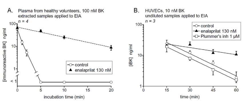
Pharmaceuticals 2020, 13, 201                                                                                            2 of 14

 Lys-BK to produce Lys-des-Arg9 -BK (des-Arg10 -kallidin), the only subnanomolar affinity agonist of the
 human form of the B1 receptor [5] (Figure 1). A fact that is often overlooked is that des-Arg9 -BK, a very
potent agonist of the B1 receptor in rodent species, has a very weak affinity for the human B1 receptor;
 thus, this receptor subtype can be said to be compartmentalized with one of the two kinin-generating
 system, tissue kallikrein (KLK-1). The release and maturation of KLK-1 are not well understood, but
 they are known to have roles in different tissues, namely intrarenal hemodynamics, flow-dependent
vasodilation and other compensatory mechanisms. They also have a role in inflammation, notably in
Pharmaceuticals 2020, 13, x FOR PEER REVIEW                                                         3 of 14
 the airways and gut [4,6–8].

                           PLG mutation
   plasminogen
              tPA                          SERPING1 mutations
                         C1-INH
     plasmin                               F12 mutations
                            FXII                                       prekallikrein
                                           contact system                                   C1-INH
           C1-INH
                            FXIIa                             plasma-kallikrein
                                        Kontact-APTT
                                                                                                       N
                                          stimulation                                              C
                                                                                                              HK

                                                                  KNG1 mutation
                               N
                           C
                                      LK

                          KLK-1
                                                 Lys-BK                                            inactive
                                                                                 BK                fragments
                     N             Lys-des-Arg9-BK                 N
                                                                                 N
                                                                                                    enalaprilat

           B1R       C
                                                            B2R    C
                                                                                 ACE        C

     Figure 1. Schematic representation of the kallikrein-kinin system, featuring the 2 validated pathways
      Figure 1. Schematic representation of the kallikrein-kinin system, featuring the 2 validated pathways
     of
      ofkinin
         kiningeneration,
               generation,thatthatof
                                   ofplasma
                                      plasmakallikrein
                                                kallikrein(part
                                                            (partof
                                                                  ofthe
                                                                      thecontact
                                                                           contactsystem)
                                                                                    system)releasing
                                                                                             releasingbradykinin
                                                                                                          bradykinin(BK)
                                                                                                                      (BK)
     from  high molecular    weight    kininogen   (HK)   and  that mediated    by  secreted  tissue  kallikrein
      from high molecular weight kininogen (HK) and that mediated by secreted tissue kallikrein (KLK-1),         (KLK-1),
     generating
      generating Lys-BK     mainlyfrom
                  Lys-BK mainly      fromlowlowmolecular
                                                 molecular    weight
                                                           weight       kininogen
                                                                    kininogen       (LK).
                                                                                (LK). SomeSome
                                                                                            of the ofknown
                                                                                                      the known   mutant
                                                                                                             mutant  genes
     genes   that  cause    HAE-nC1-INH        (dark   red   background      with    dotted   arrows
      that cause HAE-nC1-INH (dark red background with dotted arrows pointing to the corresponding      pointing   to the
     corresponding
      mutant proteins) mutant
                          and theproteins)    and the
                                   standardized         standardized
                                                   stimuli                stimuli
                                                            applied in vitro       applied
                                                                               to trigger    in vitro
                                                                                          kinin          to trigger
                                                                                                  formation          kinin
                                                                                                              (pale orange
     formation
      background) (pale   orange
                     are also       background)
                               indicated.  Elementsare    also
                                                      of the    indicated.
                                                             contact   systemElements     of theincontact
                                                                               are represented                system
                                                                                                     light blue.       are
                                                                                                                 Black and
     represented    in light  blue.  Black   and  blue   arrows   represent    biochemical    reactions.
      blue arrows represent biochemical reactions. Green arrows indicate an agonist effect on a receptor.  Green   arrows
     indicate an agonist
     “⊥” Inhibition      effect on or
                    of a protease     peptidase.“⊥”
                                   a receptor.       Inhibition
                                                 See Table       of a protease or peptidase. See Table 1 for
                                                           1 for abbreviations.
     abbreviations.

     The other kinin-generating system revolves around plasma kallikrein and its associated
components in the contact system (Figure 1, represented in light blue). HK is bound to the zymogen
prekallikrein and to coagulation factor XI (FXI) via specific domains. This complex may be loosely
bound to sites present on endothelial cells [1] but has a natural affinity for negatively charged
surfaces, such as the denuded basal membrane of damaged endothelium, sodium urate crystals,
platelet delta granule insoluble polyphosphates (see below), and in the laboratory, glass, kaolin, and
dextran sulfate. Soluble factor XII (FXII) also has an affinity to such abnormal surfaces: it and
prekallikrein mutually activate each other into active proteases, factor XIIa (FXIIa) and plasma
kallikrein, respectively. The latter releases the vasoactive mediator BK from HK and FXIIa initiates
the intrinsic coagulation pathway via FXI activation and subsequent reactions. BK is a high affinity
agonist of the B2 receptor, found in endothelial cells, sensory neurons, some epithelia and other cell
types [5]. The most immediate vascular effect of kinins is vasodilation, mediated by the endothelial
Pharmaceuticals 2020, 13, 201                                                                           3 of 14

                                       Table 1. List of abbreviations.

                     Abbreviation                                   Standing for
                          ACE                            angiotensin-I converting enzyme
                           AE                                         angioedema
                         APTT                          activated partial thromboplastin time
                          B1 R                                  bradykinin B1 receptor
                          B2 R                                  bradykinin B2 receptor
                           BK                                          bradykinin
                       C1-INH                                    C1-esterase inhibitor
                         c-Fos                                   a transcription factor
                          EIA                                   enzyme immunoassay
                       ERK1/2                      extracellular signal-regulated kinases 1 and 2
                          F12                                  gene encoding factor XII
                          FDA                           U.S. Food and Drug Administration
                          FXI                                    coagulation factor XI
                          FXII                                  coagulation factor XII
                         HAE                                   hereditary angioedema
                     HAE-C1-INH                         HAE caused by SERPING1 variants
                      HAE-FXII                              HAE caused by F12 variants
                      HAE-PLG                                HAE caused a PLG variant
                          HK                             high molecular weight kininogen
                        HPLC                        high-performance liquid chromatography
                       HUVEC                           human umbilical vein endothelial cell
                          iBK                               immunoreactive bradykinin
                           KD                              binding dissociation constant
                        KLK-1                                       tissue kallikrein
                        KNG1                                  gene encoding kininogens
                    Kontact-APTT™             a commcercial reagent that activates the contact system
                           LK                            low molecular weight kininogen
                        Lys-BK                                lysyl-bradykinin, kallidin
                            PI                                     propidium iodide
                          PLG                               gene encoding plasminogen
                         s.e.m.                              standard error of the mean
                           t1/2                                          half-life
                          tPA                               tissue plasminogen activator
                     VE-cadherin                           vascular endothelial cadherin

     The other kinin-generating system revolves around plasma kallikrein and its associated
components in the contact system (Figure 1, represented in light blue). HK is bound to the zymogen
prekallikrein and to coagulation factor XI (FXI) via specific domains. This complex may be loosely
bound to sites present on endothelial cells [1] but has a natural affinity for negatively charged surfaces,
such as the denuded basal membrane of damaged endothelium, sodium urate crystals, platelet
delta granule insoluble polyphosphates (see below), and in the laboratory, glass, kaolin, and dextran
sulfate. Soluble factor XII (FXII) also has an affinity to such abnormal surfaces: it and prekallikrein
mutually activate each other into active proteases, factor XIIa (FXIIa) and plasma kallikrein, respectively.
The latter releases the vasoactive mediator BK from HK and FXIIa initiates the intrinsic coagulation
pathway via FXI activation and subsequent reactions. BK is a high affinity agonist of the B2 receptor,
found in endothelial cells, sensory neurons, some epithelia and other cell types [5]. The most immediate
vascular effect of kinins is vasodilation, mediated by the endothelial production of nitric oxide and
prostanoids via calcium signaling, and disruption of the permeability barrier due to a contraction of the
endothelial cells and simultaneous Tyr-phosphorylation of vascular endothelial cadherin (VE-cadherin)
followed by its breakdown at the intercellular adherens junctions [9]. These effects are particularly
relevant to AE states. The fibrinolytic system, composed of plasminogen and its upstream activating
proteases, such as tissue plasminogen activator, are increasingly discussed in AE physiopathology.
Plasmin recruits the contact system via the cleavage of FXII [10] (Figure 1).
Pharmaceuticals 2020, 13, 201 4 of 14
The kallikreins are endogenously controlled by circulating serine protease inhibitors (serpins).
C1 esterase inhibitor (C1-INH; serpin G1) abates the effect of plasma kallikrein, but also of FXII,
plasmin and complement component C1 (Figure 1). Tissue kallikrein is inhibited by endogenous
kallistatin (serpin A4) [11]. Whole citrated blood was used in our initial work with iBK measurements
to verify previous claims of BK generation by the activation of neutrophil leukocytes or platelets, via
the secreted protease PR3 or NETosis in the first case, or secretory products such as polyphosphate
in the second [12]. Despite ample evidence of the activation of neutrophils and platelets following
relevant in vitro stimulation, no measurable production of iBK was evidenced. This could be explained
by the presence of the multiple endogenous protease inhibitors of plasma [12].
2. Bradykinin (BK)-Mediated Angioedema States
BK-mediated AE is a rare acquired or hereditary condition involving localized edema of subcutaneous
and submucosal tissues. This group of disorders may be life-threatening (e.g., via suffocation), very painful
and incapacitating. The mechanism is mast-cell independent and differential diagnosis must exclude various
allergic or urticarial conditions that are much more frequent [13]. The most common cause of acquired
AE is drug-induced, in the context of treatment with angiotensin-I converting enzyme (ACE) inhibitors
for cardiovascular conditions [14,15]. The basis of this disorder may be an insufficient clearance of BK,
as ACE is by far the major peptidase that inactivates kinins in blood/plasma/serum and in vivo [16–18].
However, what triggers kinin formation in affected patients is unclear [19]. Therapeutic fibrinolysis, with
recombinant tissue plasminogen activator (tPA) or comparable plasmin activators, is associated with AE or
anaphylactoid reactions. BK production initiated by plasmin-mediated FXII activation may be involved in
these cases [20,21]. Non-hereditary forms of BK-mediated AE include those associated with low levels of
C1-INH due to autoantibodies or overconsumption (lymphoproliferative disease) [22].
Hereditary angioedema (HAE), an autosomal dominant group of disorders, involves several gene
variants that are proven or postulated to be permissive for kinin production (Figure 1) [22]. Many
variants of the SERPING1 gene encoding C1-INH with impaired production (type I HAE-C1-INH)
or dysfunctional (type II HAE-C1-INH) forms of this serpin, cause the most common form of HAE.
Rarer forms of HAE with normal C1-INH levels are caused by mutation of genes encoding coagulation
FXII (F12), plasminogen (PLG), or of kininogens (KNG1) [22]. Some variants of FXII that are associated
with HAE-FXII introduce new sites of cleavage by plasmin that accelerate cleavage by this protease,
a plausible basis for a gain of function [23]. HAE associated with variants of the angiopoietin 1 gene
(ANGPT1) suggests a different general mechanism of the disease where the endothelial permeability
barrier is primarily affected [22,24]. However, for many patients, no mutation or abnormalities have as
yet found and they may differ in several aspects, including gender distribution, genetics, symptoms,
and estrogen impact.
Animal models of HAE-C1-INH do not fully recapitulate the human disease. Both homozygous
and heterozygous C1-INH-deficient mice failed to exhibit edema attacks, but nevertheless had an
increased basal microvascular permeability mediated by plasma kallikrein and the BK B2 receptor [25].
It has been argued that endothelium-specific overexpression of B2 receptors in rats, under the promoter
of VE-cadherin, resulted in a more representative model because localized BK-dependent plasma
extravasation could be triggered by the application of an irritant and spontaneous abdominal edema
was occasionally observed [26].
Ultimately, various forms of AE attacks involve the excessive stimulation of the endothelial BK B2
receptor and ensuing increased microvascular permeability. The only FDA-approved clinical use of
a BK receptor ligand is that of icatibant, an injectable peptide antagonist of the B2 receptor, to abort
attacks of HAE-C1-INH. C1-INH replenishment and various strategies to inhibit plasma kallikrein,
FXII or fibrinolysis have also been or are being developed [27]. An orally bioavailable small molecule
antagonist of the BK B2 receptor is also at an early stage of development [28].Pharmaceuticals 2020, 13, x FOR PEER REVIEW 5 of 14
FXII or fibrinolysis have also been or are being developed [27]. An orally bioavailable small molecule
antagonist of the BK B2 receptor is also at an early stage of development [28].
Pharmaceuticals 2020, 13, 201 5 of 14
3. Antibody-Based Analytical Techniques for Kinins: Importance and Pitfalls
BK and related
3. Antibody-Based peptides Techniques
Analytical are formed for
locally closeImportance
Kinins: to their site of Pitfalls
and action (autacoids). They are
extremely unstable and prone to artefactual formation when blood or tissues are sampled (for
BK and related peptides are formed locally close to their site of action (autacoids). They are
instance, following contact of blood with glass). Therefore, measuring kinins in venous blood is not
extremely unstable and prone to artefactual formation when blood or tissues are sampled (for instance,
really informative [3], contrasting with many useful peptide hormone dosages applied in
following contact of blood with glass). Therefore, measuring kinins in venous blood is not really
endocrinology. A further challenge in concentration determination is that the BK sequence is
informative [3], contrasting with many useful peptide hormone dosages applied in endocrinology.
embedded in that of precursor kininogens, with necessary separation if an antibody-based detection
A further challenge in concentration determination is that the BK sequence is embedded in that of
system is considered. Blais et al. [3] have reviewed these pitfalls and discussed extraction procedures
precursor kininogens, with necessary separation if an antibody-based detection system is considered.
and recovery in the context of the currently applied immunoassays. Professor A. Adam and
Blais et al. [3] have reviewed these pitfalls and discussed extraction procedures and recovery in the
co-workers developed separate competitive chemiluminescence enzyme immunoassays (EIAs) for
context of the currently applied immunoassays. Professor A. Adam and co-workers developed separate
BK and des-Arg9-BK based on digoxygenin-conjugated peptides [29,30]. Now several commercially
competitive chemiluminescence enzyme immunoassays (EIAs) for BK and des-Arg9 -BK based on
available kits are being used, such as that from Phoenix Pharmaceuticals for BK (96-well plate
digoxygenin-conjugated peptides [29,30]. Now several commercially available kits are being used,
format), and these have the advantage of world-wide reproducibility. The sensitivity of EIAs is
such as that from Phoenix Pharmaceuticals for BK (96-well plate format), and these have the advantage
excellent. Typical of peptide tracers with N-terminal extensions (digoxygenin, biotin), the polyclonal
of world-wide reproducibility. The sensitivity of EIAs is excellent. Typical of peptide tracers with
anti-BK antibodies react with only a few C-terminal amino acids in the kinin sequences; thus
N-terminal extensions (digoxygenin, biotin), the polyclonal anti-BK antibodies react with only a few
N-terminally extended sequences like Lys-BK and Ile-Ser-BK fully cross-react in the BK EIA, but not
C-terminal amino acids in the kinin 9sequences; thus N-terminally extended sequences like Lys-BK
the C-terminally truncated des-Arg -BK [29]. Similarly, Phoenix Pharmaceuticals states that its BK
and Ile-Ser-BK fully cross-react in the BK EIA, but not the C-terminally truncated des-Arg9 -BK [29].
EIA fully cross-reacts with Lys-BK or biotinyl-BK, but not with des-Arg -BK. Using this assay, we
9
Similarly, Phoenix Pharmaceuticals states that its BK EIA fully cross-reacts with Lys-BK or biotinyl-BK,
observed that the N-terminally extended sequence protected from aminopeptidases, D-Arg-BK, is
but not with des-Arg9 -BK. Using this assay, we observed that the N-terminally extended sequence
also equipotent with BK in the EIA, but that the C-terminally extended peptide D-Arg-BK-Arg-Arg
protected from aminopeptidases, D-Arg-BK, is also equipotent with BK in the EIA, but that the
loses much affinity, consistent with the antibody preference for an intact C-terminal BK sequence
C-terminally extended peptide D-Arg-BK-Arg-Arg loses much affinity, consistent with the antibody
(Figure 2).
preference for an intact C-terminal BK sequence (Figure 2).
Figure 2. Cross-reactivity of the BK EIA with selected kinin sequences. The inactive metabolite
Figure 2. Cross-reactivity of the BK EIA with selected kinin sequences. The inactive metabolite
des-Arg1 -BK (= BK fragment 2–9) exhibits full cross-reactivity vs. BK. N-terminal extended sequences,
des-Arg1-BK (= BK fragment 2–9) exhibits full cross-reactivity vs. BK. N-terminal extended
like Arg-BK (shown here), or Lys-BK are also fully reactive. Truncated sequences at the C-terminus,
sequences, like Arg-BK (shown here), or Lys-BK are also fully reactive. Truncated sequences at the
like the optimal B1 receptor agonist Lys-des-Arg9 -BK, do not cross9react at all [12]. An extended sequence
C-terminus, like the optimal B1 receptor agonist Lys-des-Arg -BK, do not cross react at all [12]. An
like Arg-BK-Arg-Arg, an inactive prodrug releasing active kinins via the action of carboxypeptidases [31],
extended sequence like Arg-BK-Arg-Arg, an inactive prodrug releasing active kinins via the action of
has a partial reactivity, showing the importance of a free COOH terminus. Reproduced in part from [12].
carboxypeptidases [31], has a partial reactivity, showing the importance of a free COOH terminus.
AReproduced
particular in part from
concern [12].full EIA reactivity of the BK fragment des-Arg1 -BK (Figure 2), and
is the
possibly of other N-terminally truncated BK fragment, again supporting that the antibodies recognize
only a limited C-terminal sequence. Des-Arg1 -BK is inactive as an agonist of either the B1 or B2
receptors [32] (Figure 2). Thus, the EIA signal may be contaminated with the inert metabolite generatedPharmaceuticals 2020, 13, 201 6 of 14
by aminopeptidase P, a significant peptidase that inactivates BK [17]. One way out is to couple the
EIA quantification with chromatographic separation of kinins, such as HPLC [29]. Another approach
is to verify the presence of BK-like activity in extracts using bioassays. Thus, Adam et al. [33] used
ERK1/2 signaling in cells expressing rabbit B2 receptors to confirm iBK in extracts of plasma stimulated
with contaminated heparin. Later, the accumulation of c-Fos in HEK 293a cells expressing the human
B2 receptor was successfully used as a discriminative bioassay to confirm measurements of iBK in
extracts of human blood or plasma [12,18]. Lys-BK and BK are approximately equipotent as agonists
of the B2 receptor, so they add up in EIA results as “iBK” and in bioassay results as well, if either one
or both are present in a given extract. Similarly, the published des-Arg9 -BK EIA, based on the tracer
digoxygenin-des-Arg9 -BK, was equally reactive with Lys-des-Arg9 -BK [30].
4. Probing Immunoreactive BK Degradation In Vitro
In vivo assessment of the relative importance of kininases in BK inactivation in rats, based on
specific peptidase inhibitors, has shown the major role of ACE and the significant role of aminopeptidase
P and neutral endopeptidase, but dipeptidyl peptidase IV was not quantitatively important [17].
Cyr et al. [16] used both BK and des-Arg9 -BK EIAs to assess the half-life of both synthetic peptides
in human serum. The half-life of BK was 27 sec, ACE accounting for most of the clearance activity,
as the t1/2 value increased nine-fold in the presence of the ACE inhibitor enalaprilat. We essentially
confirmed these results using the commercial EIA, a lower initial BK concentration and human
plasma [18] (Figure 3A). Arginine carboxypeptidases (kininase I), producing des-Arg9 -BK from BK,
was a very minor pathway of metabolism (11%), but more important when ACE was blocked (50%) [16].
The inactivation of des-Arg9 -BK is also mainly mediated by ACE (t1/2 doubled in the presence of
enalaprilat). Aminopeptidase P is felt to be important in the degradation of kinins, but this cannot be
Pharmaceuticals 2020, 13, x FOR PEER REVIEW 7 of 14
ascertained using the EIAs as the removal of the Arg1 residue is not detected by the exploited antibodies.
Figure
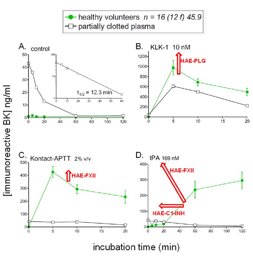
Figure 3. 3.
(A)(A) Degradationofof
Degradation syntheticBK
synthetic BK (100
(100 nM)
nM) added
added toto human
human plasma
plasma from
from healthy
healthy subjects
subjects inin
the
the presence
presence or absence
or absence of theinhibitor
of the ACE ACE inhibitor enalaprilat.
enalaprilat. Samples Samples
were incubated at 37 ◦ Catunder
were incubated 37 °Cagitation
under
foragitation for thetime
the indicated indicated
periodstime periods
before beforeand
extraction extraction and EIA determination
EIA determination of BK. The
of BK. The number number
of replicated
of replicated
from from different
different healthy donorshealthy donorsby
is indicated is “n”.
indicated
In thebypresence
“n”. In the
of presence
the ACE of the ACEthe
inhibitor, inhibitor,
data are
the data are
compatible withcompatible
a first orderwith
decaya first
withorder decay
a half-life of with a half-life
7.1 min, whereasofBK 7.1was min, whereas cleared
completely BK wasin
completely
5 min the absencecleared in 5 min (tthe
of enalaprilat absence of enalaprilat (t1/2 34 s). Reproduced from [18]. (B)
1/2 34 s). Reproduced from [18]. (B) Catabolism of BK, measured as
[iBK], by intact HUVECs as a function ofbytime.
Catabolism of BK, measured as [iBK], intactValues
HUVECs as a function
are means ± s.e.m.ofof time. Values are means
3 determinations. ±
A first
s.e.m. of 3 determinations. A first order decay model has been applied (t 1/2 values reported in main
order decay model has been applied (t1/2 values reported in main text). BK added to similar wells
text). BK
without cellsadded
(nudeto similar
plastic) waswells without
stable cells (nudeinplastic)
when incubated was stable
an identical manner when
(dataincubated
not shown).in an
identical manner (data not shown).
ACE activity in serum or blood plasma corresponds, in healthy individuals, to a form of the protein
shed from the vascular endothelial cells in an apparently regulated manner [34]. ACE, membrane
arginine carboxypeptidase (carboxypeptidase M), aminopeptidase P and other potential kininasesPharmaceuticals 2020, 13, 201 7 of 14
are expressed at the surface of vascular endothelial cells. Primary cultures of human umbilical
vein endothelial cells (HUVECs) are a standard model for which significant information is available.
These cells express at their surface a high number of ACE molecule, as judged from the saturable
binding of [3 H]enalaprilat binding (KD 0.23 nM) [35] and this binding was displaced by BK and
Lys-BK, but much less efficiently by their des-Arg9 forms [36]. Based on a radioimmunoassay of
BK, adherent HUVECs maintained at 37 ◦ C without serum cleared BK (10 ng/mL) with a half-life of
about 50 min. The ACE inhibitor was reported to halt completely the iBK breakdown, despite a minor
Figure 3.of(A)
contribution Degradation
neutral of synthetic
endopeptidase BK Using
[37]. (100 nM)theadded to human
Phoenix plasma from EIA,
Pharmaceuticals healthy
wesubjects in
have verified
the presenceoforBK
the degradation absence of the
(10 nM) ACE inhibitor
by HUVECs (see enalaprilat.
Materials andSamples were for
Methods incubated at 37 °C under
the procedure). The cells,
kept agitation for the indicated
under agitation, remainedtimeadherent
periods before extraction
and intact and EIA
(Figure 4), determination
which is importantof BK. The number
because lyzed
of replicated from different healthy donors is indicated by “n”. In the presence
HUVECs degrade BK more rapidly by unknown pathways [37]. iBK was cleared with a t1/2 of 12.9 min, of the ACE inhibitor,
the was
and this datalargely
are compatible withtoaACE
attributable first as
order decay with
enalaprilat a half-life
increased the tof 7.1 min, whereas BK was
1/2 value to 47.2 min (Figure 3B).
completely
Plummer’s clearedthat
inhibitor, in blocks
5 min arginine
the absence of enalaprilat (t1/2such
carboxypeptidases 34 s).
as Reproduced
carboxypeptidasefrom [18]. (B) not
M, did
Catabolism of BK, measured as [iBK], by intact HUVECs as a function of time. Values
exert a significant effect on the system (iBK t1/2 12.3 min). The initial recovery of BK was apparently are means ±
s.e.m. of 3 determinations. A first order decay model has been applied (t1/2 values reported in main
somewhat higher than the added amount of BK in the physiological fluid (10 nM), an artefact possibly
text). BK added to similar wells without cells (nude plastic) was stable when incubated in an
due to interference from some proteins released from the cells as the supernatants were directly applied
identical manner (data not shown).
to the EIA without extraction.
Figure 4.4.Effect
Figure Effectofofincubation
incubationunder
under agitation
agitation in Hank’s
in Hank’s balanced
balanced salt salt solution
solution containing
containing BK nM)
BK (500 (500
nM)
on on adherent
adherent HUVECs HUVECs
(matched(matched phaseand
phase contrast contrast and red fields,
red fluorescence fluorescence
originalfields, original
magnification
magnification
100×). Propidium 100×). Propidium
iodide iodide
(PI) staining was(PI) stainingaswas
performed a testperformed
of viabilityas(positive
a test of viability
control, (positive
Triton X100,
control, Triton X100, 0.5%). Total counted cell numbers are
0.5%). Total counted cell numbers are given next to the histograms. given next to the histograms.
Human healthy subjects who took two daily doses of enalapril maleate, the prodrug of enalaprilat,
exhibited a ~5-fold increase of BK t1/2 added ex vivo to their plasma relative to the pre-treatment
value [18]. Altogether, the findings illustrate that BK is highly unstable and may contribute to the
acquired AE associated with ACE inhibitors if, for some reason, important generation of kinins isPharmaceuticals 2020, 13, 201 8 of 14
triggered. The development of double peptidase inhibitors for ACE and neutral endopeptidase
(neprilysin) is still being contemplated as an anti-hypertensive after the failure of omapatrilat,
which caused relatively frequent AE attacks [38]. The possible importance of the rapid clearance of BK
for patients with HAE is the protection from the continuous production of BK, as judged from HK
consumption and kallikrein activity (see below).
Years ago, it has been found that BK can be metabolized by two cycles of ACE catalytic activity,
successively producing the fragments BK1–7 , then BK1–5; they are both inactive on B1 or B2 receptors [39].
The possible interest of BK1–5 is that it is more stable than most kinins and can be detected in clinical
samples, using mass spectrometry for instance [40]. Thus, changes in BK1–5 plasma concentrations
constitute a more discriminative proof of kallikrein–kinin system activation than those of BK in patients
undergoing cardiopulmonary bypass, whether or not ACE was pharmacologically blocked [40].
5. Profiling Immunoreactive BK Generation In Vitro: Modeling Hereditary Angioedema (HAE)
The main thrust of our investigations based on iBK measurements was the investigation of the
physiopathology of different forms of HAE to detect excessive kinin formation. This point of view is
different from that of other laboratories that evidenced continuous HK consumption or enzymatic
activity of plasma kallikrein in HAE dependent or not on C1-INH deficiency during remission [41–43],
suggesting an inherently unstable contact system. To establish profiles of iBK formation, citrated blood
or plasma obtained from healthy volunteers or HAE patients in remission was incubated at 37 ◦ C
under agitation for up to 2 h, alone or in the presence of one of three standardized stimuli (Figure 1):
the particulate material Kontact-APTT™ to activate the contact system, human recombinant KLK-1
to probe the alternate kinin generation pathway, and tPA, known to release iBK in vitro with a slow
kinetics [20].
A perhaps unexpected result was that the low background of iBK formation observed in control
plasma in the absence of stimulant is not modified in patients with HAE-C1-INH, HAE-FXII or
HAE-PLG [18,44] (Figure 5A). HAE patients are not symptomatic most of the time: the buffering action
of peptidases and protease inhibitors may protect patients from a possible low-grade BK formation
during remissions. The plasma or whole blood from HAE patients with C1-INH deficiency showed
only one consistent change relative to control iBK formation profiles: in vitro stimulation with tPA
considerably accelerated the slow formation of iBK seen in controls [12,44]. The use of specific inhibitors
showed that this accelerated response was dependent on the activities of plasmin, FXII and, ultimately,
plasma kallikrein. Thus, HAE-C1-INH plasma exhibits a form of hypersensitivity to fibrinolysis
which may be related to several of the known triggering factors of attacks, such as mechanical trauma,
surgery, infection, menstruation, physical exertion, etc., but also mental stress, the second most frequent
triggering factor [45]. Indeed, a whole body of evidence has shown that coagulation and fibrinolysis
are significantly activated by mental stress in a healthy subject [46].
HAE-FXII plasma exhibited an even more explosive early response to tPA in vitro [18] (Figure 5),
consistent with the very rapid cleavage and activation of the T328K and T328R variants of FXII by
plasmin [23]. HAE-FXII essentially affects females in an estrogen-dependent manner and males may
carry and transmit the mutation, but are not symptomatic [47]. However, the hormonal status had no
obvious influence on the effect of tPA on iBK formation, as the plasma of post-menopausal females have
the same profiles [18] and estrogen could rather alter endothelial biology. The plasma of HAE-FXII
patients also responds significantly more to the direct contact system activator Kontact-APTT™ in a
manner that is not dependant on plasmin [18], suggesting a somewhat increased activity of the variant
T328K of FXII variant in the mutual activation reaction with plasma kallikrein.
In their seminal paper about the discovery of the K330E variant of plasminogen, Bork et al. [48]
noted that the mutant form of plasmin has a normal catalytic activity, and the idiosyncratic interaction
of the mutant plasminogen with another unknown protein was raised as a hypothesis. In agreement
with this, the profile of iBK generation under tPA stimulation was not different from that of healthy
volunteers [18], although urokinase plasminogen activator has recently been reported to generateconnection is that KLK-1 is very abundant in the salivary glands and the catalytically active form of
this protease is found in human saliva [50]. Thus, the generally overlooked kinin-forming KLK-1
may be responsible, at least in part, for HAE-PLG. It has been recently reported that the
administration of C1-INH concentrates has only a variable therapeutic effect in HAE-PLG and that
some patients
Pharmaceuticals are13,completely
2020, 201 refractory [51], possibly supporting a role for kinin generation outside
9 of 14
the contact system in this form of the disease.
One stored plasma sample that was received in our laboratory was partly clotted and excluded
increased plasmin activity from the mutant form [49]. The only significantly different iBK profile
from our previous studies. However, submitting the sample to in vitro incubation and stimulation
in HAE-PLG was an increased effect of KLK-1 at the early incubation time 5 min, an unexpected
produced revealing results about the functioning of the contact system (Figure 5, overlaid curves).
finding suggesting interaction of the mutant plasminogen with KLK-1 (facilitatory) or with its serpin
Firstly, the unstimulated sample contained at time zero a sizeable iBK concentration that decreased
inhibitor kallistatin (inhibitory interaction) [18]. This has to be experimentally confirmed, but may
as a function of time with a t1/2 of 12.3 min (Figure 5A, inset), marginally superior to the value of 7.1
be related to the clinical presentation of HAE-PLG that very often involves the swelling of the lips,
min for the t1/2 measured for exogenous BK in the presence of enalaprilat (Figure 3A). Secondly, the
tongue and contiguous tissues such as the face and larynx. The possible connection is that KLK-1
partly clotted plasma sample released very little iBK in response to Kontact-APTT™ or tPA (Figure
is very abundant in the salivary glands and the catalytically active form of this protease is found in
5C,D), suggesting a major depletion of contact system components. Finally, KLK-1 released
human saliva [50]. Thus, the generally overlooked kinin-forming KLK-1 may be responsible, at least in
important quantities of iBK from the partly clotted sample (Figure 5B), within the range of healthy
part, for HAE-PLG. It has been recently reported that the administration of C1-INH concentrates has
volunteers (superior to those measured in three of the 16 healthy donors), supporting that KLK-1
only a variable therapeutic effect in HAE-PLG and that some patients are completely refractory [51],
and LK are part of a separate kallikrein–kinin system.
possibly supporting a role for kinin generation outside the contact system in this form of the disease.
Figure 5. Kinetics of iBK concentrations as a function of time and stimulation in samples of normal
plasma incubated at 37 ◦ C in the presence of enalaprilat (130 nM). Panels represent different forms of
in vitro stimulation added to plasma at time zero: (A) control; (B) KLK-1 10 nM; (C) Kontact-APTT™ 2%
v/v; (D) tPA 169 nM. The data is from the fusion of the 2 control groups (healthy volunteers) from [18,44].
The average age is 45.9 and 12 females are represented in this group of 16 subjects. Indications of
significant variations induced by specific HAE subtypes are indicated by red arrows. Measurements
made from a partly clotted plasma sample are also shown: in the presence of enalaprilat but no other
stimulant, the sizeable concentration of BK at time zero decreases according to a first-order elimination
kinetics (panel A, t1/2 12.3 min, inset).
One stored plasma sample that was received in our laboratory was partly clotted and excluded
from our previous studies. However, submitting the sample to in vitro incubation and stimulation
produced revealing results about the functioning of the contact system (Figure 5, overlaid curves).
Firstly, the unstimulated sample contained at time zero a sizeable iBK concentration that decreased as
a function of time with a t1/2 of 12.3 min (Figure 5A, inset), marginally superior to the value of 7.1 min
for the t1/2 measured for exogenous BK in the presence of enalaprilat (Figure 3A). Secondly, the partly
clotted plasma sample released very little iBK in response to Kontact-APTT™ or tPA (Figure 5C,D),Pharmaceuticals 2020, 13, 201 10 of 14
suggesting a major depletion of contact system components. Finally, KLK-1 released important
quantities of iBK from the partly clotted sample (Figure 5B), within the range of healthy volunteers
(superior to those measured in three of the 16 healthy donors), supporting that KLK-1 and LK are part
of a separate kallikrein–kinin system.
6. Areas of Uncertainty: Role of BK B1 Receptors and Tissue Kallikrein (KLK-1) in
Angioedema States
The BK B1 receptor is a peculiar G protein-coupled receptor that is strongly regulated at the
transcriptional level by tissue injury and cytokines [5]. Its selectivity to kinin metabolite generated by
arginine carboxypeptidases has been described above. There has been much speculation about the
participation of B1 receptors in attacks of AE. Let us note first that the B1 receptor is not a receptor
for peptides generated by the classical contact system, BK or des-Arg9 -BK (see above). This has been
experimentally verified: the extracts of whole blood stimulated with Kontact-APTT™ contained high
levels of iBK that were active on the recombinant B2 receptor (cFos signaling), but the extracts were not
active on the recombinant B1 receptor in replicated and controlled experiments [12]. As mentioned
above, the human B1 receptor is functionally compartmentalized with tissue kallikrein (KLK-1) that
produces Lys-BK and, secondarily, Lys-des-Arg9 -BK. Thus, B1 receptor participation in HAE forms
driven by the contact system may be less important than what several authors have described.
An elaborate in vitro simulation of the effect of HAE-C1-INH plasma on human endothelial cells
(HUVECs) has been reported [52]. A kinin content in HAE-C1-INH plasma obtained during attacks
has been postulated on the basis of an endothelial cell permeability assay based on a Transwell model:
the plasma permeabilizing effect was partially inhibited by B2 receptor or B1 receptor antagonists,
and was totally prevented by the mixture of the two antagonists. As opposed to these observations,
(1) the expression of B1 receptors by vascular cells cultured on plastic in serum is not surprising and is
largely an artefact (for HUVECs notably) [53]; (2) what was transferred from patient’s plasma to wells
to activate B1 receptors? Not necessarily peptides. Perhaps secondarily released KLK-1, explaining a
stimulation of the B1 receptors? Some other protease? Exquisitely specific biotechnological inhibitors
for several proteases now exist to address such questions. Thus, a parsimony principle may be invoked
to state that there is no proof on B1 receptor participation in attacks of HAE-C1-INH, until proper
clinical evidence is collected.
We described above the intriguing observation about KLK-1 potentiation in HAE-PLG plasma.
The highly efficient KLK-1/LK kallikrein–kinin system (Figures 1 and 3B) has been overlooked in
AE states and may be worth considering. KLK-1 is highly concentrated in the salivary glands, and
the oral/facial manifestations of AE are frequent in HAE-PLG, but also in the acquired AE state
associated with ACE inhibitors [19]. KLK-1 mediation has the potential to include the B1 receptor in
the mediation of vascular effects, as discussed above. The efficacy of icatibant, a selective B2 receptor
antagonist, is controversial in ACE inhibitor-induced AE [54], perhaps in line with this. Although
synthetic des-Arg10 -icatibant is a potent B1 receptor antagonist [5], it is not a spontaneous metabolite
of icatibant due to the non-natural residues in position 7 and 8 (D-Tic7 , Oic8 ) that preclude the binding
to arginine-carboxypeptidases. The in vivo metabolites of icatibant are well known and represent
fragments 1–5 and 7–10 of this decapeptide [55].
7. Conclusions
The systematic study of BK generation and clearance is an original point of view to investigate
the “weak spots” that may trigger attacks of BK-mediated AE states, the natural detoxifying action
of kininases being considerable. Inhibition of such a clearance mechanism is likely involved in the
acquired AE associated with ACE inhibitors. In these investigations, we stayed as close as possible to
the in vivo condition where protease inhibitors abound. All our HAE patients were seen in remission
in order to isolate excessive responses of the kallikrein–kinin system to one of the standardized
stimuli. HAE-C1-INH and HAE-FXII plasmas are both hyperresponsive to fibrinolysis activation.Pharmaceuticals 2020, 13, 201 11 of 14
Other intriguing observations suggest a possible role of the alternate KLK-1/LK kallikrein–kinin system
in specific forms of AE. However, the present approach says little about important local endothelial
factors that may modulate HAE states.
8. Materials and Methods (for Original Results)
8.1. Degradation of BK by Human Endothelial Cells
The local ethics committee (CHU de Québec-Université Laval) had approved the anonymous use of
human umbilical cord segments obtained after normal deliveries (File no. 2017-3720). Primary cultures
of endothelial cells from the human umbilical vein (HUVECs) were obtained and propagated as
described [53]. These cells were characterized in immunofluorescence experiments by their morphology
and expression of von Willebrand factor using primary antibodies from Dako (polyclonals) and the
appropriate fluorophore-conjugated antibodies.
HUVECs were cultured until confluency in 6-well plates; the culture medium was then removed,
rinsed and refilled (3 mL/well) with preheated (37 ◦ C) Hank’s balanced salt solution (pH 7.4). Synthetic
BK (final concentration 10 nM) was added, optionally with an enzyme inhibitor, and the plates were
incubated for up to 60 min in a warmed chamber (37 ◦ C) under continuous oscillating agitation.
The cells remained viable for 2 h (see below). Control plates without cells (nude plastic) were used as
controls. After the incubation, 300 µL fluid from each well was removed, added to 1.5 mL ice-cold
ethanol and the mixture was further incubated on ice for 1 h. At the end, each tube was centrifuged
and the supernatants set aside in a new set of tube submitted to complete evaporation in a SpeedVac
apparatus and preserved at −80 ◦ C. The dried residues were later resuspended in 300 µL with the
supplied enzyme immunoassay (EIA) buffer; undiluted 50 µL samples were directly applied in
duplicate to the BK EIA as recommended by the manufacturer (Phoenix Pharmaceuticals, Burlingame,
CA; cat. no. EK-009-01; 96-well plate format).
In addition, a microscopic study of the morphology of HUVECs maintained in Hank’s balanced
salt solution containing 500 nM BK under agitation for 0–2 h was conducted. The study included a
staining with propidium iodide (2.5 µM, 4 min, room temperature) as a further test for endothelial
cell viability under the conditions applied to measure BK breakdown. The proportion of propidium
iodide stained nucleus (red fluorescence) was calculated from a large photographic record (100×).
Cells acutely treated with Triton X-100 (0.5%) were positive controls.
8.2. Generation and Degradation of BK by Human Plasma in Health and Disease
The local ethical review board (Comité d’éthique de la recherche, CHU de Québec-Université
Laval) granted ethical approval to carry out the study involving blood donations from adult healthy
volunteers and HAE patients 16 years old or older (files no. 2018-3857, 2020-4696). Citrated plasma was
obtained, processed, and extracted precisely as described [18,44] for the determination of synthetic BK
degradation and iBK generation under standardized in vitro stimulation. The same EIA was applied
to plasma extracts (1:100 or 1:1000) diluted with the assay buffer.
Author Contributions: F.M. wrote the first draft of the manuscript and input and advice was received from all
the other authors (H.B., X.C.-M., J.H., and G.E.R.). All authors have read and agreed to the published version of
the manuscript.
Funding: The published or original work from our laboratory has been supported by Shire, now part of Takeda
(Investigator-Initiated Research grants IIR-CAN-000902, to F.M. and G.E.R., and IIR-CAN-001615 to F.M., J.H.
and G.E.R), by Angio-Œdème Héréditaire du Québec (grant to F.M., H.B. and J.H.), by the Canadian Institutes
of Health Research (operating grant MOP-93773 to F.M, and H.B.) and by the Fonds de recherche Santé du Québec
(Studentship award to X.C.-M.).
Acknowledgments: We thank Johanne Bouthillier for technical help.
Conflicts of Interest: F.M. served as a consultant for Pharvaris B.V. and received research funds from Shire/Takeda
and Pharvaris B.V. G.E.R. has been a member of advisory boards (Baxalta, Bayer, Biogen Idec, CSL Berhing,
Novo Nordisk, Octapharma, Pfizer) and received funding from Bayer, CSL Behring and Pfizer (unrelated to thePharmaceuticals 2020, 13, 201 12 of 14
submitted work). J.H. has been a speaker/teacher for CLS Behring, Novartis, and Shire; he has been a member of
advisory committees (CLS Behring, Shire, and Novartis) and a clinical investigator for Merck (ALK), Stallergene,
Boehringer-Ingelheim, GlaxoSmithKline (GSK), Novartis, Sanofi, AstraZeneca, CLS Behring, Shire, Roche, and
Griffols (unrelated to the submitted work). The other authors have no conflict of interest to disclose. The funders
had no role in the design of the study; in the collection, analyses, or interpretation of data; in the writing of the
manuscript, or in the decision to publish the results.
References
1. Kaplan, A.P. The bradykinin-forming cascade: A historical perspective. Chem. Immunol. Allergy 2014, 100,
205–213. [PubMed]
2. Bhoola, K.D.; Figueroa, C.D.; Worthy, K. Bioregulation of kinins: Kallikreins, kininogens, and kininases.
Pharmacol. Rev. 1992, 44, 1–80. [PubMed]
3. Blais, C.; Marceau, F.; Rouleau, J.L.; Adam, A. The kallikrein-kininogen-kinin systems: Lessons from the
quantification of endogenous kinins. Peptides 2000, 21, 1903–1940. [CrossRef]
4. Alhenc-Gelas, F.; Bouby, N.; Girolami, J.P. Kallikrein/K1, Kinins, and ACE/Kininase II in homeostasis and in disease
insight from human and experimental genetic studies, therapeutic implication. Front. Med. (Lausanne) 2019, 6, 136.
[CrossRef]
5. Leeb-Lundberg, L.M.; Marceau, F.; Müller-Esterl, W.; Pettibone, D.J.; Zuraw, B.L. International Union
of Pharmacology. XLV. Classification of the kinin receptor family: From molecular mechanisms to
pathophysiological consequences. Pharmacol. Rev. 2005, 57, 27–77. [CrossRef]
6. Bergaya, S.; Matrougui, K.; Meneton, P.; Henrion, D.; Boulanger, C.M. Role of tissue kallikrein in response to
flow in mouse resistance arteries. J. Hypertens. 2004, 22, 745–750. [CrossRef]
7. Stadnicki, A.; Mazurek, U.; Plewka, D.; Wilczok, T. Intestinal tissue kallikrein-kallistatin profile in
inflammatory bowel disease. Int. Immunopharmacol. 2003, 3, 939–944. [CrossRef]
8. Sexton, D.J.; Chen, T.; Martik, D.; Kuzmic, P.; Kuang, G.; Chen, J.; Nixon, A.E.; Zuraw, B.L.; Forteza, R.M.;
Abraham, W.M.; et al. Specific inhibition of tissue kallikrein 1 with a human monoclonal antibody reveals a
potential role in airway diseases. Biochem. J. 2009, 422, 383–392. [CrossRef]
9. Bouillet, L.; Mannic, T.; Arboleas, M.; Subileau, M.; Massot, C.; Drouet, C.; Huber, P.; Vilgrain, I. Hereditary
angioedema: Key role for kallikrein and bradykinin in vascular endothelial-cadherin cleavage and edema
formation. J. Allergy Clin. Immunol. 2011, 128, 232–234. [CrossRef]
10. Kaplan, A.P.; Austen, K.F. A prealbumin activator of prekallikrein. II. Derivation of activators of prekallikrein
from active Hageman factor by digestion with plasmin. J. Exp. Med. 1971, 133, 696–712. [CrossRef]
11. Chao, J.; Chao, L. Biochemistry, regulation and potential function of kallistatin. Biol. Chem. Hoppe Seyler 1995,
376, 705–713. [PubMed]
12. Charest-Morin, X.; Hébert, J.; Rivard, G.É.; Bonnefoy, A.; Wagner, E.; Marceau, F. Comparing pathways of
bradykinin formation in whole blood from healthy volunteers and patients with hereditary angioedema due
to C1 inhibitor deficiency. Front. Immunol. 2018, 9, 2183. [CrossRef] [PubMed]
13. Maurer, M.; Magerl, M.; Metz, M.; Siebenhaar, F.; Weller, K.; Krause, K. Practical algorithm for diagnosing
patients with recurrent wheals or angioedema. Allergy 2013, 68, 816–819. [CrossRef] [PubMed]
14. Bezalel, S.; Mahlab-Guri, K.; Asher, I.; Werner, B.; Sthoeger, Z.M. Angiotensin-converting enzyme
inhibitor-induced angioedema. Am. J. Med. 2015, 128, 120–125. [CrossRef] [PubMed]
15. Aygören-Pürsün, E.; Magerl, M.; Maetzel, A.; Maurer, M. Epidemiology of Bradykinin-mediated angioedema:
A systematic investigation of epidemiological studies. Orphanet J. Rare Dis. 2018, 13, 73. [CrossRef] [PubMed]
16. Cyr, M.; Lepage, Y.; Blais, C.; Gervais, N.; Cugno, M.; Rouleau, J.L.; Adam, A. Bradykinin and
des-Arg9 -bradykinin metabolic pathways and kinetics of activation of human plasma. Am. J. Physiol.
Heart Circ. Physiol. 2001, 281, H275–H283. [CrossRef]
17. Fryer, R.M.; Segreti, J.; Banfor, P.N.; Widomski, D.L.; Backes, B.J.; Lin, C.W.; Ballaron, S.J.; Cox, B.F.;
Trevillyan, J.M.; Reinhart, G.A.; et al. Effect of bradykinin metabolism inhibitors on evoked hypotension in
rats: Rank efficacy of enzymes associated with bradykinin-mediated angioedema. Br. J. Pharmacol. 2008, 153,
947–955. [CrossRef]
18. Marceau, F.; Rivard, G.E.; Gauthier, J.M.; Binkley, K.; Bonnefoy, A.; Boccon-Gibod, I.; Bouillet, L.; Picard, M.;
Levesque, G.; Elfassy, H.L.; et al. Measurement of bradykinin formation and degradation in blood plasma:
Relevance for acquired angioedema associated with angiotensin converting enzyme inhibition and forPharmaceuticals 2020, 13, 201 13 of 14
hereditary angioedema due to factor XII or plasminogen gene variants. Front. Med. (Lausanne) 2020, 7, 358.
[CrossRef]
19. Hoover, T.; Lippmann, M.; Grouzmann, E.; Marceau, F.; Herscu, P. Angiotensin converting enzyme inhibitor
induced angio-oedema: A review of the pathophysiology and risk factors. Clin. Exp. Allergy 2009, 40, 50–61.
[CrossRef]
20. Molinaro, G.; Gervais, N.; Adam, A. Biochemical basis of angioedema associated with recombinant tissue
plasminogen activator treatment: An in vitro experimental approach. Stroke 2002, 33, 1712–1716. [CrossRef]
21. Gauberti, M.; Potzeha, F.; Vivien, D.; Martinez de Lizarrondo, S. Impact of bradykinin generation during
thrombolysis in ischemic stroke. Front. Med. (Lausanne) 2018, 5, 195. [CrossRef] [PubMed]
22. Marcelino-Rodriguez, I.; Callero, A.; Mendoza-Alvarez, A.; Perez-Rodriguez, E.; Barrios-Recio, J.;
Garcia-Robaina, J.C.; Flores, C. Bradykinin-mediated angioedema: An update of the genetic causes and the
impact of genomics. Front. Genet. 2019, 10, 900. [CrossRef] [PubMed]
23. de Maat, S.; Björkqvist, J.; Suffritti, C.; Wiesenekker, C.P.; Nagtegaal, W.; Koekman, A.; van Dooremalen, S.;
Pasterkamp, G.; de Groot, P.G.; Cicardi, M.; et al. Plasmin is a natural trigger for bradykinin production in
patients with hereditary angioedema with factor XII mutations. J. Allergy Clin. Immunol. 2016, 138, 1414–1423.
[CrossRef]
24. Bafunno, V.; Firinu, D.; D’Apolito, M.; Cordisco, G.; Loffredo, S.; Leccese, A.; Bova, M.; Barca, M.P.;
Santacroce, R.; Cicardi, M.; et al. Mutation of the angiopoietin-1 gene (ANGPT1) associates with a new type
of hereditary angioedema. J. Allergy Clin. Immunol. 2018, 141, 1009–1017. [CrossRef] [PubMed]
25. Han, E.D.; MacFarlane, R.C.; Mulligan, A.N.; Scafidi, J.; Davis, A.E., 3rd. Increased vascular permeability in
C1 inhibitor-deficient mice mediated by the bradykinin type 2 receptor. J. Clin. Investig. 2002, 109, 1057–1063.
[CrossRef] [PubMed]
26. Veronez, C.L.; Maghsodi, S.; Todiras, M.; Popova, E.; Rodrigues, A.F.; Qadri, F.; Pesquero, J.B.; Bader, M.
Endothelial B2-receptor overexpression as an alternative animal model for hereditary angioedema. Allergy
2019, 74, 1998–2002. [CrossRef] [PubMed]
27. Nicola, S.; Rolla, G.; Brussino, L. Breakthroughs in hereditary angioedema management: A systematic review
of approved drugs and those under research. Drugs Context. 2019, 8, 212605. [PubMed]
28. Lesage, A.; Gibson, C.; Marceau, F.; Ambrosi, H.-D.; Saupe, J.; Katzer, W.; Loenders, B.; Charest-Morin, X.;
Knolle, J. In vitro pharmacological profile of new small molecule bradykinin B2 receptor antagonists.
Front. Pharmacol. 2020, 11, 916. [CrossRef]
29. Décarie, A.; Drapeau, G.; Closset, J.; Couture., R.; Adam, A. Development of digoxigenin-labeled peptide:
Application to chemiluminoenzyme immunoassay of bradykinin in inflamed tissues. Peptides 1994, 5, 511–518.
30. Raymond, P.; Drapeau, G.; Raut, R.; Audet, R.; Marceau, F.; Ong, H.; Adam, A. Quantification of
des-Arg9 -bradykinin using chemiluminescence enzyme immunoassay: Application to its kinetic profile
during plasma activation. J. Immunol. Methods 1995, 180, 247–257. [CrossRef]
31. Bachelard, H.; Charest-Morin, X.; Marceau, F. D-Arg0 -bradykinin-Arg-Arg, a latent vasoactive bradykinin B2
receptor agonist metabolically activated by carboxypeptidases. Front. Pharmacol. 2018, 9, 273. [CrossRef]
[PubMed]
32. Regoli, D.; Barabé, J. Pharmacology of bradykinin and related kinins. Pharmacol. Rev. 1980, 32, 1–46. [PubMed]
33. Adam, A.; Montpas, N.; Keire, D.; Désormeaux, A.; Brown, N.J.; Marceau, F.; Westenberger, B. Bradykinin
forming capacity of oversulfated chondroitin sulfate contaminated heparin in vitro. Biomaterials 2010, 31,
5741–5748. [CrossRef]
34. Ehlers, M.R.W.; Gordon, K.; Schwager, S.L.U.; Sturrock, E.D. Shedding the load of hypertension:
The proteolytic processing of angiotensin-converting enzyme. S. Afr. Med. J. 2012, 102, 461–464. [CrossRef]
35. Morissette, G.; Couture, J.P.; Désormeaux, A.; Adam, A.; Marceau, F. Lack of direct interaction between
enalaprilat and the kinin B1 receptors. Peptides 2008, 29, 606–612. [CrossRef] [PubMed]
36. Koumbadinga, G.A.; Bawolak, M.T.; Marceau, E.; Adam, A.; Gera, L.; Marceau, F. A ligand-based approach
to investigate the expression and function of angiotensin converting enzyme in intact human umbilical vein
endothelial cells. Peptides 2010, 31, 1546–1554. [CrossRef]
37. Graf, K.; Gräfe, M.; Auch-Schwelk, W.; Baumgarten, C.R.; Bossaler, C.; Fleck, E. Bradykinin degrading
activity in cultured human endothelial cells. J. Cardiovasc. Pharmacol. 1992, 20 (Suppl. 9), S16–S20. [CrossRef]Pharmaceuticals 2020, 13, 201 14 of 14
38. Sharma, U.; Cozier, G.E.; Sturrock, E.D.; Acharya, K.R. Molecular basis for omapatrilat and sampatrilat binding
to neprilysin—Implications for dual inhibitor design with angiotensin converting enzyme. J. Med. Chem.
2020, 63, 5488–5500.
39. Sheikh, I.A.; Kaplan, A.P. Studies of the digestion of bradykinin, Lys-bradykinin, and des-Arg9 -bradykinin
by angiotensin converting enzyme. Biochem. Pharmacol. 1986, 35, 1951–1956. [CrossRef]
40. Pretorius, M.; McFarlane, J.A.; Vaughan, D.E.; Brown, N.J.; Murphey, L.J. Angiotensin-converting enzyme
inhibition and smoking potentiate the kinin response to cardiopulmonary bypass. Clin. Pharmacol. Ther.
2004, 76, 379–387. [CrossRef]
41. Suffritti, C.; Zanichelli, A.; Maggioni, L.; Bonanni, E.; Cugno, M.; Cicardi, M. High-molecular-weight
kininogen cleavage correlates with disease states in the bradykinin-mediated angioedema due to hereditary
C1-inhibitor deficiency. Clin. Exp. Allergy 2014, 44, 1503–1514. [CrossRef] [PubMed]
42. Baroso, R.; Sellier, P.; Defendi, F.; Charignon, D.; Ghannam, A.; Habib, M.; Drouet, C.; Favier, B. Kininogen
cleavage assay: Diagnostic assistance for kinin-mediated angioedema conditions. PLoS ONE 2016,
11, e0163958. [CrossRef] [PubMed]
43. Lara-Marquez, M.L.; Christiansen, S.C.; Riedl, M.A.; Herschbach, J.; Zuraw, B.L. Threshold-stimulated
kallikrein activity distinguishes bradykinin- from histamine-mediated angioedema. Clin. Exp. Allergy 2018,
48, 1429–1438. [CrossRef] [PubMed]
44. Marceau, F.; Bachelard, H.; Rivard, G.É.; Hébert, J. Increased fibrinolysis-induced bradykinin formation in
hereditary angioedema confirmed using stored plasma and biotechnological inhibitors. BMC Res. Notes
2019, 12, 291. [CrossRef]
45. Zotter, Z.; Csuka, D.; Szabó, E.; Czaller, I.; Nébenführer, Z.; Temesszentandrási, G.; Fust, G.; Varga, L.;
Farkas, H. The influence of trigger factors on hereditary angioedema due to C1-inhibitor deficiency.
Orphanet J. Rare Dis. 2014, 9, 44. [CrossRef]
46. von Känel, R. Acute mental stress and hemostasis: When physiology becomes vascular harm. Thromb. Res.
2015, 135, S52–S55. [CrossRef]
47. Duan, Q.L.; Binkley, K.; Rouleau, G.A. Genetic analysis of Factor XII and bradykinin catabolic enzymes in a family
with estrogen-dependent inherited angioedema. J. Allergy Clin. Immunol. 2009, 123, 906–910. [CrossRef]
48. Bork, K.; Wulff, K.; Steinmüller-Magin, L.; Braenne, I.; Staubach-Renz, P.; Witzke, G.; Hardt, J. Hereditary
angioedema with a mutation in the plasminogen gene. Allergy 2018, 73, 442–450. [CrossRef]
49. Parsopoulou, F.; Charignon, D.; Tengo, M.; Psarros, F.; Maas, C.; Gonzalez-Quevedo, T.; Drouet, C.;
Germenis, A.E.; Ghannam, A. Plasminogen glycoforms alteration and activation susceptibility associated
with the missense variant p.Lys330Glu in HAE-PLG patients. Allergy 2020. Online ahead of print. [CrossRef]
50. Pantano, E.; Marchi, S.; Biagini, M.; Di Fede, M.; Nardi Dei, M.; Rossi Paccani, S.; Pizza, M.; Cartocci, E.
NHBA is processed by kallikrein from human saliva. PLoS ONE 2019, 14, e02003234. [CrossRef]
51. Bork, K.; Wulff, K.; Witzke, G.; Machnig, T.; Hardt, J. Treatment of patients with hereditary angioedema with the
c.988A>G (p.Lys330Glu) variant in the plasminogen gene. Orphanet J. Rare Dis. 2020, 15, 52. [CrossRef] [PubMed]
52. Bossi, F.; Fischetti, F.; Regoli, D.; Durigutto, P.; Frossi, B.; Gobeil, F.; Ghebrehiwet, B.; Peerschke, E.I.;
Cicardi, M.; Tedesco, F. Novel pathogenic mechanism and therapeutic approaches to angioedema associated
with C1 inhibitor deficiency. J. Allergy Clin. Immunol. 2009, 124, 1303–1310. [CrossRef]
53. Koumbadinga, G.A.; Désormeaux, A.; Adam, A.; Marceau, F. Effect of interferon-γ on inflammatory
cytokine-induced bradykinin B1 receptor expression in human vascular cells. Eur. J. Pharmacol. 2010, 647,
117–125. [CrossRef]
54. Montinaro, V.; Cicardi, M. ACE inhibitor-mediated angioedema. Int. Immunopharmacol. 2020, 78, 106081.
[CrossRef] [PubMed]
55. Leach, J.K.; Spencer, K.; Mascelli, M.; McCauley, T.G. Pharmacokinetics of single and repeat doses of icatibant.
Clin. Pharmacol. Drug Dev. 2015, 4, 105–111. [CrossRef] [PubMed]
© 2020 by the authors. Licensee MDPI, Basel, Switzerland. This article is an open access
article distributed under the terms and conditions of the Creative Commons Attribution
(CC BY) license (http://creativecommons.org/licenses/by/4.0/).You can also read