Neural correlates of mating system diversity: oxytocin and vasopressin receptor distributions in monogamous and non monogamous Eulemur - Nature
←
→
Page content transcription
If your browser does not render page correctly, please read the page content below
www.nature.com/scientificreports

                OPEN             Neural correlates of mating
                                 system diversity: oxytocin
                                 and vasopressin receptor
                                 distributions in monogamous
                                 and non‑monogamous Eulemur
                                 Nicholas M. Grebe1*, Annika Sharma1, Sara M. Freeman2,3, Michelle C. Palumbo2,4,
                                 Heather B. Patisaul5, Karen L. Bales2 & Christine M. Drea1

                                 Contemporary theory that emphasizes the roles of oxytocin and vasopressin in mammalian sociality
                                 has been shaped by seminal vole research that revealed interspecific variation in neuroendocrine
                                 circuitry by mating system. However, substantial challenges exist in interpreting and translating
                                 these rodent findings to other mammalian groups, including humans, making research on nonhuman
                                 primates crucial. Both monogamous and non-monogamous species exist within Eulemur, a genus of
                                 strepsirrhine primate, offering a rare opportunity to broaden a comparative perspective on oxytocin
                                 and vasopressin neurocircuitry with increased evolutionary relevance to humans. We performed
                                 oxytocin and arginine vasopressin 1a receptor autoradiography on 12 Eulemur brains from seven
                                 closely related species to (1) characterize receptor distributions across the genus, and (2) examine
                                 differences between monogamous and non-monogamous species in regions part of putative “pair-
                                 bonding circuits”. We find some binding patterns across Eulemur reminiscent of olfactory-guided
                                 rodents, but others congruent with more visually oriented anthropoids, consistent with lemurs
                                 occupying an ‘intermediary’ evolutionary niche between haplorhine primates and other mammalian
                                 groups. We find little evidence of a “pair-bonding circuit” in Eulemur akin to those proposed in previous
                                 rodent or primate research. Mapping neuropeptide receptors in these nontraditional species questions
                                 existing assumptions and informs proposed evolutionary explanations about the biological bases of
                                 monogamy.

                                 Abbreviations
                                 Arc	Arcuate nucleus
                                 BLA	Basolateral amygdala
                                 BNST	Bed nucleus of the stria terminalis
                                 CeA	Central amygdala
                                 Cd	Caudate
                                 DG	Dentate gyrus
                                 DR	Dorsal raphe nucleus
                                 EC	Entorhinal cortex
                                 GPe	Globus pallidus external segment
                                 GPi	Globus pallidus internal segment
                                 Hipp	Hippocampal formation
                                 IO	Inferior olive
                                 LA	Lateral amygdala

                                 1
                                  Department of Evolutionary Anthropology, Duke University, Durham, NC, USA. 2Department of Psychology,
                                 California National Primate Research Center, University of California-Davis, Davis, CA, USA. 3Department of
                                 Biology, Utah State University, Logan, UT, USA. 4Department of Behavioral Neuroscience, Oregon Health and
                                 Science University, Portland, OR, USA. 5Department of Biological Sciences, North Carolina State University,
                                 Raleigh, NC, USA. *email: nicholas.grebe@duke.edu

Scientific Reports |   (2021) 11:3746              | https://doi.org/10.1038/s41598-021-83342-6                                           1

                                                                                                                                      Vol.:(0123456789)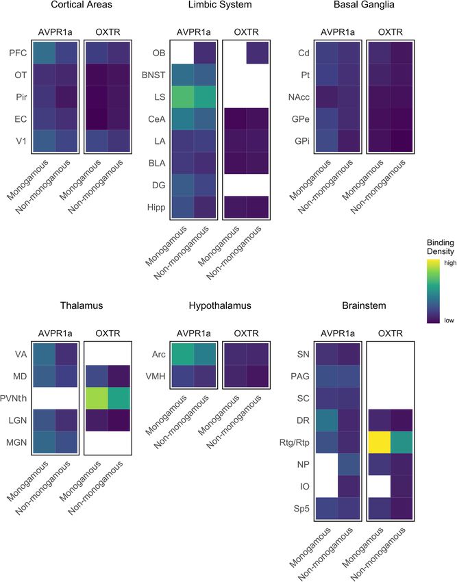
www.nature.com/scientificreports/
LGN Lateral geniculate nucleus
LS Lateral septum
MD Medial dorsal thalamus
MGN Medial geniculate nucleus
NAcc Nucleus accumbens
NP Nucleus prepositus
OB Olfactory bulb
OT Olfactory tubercle
PAG Periaqueductal gray
PFC Prefrontal cortex
Pir Piriform cortex
Pt Putamen
PVNth Paraventricular nucleus of the thalamus
Rtg/Rtp Reticulotegmental/reticulopontine nucleus
SC Superior colliculus
SN Substantia nigra
V1 Primary visual cortex
VA Ventral anterior thalamus
VMH Ventromedial hypothalamus
“Undoubtedly there are numerous molecular and neurobiological pathways that could evolve to support pair-
bond formation between mates, and different species may have achieved similar behaviors through a process
of convergent evolution involving different circuits… Nevertheless, it is intriguing to consider the possibility
that similar mechanisms may underlie the formation of pair bonds in both humans and rodents.” –Young and
Wang (2004, p. 1052).
Behavioral biologists are keenly interested in the evolved mechanisms that underlie diversity in social systems.
Oxytocin and vasopressin, two closely related neuropeptides, have been promising hormonal candidates in the
study of social systems due to their socioregulatory functions spanning a range of behavior in a wide variety
of taxa1. Beyond their conserved physiological roles, key behavioral and cognitive processes modulated by
these nonapeptides include pair bonding and mating2–5, parental c are6,7, stress coping8,9, aggression10,11, social
reward12, and social recognition13,14. Many hypotheses regarding the mechanisms by which oxytocin and vaso-
pressin modulate attachment behavior, and even mating systems, stem from comparative rodent work examining
underlying neural circuits. Hypotheses arising from this work posed the exciting possibility that mechanisms of
neuropeptide function might generalize to humans; nevertheless, primate models that would provide important,
evolutionarily relevant tests of these mechanisms have been underutilized15,16. Here, we examine neuroanatomical
distributions of oxytocin and vasopressin receptors for the first time in strepsirrhine primates, taking advantage
of the distinct interspecific variation in social system within a single genus to test the influential hypothesis that
differences in receptor distributions reflect differences in mating system.
Only one type of oxytocin receptor, the G protein-coupled receptor OXTR, has been characterized thus far,
but vasopressin has three known receptors (AVPR1a, AVPR1b and AVPR2;17). Of these receptors, AVPR1a is
present centrally and is thought to mediate the majority of vasopressin’s social effects17. In foundational neuro-
anatomical studies on the Microtus genus of voles, researchers used in vitro receptor autoradiography to examine
both OXTR and AVPR1a. Voles emerged as exceptional comparative models because both monogamous and
non-monogamous species exist within the same genus, allowing researchers to examine neurobiological dif-
ferences between phylogenetically proximate, yet socially divergent species. While the neural distributions of
vasopressin and oxytocin immunoreactive fibers are relatively conserved across v ertebrates1, receptor distribu-
tions can vary substantially between even closely related species. Indeed, researchers found striking species-level
differences in the distribution of OXTR and AVPR1a between monogamous prairie voles (Microtus ochrogaster)
and promiscuous montane and meadow voles (Microtus montanus and Microtus pennsylvanicus, respectively)18–21,
linking these differences in neuropeptide receptor distributions to interspecific differences in the ability to form
pair bonds4,18. This approach has since been broadly applied to examine the evolved functions of oxytocin and
vasopressin in rodent social behavior (e.g.22,23) and has established rodent models as central to understanding
oxytocin’s purportedly conserved social functions across mammals, including h umans4,24,25.
Developments within the field of oxytocin and vasopressin research have also revealed substantial challenges
to the interpretation and translation of findings from rodent models to other mammalian groups. Prairie voles
exhibit substantial diversity in their mating t actics26 and their central distribution patterns of nonapeptide
receptors27–29. Reducing any species’ socioecology and neurobiology to sets of strictly canalized components
might oversimplify the underlying mechanisms and limit insights. More generally, behavioral endocrinologists
and neuroscientists have long raised concerns about the field’s reliance on a small set of model o rganisms30–32.
33,34
Additionally, research identifying unique aspects of human neurobiology challenges the potential translat-
ability of rodent models. Nonhuman primates might thus serve as valuable bridges from rodent to human biology
and sociality15,16.
The Eulemur genus of strepsirrhine primates represents a unique and powerful test system for this research
area for two reasons. First, with regard to their morphology and sensory adaptations, lemurs are seen as occu-
pying a ‘transitional’ evolutionary niche between rodents and the more-often studied anthropoid p rimates35,36.
Features shared with rodents include enhanced olfactory reliance, acuity, and chemical communication, includ-
ing a functional vomeronasal organ; features shared with haplorhine primates include forward-facing eyes and
visual elaboration35. Despite being our most distant primate relatives, lemurs are approximately only half the
Scientific Reports | (2021) 11:3746 | https://doi.org/10.1038/s41598-021-83342-6 2
Vol:.(1234567890)www.nature.com/scientificreports/
genetic distance away from humans as are rodents37. Second, and crucially, Eulemur is the sole primate analogue
to Microtus in terms of containing both monogamous and non-monogamous species in a single genus. The 12
extant species of Eulemur are all cathemeral, arboreal, and seasonal breeders; they are generally frugivorous and
sympatric; and they are collectively found in almost all forest habitats across Madagascar38. Phylogenetic recon-
structions have revealed group-living (with male dispersal and female philopatry) as the ancestral form of social
organization in this clade39). Social monogamy and pair-bonding (which need not entail genetic monogamy)40,
a year-round arrangement whereby a male–female pair lives in a small family group, defends a shared territory
via mutual scent-marking, and jointly cares for young across several seasons, has evolved either once or twice,
giving rise to E. rubriventer and E. mongoz as the two monogamous species in this genus39,41–43. All other species
are non-monogamous, live in larger social groups, and exhibit varying degrees of promiscuous mating; they
also lack the behavioral signatures of social pair-bonds seen in monogamous species39,42. Along with evidence of
behavioral bifurcation, phylogenetic evidence of recent species divergence in Eulemur39 supports a consideration
of two distinct categories of social systems across the genus. Thus, Eulemur presents us with the opportunity
to examine how a conspicuous split in mating systems between closely related primate species is predicted by
neuropeptide circuitry.
Because the neurobiology of the oxytocin and vasopressin systems has yet to be characterized in any strepsir-
rhine primate, we begin with a broad ‘discovery’ aim (1). Across species, we expect to find conserved binding
to both neuropeptide receptors in certain specific regions. Parallel to the findings of OXTR/AVPR1a expression
along regions of the olfactory pathway in r odents19,20,44, OXTRs in regions of visual processing and attention have
consistently been found in anthropoid p rimates45. In strepsirrhines, we expect to find binding in nuclei involved
in both perceptual modalities. Additionally, in anthropoid primates, AVPR1a generally has been found to be
more widely distributed than O XTRs46–48—we predict a similar pattern in strepsirrhines.
With foundational neuroanatomical information in hand, we address our more targeted aim (2) to test vari-
ation in these receptors as a function of mating system. We investigate whether binding patterns in the brains
of monogamous versus non-monogamous Eulemur differ in several regions of hypothesized “pair-bonding cir-
cuits”. Based on correlational and experimental evidence in v oles4,18,19 linking neuropeptide receptor expression
in specific nuclei to pair-bond formation, key OXTR regions include the medial amygdala, nucleus accumbens,
and prefrontal cortex, and key AVPR1a regions include the ventral pallidum, lateral septum, and bed nucleus
of the stria terminalis. Based on reported neuropeptide receptor distributions and patterns of central glucose
uptake upon partner separation in monogamous coppery titi monkeys (Plecturocebus cupreus)49, additional key
regions for either OXTR or AVPR1a include the hippocampus, lateral septum, and central amygdala. Examin-
ing differences in these predicted regions as a function of mating system, as well as any additional differences
specific to Eulemur, will provide a powerful test of the role of neuropeptide receptor organization in predicting
social diversity.
Methods
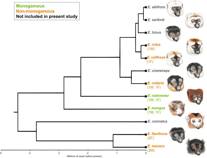
Specimens. Frozen, unfixed brain specimens derived from 12 individual Eulemur subjects (6 M, 6 F), rep-
resenting two distinct mating systems among seven closely related species (see Fig. 1 for numbers of specimens
per sex and species). Members of Eulemur range from vulnerable to critically endangered50. The specimens
were thus obtained from the tissue bank of the Duke Lemur Center (DLC) in Durham, NC, which is the only
facility outside of Madagascar to house and/or breed several of these species. All individuals whose brains were
obtained had been housed socially, primarily in adult male–female pairs, until their death from natural causes or
veterinary euthanization (all for non-neurological reasons; age of death ranged from 16.6 to 34.0 years of age).
Of the 12 specimens, 11 were hemispheres, and one was a whole brain. Owing to variability in location of
the midline bisections, and tissue integrity around the edges, some brain regions (such as the midline thalamic
nuclei and reticulotegmental nucleus; see below) were either absent or non-quantifiable in some specimens.
Additionally, the olfactory bulbs and brainstem nuclei (such as the inferior olive and spinal trigeminal nucleus)
were only quantifiable in a subset of subjects (see below).
Tissue preparation. We blocked brain specimens coronally on dry ice, before wrapping them tightly in
aluminum foil and storing them at − 80 °C until sectioning. We brought hemisphere blocks up to − 20 °C for sec-
tioning at 20 µm on one of two cryostats. We mounted tissue sections on SuperFrost Plus slides (Brain Research
Labs, Newton, MA) and stored them in a sealed slide box with a desiccant packet at − 80 °C until their use in
receptor autoradiography.
Receptor autoradiography. For receptor autoradiography, we used a competitive oxytocin and vasopres-
sin receptor binding protocol developed and optimized for primate tissue by Freeman et al.46 in rhesus macaques
(Macaca mulatta) and validated to selectively reveal OXTR or AVPR1a binding sites in postmortem brain tissue
from common marmosets (Callithrix jacchus;45), coppery titi monkeys [ 47], and h umans48,52. After lightly fixing
the tissue sections in 0.1% paraformaldehyde and rinsing with Tris buffer, we incubated them with either the
OXTR radioligand 125I-ornithine vasotocin analogue (125I-OVTA; PerkinElmer, Waltham, MA) or the AVPR1a
radioligand 125I-linear vasopressin antagonist (125I-LVA; PerkinElmer, Waltham, MA). We co-incubated sets of
three adjacent sections in three different conditions: (i) 50 pM radioligand alone, (ii) 50 pM radioligand plus
1 nM SR49059 (Tocris, Minneapolis, MN), an AVPR1a antagonist, and (iii) 50 pM radioligand plus 100 nM
ALS-II-69 (donated by ALS; see53), an OXTR antagonist. Accordingly, set (i) could be compared to sets (ii) and
(iii) to show regions of selective binding. After incubation, we washed the slides with Tris buffer, dipped them in
ddH2O, air dried them, and exposed them to BioMax MR film (Kodak, Rochester, NY) for four days with a set of
Scientific Reports | (2021) 11:3746 | https://doi.org/10.1038/s41598-021-83342-6 3
Vol.:(0123456789)www.nature.com/scientificreports/
Figure 1. The mating system classification of seven Eulemur species at the Duke Lemur Center and their
phylogenetic relationships, adapted from39,51. The number and sex of specimens from each focal species is
gure51 is published under a creative commons attribution license.
denoted in parentheses. The source fi
ten125I autoradiographic standards (American Radiolabeled Chemicals, St. Louis, MO). After film development,
we quantified receptor density directly from films without image enhancement.
Because no labelled brain atlas exists for any Eulemur species (or any member of the Lemuridae family), we
delineated brain regions for image analysis by counterstaining slides for acetylcholinesterase (AChE) following
a modified protocol from Lim et al.20 that has been shown to amplify signal in tissue previously used for recep-
tor autoradiography.
We quantified the optical binding density (OBD) of the autoradiogram images on a light box with MCID
Core Digital Densitometry software (Cambridge, UK). First, we determined a flat field correction for luminosity.
Then, we loaded the optical binding values from the set of125I autoradiographic standards into the software and
used them to generate a standard curve from which OBD values of brain regions of interest could be interpo-
lated. To determine neuroanatomical landmarks and identify regions, we compared images to the sets of AChE
counterstained slides, as well as to two atlases of rhesus macaque brains (54,55; www.brainmuseum.org) and an
rain56. We made three separate measurements per brain region with identifiable OXTR/
atlas of the adult human b
AVPR1a binding.
Analyses. Our statistical analyses proceeded in three stages. First, we validated the competitive binding pro-
tocol using paired t-tests to compare results from each of the ‘competitor binding’ conditions to the ‘radioligand
alone’ condition, in four representative regions as well as across all measured regions. In these analyses, we con-
sidered measurements from monogamous and non-monogamous animals together. We next performed Welch’s
t-tests to identify regions with appreciable selective binding of OXTR (125I-OVTA + SR49059) or AVPR1a (125I-
LVA + ALS-II-69). Results in this section are presented as mean ± SEM estimated disintegrations per minute per
milligram (dpm/mg). Lastly, to examine differences as a function of mating system, we used linear mixed models
that contained replicate OBD measurements nested within individual animals as a random effect. These analyses
included sex as a factor in the mixed model; however, the exclusion of sex had no substantive effect on any results
presented below, indicating a lack of significant differences in binding profiles between the sexes. We performed
separate models for individual regions that had either (a) been previously implicated as key areas for rodent
and/or primate pair bonding, or (b) showed dense neuropeptide binding in our exploratory analyses. We report
results for mating system differences as effect sizes in Cohen’s d, with positive values of d representing greater
binding in specimens from monogamous lemurs. The data and corresponding R code needed to reproduce our
results are publicly available at https://osf.io/rymz5/.
Scientific Reports | (2021) 11:3746 | https://doi.org/10.1038/s41598-021-83342-6 4
Vol:.(1234567890)www.nature.com/scientificreports/
Results
Selectivity of radioligands. The radioligands125I-OVTA and125I-LVA produced distinct patterns of bind-
ing in Eulemur brains (Fig. S1). As in anthropoids, strepsirrhine brains required competitive binding with the
AVPR1a antagonist to allow accurately identifying regions of OXTR binding. At the concentration used in our
assay,125I-OVTA labelled both OXTR and AVPR1a. Both the AVPR1a antagonist, SR49059, and the OXTR
antagonist, ALS-II-69, significantly reduced125I-OVTA binding in the central amygdala (CeA), nucleus accum-
bens (NAcc), and spinal trigeminal nucleus (Sp5) (Table S1). In contrast, the AVPR1a antagonist significantly
reduced125I-LVA binding in the CeA, Sp5, and primary visual cortex (V1), whereas the OXTR antagonist did
not reduce125I-LVA binding in these regions (Table S2). This selective reduction in125I-LVA binding by the
AVPR1a antagonist showed that125I-LVA binds selectively to AVPR1a and not to OXTR in Eulemur species,
while125I-OVTA appears to be able to bind to both receptor subtypes in some regions. Alternatively, SR49059
and ALS-II-69 may have different affinities for Eulemur OXTR and AVPR1a at the concentrations used in this
study. Fig. S1 shows the overall efficacy of the antagonists for displacing radioligand binding. Based on these
results, below we present values for sections incubated with both the radioligand and the opposing receptor
antagonist:125I-OVTA + SR49059, and125I-LVA + ALS-II-69.
OXTR distribution Across Eulemur brains. Strong125I-OVTA binding in the presence of the AVPR1a
antagonist was restricted to few areas (Figs. 2A, 3A,D), including the paraventricular nucleus of the thalamus
(PVNth; 343.48 ± 39.17), V1 (86.66 ± 17.33), prefrontal cortex (PFC; 76.82 ± 12.50), mediodorsal thalamus (MD
Thal; 72.66 ± 32.97), and olfactory bulb (Olf; 61.99 ± 26.88; this region was only present in specimens from non-
monogamous species; Figs. 2, 4). We observed modest OXTR binding in the hypothalamus (arcuate nucleus
[Arc]: 55.00 ± 19.40; ventromedial hypothalamus [VMH]: 34.36 ± 12.53), striatum (caudate [Cd]: 37.58 ± 7.56;
putamen [Pt]: 33.43 ± 6.26; NAcc: 19.20 ± 6.31), and assorted brainstem nuclei (nucleus prepositus [NP]:
52.07 ± 8.37; Sp5: 43.86 ± 9.51). We found low levels of binding in the olfactory tubercle (OT), piriform cortex
(Pir), entorhinal cortex (EC), globus pallidus external and internal segments (GPe / GPi), various amygdalar
nuclei (CeA; LA; BLA), hippocampal formation (Hipp), lateral geniculate nucleus (LGN), and dorsal raphé
nucleus (DR). Lastly, there were also notable null results: Unlike binding in vole species18, we observed no OXTR
radioligand binding in the lateral septum (LS) or bed nucleus of the stria terminalis (BNST) of any Eulemur
specimen (Figs. 2A, 3A). Unlike previous findings in multiple non-human primate species46,47, we did not detect
OXTR radioligand binding in the nucleus basalis of Meynert.
AVPR1a distribution across Eulemur brains. Relative to OXTR binding patterns in Eulemur brains,
AVPR1a binding was much more widespread and showed greater average binding across regions (Figs. 2B,
3B,E). In the presence of the OXTR antagonist, we found d ense125I-LVA binding in specimens across mating
systems in the PFC (126.33 ± 17.43) and V1 (116.80 ± 7.92), the Arc (231.13 ± 43.40), along with several areas of
the limbic system (LS: 299.54 ± 45.03; BNST: 162.10 ± 13.73; CeA: 170.28 ± 13.38), thalamus (MD 111.70 ± 20.97;
medial geniculate [MGN]: 137.80 ± 18.57), and brainstem (periaqueductal gray [PAG]: 119.40 ± 15.77; Sp5:
88.38 ± 18.56). We found moderate binding in the basal ganglia (Cd: 78.20 ± 13.37; Pt: 77.75 ± 11.57; NAcc:
53.76 ± 12.44; GPe: 72.39 ± 16.95; GPi: 64.18 ± 19.73), LGN (88.26 ± 23.38), olfactory cortex (OT: 54.43 ± 12.44;
EC: 73.83 ± 17.20; Pir: 39.41 ± 10.24), VMH (79.18 ± 13.56), and other areas of the limbic system (LA: 90.97 ± 7.94;
BLA: 67.99 ± 14.23; Hipp: 72.46.31 ± 17.36) and brainstem (SC; 78.80 ± 10.38; SN; 53.23 ± 15.32) (Figs. 2 and 3).
Binding patterns as a function of mating system. We targeted candidate regions of hypothesized
‘pair-bonding circuits’ in rodents (MeA, NAcc, PFC, LS and BNST;4) and titi monkeys (LS, CeA, and Hipp;49)
in our comparisons of mating system-related differences in Eulemur OXTR/AVPR1a binding. Contra Insel and
Shapiro’s18 consistent findings of greater OXTR binding in monogamous specimens, we found no evidence that
OXTR binding patterns in the Eulemur amygdala differed significantly between specimens from monogamous
vs. non-monogamous species (Figs. 2, 3, 4). We did not observe significant binding in the medial amygdala of
any specimens, and in other amygdalar nuclei where OXTR was present, differences between mating systems
were non-significant and inconsistent in direction (d ranging from − 0.25 to 0.33). Similarly, and somewhat
surprisingly, we observed no significant differences in OXTR binding patterns in the NAcc (d = 0.13), Hipp
(d = 0.43), or PFC (d = 0.68).
We also observed no statistically significant differences in AVPR1a binding in the regions of interest that were
targeted in this analysis for their hypothesized roles in rodent or titi monkey pair-bonding4,49. Also divergent from
findings in rodents, but consistent with findings in haplorhine primates (e.g.,47), we observed no binding in the
ventral pallidum of any Eulemur specimen. Furthermore, in regions where we observed binding, including the LS,
BNST and Hipp, there were no significant differences as a function of mating system. Differences were small to
medium for the LS (d = 0.41) and BNST (d = 0.59), and larger for the Hipp (d = 1.03), but all p > 0.05 (Figs. 2, 3, 4).
We next examined if there were differences by mating system in any of the regions in which we observed
OXTR and/or AVPR1a binding. The only region where we observed significant OXTR differences by mating
system was the reticulotegmental nucleus, which showed stronger binding in specimens from monogamous
than non-monogamous species (d = 3.71, p = 0.021; Fig. 4). For AVPR1a, we observed a significant difference in
the ventral anterior thalamus (VA; d = 1.28, p = 0.025), dorsal raphé nucleus (DR; d = 1.49, p = 0.010), and PFC
(d = 1.45, p = 0.028), with specimens from monogamous species again showing stronger binding in all three
regions compared to their counterparts from non-monogamous species (Fig. 4).
Scientific Reports | (2021) 11:3746 | https://doi.org/10.1038/s41598-021-83342-6 5
Vol.:(0123456789)www.nature.com/scientificreports/
Figure 2. Distribution of OXTR (A) and AVPR1a (B) in sequential coronal sections from the brain of one
representative non-monogamous Eulemur individual (E. macaco), aligned with acetylcholinesterase (AChE)
counterstain (C). Panels 1–2.
Scientific Reports | (2021) 11:3746 | https://doi.org/10.1038/s41598-021-83342-6 6
Vol:.(1234567890)www.nature.com/scientificreports/
Figure 2. (continued)
Discussion
As the first study to investigate neuropeptide receptor distribution in strepsirrhine primates, we document bind-
ing patterns of both oxytocin and vasopressin in members of the Eulemur clade that fall between those of classic
rodent models (e.g.18,19) and those of more recently characterized haplorhine primates46,47,57. This intermediacy
may have functional implications for lemurs’ evolutionary specializations, potentially reflecting the comparatively
variable role of these neuropeptides in sensory ecology (e.g.13,58). As the first primate study to directly compare
Scientific Reports | (2021) 11:3746 | https://doi.org/10.1038/s41598-021-83342-6 7
Vol.:(0123456789)www.nature.com/scientificreports/
Figure 3. Distribution of OXTR (A, D) and AVPR1a (B, E) in sequential coronal sections from the brain of
one representative monogamous Eulemur individual (E. rubriventer), aligned with acetylcholinesterase (AChE)
counterstain (C, F).
neuropeptide receptor binding between brain specimens from monogamous and non-monogamous species
of the same genus, our findings also fill a critical gap in knowledge of how variation in neuroanatomy reflects
variation in primate mating systems or sociality. Beyond simply representing another data point in the domain
of comparative neurology, findings from our study of Eulemur question the universality of classic vole models
and suggest a revisitation of their implications for humans.
Like rodents, lemurs show olfactory s pecialization59, which is prominently displayed in their use of scent to
convey a wide array of reproductive and social information60,61. Some degree of similarity in the involvement
Scientific Reports | (2021) 11:3746 | https://doi.org/10.1038/s41598-021-83342-6 8
Vol:.(1234567890)www.nature.com/scientificreports/
Figure 4. Summary of relative binding densities by brain region in Eulemur species. Columns depict average
binding density across brain specimens from monogamous and non-monogamous species, respectively. Blank
cells indicate no measurement available for radioligand binding in that region.
of OXTRs in processing chemically encoded socio-reproductive information in these taxa is suggested by the
diffuse binding of both OXTR and AVPR1a in the olfactory bulbs and olfactory tubercle, and by dense bind-
ing of AVPR1a in the CeA and BNST (across mating systems). Likewise, AVPR1a binding has been found in
the olfactory bulb of platyrrhine primates (e.g. common marmosets;57) that also rely extensively on olfactory
communication62; similar binding has not been reported in less olfactory-oriented catarrhine primates.
Relative to other placental mammals, vision is exceptionally well-developed in primates, but less so in strepsir-
rhines than in haplorhines. In catarrhines, for example, trichromacy63,64 and visual gaze are particularly impor-
tant in reproductive and social c ommunication65,66. Consistent with previous work in haplorhine primates, we
found OXTR expression in V1 and the LGN across species, and AVPR1a expression in these and additional
Scientific Reports | (2021) 11:3746 | https://doi.org/10.1038/s41598-021-83342-6 9
Vol.:(0123456789)www.nature.com/scientificreports/
areas related to visual attention (i.e., Rtg, SC, and amygdalar nuclei); nevertheless, binding in Eulemur was less
widespread than that observed in haplorhine primates (e.g.46,47). With regard to sensory pathways, therefore,
our results are consistent with lemur neuroanatomy representing a bridge between odor-reliant rodents and
vision-reliant haplorhines.
Intermediary patterns were also evident in other pathways. For instance, consistent with findings in some
rodent species (singing mice:23; prairie and montane voles:19), but unlike findings in haplorhine primates, we
observed dense AVPR1a expression in the MGN of lemurs. Because the MGN is an essential auditory relay
nucleus—receiving input from the inferior colliculus and projecting to the auditory cortices—our findings
potentially implicate vasopressin in another sensory modality in Eulemur; it is possible that vasopressin plays a
modulatory role in the processing of vocal communication or emotionally valent sounds.
Relative to haplorhines, additional patterns of receptor binding in Eulemur show both similarities and striking
reversals. In Eulemur, we observed strong AVPR1a binding, but diffuse or modest OXTR binding, in both the
striatum and hippocampal regions. This striatal pattern is comparable to that seen in coppery titi monkeys47, but
it contrasts with the dense OXTR expression found in both r odents67 and m armosets57. Hippocampal patterns
in Eulemur are reversed from that observed in titi m 47
onkeys . Oxytocin acting on OXTRs in the NAcc is neces-
sary for pair-bond formation in v oles4. Precise functions of oxytocin or vasopressin within the hippocampal
formation remain to be identified68, but there is some evidence that they modulate the encoding and consolida-
tion of socially relevant m emories69,70. In any event, our divergent results in these regions suggest that neural
mechanisms of pair bonding in lemurs may differ substantially from other mammalian groups studied thus far.
Regarding the influential hypothesis that interspecific variation in specific populations of receptors reflects
variation in social organization or mating system, our results did not reveal comparable differences to the strik-
ing findings previously reported for monogamous and non-monogamous vole species. For instance, in Insel and
Shapiro18, the effect size for a mating system difference in OXTR was d = 2.23 in the NAcc and d = 2.06 for the
LA; in Insel et al.19, the mating system difference in AVPR1a was d = 3.66 for the LS and d = 2.81 for the BNST.
In Eulemur, despite a sample size that matched these classic vole studies, differences between mating systems,
for either neuropeptide, were almost uniformly non-significant (with much smaller effect sizes; all d < 0.8) in all
regions of a hypothesized rodent ‘pair-bonding circuit’. Our results do not support the suggestion that OXTR/
AVPR1a differences in key dopaminergic areas separate monogamous from non-monogamous s pecies4. When
expanding our comparisons across the entire brain, however, we observed some significant differences between
mating systems, including in the Rtp for OXTR and the VA Thal, DR, and PFC for AVPR1a.
How should one interpret these mixed results? Regarding null findings, we note that exhausting the avail-
able bank of Eulemur brain tissue at the Duke Lemur Center nevertheless left us with limited statistical power
to detect differences in individual regions as a function of mating system. While large differences comparable
in magnitude to those reported in Insel and Shapiro18 and Insel et al.19 would be detectable with our sam-
ple—indeed, we matched the sample size from these classic studies—more modest differences may have been
missed. Regarding exploratory positive findings, we first caution that examining numerous regions increases the
potential for false-positives, and that there is a lack of information about the functional significance for many
of these differences. For instance, whereas the presence of OXTR in the pontine reticular areas of Eulemur and
rhesus macaques46 suggests a possible conserved function of oxytocin in this region, it is unclear how differ-
ences in this region that controls horizontal gaze and saccadic eye movement would be involved in differences
in social bonding behavior. Although the ventral anterior thalamus has important functions in spatial memory
and learning71, it has not been specifically implicated in pair-bonding processes. That said, other findings more
readily yield potential interpretations. First, the AVPR1a difference we observed in the DR, a source of serotonin
and a region involved in reward-seeking and reward-tracking behavior72, suggests that some of the effects of
vasopressin on social behavior may owe to activation of the DR serotonin system73. If so, monogamous Eulemur
may have developed denser populations of AVPR1a to bolster serotonergic functions of social reward behavior
that foster the creation of pair bonds. Second, rather than observing OXTR binding differences in the PFC—a
key area generating the reinforcing, hedonic properties of pair-bonding behavior and mating in r odents4—we
instead found a difference in AVPR1a binding in this region. Perhaps some of the mechanisms mediated by
oxytocin in rodents are carried out by the structurally similar vasopressin in primates—a suggestion that has
been hypothesized and substantiated in several previous s tudies45.
Collectively, mixed findings for mating system differences, like the aforementioned binding patterns found
across lemur species, are consistent with the existence of distinctive mechanisms for the formation of monog-
amous mating systems in Eulemur. In questioning the universality of these mechanisms across mammalian
groups, our findings in this domain can also be considered within the broader context of psychological oxytocin
research, which is similarly marked by interpretive challenges and heterogeneous findings (e.g.74). We suggest
that expanding the toolkits available to researchers, including broadening the animal models studied, will likely
continue to reveal unexpected findings that require modification to existing theory (a point echoed by behavioral
ecologists; e.g.75).
Providing context to our results is the fact that numerous factors other than species identity influence an
individual’s oxytocin and vasopressin neurocircuitry. Neurobiology is not static throughout the lifespan, but
rather may vary seasonally, with social circumstance, and with age or life-history stage (e.g.52). Thus, while
receptor distributions can differ widely between species and social s ystems18,19,22, they might also differ substan-
tially within individuals of the same species or mating system. Indeed, Phelps and Y oung27 report intraspecific
variation in AVPR1a binding among prairie voles often comparable to or greater than interspecific variation
(for a recent example of experience-dependent, intraspecific OXTR patterns in a primate model, s ee68). Nev-
ertheless, these same authors also report less variation in regions regulating social bonding, relative to those
unrelated to social bonding—a pattern consistent with natural selection winnowing neuropeptide expression in
these former regions. We also observed substantial intraspecific and within-mating system variation in Eulemur
Scientific Reports | (2021) 11:3746 | https://doi.org/10.1038/s41598-021-83342-6 10
Vol:.(1234567890)www.nature.com/scientificreports/
(see individual-level estimates of receptor profiles in Table S3)—given our limited sample size per species, it is
unclear to what extent this might be explained by season-level, individual-level, and/or species-level differences.
In Eulemur, some areas previously identified as key to social bonding—such as nuclei of the amygdala and the
BNST—showed relatively small coefficients of variation within mating systems, consistent w ith27, even though
they did not differ significantly between mating systems. Other regions that showed relatively little variation
within Eulemur mating systems, such as the primary visual cortex and SC, were not the same ones identified as
part of a pair-bonding circuit in rodent studies, but they are consistently identified as sites of OXTR and AVPR1a
in nonhuman primate studies46,47. Perhaps neuropeptide binding in regions responsible for processing visual
information are important targets of stabilizing selection in primates, regardless of the underlying mating system.
As in the classic vole studies18,19, we categorized our Eulemur species as belonging to one of two broad mating
systems, based on extant information about their wild c ounterparts39. On the one hand, we cannot rule out the
possibility that group size reductions, selective reproduction, or long-term pair housing in captivity may have
contributed to ‘monogamous-like’ receptor binding profiles across species in our sample, potentially minimizing
differences by mating-system category. On the other hand, one might expect such a ‘flattening’ influence to lead
to similar receptor profiles across individuals and species, but this does not reflect our results, which are more
accurately characterized by a large degree of within-mating system variation. More generally, we believe our
results complement the recognition of substantial, natural heterogeneity in social behavior, within or between
species, under the general umbrella of ‘monogamous’ or ‘non-monogamous’. Pair-living, pair-bonding, and
genetic monogamy are overlapping, yet constitute distinct components of a monogamous mating system that are
often conflated40,76. Different configurations of these components across ‘monogamous’ species could conceivably
create different neuropeptide receptor distributions. Importantly, we note that flexibility in putative mating sys-
tems is likely the norm, rather than the exception in animal models. Even the seemingly well-characterized mat-
ing system of prairie voles contains surprises revealed only upon extensive observation in naturalistic s ettings26.
In some cases, differences in neuropeptide receptor distributions may be detectable in spite of intraspecific (or
within-mating system) social variation, but this may less common than previously assumed.
Conclusion
Our analyses of the oxytocin and vasopressin receptor distributions throughout the Eulemur genus break ground
into a previously unstudied neurobiological system and question a popular and foundational neurobiological
explanation for the differences between monogamous and non-monogamous species. We find in lemurs some
elements of neuropeptide expression seen in rodents (e.g. binding in olfactory regions) and other elements more
commonly found in haplorhine primates (e.g. binding along visual pathways), consistent with other lines of
evidence suggesting the intermediary evolutionary niche occupied by lemurs between other mammalian groups
and haplorhine primates. While previous researchers often note the possibility that different mammalian line-
ages have developed mating systems via distinct neurobiological mechanisms (e.g. 4), much of the impact and
appeal of rodent studies has come from the enticing possibility that conserved mechanisms related to oxytocin
and vasopressin may help explain how human pair bonds are formed. We show that circuits identified as key to
pair bonding in rodents cannot simply be invoked to explain primate pair bonding. Our research on the lemur
oxytocin system, as part of burgeoning body of work across a range of nonhuman primates, also has important
implications for translational research, as it provides a glimpse into the diversity by which these neuropeptides
may have their manifold effects on social behavior.
Data availability
The data and corresponding R code needed to reproduce our results are publicly available at https: //osf.io/rymz5/ .
Received: 17 November 2020; Accepted: 1 February 2021
References
1. Goodson, J. L. The vertebrate social behavior network: evolutionary themes and variations. Horm. Behav. 48(1), 11–22 (2005).
2. Carter, C. S. Oxytocin and sexual behavior. Neurosci. Biobehav. Rev. 16(2), 131–144 (1992).
3. Cho, M. M., DeVries, A. C., Williams, J. R. & Carter, C. S. The effects of oxytocin and vasopressin on partner preferences in male
and female prairie voles (Microtus ochrogaster). Behav. Neurosci. 113(5), 1071–1079 (1999).
4. Young, L. J. & Wang, Z. The neurobiology of pair bonding. Nat. Neurosci. 7(10), 1048–1054 (2004).
5. Borrow, A. P. & Cameron, N. M. The role of oxytocin in mating and pregnancy. Horm. Behav. 61(3), 266–276 (2012).
6. Wang, Z., Young, L. J., De Vries, G. J. & Insel, T. R. Voles and vasopressin: a review of molecular, cellular, and behavioral studies
of pair bonding and paternal behaviors. Prog. Brain Res. 119, 483–499 (1998).
7. Feldman, R. Oxytocin and social affiliation in humans. Horm. Behav. 61(3), 380–391 (2012).
8. Neumann, I. D. & Landgraf, R. Balance of brain oxytocin and vasopressin: implications for anxiety, depression, and social behaviors.
Trends Neurosci. 35(11), 649–659 (2012).
9. Cavanaugh, J., Carp, S. B., Rock, C. M. & French, J. A. Oxytocin modulates behavioral and physiological responses to a stressor in
marmoset monkeys. Psychoneuroendocrinology. 66, 22–30 (2016).
10. Potegal, M. & Ferris, C. F. Intraspecific aggression in male hamsters is inhibited by intrahypothalamic vasopressin-receptor
antagonist. Aggress. Behav. 15(4), 311–320 (1989).
11. De Dreu, C. K. W. et al. The neuropeptide oxytocin regulates parochial altruism in intergroup conflict among humans. Science
328(5984), 1408–1411 (2010).
12. Dölen, G., Darvishzadeh, A., Huang, K. W. & Malenka, R. C. Social reward requires coordinated activity of nucleus accumbens
oxytocin and serotonin. Nature 501(7466), 179–184 (2013).
13. Ferguson, J. N., Aldag, J. M., Insel, T. R. & Young, L. J. Oxytocin in the medial amygdala is essential for social recognition in the
mouse. J. Neurosci. 21(20), 8278–8285 (2001).
Scientific Reports | (2021) 11:3746 | https://doi.org/10.1038/s41598-021-83342-6 11
Vol.:(0123456789)www.nature.com/scientificreports/
14. Lopatina, O. L., Komleva, Y. K., Gorina, Y. V., Higashida, H. & Salmina, A. B. Neurobiological aspects of face recognition: the role
of oxytocin. Front. Behav. Neurosci. 12, 195 (2018).
15. Freeman, S. M. & Bales, K. L. Oxytocin, vasopressin, and primate behavior: diversity and insight. Am. J. Primatol. 80(10), e22919
(2018).
16. Putnam, P. T., Young, L. J. & Gothard, K. M. Bridging the gap between rodents and humans: the role of non-human primates in
oxytocin research. Am. J. Primatol. 80(10), e22756 (2018).
17. Caldwell, H. K., & Young, W. S. Oxytocin and vasopressin: genetics and behavioral implications. In Handbook of Neurochemistry
and Molecular Neurobiology 573–607 (Boston, MA, Springer US, 2006).
18. Insel, T. R. & Shapiro, L. E. Oxytocin receptor distribution reflects social organization in monogamous and polygamous voles.
Proc. Natl. Acad. Sci. USA 89(13), 5981–5985 (1992).
19. Insel, T. R., Wang, Z. X. & Ferris, C. F. Patterns of brain vasopressin receptor distribution associated with social organization in
microtine rodents. J. Neurosci. 14(9), 5381–5392 (1994).
20. Lim, M. M., Murphy, A. Z. & Young, L. J. Ventral striatopallidal oxytocin and vasopressin V1a receptors in the monogamous prairie
vole (Microtus ochrogaster). J. Comp. Neurol. 468(4), 555–570 (2004).
21. Smeltzer, M. D., Curtis, J. T., Aragona, B. J. & Wang, Z. Dopamine, oxytocin, and vasopressin receptor binding in the medial
prefrontal cortex of monogamous and promiscuous voles. Neurosci. Lett. 394(2), 146–151 (2006).
22. Beery, A. K., Lacey, E. A. & Francis, D. D. Oxytocin and vasopressin receptor distributions in a solitary and a social species of
tuco-tuco (Ctenomys haigi and Ctenomys sociabilis). J. Comp. Neurol. 507(6), 1847–1859 (2008).
23. Campbell, P., Ophir, A. G. & Phelps, S. M. Central vasopressin and oxytocin receptor distributions in two species of singing mice.
J. Comp. Neurol. 516(4), 321–333 (2009).
24. Carter, C. S., DeVries, A. C. & Getz, L. L. Physiological substrates of mammalian monogamy: the prairie vole model. Neurosci.
Biobehav. Rev. 19(2), 303–314 (1995).
25. Insel, T. R. The challenge of translation in social neuroscience: A review of oxytocin, vasopressin, and affiliative behavior. Neuron
65(6), 768–779 (2010).
26. Madrid, J. E., Parker, K. J., & Ophir, A. G. Variation, plasticity, and alternative mating tactics: Revisiting what we know about the
socially monogamous prairie vole. In Advances in the Study of Behavior 203–42 (Elsevier, 2020).
27. Phelps, S. M. & Young, L. J. Extraordinary diversity in vasopressin (V1a) receptor distributions among wild prairie voles (Microtus
ochrogaster): patterns of variation and covariation. J. Comp. Neurol. 466(4), 564–576 (2003).
28. King, L. B., Walum, H., Inoue, K., Eyrich, N. W. & Young, L. J. Variation in the oxytocin receptor gene predicts brain region–specific
expression and social attachment. Biol. Psychiatry. 80(2), 160–169 (2016).
29. Walum, H. & Young, L. J. The neural mechanisms and circuitry of the pair bond. Nat. Rev. Neurosci. 19(11), 643–654 (2018).
30. Beach, F. A. The Snark was a Boojum. Am. Psychol. 5(4), 115–124 (1950).
31. Preuss, T. M. Taking the measure of diversity: comparative alternatives to the model-animal paradigm in cortical neuroscience.
Brain Behav. Evol. 55(6), 287–299 (2000).
32. Thompson, R. R. An updated field guide for snark hunting: Comparative contributions to behavioral neuroendocrinology in the
era of model organisms. Horm. Behav. 122(104742), 104742 (2020).
33. Boldog, E. et al. Transcriptomic and morphophysiological evidence for a specialized human cortical GABAergic cell type. Nat.
Neurosci. 21(9), 1185–1195 (2018).
34. Hodge, R. D. et al. Conserved cell types with divergent features in human versus mouse cortex. Nature 573(7772), 61–68 (2019).
35. Fleagle, J. G. Primate Adaptation and Evolution 3rd edn. (CA, Academic Press, San Diego, 2013).
36. Hozer, C., Pifferi, F., Aujard, F. & Perret, M. The biological clock in gray mouse lemur: Adaptive, evolutionary and aging considera-
tions in an emerging non-human primate model. Front Physiol. 10, 1033 (2019).
37. Ezran, C. et al. The mouse lemur, a genetic model organism for primate biology, behavior, and health. Genetics 206(2), 651–664
(2017).
38. Ossi, K. & Kamilar, J. M. Environmental and phylogenetic correlates of Eulemur behavior and ecology (Primates: Lemuridae).
Behav. Ecol. Sociobiol. 61(1), 53–64 (2006).
39. Kappeler, P. M. & Fichtel, C. The evolution of Eulemur social organization. Int. J. Primatol. 37(1), 10–28 (2016).
40. Tecot, S. R., Singletary, B. & Eadie, E. Why, “monogamy” isn’t good enough: pair-living, pair-bonding, and monogamy. Am. J.
Primatol. 78(3), 340–354 (2016).
41. Lukas, D. & Clutton-Brock, T. H. The evolution of social monogamy in mammals. Science 341(6145), 526–530 (2013).
42. Singletary, B. & Tecot, S. Signaling across the senses: a captive case study in pair-bonded red-bellied lemurs (Eulemur rubriventer)
at the Duke Lemur Center, NC. USA. Primates. 60(6), 499–505 (2019).
43. Shultz, S., Opie, C. & Atkinson, Q. D. Stepwise evolution of stable sociality in primates. Nature 479(7372), 219–222 (2011).
44. Tobin, V. A. et al. An intrinsic vasopressin system in the olfactory bulb is involved in social recognition. Nature 464(7287), 413–417
(2010).
45. Freeman SM, Young LJ. Comparative perspectives on oxytocin and vasopressin receptor research in rodents and primates: trans-
lational implications. J. Neuroendocrinol. 2016;28(4).
46. Freeman, S. M., Inoue, K., Smith, A. L., Goodman, M. M. & Young, L. J. The neuroanatomical distribution of oxytocin receptor
binding and mRNA in the male rhesus macaque (Macaca mulatta). Psychoneuroendocrinology. 45, 128–141 (2014).
47. Freeman, S. M. et al. Neuroanatomical distribution of oxytocin and vasopressin 1a receptors in the socially monogamous coppery
titi monkey (Callicebus cupreus). Neuroscience 273, 12–23 (2014).
48. Freeman, S. M., Smith, A. L., Goodman, M. M. & Bales, K. L. Selective localization of oxytocin receptors and vasopressin 1a recep-
tors in the human brainstem. Soc. Neurosci. 12(2), 113–123 (2017).
49. Bales, K. L. et al. Titi monkeys as a novel non-human primate model for the neurobiology of pair bonding. Yale J. Biol. Med. 90(3),
373–387 (2017).
50. The IUCN Red List of Threatened Species. 2020–2 [cited 2020 Jul 9]. Available from: https://www.iucnredlist.org
51. Markolf, M. & Kappeler, P. M. Phylogeographic analysis of the true lemurs (genus Eulemur) underlines the role of river catchments
for the evolution of micro-endemism in Madagascar. Front Zool. 10(1), 70 (2013).
52. Freeman, S. M. et al. Effect of age and autism spectrum disorder on oxytocin receptor density in the human basal forebrain and
midbrain. Transl. Psychiatry. 8(1), 257 (2018).
53. Smith, A. L. et al. Initial investigation of three selective and potent small molecule oxytocin receptor PET ligands in New World
monkeys. Bioorg. Med. Chem. Lett. 26(14), 3370–3375 (2016).
54. Bakker, R., Tiesinga, P. & Kötter, R. The Scalable Brain Atlas: Instant web-based access to public brain atlases and related content.
Neuroinformatics. 13(3), 353–366 (2015).
55. Rohlfing, T. et al. The INIA19 template and NeuroMaps atlas for primate brain image parcellation and spatial normalization. Front.
Neuroinform. 6, 27 (2012).
56. Ding, S.-L., Royall, J. J., Sunkin, S. M., Ng, L., Facer, B. A. C., Lesnar, P. et al. Comprehensive cellular-resolution atlas of the adult
human brain: adult human brain atlas. J. Comp. Neurol. 524(16), Spc1–Spc1 (2016).
57. Schorscher-Petcu, A., Dupré, A. & Tribollet, E. Distribution of vasopressin and oxytocin binding sites in the brain and upper spinal
cord of the common marmoset. Neurosci. Lett. 461(3), 217–222 (2009).
58. Wacker, D. & Ludwig, M. The role of vasopressin in olfactory and visual processing. Cell Tissue Res. 375(1), 201–215 (2019).
Scientific Reports | (2021) 11:3746 | https://doi.org/10.1038/s41598-021-83342-6 12
Vol:.(1234567890)www.nature.com/scientificreports/
59. Heritage, S. Modeling olfactory bulb evolution through primate phylogeny. PLoS ONE 9(11), e113904 (2014).
60. Drea, C. M. D’scent of man: a comparative survey of primate chemosignaling in relation to sex. Horm. Behav. 68, 117–133 (2015).
61. Drea, C. M. Design, delivery and perception of condition-dependent chemical signals in strepsirrhine primates: implications for
human olfactory communication. Philos. Trans. R. Soc. Lond. B Biol. Sci. 2020(375), 20190264 (1800).
62. Lazaro-Perea, C., Snowdon, C. T. & de Fátima, A. M. Scent-marking behavior in wild groups of common marmosets (Callithrix
jacchus). Behav. Ecol. Sociobiol. 46(5), 313–324 (1999).
63. Changizi, M. A., Zhang, Q. & Shimojo, S. Bare skin, blood and the evolution of primate colour vision. Biol. Lett. 2(2), 217–221
(2006).
64. Fernandez, A. A. & Morris, M. R. Sexual selection and trichromatic color vision in primates: statistical support for the preexisting-
bias hypothesis. Am. Nat. 170(1), 10–20 (2007).
65. Emery, N. J. The eyes have it: the neuroethology, function and evolution of social gaze. Neurosci. Biobehav. Rev. 24(6), 581–604
(2000).
66. Shepherd, S. V. & Platt, M. L. Spontaneous social orienting and gaze following in ringtailed lemurs (Lemur catta). Anim. Cogn.
11(1), 13–20 (2008).
67. Freeman, A. R., Aulino, E. A., Caldwell, H. K. & Ophir, A. G. Comparison of the distribution of oxytocin and vasopressin 1a
receptors in rodents reveals conserved and derived patterns of nonapeptide evolution. J. Neuroendocrinol. 32(4), e12828 (2020).
68. Baxter, A. et al. Oxytocin receptor binding in the titi monkey hippocampal formation is associated with parental status and partner
affiliation. Sci. Rep. 10(1), 1–14 (2020).
69. Cilz, N. I., Cymerblit-Sabba, A. & Young, W. S. Oxytocin and vasopressin in the rodent hippocampus. Genes Brain Behav. 18(1),
e12535 (2019).
70. Pagani, J. H. et al. Role of the vasopressin 1b receptor in rodent aggressive behavior and synaptic plasticity in hippocampal area
CA2. Mol. Psychiatry. 20(4), 490–499 (2015).
71. Jankowski, M. M. et al. The anterior thalamus provides a subcortical circuit supporting memory and spatial navigation. Front.
Syst. Neurosci. 7, 45 (2013).
72. Nakamura, K. The role of the dorsal raphé nucleus in reward-seeking behavior. Front. Integr. Neurosci. 7, 60 (2013).
73. Rood, B. D. & Beck, S. G. Vasopressin indirectly excites dorsal raphe serotonin neurons through activation of the vasopressin1A
receptor. Neuroscience 260, 205–216 (2014).
74. Mierop, A. et al. How can intranasal oxytocin research be trusted? A systematic review of the interactive effects of intranasal
oxytocin on psychosocial outcomes. Perspect. Psychol. Sci. 15(5), 1228–1242 (2020).
75. Rosenthal, M. F., Gertler, M., Hamilton, A. D., Prasad, S. & Andrade, M. C. B. Taxonomic bias in animal behavior publications.
Anim. Behav. 127, 83–89 (2017).
76. Huck, M., Di Fiore, A. & Fernandez-Duque, E. Of apples and oranges? The evolution of “monogamy” in non-human primates.
Front. Ecol. Evol. 7, 472 (2020).
Acknowledgements
We are indebted to Brian Horman, Genna St. Armour, Jordan Walker, and Tyler Beauchamp for their assistance
with tissue sectioning and quantification, Leonard White and Jenna McHenry for their comments on a previous
version of this manuscript, and staff at the Duke Lemur Center for assistance with acquiring brain specimens. This
research was supported by: the National Science Foundation (SBE-1808803 to NMG and CMD); the National
Institute of Mental Health (NIMH R21MH115680 to SMF and KLB); Duke University Research Support (to
CMD); the Josiah Charles Trent Memorial Foundation Endowment Fund, Duke Institute for Brain Sciences, and
the Duke Lemur Center Director’s Fund (to NMG); and the Duke Office of Undergraduate Research Support
and Charles Lafitte Foundation for Research (to AS). This is DLC publication #1471.
Author contributions
N.M.G., S.M.F., and C.M.D. designed research; N.M.G. and A.S. prepared specimens; N.M.G., A.S., S.M.F.,
and M.C.P. conducted experiments; H.B.P. and K.L.B. provided equipment and analytic tools; N.M.G. and A.S.
analyzed data; all authors contributed to the writing of the manuscript.
Competing interests
The authors declare no competing interests.
Additional information
Supplementary Information The online version contains supplementary material available at https://doi.
org/10.1038/s41598-021-83342-6.
Correspondence and requests for materials should be addressed to N.M.G.
Reprints and permissions information is available at www.nature.com/reprints.
Publisher’s note Springer Nature remains neutral with regard to jurisdictional claims in published maps and
institutional affiliations.
Open Access This article is licensed under a Creative Commons Attribution 4.0 International
License, which permits use, sharing, adaptation, distribution and reproduction in any medium or
format, as long as you give appropriate credit to the original author(s) and the source, provide a link to the
Creative Commons licence, and indicate if changes were made. The images or other third party material in this
article are included in the article’s Creative Commons licence, unless indicated otherwise in a credit line to the
material. If material is not included in the article’s Creative Commons licence and your intended use is not
permitted by statutory regulation or exceeds the permitted use, you will need to obtain permission directly from
the copyright holder. To view a copy of this licence, visit http://creativecommons.org/licenses/by/4.0/.
© The Author(s) 2021
Scientific Reports | (2021) 11:3746 | https://doi.org/10.1038/s41598-021-83342-6 13
Vol.:(0123456789)You can also read