The Biology and Immature Stages of the Moss-Eating Flea Beetle Cangshanaltica fuanensis sp. nov. Coleoptera, Chrysomelidae, Galerucinae ...
←
→
Page content transcription
If your browser does not render page correctly, please read the page content below
insects
Article
The Biology and Immature Stages of the Moss-Eating
Flea Beetle Cangshanaltica fuanensis sp. nov.
(Coleoptera, Chrysomelidae, Galerucinae, Alticini),
with Description of a Fan-Driven High-Power
Berlese Funnel
Yongying Ruan 1, *, Alexander S. Konstantinov 2 and Albert F. Damaška 3
 1    School of Applied Chemistry and Biological Technology, Shenzhen Polytechnic, Shenzhen 518055, China
 2    Systematic Entomology Laboratory, USDA, Smithsonian Institution, National Museum of Natural History,
      P.O. Box 37012, Washington, DC 20013-7012, USA; alex.konstantinov@ars.usda.gov
 3    Department of Zoology, Faculty of Science, Charles University, Viničná 7, 128 00 Prague, Czech Republic;
      albert.damaska@natur.cuni.cz
 *    Correspondence: yongyingruan@szpt.edu.cn

 Received: 21 July 2020; Accepted: 20 August 2020; Published: 26 August 2020

 Simple Summary: The immature stages and the biology of the moss inhabiting flea beetles are poorly
 understood. In this study, a new species of moss-eating flea beetles—Cangshanaltica fuanensis sp. nov.
 is described; the morphology of the adult and immature stages is described and illustrated. The life
 history and remarkable biological features of this species are revealed. Females deposit one large egg
 at a time; egg length equals 0.4–0.5 times the female body length. Females lay and hide each egg
 under a spoon-shaped moss leaf. There are only two ovarioles on each side of the ovary in the female
 reproductive system, which has not been reported before in Chrysomelidae. Besides, a modified
 fan-driven Berlese funnel is designed for faster extraction of moss inhabiting flea beetles. We suggest
 this improved device could also be useful for collecting other ground-dwelling arthropods.

 Abstract: The biology of the moss and leaf litter inhabiting flea beetles is poorly understood. In this
 study, a new species of moss-eating flea beetles Cangshanaltica fuanensis sp. nov. is described;
 the morphology of adult and immature stages is examined and illustrated. Its life history and biology
 are studied. The remarkable and unique biological features are revealed: (1) females deposit one
 large egg at a time, egg length equals 0.4–0.5 times the female body length, these are unusual in
 Chrysomelidae; (2) females have only two ovarioles on each side of the ovary, which has not been
 reported in other Chrysomelidae species; (3) females lay and hide each egg under a spoon-shaped
 moss leaf; (4) cannibalism of a second instar larva on an egg was observed. Both adults and larvae
 feed on moss and are polyphagous; their feces mainly consist of un-digested moss fragments;
 high humidity is essential for the survival of eggs and larvae and expedites the hatching. In addition,
 a modified fan-driven Berlese funnel is designed for faster extraction of moss inhabiting flea beetles.
 This device could also be used for collecting other ground-dwelling arthropods. Its working diagram
 is illustrated and described.

 Keywords: Berlese extraction; Berlese-Tullgren funnel; biodiversity; bryobiont; China; larva;
 moss inhabiting

Insects 2020, 11, 571; doi:10.3390/insects11090571                                 www.mdpi.com/journal/insects
Insects 2020, 11, 571                                                                                2 of 27

1. Introduction
      The moss and leaf litter inhabiting flea beetles (Alticini) are not well studied due to their
relatively cryptic living environment. Their tremendous diversity has been highlighted by recent
studies (e.g., [1–11]). Most known moss inhabiting leaf beetles belong to the tribe Alticini [12].
Recently, an unusual Galerucini species was discovered feeding on and inhabiting moss cushions [12].
In New Zealand, several genera in the subfamily Chrysomelinae (i.e., Nanomela, Zeaphilon, Aphilon
and Maurodus) are associated with mosses and liverworts [13]. These studies suggest that other
Chrysomelidae groups could also inhabit and feed on moss. It also means our current knowledge of
the diversity of the moss inhabiting leaf beetles is still insufficient.
      The moss-eating genus Cangshanaltica was described from Yunnan, China by Konstantinov et al. [2];
subsequently, four more species were described from Thailand, China (Hongkong) and
Philippines [3,7,9]. The wide distribution and the small number of described species suggested
a hidden diversity in this genus.
      Three species of moss inhabiting flea beetle larvae were already described by previous studies:
Ivalia korakundah Prathapan, Konstantinov and Duckett [14]; Mniophila muscorum (Koch) [15];
Distigmoptera borealis Blake [10]. Despite these studies, the biology and life history of moss inhabiting
flea beetles are not well understood. In this study, we describe a new species of Cangshanaltica, with data
on its biology, life history, and morphology of the immature stages.
      Prior to this study, traditional Berlese funnels were frequently used to collect moss inhabiting
flea beetles (for methods see [11]). These devices usually use light bulbs (40–150 W) as heating
elements. Processing moss with moderate-to-high moisture using the light-bulb-driven funnels is
very time consuming (usually more than 12 h for a standard size load). This process often yields only
a few specimens considering that flea beetles are usually scarce in the moss, especially in daylight.
In 2012, we used the traditional light-bulb-driven Berlese funnels to process moist moss cushions in
Shuyang, Fuan, Fujian Province, China. Despite our endeavors for several days, only one individual of
Cangshanaltica fuanensis sp. nov. was extracted. In 2014, an attempt to collect more specimens for the
taxonomical study had failed as this species has a very low population density. To process more moss
and therefore to obtain more individuals within a limited time, a modified Berlese funnel applying
a high-power heating element and a fan was developed and tested. In 2019, using the modified
fan-driven Berlese funnel, we extracted numerous living larvae and adults of Cangshanaltica fuanensis
sp. nov. and several other flea beetle species that dwell in the moss cushions. The adults of C. fuanensis
sp. nov. were then transferred to rearing containers, and their biology was observed. The modified
fan-driven Berlese funnel was also tested and proven suitable for collecting other ground-dwelling
arthropods. A working diagram of the device is illustrated and described here.

2. Materials and Methods

2.1. Morphological Methods
     Observations of the habitus and diagnostic characters of flea beetles were made using the Nikon
SMZ645 stereomicroscope and Nikon OPTIPHOT microscope. The genitalia with the last few tergites
was torn off using sharp insect pins attached to plastic sticks. The tissues surrounding the aedeagus
were cleared. Female genitalia and accompanied structures (the last tergites) were immersed into hot
10% NaOH solution for 30 s (or the appropriate time required to soften irrelevant tissue). The extra
tissues surrounding the genitalia were carefully removed using insect pins. For photography, the female
genitalia were mounted on slides with glycerin; male genitalia were glued to paper points. Digital
images were taken with Canon D800 camera attached to Canon MP-E 65 mm Lens or a modified lens
(consists of a Nikon M Plan lens, a manual aperture unit, and an adjustable extension tube).
     Flea beetles are treated as “Alticini” in this study.
     Adult morphological terminology follows Konstantinov et al. (2013) [2]. The terminology of larval
tubercles follows Kimoto (1962) [16] and Takizawa (2005) [17]. Abbreviations of tubercles: D, dorsal;
Insects 2020, 11, 571                                                                                        3 of 27

DL, dorsolateral; EP, epipleural; ES, eusternal; P, pleural; PS, parasternal; SS, sternellar. Position of tubercles
denoted by lowercase letter: a, anterior; p, posterior; i, interior; e, exterior.
     Abbreviations for insect collections. ADPC: personal collection of A. Damaška. IZCAS: Institute
of Zoology, Chinese Academy of Sciences, Beijing, China. SZPT: School of Applied Chemistry and
Biological Technology, Shenzhen Polytechnic, Shenzhen, Guangdong, China.
     Mosses were identified using “Flora Bryophytorum Sinicorum” [18]. A bryophyte taxonomist
was consulted.

2.2. Biological Methods
      The activities of Cangshanaltica fuanensis sp. nov. are hard to observe in nature; most of the
biological data were collected based on lab-reared individuals. A rearing container was designed for
rearing C. fuanensis sp. nov. Transparent plastic containers (15 cm × 7 cm × 5 cm) were selected and
placed in a north-facing room to avoid direct sunlight. Two rectangle openings were carved in the sides
of the container; they were sealed by non-woven fabrics using adhesive, allowing for air circulating
and preventing other organisms from coming into the container. A thick layer of moist paper towel
was placed at the bottom of the container in order to maintain proper humidity and avoid larvae from
drowning in water drops; a thin layer of soil was placed above the paper towel in order to provide
nutrition for host plant and site for larvae to pupate; fresh host plant moss was collected and placed
loosely above the soil layer. Distilled water was sprayed on the moss once a day to maintain humidity
using a small spraying device.
      Adults extracted from moss were transferred to the rearing container (ca. 25 ◦ C, 90–100% air
humidity were maintained). Approximately 30 parent (F0 ) adults were collected from wild and raised
until they were dead. Their first filial generation (F1 ) was also maintained alive in the same container
for the remainder of their lives. An F2 generation was not successfully bred. The biology and behavior
of numerous individuals of different stages were observed (ca. 30 F0 adults, 35 F1 eggs, 30 F1 larvae,
10 F1 pupae, and 6 F1 adults).
      The genitalia of the adults of F1 were dissected to confirm that they were conspecific with their
parent generation.

2.3. DNA Barcoding
      Specimens used for the DNA barcoding were treated as follows: DNA was extracted by
Qiagen DNEasy Blood and Tissue kit or GenAid Genomic DNA Mini kit in a thermo-shaker,
and 50 µL of elution buffer was used in the final step. In every specimen, the body was
perforated by breaking abdominal tergites for better DNA yield during the extraction. After DNA
extraction, the voucher specimens were mounted on cards and housed in ADPC (see Type
Material section). For PCR reactions, we used a modified protocol with a commercially prepared
premix (PPP Mix with MgCl2 added, Top-Bio Czech Republic). We used cox1 barcoding
primers: forward LCO1490 (50 -GGTCAACAAATCATAAAGATATTGG-30 ) and reverse HCO2198
(50 -TAAACTTCAGGGTGACCAAAAAATCA-30 ) [19]. PCR was performed in a 13 µL total volume of
the mixture, containing 6.25 µL of PPP Mix, 4.75 µL of PCR ddH2 O, 1.0 µL of each primer and 1.0 µL
of the DNA extract. The following PCR program was used: 94 ◦ C for 180 s + 35 × (94 ◦ C for 30 s, 48 ◦ C
for 45 s, 72 ◦ C for 60 s) + 72 ◦ C for 480 s. PCR products were purified by adding 0.5 µL Exonuclease 1
(Exo1 (20 U/µL)) (ThermoFisherScientific) and 1.0 µL Thermosensitive Alkaline Phosphatase (FastAP
(1 U/µL)) (ThermoFisherScientific); the mixture was incubated in a thermocycler for 37 ◦ C for 15 min
and 80 ◦ C for 15 min. Samples were sequenced by using Sanger sequencing. Raw sequence data
were edited by using Geneious 9.1.7 software (Biomatters). Sequences were submitted to GenBank,
accession numbers: MT891188.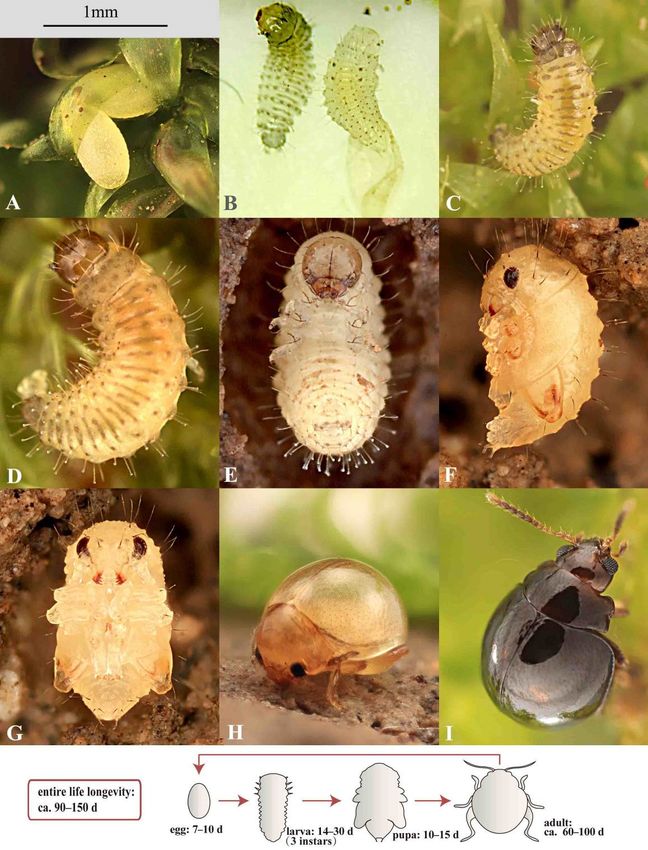
Insects 2020, 11, 571                                                                               4 of 27

2.4. The Modified Fan-Driven Berlese Funnel
      The majority of the specimens used in this study were extracted from moss using the modified
Berlese funnel. A portable fan heater was used as the heating source, 500–1800 W, switchable for
high-power and low-power modes depends on the moisture contained in the samples; it mainly
consists of a PTC (positive temperature coefficient) ceramic heating elements, and a fan installed above
it. The fan heater was hung 20 cm above the funnel using a tripod or other supporters; it generated and
propelled the warm airflow down to the samples (soil, moss, or leaf litter) placed in the funnel. By this
means, the warm airflow brought away the moisture, dried up the samples and drove the arthropods
down to the beaker placed under the funnel. One or two pieces of plastic mesh screens were placed at
the bottom of the funnel; soil, moss, or leaf litter was loosely placed above the mesh screen. A large
cylindrical plastic beaker was placed on the ground and attached under the funnel in order to collect
arthropod specimens and keep the funnel from toppling. The gap between funnel and beaker was
sealed by tape; a layer of thick moist paper towel was placed in the beaker to keep organisms alive
(paper towel could be replaced with ethanol to kill specimens). The moist paper towel was critical for
keeping specimens alive: in the earlier stage of the extraction process, it provided humidity for the
survival of organisms (especially insect larvae); while in a later stage, it absorbed the condensed water
aggregated at the bottom of the beaker and prevented organisms from drowning.
      The extracting process required approximately 2–4 h for lightly moist moss (moss volume = 6–8 L,
loosely placed in the funnel); around 4–8 h for entirely wet moss (moss volume = 6–8 L, loosely placed in
the funnel); around 4–5 h for moist and fluffy humus soil (volume = 4–5 L, loosely placed in the funnel).

3. Results

3.1. Taxonomy
     Genus Cangshanaltica Konstantinov et al. 2013.
     Cangshanaltica Konstantinov, Chamorro, Prathapan, Ge and Yang, 2013: 6.
     Type species. Cangshanaltica nigra Konstantinov, Chamorro, Prathapan, Ge and Yang, 2013: 16,
by original designation.
     Distribution. China (Yunnan, Hongkong, Fujian); Thailand (Chiang Mai), Philippines (Luzon,
Mindanao).
     Cangshanaltica fuanensis Ruan, Konstantinov and Damaška, new species (Figures 1 and 2)
     Type locality. Shuyang, Fuan, Fujian Prov., China.
     Distribution. China (Fujian).
     Etymology. This species is named after the type locality—Fuan city. Specific epithet is a noun
in apposition.
     Diagnosis. This new species is assigned to the genus Cangshanaltica based on its pronotal
anterolateral setiferous pore situated at the middle of the lateral margin of pronotum, and a longitudinal
ridge on the first abdominal ventrite is strongly developed, extending to near posterior margin. It may
be distinguished from other known species of Cangshanaltica by the shape of the aedeagus and female
genitalia, particularly the distinct U-shaped unsclerotized area on the anterior part of the vaginal palpi
(Figure 2L, indicated by arrow).
     Description of adult. Male body length 1.35–1.55 mm, width 1.10–1.20 mm; female body length
1.50–1.70 mm, width 1.25–1.35 mm. Ratio of body length to body width: 1.20–1.30. Dorsum and head
smooth, not reticulate, dark brown. Venter slightly lighter than dorsum: chestnut brown to dark brown.
Antennae: antennomeres I–II yellow-brown, II–IV yellow-brown to chestnut brown, V–X chestnut
brown, XI yellow-brown. Legs chestnut brown. Legs and antennae covered with yellow setae.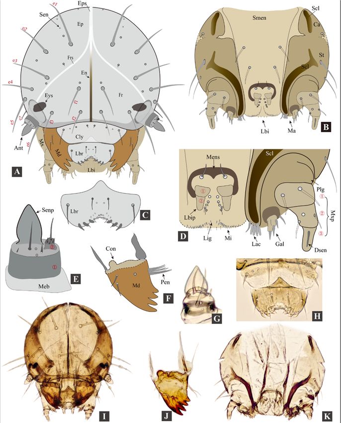
Insects 2020, 11, 571                                                                                             5 of 27
Insects 2020, 11, x FOR PEER REVIEW                                                                               5 of 28

     Figure
      Figure1.1.Adult morphology
                  Adult  morphology  of of
                                         Cangshanaltica
                                           Cangshanaltica  fuanensis
                                                              fuanensissp.sp.
                                                                           nov.nov.
                                                                                (A): Holotype,    dorsaldorsal
                                                                                     (A): Holotype,      view.view.
                                                                                                               (B):
     Holotype,   ventral
      (B): Holotype,      view.
                     ventral    (C):(C):
                              view.  Holotype,  lateral
                                         Holotype,      view.
                                                    lateral     (D):
                                                            view.    Pronotum,
                                                                  (D):  Pronotum,holotype.
                                                                                    holotype.(E):
                                                                                               (E):Head,
                                                                                                    Head,paratype.
                                                                                                           paratype.
     (F):
      (F): Aedeagus
           Aedeagusofof holotype,
                         holotype,ventral
                                    ventralview.
                                            view. (G):
                                                   (G):Aedeagus
                                                        Aedeagusof   ofholotype,
                                                                        holotype,lateral
                                                                                  lateralview.
                                                                                          view. (H):
                                                                                                  (H):Aedeagus
                                                                                                      Aedeagusof of
     holotype,
      holotype,dorsal
                 dorsalview.
                        view.

     Head (Figure 1E). Head hypognathous. Vertex smooth and shiny, with small and sparse punctures
bearing short setae. Antennal calli poorly delimited, smooth, triangular, with lighter color than
vertex. Supracallinal sulcus absent, midfrontal and suprafrontal sulci barely visible, supraantennal
and supraorbital sulci moderately deep and straight. Frontal ridge wide, slightly wider than width
of eye. Labrum with three pairs of setae. Mandibles symmetrical, palmate; each mandible with
five sharp teeth, mesal side with a membranous lobe bearing dense microtrichia. Antennomere
VII with weak distal protrusion (almost invisible) (Figure 2E). Proportions of antennomere lengths:
100:70–72:57–59:38–39:44–46:52–54:59–61:61–63:61–63:61–63:119–121.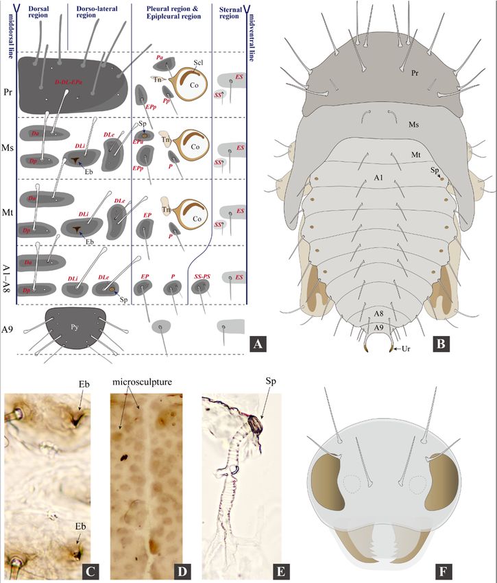
Insects 2020, 11, 571 6 of 27
Insects 2020, 11, x FOR PEER REVIEW 6 of 28
Figure
Figure2.2.AdultAdult morphology
morphology of of
genus
genusCangshanaltica.
Cangshanaltica. (A–L), Cangshanaltica
(A–L), fuanensis
Cangshanaltica sp. nov.;
fuanensis sp. (M–
nov.;
(M–O),
O), otherother Cangshanaltica
Cangshanaltica species.
species. (A): (A): Labrum.
Labrum. (B): (B): Maxilla.
Maxilla. (C): (C): Mandible.
Mandible. (D):(D): Metafemoral
Metafemoral spring
spring of
of hind
hind leg.leg.
(E):(E): Antenna,
Antenna, indicating
indicating thethe apical
apical part
part of of
thethe
7th7th antennomere
antennomere onlyvery
only veryslightly
slightlyproduced.
produced.
(F):Elytra
(F): Elytraunder
underhighhighmagnification
magnification(immersed
(immersedin inglycerin,
glycerin,showing
showingkidney-shaped
kidney-shapedelytral
elytralpunctures
punctures
and small granules. (G): Spermatheca. (H): Vaginal palpi; the arrow indicates the unsclerotized
and small granules. (G): Spermatheca. (H): Vaginal palpi; the arrow indicates the unsclerotized area. area.
(I): Tignum.
(I): Tignum. (J):(J): Female
Female pygidium.
pygidium.(K):(K):Female
Femalereproductive
reproductivesystem,
system,only
onlytwo
twoovarioles
ovariolespresent
presentonon
eachside
each sideofofovary;
ovary;thethearrows
arrowsindicate
indicatethethetwo
twoovarioles
ovarioleson onthe
theright
rightovary.
ovary. (L–O):
(L–O):Vaginal
Vaginalpalpi
palpiof
of
Cangshanalticaspecies,
Cangshanaltica species, showing
showing interspecific
interspecific variation,
variation, insets
insets M–O
M–O are
are redrawn based on [2,3,7].
Thorax.ofPronotum
Description adult. Male(Figure
body1D) strongly
length convex,
1.35–1.55 mm,ratio of 1.10–1.20
width pronotummm;width (measured
female at posterior
body length 1.50–
1.70 mm, width 1.25–1.35 mm. Ratio of body length to body width: 1.20–1.30. Dorsum midlength.
edge) to length: 2.0–2.2. Lateral margin with anterolateral setiferous pore situated nearly in and head
Elytra strongly
smooth, convex,dark
not reticulate, humeral calliVenter
brown. absent.slightly
Elytralighter
with punctures confused,
than dorsum: sparse,
chestnut minute
brown and
to dark
shallow in dorsal view, kidney-shaped sculpture present when immersed in glycerin
brown. Antennae: antennomeres I–II yellow-brown, II–IV yellow-brown to chestnut brown, V–X (Figure 2F).
Hind wings absent. Procoxal cavity open externally. Dorsal side of metatibia with
chestnut brown, XI yellow-brown. Legs chestnut brown. Legs and antennae covered with yellow a row of minute
setae.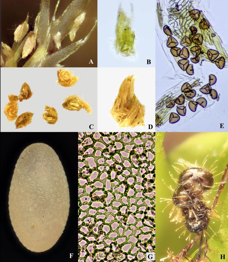
Insects 2020, 11, 571                                                                              7 of 27

spines situated between basal 1/3 and tibial apex. Length of metatibial apical spur to length of
metatarsomere: 0.50–0.60. Proportions of metatarsomere lengths: 100: 6–37:44–45:70–72. First male
protarsomere as large as that of female.
      Abdomen. Longitudinal ridge on first abdominal ventrite strongly developed, extending to near
posterior margin. Last visible abdominal ventrite of male has same shape as female.
      Genitalia. Median lobe of aedeagus (Figure 1F–H) in ventral view: widest at middle or base; ventral
surface with middle longitudinal area raised; sides narrowing from middle to apex; apex rounded,
without denticle; widest near basal opening. Median lobe of aedeagus in lateral view: evenly curved
ventrad, apex slightly bent dorsad, widest near middle.
      Receptacle of spermatheca with external side convex, internal side almost straight to slightly
convex (Figure 2G). Spermathecal pump much shorter and narrower than receptacle, slightly
curved, apex without denticle. Tignum spear-shaped; posterior part short, oval to diamond-shaped,
weakly sclerotized, much wider than anterior part; anterior part narrow and long, crooked near apex
(Figure 2I, indicated by arrow). Vaginal palpi fused at base (anterior part), twice as long as wide,
with base partly unsclerotized (Figure 2H, indicated by arrow); vaginal palpus: posterior half with
lateral sides sinuate and gradually narrowed, posterior apex acute to narrowly rounded.
      Variation. Adults with external characters almost invariable except for a slight variation of body
size. Shape of median lobe of aedeagus: with middle part slightly dilated at middle part in ventral and
lateral views in some cases; apex invariable, always slightly bending dorsally in lateral view. Shape of
female genitalia: internal side of spermathecal receptacle slightly varied from slightly convex to almost
straight; vaginal palpi invariable including the unsclerotized area; tignum invariable including the
crooked anterior part (Figure 2I, indicated by arrow) near anterior apex.
      Barcode sequence. The cox1 barcode is accessible on GenBank, accession numbers: MT891188.
      Type Material (information is recorded verbatim from labels). Holotype: ♂(IZCAS), labels:
(1) China, Fujian Prov., Fuan, Shuyang, 250 m, 27.159◦ N 119.677◦ E, 27-IV-2019, sifted from moss.
(2) HOLOTYPE Cangshanaltica fuanensis sp. nov. Des. Ruan et al. 2019.
      Paratypes: 1♀(IZCAS), labels: (1) Fujian Prov., Fuan, Shuyang Village, 368 m, 27◦ 090 32” N,
119 400 34” E, 2012.VIII.13, Host: Hypnaceae? Leg. Yongying R [label partly written in Chinese];
    ◦

(2) PARATYPE Cangshanaltica fuanensis sp. nov. Des. Ruan et al. 2019. • 1♀(IZCAS), labels: (1) China,
Fujian Prov., Fuan, Shuyang, 250 m, 27.159◦ N 119.677◦ E, 27-IV-2019, sifted from moss; (2) PARATYPE
Cangshanaltica fuanensis sp. nov. Des. Ruan et al. 2019. • 2♂2♀(IZCAS), labels: (1) China, Fujian Prov.,
Fuan, Shuyang, 290 m, 27.1611◦ N 119.6763◦ E, 16-VIII-2019, Extracted from moss, Leg. Y. Ruan;
(2) PARATYPE Cangshanaltica fuanensis sp. nov. Des. Ruan et al. 2019. • 1♂(ADPC), labels: (1) China,
Fujian Prov., Fuan, Shuyang, 290 m, 27.1611◦ N 119.6763◦ E, 3-X-2019, Extracted from moss, Leg. Y.
Ruan; (2) Cangshanaltica fuanensis des. Ruan, 2019; (3) PARATYPE Cangshanaltica fuanensis sp. nov.
Des. Ruan et al. 2019; (4) VOUCHER SPECIMEN A. F. Damaška coll., AFD-265.• 1♂(ADPC), labels:
(1) China, Fujian Prov., Fuan, Shuyang, 290 m, 27.1611◦ N 119.6763◦ E, 3-X-2019, Extracted from moss,
Leg. Y. Ruan; (2) Cangshanaltica fuanensis des. Ruan, 2019; (3) PARATYPE Cangshanaltica fuanensis
sp. nov. Des. Ruan et al. 2019. • 3♀5♂(SZPT), labels: (1) China, Fujian Prov., Fuan, Shuyang, 290 m,
27.1611◦ N 119.6763◦ E, 3-X-2019, Extracted from moss, Leg. Y. Ruan; (2) PARATYPE Cangshanaltica
fuanensis sp. nov. Des. Ruan et al. 2019. • 3♂10♀(SZPT), labels: (1) China, Fujian Prov., Fuan,
Shuyang, 290 m, 27.1611◦ N 119.6763◦ E, 13-II-2020, Extracted from moss, Leg. Y. Ruan; (2) PARATYPE
Cangshanaltica fuanensis sp. nov. Des. Ruan et al. 2020.
      Host plants (see “Biology” section for more information). Adults feed on Hypnum plumaeforme
(Hypnaceae) and Racopilum cf. aristatum (Racopilaceae) in nature, they also casually feed on
Bazzania tridens (Lepidoziaceae), Hylocomiaceae sp. and Thuidiaceae sp. In a lab environment;
moss spores were found in their intestinal tract. Larvae: feed on both Hypnum plumaeforme and
Racopilum cf. aristatum in a lab environment.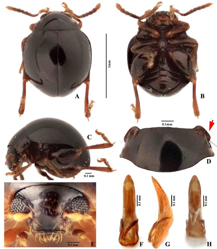
Insects 2020, 11, 571                                                                               8 of 27

      Remarks. By the external habitus, Cangshanaltica fuanensis sp. nov. is similar to other non-metallic
species of Cangshanaltica (C. nigra Konstantinov et al. [2]; C. siamensis Damaška and Konstantinov [3];
and C. sprynari Damaška and Aston [7]).
      Cangshanaltica fuanensis sp. nov. may be distinguished from C. nigra by the following characters:
dorsum dark brown throughout (in C. nigra, dorsum black with slight green-blue luster on elytra);
protrusion at distal part of antennomere VII barely visible (in C. nigra, protrusion at distal part of
antennomere VII strongly developed, clearly visible); in lateral view, apex of median lobe of aedeagus
very slightly bent dorsally (in C. nigra, apex of median lobe of aedeagus bent ventrally).
      Cangshanaltica fuanensis sp. nov. may be distinguished from C. siamensis by the following
characters: body length 1.35–1.70 mm, width 1.10–1.35 mm (in C. siamensis, body length 1.94–2.21 mm,
width 1.45–1.62 mm); middle-longitudinal ridge on first abdominal ventrite long and strongly
developed, extending to near posterior margin (in C. siamensis, middle-longitudinal ridge on first
abdominal ventrite short, weakly developed, not extending past midlength of first abdominal ventrite);
in lateral view, apex of median lobe of aedeagus very slightly bent dorsally (in C. siamensis, apex of
median lobe of aedeagus very slightly bent ventrally).
      Cangshanaltica fuanensis sp. nov. may be distinguished from C. sprynari in by the following
characters: surface of pronotum and elytra smooth, with fine punctures (in C. sprynari, dorsum with
coarse punctures); middle-longitudinal ridge on first abdominal ventrite almost extending to posterior
margin (in C. sprynari, middle-longitudinal ridge on first abdominal ventrite short, not extending to
posterior margin); median lobe of aedeagus in ventral view, apical fourth of median lobe gradually
narrowed, without an abruptly narrowing step (in C. sprynari, apical quarter of median lobe has
an abruptly narrowing step); median lobe of aedeagus in lateral view, apex slightly bent dorsally
(in C. sprynari, apex straight).
      The main sexually dimorphic features present in other genera of flea beetles are absent in some
Cangshanaltica, e.g., in C. nigra: male does not have larger first pro- and meso- tarsomeres; apex of last
visible abdominal ventrite has the same shape in female and male [2]. This condition also occurs in
C. fuanensis sp. nov.: females are almost indistinguishable from males by external characters, except for
the slightly larger body size. However, in C. fuanensis sp. nov., the female tignum could be seen
through abdominal ventrites when they are alive or preserved in ethanol (but hardly visible when the
specimens are dry).
      Although the four non-metallic species of Cangshanaltica are very similar to each other in external
characters, the shape of genitalia is useful to separate species, especially the female vaginal palpi
(see Figure 2L–O).

3.2. Morphology of Immature Stages

3.2.1. Larval Morphology
     Measurements of larvae. Body length 0.90–2.80 mm, body width 0.30–0.80 mm. Head width
0.27–0.46 mm (Figures 3–5; Table 1).

      First instar: head width 0.27–0.30 mm; body length 0.90–1.50 mm, body width 0.30–0.40 mm.
      Second instar: head width 0.34–0.37 mm; body length 1.50–2.30 mm, body width 0.45–0.61 mm.
      Third instar: head width 0.42–0.46 mm; body length 2.00–2.80 mm, body width 0.50–0.80 mm.

     Head width of second instar to that of the first instar ratio: 1.2–1.3. Head width of third instar to
that of second instar ratio: 1.2–1.3.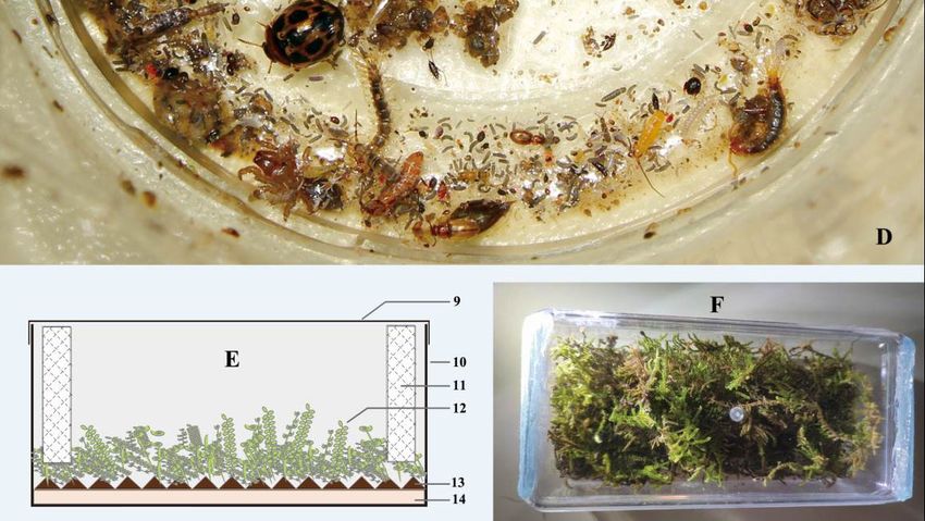
pronotum and body tubercles grey-black; ovoid micro sculpture present on body surface (only visible
under high magnification, see Figure 5D). Head and pronotum with long setae (not capitate).
Mesothorax, metathorax, and abdomen: dorsal and dorso-lateral region with long capitate setae,
epipleural region with shorter setae (not capitate), pleural and sternal region with shorter spine-
Insects 2020,
shaped        11, 571
          setae.                                                                                 9 of
                   Spiracles present on mesothorax and abdominal segments I–VIII (Figure 3A: Sp-ms,   27
                                                                                                    Sp-
a1 to Sp-a8). Larvae preserved in ethanol: body “C”-shaped, pale yellow to whitish.

    Figure
    Figure 3.3. Larval
                Larval morphology
                        morphology ofofCangshanaltica
                                        Cangshanaltica fuanensis
                                                          fuanensissp.
                                                                     sp.nov.
                                                                         nov.(specimens
                                                                               (specimensmounted
                                                                                            mountedin   inglycerin).
                                                                                                           glycerin).
    (A):
    (A): Habitus of 3rd instar, dorsal view. (B): Habitus of 3rd instar, ventral view. (C): Habitus of
         Habitus   of  3rd instar, dorsal view. (B): Habitus  of  3rd instar, ventral   view.  (C):  Habitus  of 3rd
                                                                                                                 3rd
    instar, lateral view.
    instar, lateral view. (D):
                            (D): Pygidium
                                 Pygidium and
                                            and pygopod.
                                                pygopod. (E):
                                                            (E): Thoracic
                                                                 Thoracic leg.
                                                                           leg. (F):  Pronotum. (G):
                                                                                 (F): Pronotum.     (G): Meso-
                                                                                                         Meso- and
                                                                                                                and
    metanotum.
    metanotum. (Abbreviations:
                   (Abbreviations: A1–A10,
                                     A1–A10, abdominal
                                              abdominal segments
                                                         segments I–X;
                                                                   I–X;Co,
                                                                        Co, coxa;
                                                                             coxa; Fe,
                                                                                   Fe, femur;
                                                                                        femur; Ms,
                                                                                               Ms, mesothorax;
                                                                                                     mesothorax; Mt,
                                                                                                                 Mt,
     metathorax; Pd, pygopod; Pr, prothorax; Pu, Pulvillus; Py, pygidium; Scl, sclerotization; Sp, spiracle; Sp-a1,
     1st abdominal spiracle; Sp-a8, 8th abdominal spiracle; Sp-ms, mesothoracic spiracle; Ti, Tibia (Tibia-tarsus); Tn,
     trochantin; Tr, trochanter.)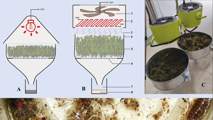
1st abdominal spiracle; Sp-a8, 8th abdominal spiracle; Sp-ms, mesothoracic spiracle; Ti, Tibia (Tibia-tarsus);
     Tn, trochantin; Tr, trochanter.)

       Head (Figure 4). Head not retracted into prothorax, hypognathous, posterior part not ‘V’-
shaped, heavily sclerotized; in frontal view globular, 1.15–1.25 times as long as wide; ovoid in lateral
Insects 2020,
view.         11, 571 absent, one pair of black eyespots (Figure 4A: Eys) present behind antenna (inside
         Stemmata                                                                                 10 of 27

head capsule).

     Figure 4. Larval morphology of Cangshanaltica fuanensis sp. nov. (A): Head; (B): maxilla and
     Figure 4. Larval morphology of Cangshanaltica fuanensis sp. nov. (A): Head; (B): maxilla and labium;
     labium;
     (C):      (C): (D):
          labrum;   labrum;   (D):
                         apical    apical
                                part       part of
                                     of labium  andlabium    and
                                                      maxilla;    maxilla;
                                                               (E): antenna;(E): antenna;
                                                                              (F): mandible.(F):(G):
                                                                                                 mandible.
                                                                                                     antenna;(G):
                                                                                                               (H):antenna;
                                                                                                                    labrum
     (H):  labrum    and   clypeus;  (I): head;  (J):  mandible;    (K):  maxilla   and  labium.    (Abbreviations:
     and clypeus; (I): head; (J): mandible; (K): maxilla and labium. (Abbreviations: Ant, antenna; Ca, cardo;           Ant,
     antenna;
     Cly,       Ca,Con,
           clypeus;  cardo;  Cly, clypeus;
                          condyle;          Con, condyle;
                                   Dsen, digitiform           Dsen,
                                                      sensillum;     digitiform
                                                                 e1–e7,          sensillum;
                                                                         epicranial setae; En,e1–e7,  epicranial
                                                                                               endocarina;        setae; En,
                                                                                                            Ep, epicranium;
     endocarina;  Ep,  epicranium;   Eps,  epicranial  suture; Eys,  eyespot; f1–f3,  frontal setae; Fr, frons; Frs, frontal
     Eps, epicranial suture; Eys, eyespot; f1–f3, frontal setae; Fr, frons; Frs, frontal suture; Gal, galea; Lac, lacinia;
     Lbi, labium; Lbr, labrum; Lbip, labial palpus; Lig, ligula; Ma, mala; Md, mandible; Meb, membrane; Mens,
     mental sclerite; Mi, microtrichia; Mx, maxilla; Mxp, maxillary palpus; Pen, penicillus; Plg, palpiger; Scl,
     sclerotization; Sen, sensillum; Senp, sensory papilla; Smen, submentum; St, stipes.).
Insects 2020, 11, x FOR PEER REVIEW 11 of 28
suture; Gal, galea; Lac, lacinia; Lbi, labium; Lbr, labrum; Lbip, labial palpus; Lig, ligula; Ma, mala; Md,
mandible; Meb, membrane; Mens, mental sclerite; Mi, microtrichia; Mx, maxilla; Mxp, maxillary palpus; Pen,
Insectspenicillus;
2020, 11, 571 11 of 27
Plg, palpiger; Scl, sclerotization; Sen, sensillum; Senp, sensory papilla; Smen, submentum; St,
stipes.).
Figure
Figure 5.
5. Larval
Larvalandandpupal
pupalmorphology
morphologyof ofCangshanaltica
Cangshanalticafuanensis
fuanensissp. sp.nov.
nov.(A):
(A):Tubercular
Tubercular pattern
pattern
of
of mature larva, terminology follows Kimoto (1962) [16] and Takizawa (2005) [17]. (B): Dorsal
mature larva, terminology follows Kimoto (1962) [16] and Takizawa (2005) [17]. (B): Dorsal view
view of
of
Pupa.
Pupa. (C):
(C): Egg-bursters
Egg-bursters on on meso-
meso- and
and metathorax
metathorax of of larva.
larva. (D):
(D): Ovoid
Ovoid micro
micro sculpture
sculpture onon pronotum
pronotum
of larva. (E):
of larva. (E): Mesothoracic
Mesothoracic spiracle.
spiracle. (F):
(F): Head
Head ofof pupa.
pupa. (Abbreviations
(Abbreviations of of tubercles:
tubercles: D, D, dorsal;
dorsal; DL,
DL,
dorsolateral;
dorsolateral; EP,
EP, epipleural;
epipleural; ES,
ES, eusternal;
eusternal; P,
P, pleural;
pleural; PS,
PS, parasternal;
parasternal; SS,
SS, sternellar.
sternellar. Position
Position of
of tubercles
tubercles is
is
denoted by lowercase letters: a, anterior; p, posterior; i, interior; e, exterior. A1–A9, abdominal
denoted by lowercase letters: a, anterior; p, posterior; i, interior; e, exterior. A1–A9, abdominal segments segments I–
IX;
I–IX;Co,
Co,coxa;
coxa;Scl,
Scl,sclerotization;
sclerotization;Eb,
Eb,egg-burster;
egg-burster;Ms,Ms,mesothorax;
mesothorax;Mt, Mt, metathorax; Pr, prothorax;
metathorax; Pr, prothorax; Py,
Py,
pygidium;
pygidium; Sp,
Sp, spiracle;
spiracle; Tn,
Tn, trochantin.).
trochantin.).
Epicranium (Figure 4A: Ep): with six pairs of long (e1–e6) and one pair of short (e7) setae; e5–e7
situated at posterolateral part of epicranial halves. The epicranium has 12–13 pairs of sensilla (Sen),Insects 2020, 11, 571 12 of 27
The following descriptions are based on the third instar larvae:
Larval morphology. Live specimens: body eruciform; lemon yellow, semi-transparent (intestinal
tract and content visible from exterior), head brown, eye spots black; legs blackish, semi-transparent;
pronotum and body tubercles grey-black; ovoid micro sculpture present on body surface (only visible
under high magnification, see Figure 5D). Head and pronotum with long setae (not capitate).
Mesothorax, metathorax, and abdomen: dorsal and dorso-lateral region with long capitate setae,
epipleural region with shorter setae (not capitate), pleural and sternal region with shorter spine-shaped
setae. Spiracles present on mesothorax and abdominal segments I–VIII (Figure 3A: Sp-ms, Sp-a1 to
Sp-a8). Larvae preserved in ethanol: body “C”-shaped, pale yellow to whitish.
Head (Figure 4). Head not retracted into prothorax, hypognathous, posterior part not ‘V’-shaped,
heavily sclerotized; in frontal view globular, 1.15–1.25 times as long as wide; ovoid in lateral
view. Stemmata absent, one pair of black eyespots (Figure 4A: Eys) present behind antenna
(inside head capsule).
Epicranium (Figure 4A: Ep): with six pairs of long (e1–e6) and one pair of short (e7) setae;
e5–e7 situated at posterolateral part of epicranial halves. The epicranium has 12–13 pairs of sensilla (Sen),
including four pairs of larger ones near frontal sutures. Epicranial suture (Eps) short, approximately
half as long as endocarina (En).
Frons (Figure 4A: Fr): with three pairs of long setae (f1–f3) and one pair of sensilla; frontal sutures
(Frs) reaching antennal sockets; median endocarina narrow ridge-shaped, extending from base of
frontal sutures to clypeus.
Clypeus (Figure 4A: Cly): sclerotized, transverse, band-shaped, bearing 1 pair of long setae
and three pairs of sensilla. Clypeus and frons divided by epistomal sulcus; epistomal ridge
strongly sclerotized.
Labrum (Figure 4C,H: Lbr): sclerotized, transverse; bearing one pair of setae medially, one pair of
setae laterally and one pair of sensilla near midline; anterior edge deeply incised (i.e., anteromedial
notch), bearing numerous microtrichia; posterior edge with middle part produced posteriorly.
Mandibles (Figure 4F,J: Md): symmetrical, palmate, mesal membranous lobe absent (present in
adults); each mandible with four sharp teeth, one sensillum near base and two setae on lateral edge,
penicillus with 7–8 stiff setae, first tooth largest, remainder decrease in size.
Antenna (Figure 4E,G): weakly sclerotized, two segmented, attached to membranous area at end
of frontal suture; first antennomere partly membranous, bearing one large conical sensory papilla and
several sensilla; second antennomere small, bearing about four sensilla.
Maxilla (Figure 4B,D): cardo (Ca) small, triangular, bearing one seta, with a longitudinal
band-shaped sclerotization (Scl); stipes (St) elongate with a long and curved sclerotization (Scl), bearing
two long setae near lateral edge. Mala with galea (Gal) and lacinia (Lac) not fused; apical part of galea
with approximately 5–6 setae arranged around a stout pedunculate seta (appearing two-segmented);
apical part of lacinia bearing a row of peg-shaped stout setae; maxillary palpus (Mxp) three-segmented:
first palpomere with two setae, second palpomere with two setae and one sensillum, third palpomere
with one digitiform sensillum (Dsen).
Labium (Figure 4B,D: Lbi): submentum (Smen) trapezoid, membranous, bearing two pairs of long
setae; mentum not well defined; prementum short and transparent, with prominent horseshoe-shaped
mental sclerite, bearing one pair of setae at base; ligular membranous, not separated from prementum,
anterior edge broadly rounded to straight, bearing numerous microtrichia; labial palpi small,
two segmented; five pairs of sensilla present near labial palpi.Insects 2020, 11, 571 13 of 27
Thorax. The tubercular pattern on the right side of the thorax is illustrated in Figure 5A and
described as follows (tubercle names are italicized).
Prothorax: dorsum with large D-DL-EPa bearing seven long setae and about five sensilla,
well sclerotized. Tubercles on epipleural, pleural, and sternal regions weakly sclerotized, with poorly
defined edge. Epipleural region with EPp bearing one long seta (not capitate). Pleural region with Pa
and Pp each bearing one spine-shaped seta. Sternal region with ES and SS obsolete, each bearing one
spine-shaped seta.
Mesothorax: tubercles weakly sclerotized, with poorly defined edge. Dorsal region with Da and
Dp each bearing one long capitate seta and one sensillum. Dorso-lateral region with DLi bearing one
long capitate seta and one egg-burster; DLe bearing two capitate setae, and two sensilla. Epipleural
region with EPa bearing one spiracle and EPp bearing one long seta (not capitate). Pleural region
with P bearing one spine-shaped seta. Sternal region with ES and SS obsolete, each bearing one
spine-shaped seta.
Metathorax has the same tubercular pattern as mesothorax, except for the absence of Epa and
spiracle; one egg-burster present on DLi.
Leg (Figure 3E). One seta present on base of claw. Pulvillus present, membranous, round to ovoid,
as long as tarsungulus.
Abdomen. Abdomen with 10 abdominal segments. The tubercular pattern on the right side of
the abdomen is illustrated in Figure 5A and described as follows (tubercle names are italicized).
Segments I–VIII: with same tubecular pattern and chaetotaxy, tubercles weakly sclerotized,
with poorly defined edge. Dorsal region with Da and Dp each bearing one long capitate seta and one
sensillum. Dorso-lateral region with DLi bearing 1 long capitate seta; DLe bearing one long capitate seta,
one setose sensillum and one spiracle. Epipleural region with Ep bearing one long seta (not capitate)
and one setose sensillum. Pleural region with P bearing one long and one short spine-shaped setae.
Sternal region with SS-PS bearing one long and one short spine-shaped setae; ES obsolete, bearing one
spine-shaped seta.
Segment IX. Pygidium (Figure 3D) moderately sclerotized, bearing two pairs of capitate setae
close to lateral edge, another two pairs of un-capitate setae on lateral edge, and about three pairs of
sensilla. Lateral side with one pair of obsolete tubercles bearing one spine-shaped seta. Venter with
one pair of obsolete tubercles bearing one spine-shaped seta.
Segment X not visible in dorsal view, bearing prominent eversible pygopod (Figure 3C: Pd).
Remarks on larval characters. The general appearance of Cangshanaltica fuanensis sp. nov. is
similar to some free-living flea beetle larvae described by previous studies (see Table 1), such as Altica
Geoffroy [20], Ivalia Jacoby [14], Distigmoptera Blake [10]. However, the larval tubercular pattern
(Figure 5A) of C. fuanensis sp. nov. is unique among flea beetles. For instance, pleural tubercle of
prothorax divided into two parts (Pp and Pa); Epa present on mesothorax and absent on metathorax;
pygidium with four long setae; only two tubercles (DLi and DLe) present on dorso-lateral region of
mesothorax, metathorax, and abdomen.Insects 2020, 11, 571 14 of 27
Table 1. Comparison of larval morphology between Cangshanaltica fuanensis sp. nov. and other flea beetles.
Cangshanaltica Ivalia korakundah, Distigmoptera Agasicles hygrophila Podagricomela Aphthona russica
fuanensis sp. nov. Parathapan et al. borealis Blake Selman and Vogt shirahatai (Chûjô) Konstantinov et al.
Leaf feeding Leaf mining Root feeding
Feeding type Leaf feeding (moss) Leaf feeding (moss) Leaf feeding (moss)
(angiosperm) (angiosperm) (angiosperm)
Body color
Lemon yellow White Yellow Green-black to black Yellow Whitish
(mature larva)
Slightly flattened
Eruciform; Eruciform; Eruciform; Eruciform; Subcylindrical;
Body shape dorso-ventrally;
short and robust short and robust short and robust short and robust elongate and slender
robust
Sclerotization Weak, Moderate, Strong,
Absent Absent Absent
of tubercles edge poorly defined edge well defined edge well defined
Long capitate
Present Present Present Absent Unknown Absent
setae on dorsum
Head orientation Hypognathous Hypognathous Hypognathous Hypognathous Prognathous Hypognathous
Flattened Almost parallel-sided;
Head shape Globular Globular Globular Globular
dorso-ventrally slightly elongated
Posterior Strongly developed
Shallow Shallow Moderately deep Shallow Moderately deep
emargination of head and V-shaped
Stemmata Absent Absent Absent 1 pair Absent Absent
Present
Eyespot Present Unknown Absent Present Unknown
(based on image)
Epicranial suture Short Short Short Short Absent Short
Strongly developed
Shape of Endocarina Narrow ridge Unknown Narrow ridge Narrow ridge Narrow ridge
Median bulge
Anterior edge Slightly concave
Deeply concave Deeply concave Moderately concave Convex Slightly concave
of labrum at middle
Number and shape of 4; sharp, 4; largest tooth 4 sharp long
3 sharp and 1 blunt 3 large and 1 small 4; all blunt
mandibular teeth with similar size bearing minute serration and 1 small
Source of data: Cangshanaltica fuanensis sp. nov.—current study; Ivalia korakundah [14]; Distigmoptera borealis Blake [10]; Agasicles hygrophila—observed in the current study;
Podagricomela shirahatai [21]; Aphthona russica [22].Insects 2020, 11, 571 15 of 27
The adult morphology of Cangshanaltica Konstantinov et al. is close to Ivalia Jacoby [2].
Larval morphology of C. fuanensis sp. nov. also shows extreme similarity with Ivalia korakundah
Parathapan et al., especially in the body shape, tubercular pattern, and chaetotaxy. Both larval and
adult morphology suggests that they are very close lineages. Despite the resemblance in general
morphological features, the larvae of Cangshanaltica fuanensis sp. nov. and Ivalia korakundah can
be differentiated by the following characters: in C. fuanensis sp. nov. body tubercles with weak
sclerotization and poorly defined edge (tubercles with moderate sclerotization and well-defined edge
in I. korakundah), tubercle EPa of mesothorax absent (EPa of mesothorax present in I. korakundah),
maxilla with triangular cardo (cardo narrow and transverse in I. korakundah).
The larval mandible of C. fuanensis sp. nov. is very similar to that of adults in the general palmate
shape, sharp teeth, decreasing in size from the first to last tooth. The significant difference is that the
adult mandible has one more tooth, and a mesal membranous lobe with dense microtrichia, which are
absent in the larvae. The resemblance of their mandible shape may be related to similar feeding
behavior, both are eating the leaves of the same host plant.
3.2.2. Pupal Morphology
Body length 1.60–1.75 mm; width 0.90–1.10 mm; bent ventrally in lateral view. Body white–yellow
to yellow–brown, bearing long, brownish, and spine-shaped setae. Head invisible from above, bearing
3 pairs of long setae. Mesothorax and abdominal segment I–V bearing one spiracle each. Pronotum
bearing seven pairs of long and one pair of short setae; meso- and metanotum each bearing one pair of
setae near middle. Abdominal segments I–VIII each bearing one pair of dorsal and one pair of lateral
setae. Abdominal segment IX bearing two pairs of setae and one pair of distal urogomphi. Abdominal
segment X invisible in dorsal view, bearing prominent eversible pygopods (Figure 5B,F).
3.2.3. Egg Morphology
Length 0.68–0.74 mm, width 0.36–0.40 mm. Egg large, egg length to female adult body length
ratio: 0.4–0.5; egg width to female adult body width ratio: 0.29–0.33. Egg oval and lemon yellow.
Surface with minute reticulated pattern consists of irregular polygonal cells (mostly hexagonal or
pentagonal when viewed under high magnification) (Figure 6D,E, Figure 7F,G and Figure 8A; Table 2).
Remarks. The polygonal pattern on egg chorion resembles those in some other flea beetles,
e.g., the polygonal pattern in Agasicles hygrophila Selman and Vogt (observed in current study), and the
hexagonal cells further subdivided into 4 to 6 smaller ones in Disonycha leptolineata Blatchley [23].
3.3. Biology
3.3.1. Life History
Approximately 30 F0 parent adults, 35 F1 eggs, 30 F1 larvae, 10 F1 pupae, and 6 F1 adults were
observed in this study (in a lab environment, ca. 25 ◦ C, 90–100% air humidity). The following biological
data were collected based on these lab-reared individuals (Figure 8).
Life longevity lasts approximately 90–150 days. Approximately 3–4 generations per year are
predicted in the type locality (Fujian, China).
Egg stage (Figures 7F and 8A). The fecundity is approximately 2–4 eggs per female. The egg stage
lasts approximately 7–16 days on slightly moist to entirely wet moss. The hatching of the eggs on dry
moss usually took more time than those on wet moss. It indicates that higher humidity may expedite
the development of eggs.
Larval stage (Figure 8B–E). Duration: ca. 14–30 days. Chorion ingestion is absent based on
our observation. About 1–2 days before hatching, larvae are active and visible from the exterior;
apex of mandibles are sclerotized and red-brown; eyespots and egg-bursters are black and prominent;
larvae frequently move and rotate inside the egg chorion in this period. Before emerging, larvae contract
their body and push the dilated thorax (dorsum) against egg chorion, after several times of contraction,Insects 2020, 11, 571 16 of 27
the egg-bursters slit the chorion. Newly hatched larvae usually stay at the base of moss, they feed
on both rotten and fresh leaves of the host plant. Two larvae were spotted feeding on adult feces
(their feeding last ca. 2–3 min, in a lab environment). As the adult feces consist of un-digested moss
leaf fragments (see Figure 7D), they are probably suitable for a food source for young larvae. It is still
unknown if the feces-feeding behavior also occurs in nature.
Insects 2020, 11, x FOR PEER REVIEW 16 of 28
6. Biology of Cangshanaltica
Figure 6. Cangshanaltica fuanensis
fuanensis sp.
sp. nov.
nov. (A):
(A): Habitat
Habitat environment—a
environment—a north-facing
north-facing
valley,arrow
valley, arrowshowing
showingcollecting
collecting spot—a
spot—a deserted
deserted plum
plum orchard
orchard withwith a creek
a creek flowing
flowing nearby.nearby. (B):
(B): Host
Host plant Hypnum plumaeforme on the stem of a plum tree; they usually grow on
plant Hypnum plumaeforme on the stem of a plum tree; they usually grow on soil, rocks, and soil, rocks, and tree
stem. (C): An adult on the host plant. (D):
(D): A 2nd instar larva is eating an egg in a lab lab environment
environment
(cannibalism); arrow indicating an egg hidden under under aa moss
moss leaf
leaf byby female.
female. (E): A damaged
damaged egg
species of
consumed by an unknown species of springtail
springtail (collembola)
(collembola) in
in aa lab
lab environment.
environment.Insects 2020, 11, 571 17 of 27
Insects 2020, 11, x FOR PEER REVIEW 17 of 28
Figure 7.
Figure 7. Biology
BiologyofofCangshanaltica
Cangshanalticafuanensis
fuanensissp.sp.nov.
nov.(A,B): Larval
(A,B): feces.
Larval (C,D):
feces. Adult
(C,D): feces.feces.
Adult (E):
Contents in the intestinal tract of an adult, showing moss cells and spores. (F): Egg. (G):
(E): Contents in the intestinal tract of an adult, showing moss cells and spores. (F): Egg. (G): MicroMicro
sculpture on
sculpture on egg
egg chorion.
chorion. (H):
(H): Excuvium
Excuvium ofof larva.
larva.
Because the larvae are lemon yellow and semi-transparent, they are challenging to discover on the
host plant (yellow-green). Furthermore, the host plant tissue in their intestinal tract is only partially
digested and highly visible externally, which makes their color much closer to that of moss.
First instar. Duration: ca. 4–7 days. Head width 0.27–0.30 mm; body length 0.90–1.50 mm,
body width 0.30–0.40 mm. Newly hatched individuals appear unsclerotized (Figure 8B, the individual
on the right side), transparent except for red mandibles, black eyespots and egg-bursters, and light-black
setae. Old individuals (Figure 8C): body becomes semi-transparent and lemon yellow; pronotum,
pygidium, and tubercles light-black; mandibles brown. The number and shape of egg-bursters are
similar to some other flea beetles we examined (e.g., Agasicles hygrophila Selman and Vogt).Insects 2020, 11, 571 18 of 27
Insects 2020, 11, x FOR PEER REVIEW 18 of 28
Figure 8. Life cycle of Cangshanaltica fuanensis sp. nov. (1 mm
sp. nov. mm Scale
Scale bar
barisissuitable
suitablefor
forinsets
insets(A–I)).
A–I).
(A): Two
Two eggs
eggsare
arelaid
laidunder
underone
onemoss
mossleaf (this
leaf is is
(this a rare case;
a rare usually,
case; there
usually, is only
there one one
is only egg egg
under one
under
leaf). (B): Newly hatched larvae; the individual on the right is just crawling out of the egg.
one leaf). (B): Newly hatched larvae; the individual on the right is just crawling out of the egg. (C): (C): First
instarinstar
First larva. larva.
(D): Third instar larva.
(D): Third instar (E): Prepupal
larva. larva (with
(E): Prepupal larvamilky-white color) in color)
(with milky-white pupal inchamber.
pupal
(F): Pupa, lateral
chamber. view.lateral
(F): Pupa, (G): Pupa,
view.ventral view. ventral
(G): Pupa, (H): Newlyview.emerged adult. emerged
(H): Newly (I): Well-sclerotized
adult. (I): adult.
Well-
sclerotized adult.
Second instar. Duration: ca. 4–6 days. Head width 0.34–0.37 mm. Head width of second instar to
that of first instar ratio: 1.2–1.3. Body length 1.50–2.30 mm, body width 0.45–0.61 mm. The newly
molted individual with body transparent and soft. Old individual: body lemon yellow; pronotum,
pygidium, and tubercles light black; mandibles reddish-brown to dark brown. Egg-bursters obsolete.Insects 2020, 11, 571 19 of 27
Third instar. Duration: ca. 8–13 days (including a prepupal period). Head width 0.42–0.46 mm.
Head width of third instar to that of second instar ratio: 1.2–1.3. Body length 2.00–2.80 mm, body width
0.50–0.80 mm. With similar coloration as second instar. Egg-bursters obsolete.
Prepupal period (pupating period): 3–5 days, body color turned milky white. In the first 1–2 days:
prepupal larvae stop feeding, crawl down to soil surface near the base of moss, excrete residual contents
in the intestinal tract, and search for pupation location. Day 2–3: they build an earthen chamber,
use legs to collect moist earth patches, bite off minute earth granules one by one using mouthpart,
then attach them to the chamber wall; by this means, they construct the oval and smooth inner wall of
the earthen chambers. Day 3–5: earthen chambers are completed, they cease movement and are not
responding to minor stimuli.
Pupal stage (Figure 8F,G). Duration: 10–15 days, abdominal segments are able to move,
with eversible pygopod. In the first 1–3 days: body light-yellow; surface glabrous and transparent.
Day 4–6: body yellow, semi-transparent; mandibles and compound eyes red. Day 7–8: body light
yellow-brown, semi-transparent, mandibles and metafemoral springs brown, compound eyes red.
Day 9–15: body yellow-brown, not transparent; eyes, legs, antennae, mouthparts and metafemoral
springs well sclerotized and with brown color.
Adult stage (Figure 8H,I). Adult longevity: ca. 60–100 days. After emergence from pupae,
adults stay in the earthen cell (for approximately one day), with yellow and unsclerotized integument,
and are unable to jump upon stimulation. After a few days (ca. 2–3 days), their integuments gradually
turn brown, and they can perform minor jumps. After approximately 5–6 days, adults can perform
explosive jumps.
3.3.2. Reduction of Ovarioles and Large Egg Size
The ovaries of seven females of Cangshanaltica fuanensis sp. nov. were dissected in this study
(Figure 2K): females have two ovaries, each ovary has only two ovarioles (Figure 2K, indicated by
two arrows), which is very unusual in leaf beetles. According to [24], the ovariole number per ovary
usually varies from 4 to 46 in Chrysomelidae.
Table 2. Larger egg size and smaller egg number in Cangshanaltica fuanensis sp. nov. compared with
several other flea beetles.
Egg Length to Egg Width to
Egg Length Adult Body Egg Numbers
Adult Body Adult Body
and Width Length and Width Laid by Female
Length Ratio Width Ratio
1.5–1.7 mm;
0.68–0.74 mm;
Cangshanaltica fuanensis sp. nov. 1.2–1.3 mm 0.40–0.50 0.28–0.33 Ca. 2–4, laid separately
0.36–0.40 mm
(female)
Agasicles hygrophila 1.26 mm;
6 mm; 3 mm 0.21 0.18 >50, laid in clusters
Selman and Vogt 0.54 mm
0.85 mm;
Altica caerulescens (Baly) 4 mm; 2 mm 0.21 0.18 Unknown
0.35 mm
0.84 mm;
Altica fragariae Nakane 3.8 mm; 2 mm 0.22 0.19 >100, laid in clusters
0.37 mm
0.75 mm; 1.9–3 mm;
Chaetocnema ingenua Baly 0.25–0.39 0.22–0.39 Ca. 100, laid in clusters
0.35 mm 0.9–1.57 mm
1.77–2.23 mm; 6.2–7.5 mm; Unknown,
Disonycha leptolineata Blatchley 0.24–0.36 0.15–0.32
0.66–1.09 mm 3.4–4.5 mm laid in clusters
0.37 mm; 1.8–2.4 mm;
Phyllotreta striolata (Fabricius) 0.15–0.21 0.23 >25, laid in clusters
0.21 mm 0.9 mm
Source of data for egg length and adult body length: Agasicles hygrophila—measured in current study; Altica spp. [25];
Chaetocnema ingenua Baly [26–28]; Disonycha leptolineata Blatchley [23]; Phyllotreta striolata [29,30].
Females lay significantly larger and fewer eggs than many other flea beetles (see Table 2),
the maximal egg length reaches up to half of the female body length.Insects 2020, 11, 571 20 of 27
3.3.3. Egg Hiding Behavior of Females
The females deposit one egg at a time. Eggs are laid independently and at a distance from each
other. Each egg is hidden under a single leaf of the host plant (see Figure 6D, indicated by arrow).
As the moss leaves are strongly convex dorso-ventrally, spoon-shaped, and slightly larger than the
egg, they could well contain and cover the eggs. The eggs are not garnished with feces or litter.
However, they are cryptic under moss leaves and very difficult to discover, even when viewed under
the stereomicroscope. It would be more difficult to spot them when the moss is wet, and the eggs are
partly immersed in water. By this means, they may be protected from potential predators (see the
following text in the “Natural enemies” section). In a sporadic case, two eggs were laid adhering to
each other under the same moss leaf (Figure 8A).
3.3.4. Cannibalism
The cannibalism of a second instar larva on an egg was observed (see Figure 6D). The entire
feeding process lasted for approximately 10 min; in the end, most of the egg contents were eaten by
the larva.
3.3.5. Host Plant and Feeding Habit
Both adults and larvae are polyphagous; they can feed on different host plants of different moss
families. In nature, adults primarily inhabit and feed on Hypnum plumaeforme (Hypnaceae), which is
a common moss in East Asia (including both north and south China) and grows in various habitats
at different altitudes (50–4000 m above sea level) [18]. Adults also feed on Racopilum cf. aristatum
(Racopilaceae) in nature. However, at the type locality, Racopilum cf. aristatum is rather scarce compared
to the dominant moss species Hypnum plumaeforme. In a lab environment, when Hypnum plumaeforme
and Racopilum cf. aristatum are absent, adults also casually feed on Bazzania tridens (Lepidoziaceae),
Hylocomiaceae sp. and Thuidiaceae sp. Larvae feed on both Hypnum plumaeforme and Racopilum cf.
aristatum in a lab environment; their feeding behavior in the natural environment was not studied.
The food source is abundant to C. fuanensis sp. nov. An investigation was conducted on a 30 m2
sample plot at the type locality in July 2020. It turned out only 15 adults were discovered on the
surface of moss from 9–11 p.m. in a single night. Based on our field investigation, the host plant
Hypnum plumaeforme is abundant, and the feeding injury on the host plant (e.g., damages on the distal
end of young shoots of moss) is scarce. All these indicate the population density of this species may
be low.
Adults usually feed on the distal end of moss branches, the end of young shoots are usually
chopped off by their feeding, which is destructive to the host plant. In a lab environment, adults can
feed on other parts of the host plant after most of the end of young shoots are eaten. Dissection of the
intestinal tract of adults collected from nature shows that they also consume moss spores (Figure 7E).
At the type locality of C. fuanensis sp. nov., another flea beetle species-Benedictus sp. was
discovered. In both natural and lab environment, it feeds on H. plumaeforme, and has very similar
feeding habits, eating the distal end of young branches. The population density of Benedictus sp. is
probably lower than C. fuanensis sp. nov. based on the number of specimens collected (less than 10).
The competition between the two species in nature is still unknown.
Feces of larvae and adults mainly consist of undigested fragments of host plant leaf
(see Figure 7B,D), which is unique in flea beetles.
3.3.6. Habitat Environment
Most specimens collected in this study were extracted from moist moss in a north-facing valley with
a creek flowing nearby at the type locality (Fujian, China) (Figure 6A). The surrounding mountain has
north-facing steep slopes; they prevent long-time direct sunlight; therefore, they create a comparatively
humid environment. There are various mosses in this area, and they are able to grow all year-round.You can also read