Temperature controlled high-throughput magnetic tweezers show striking difference in activation energies of replicating viral RNA-dependent RNA ...
←
→
Page content transcription
If your browser does not render page correctly, please read the page content below
Published online 14 April 2020                                                     Nucleic Acids Research, 2020, Vol. 48, No. 10 5591–5602
                                                                                                                   doi: 10.1093/nar/gkaa233

Temperature controlled high-throughput magnetic
tweezers show striking difference in activation
energies of replicating viral RNA-dependent RNA
polymerases

                                                                                                                                                                  Downloaded from https://academic.oup.com/nar/article-abstract/48/10/5591/5819939 by MPI Science of Light user on 10 July 2020
Mona Seifert1 , Pauline van Nies1 , Flávia S. Papini1 , Jamie J. Arnold2 , Minna M. Poranen3 ,
Craig E. Cameron2 , Martin Depken4,* and David Dulin 1,*
1
  Junior Research Group 2, Interdisciplinary Center for Clinical Research, Friedrich-Alexander-University
Erlangen-Nürnberg (FAU), Cauerstr. 3, 91058 Erlangen, Germany, 2 Department of Microbiology and Immunology,
School of Medicine, The University of North Carolina Chapel Hill, 6012 Marsico Hall, CB 7290 Mason Farm Road, NC
27599, USA, 3 Molecular and Integrative Biosciences Research Program, Faculty of Biological and Environmental
Sciences, University of Helsinki, Viikki Biocenter 1, P.O. Box 56 (Viikinkaari 9), 00014 Helsinki, Finland and
4
  Department of Bionanoscience, Kavli Institute of Nanoscience, Delft University of Technology, Van der Maasweg 9,
2629 HZ Delft, The Netherlands

Received January 13, 2020; Revised March 10, 2020; Editorial Decision March 26, 2020; Accepted March 30, 2020

ABSTRACT                                                                          temperature controlled study of biomolecular com-
                                                                                  plex at the single molecule level.
RNA virus survival depends on efficient viral genome
replication, which is performed by the viral RNA de-                              INTRODUCTION
pendent RNA polymerase (RdRp). The recent devel-
opment of high throughput magnetic tweezers has                                   Genome replication is essential to any organism. To achieve
enabled the simultaneous observation of dozens of                                 this task, every RNA virus encodes an RNA dependent
viral RdRp elongation traces on kilobases long tem-                               RNA polymerase (RdRp) that synthesizes all the viral
                                                                                  RNA, either to form messenger RNA for viral protein
plates, and this has shown that RdRp nucleotide ad-
                                                                                  translation, or to produce new viral genomes that are en-
dition kinetics is stochastically interrupted by rare                             closed into viral particles as they mature into infectious
pauses of 1–1000 s duration, of which the short-                                  virions. Furthermore, the viral RdRp also evolves the vi-
lived ones (1–10 s) are the temporal signature of a                               ral genome either by incorporating mutations (1,2) while
low fidelity catalytic pathway. We present a simple                               replicating the viral genome, or by assisting RNA recom-
and precise temperature controlled system for mag-                                bination (3). The viral RdRp is a key player in success-
netic tweezers to characterize the replication kinet-                             ful infection, and is therefore the target of many antiviral
ics temperature dependence between 25◦ C and 45◦ C                                drugs (4,5). Of special interest is the mechanochemical cy-
of RdRps from three RNA viruses, i.e. the double-                                 cle of nucleotide incorporation occurring at the RdRp cat-
stranded RNA bacteriophage 6, and the positive-                                  alytic site (6). This site is conserved among RNA viruses
sense single-stranded RNA poliovirus (PV) and hu-                                 (7), and resembles the one of the DNA and RNA poly-
                                                                                  merases in the A family with their typical cupped right hand
man rhinovirus C (HRV-C). We found that 6 RdRp is
                                                                                  shape (7–9). PV and 6 RdRps are model for the RdRps of
largely temperature insensitive, while PV and HRV-C                               positive-sense single-stranded (ss) and double-stranded (ds)
RdRps replication kinetics are activated by temper-                               viruses, respectively. Pre-steady-state kinetics, structural bi-
ature. Furthermore, the activation energies we mea-                               ology, molecular dynamics and next generation sequencing
sured for PV RdRp catalytic state corroborate previ-                              studies on PV RdRp and its genome have largely shaped
ous estimations from ensemble pre-steady state ki-                                our understanding on nucleotide selection and incorpora-
netic studies, further confirming the catalytic origin                            tion kinetics by viral RdRps (10), while structural and bio-
of the short pauses and their link to temperature in-                             chemical analyses on the 6 RdRp have provided insights
dependent RdRp fidelity. This work will enable future                             into the de novo initiation mechanism (11–13). However,

* To
   whom correspondence should be addressed. Tel: +49 9131 85 70347; Fax: +49 9131 85 35903; Email: david.dulin@uk-erlangen.de
Correspondence may also be addressed to Martin Depken. Tel: +31 15 27 81305; Email: s.m.depken@tudelft.nl

C The Author(s) 2020. Published by Oxford University Press on behalf of Nucleic Acids Research.
This is an Open Access article distributed under the terms of the Creative Commons Attribution Non-Commercial License
(http://creativecommons.org/licenses/by-nc/4.0/), which permits non-commercial re-use, distribution, and reproduction in any medium, provided the original work
is properly cited. For commercial re-use, please contact journals.permissions@oup.com
5592 Nucleic Acids Research, 2020, Vol. 48, No. 10
a complete description of the kinetic cycle of viral RdRp PV and HRV-C RdRps, as both are human enteroviruses.
elongation is still lacking. Indeed, the ensemble techniques Indeed, we show that PV and HRV-C RdRps present a
are unable to access RdRp kinetics on genome-long tem- steep and similar increase in nucleotide addition rate and
plates, i.e. kilobases (kb), and cannot interrogate rare asyn- in the exit rates for the short pauses (0.3–10 s), while the
chronous events, such as nucleotide misincorporation or an- same kinetic states are lesser affected for 6 RdRp. Fur-
tiviral nucleotide analogue incorporation in the presence of thermore, we noticed that the long pauses (>20 s) prob-
cognate canonical NTPs. Our recent single molecule studies ability is largely unaffected for all RdRp. For the three
on 6 and PV RdRps elongation kinetics have partly filled RNA viruses we studied, we show that RNA virus repli-
this gap, shedding light on the kinetic of elongation pauses cation rate is optimum near the optimum growth tempera-
Downloaded from https://academic.oup.com/nar/article-abstract/48/10/5591/5819939 by MPI Science of Light user on 10 July 2020
of various biochemical origins (14–16). This work relied on ture of the host cell. Moreover, we believe that the easy im-
the concomitant development of high throughput magnetic plementation and low-cost of the temperature control sys-
tweezers, a powerful single molecule force and torque spec- tem we have characterized makes it very attractive and will
troscopy technique enabling the characterization of protein- therefore be of large interest to the single molecule commu-
nucleic acids interactions at both high throughput (15,17– nity using instruments that include an oil immersion ob-
20) and high-resolution (21–23), and of a new analysis ap- jective, as well as for fluorescence microscopy and optical
proach based on First-Passage statistics (24,25). However, tweezers.
the home-built magnetic tweezers instrument used for these
studies did not include a temperature control system, and MATERIALS AND METHODS
the experiments were therefore performed at room temper-
ature. Ensemble kinetic assays have demonstrated the tem- High throughput magnetic tweezers apparatus
perature dependence of RdRps activity, either in initiation The high throughput magnetic tweezers apparatus has been
for dengue and Zika virus RdRps (26), or in elongation previously described (15,18,35). Shortly, it is a custom in-
for PV RdRp (27). Furthermore, temperature controlled ex- verted microscope with a 50× oil immersion objective (CFI
periments performed on Escherichia coli RNA polymerase Plan Achro 50 XH, NA 0.9, Nikon, Germany), on top of
(RNAP) at the single molecule level using optical tweezers which a flow cell is mounted. The assembly, surface prepa-
have further informed on the mechanochemical cycle of nu- ration of the flow cell and nucleic acid tethering of the mag-
cleotide addition and on the nature of RNAP pauses, show- netic beads is described in the paragraph below. To apply an
ing that off-pathway pauses have no enthalpic contribution, attractive force to the magnetic beads and stretch the nucleic
i.e. pause exit rate demonstrating no temperature depen- acid tether, a pair of vertically aligned permanent magnets
dence, whereas nucleotide addition rate is dominated by en- (5 mm cubes, SuperMagnete, Switzerland) separated by a
thalpy, i.e. having strong temperature dependence (28,29). A 1 mm gap (36) are positioned above the objective; the ver-
temperature dependent study of viral RdRps elongation ki- tical position and rotation of the beads are controlled by
netics would thus significantly complement our current un- the M-126-PD1 and C-150 motors (Physik Instrumente PI,
derstanding of their mechanochemical cycle, and warrants GmbH & Co. KG, Karlsruhe, Germany), respectively (Fig-
the development of temperature controlled high throughput ure 1A). The field of view is illuminated through the mag-
magnetic tweezers. nets gap by a collimated LED-light source located above
Recently, several studies have reported on the develop- it, and is imaged onto a large chip CMOS camera (Dalsa
ment of custom temperature controlled magnetic tweez- Falcon2 FA-80-12M1H, Stemmer Imaging, Germany). The
ers assays, i.e. relying either on a totally custom ap- temperature control system is made of a flexible resistive foil
proach (home-built proportional-integral-derivative (PID) heater with an integrated 10 M thermistor (HT10K, Thor-
controller, heating elements and thermistors disposed at labs) wrapped around an oil immersion objective (CFI Plan
several locations on the microscope, and a custom graphic Achro 50 XH, NA 0.9, Nikon, Germany) and further insu-
user interfaces (GUI)), or a dedicated commercial device lated by several layers of kapton tape (KAP22-075, Thor-
(30–34). In the present study, we take advantage of a simple labs). The heating foil is connected to a PID temperature
and robust commercial device from Thorlabs (originally de- controller (TC200 PID controller, Thorlabs) to adjust the
signed to control the temperature on 1 inch diameter optical temperature within ∼0.1◦ C. We used the Thorlabs GUI to
tubes and provided with its own GUI) to precisely control control the heating system via USB on the data acquisition
the temperature on an oil immersion microscope objective. computer.
Doing so, we are able to maintain the temperature within
∼0.1◦ from room temperature up to 60◦ C in the whole field
Flow cell fabrication
of view, i.e. with dimensions of (0.5 × 0.4) mm, of a home-
built high throughput magnetic tweezers. The flow cell assembly has been described previously (35).
We applied this temperature controlled high through- The flow cell was mounted on the magnetic tweezers setup
put magnetic tweezers to study the temperature depen- and rinsed with 1 ml of 1× phosphate buffered saline
dence of the replication kinetics of three viral RdRps, i.e. (PBS). 100 l of a 1:1000 dilution of 3 m polystyrene
6, PV and HRV-C RdRps. On the one hand, we ex- beads (LB30, Sigma Aldrich, Germany, stock concentra-
pect a different response to temperature for 6 and PV tion 1.828 × 10¹¹ particles per milliliter) were added and
RdRps, as 6 and PV are respectively a plant pathogenic after a 3-minute incubation, the flow cell was rinsed with 1
bacteria bacteriophage and a human virus, with respec- ml PBS. 40 l of anti-digoxigenin Fab fragments (1 mg/ml)
tive host optimal temperature of 28◦ C and 37◦ C. On the were added and the excess rinsed away with 1 ml PBS
other hand, we expect a similar response to temperature for after 30 min incubation. The flow cell was then treated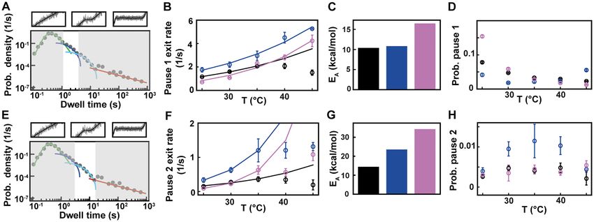
Nucleic Acids Research, 2020, Vol. 48, No. 10 5593
Downloaded from https://academic.oup.com/nar/article-abstract/48/10/5591/5819939 by MPI Science of Light user on 10 July 2020
Figure 1. Description and calibration of the temperature control device installed in the high throughput magnetic tweezers assay. (A) Schematic of the
magnetic tweezers assay. (B) Temperature measured at the surface of the flow cell as a function of the temperature measured at the objective by the
thermistor of the heating foil. Red triangles: temperature at the surface of the flow cell extracted from the extension-rotation experiments (Material and
methods; error bars are the standard deviation for seven tethers). Black circles: temperature at the surface of the flow cell measured using a macroscopic
thermistor (Supplementary Figure S1B). The dashed lines represent the linear fits. (C) Schematic of supercoiling of linear dsDNA. (D) The median rotation–
extension of seven different MyOne magnetic beads tethered by 20.6 kb dsDNA at either 30◦ C (light blue), 35◦ C (green), 40◦ C (yellow), 45◦ C (red) or 50◦ C
(black), experiencing a 0.3 pN force. Each rotation–extension experiment for a given temperature is fitted by a Gaussian. (E) A magnification of the shift
of the maximum extension position. Arrows indicate maxima.
with bovine serum albumin (BSA, New England Biolabs) Coilable DNA construct for temperature calibration
(100 mg/ml) for 10 min and rinsed with 1 ml PBS. To re-
The 20.6 kb coilable DNA construct fabrication was de-
move BSA excess from the surface, high salt buffer (10 mM
scribed in (35).
Tris, 1 mM EDTA pH 8.0, supplemented with 750 mM
NaCl and 2 mM sodium azide) was flushed through the
flow cell and rinsed 10 min later with TE buffer (TE sup- dsRNA construct
plemented with 150 mM NaCl and 2 mM sodium azide). The construct employed here, which has been previously de-
To establish the temperature dependent rotation-extension scribed in detail (15), a 4 kb long single-stranded splint to
experiments, we carefully mixed 10 l of streptavidin coated which four ssRNAs are annealed: a biotin-labeled strand to
MyOne magnetic beads (Thermofisher, Germany) and with attach to the streptavidin-coated magnetic bead, a spacer,
∼0.25 ng of 20.6 kb long coilable double-stranded DNA ∼2.9 kb template, and a digoxygenin-labeled strand to at-
tether, diluted it to 40 l, and flushed the solution into the tach the magnetic bead to the surface glass surface. The tem-
flow cell (35). For the RdRp experiment, we mixed 5 l plate strand ends in 3 with either a ssRNA flap made of 3
of dsRNA at ∼0.1 ng/l with 20 l of streptavidin coated C residues followed by 15 U residues––used for 6 RdRp
M270 magnetic beads (Invitrogen), diluted it to 40 l and to catalyze de novo initiation, or a small hairpin with the se-
flushed the solution into the flow cell. For both experiments, quence ACGCUUUCGCGT followed by 15 U residues to
the flow cell was rinsed to remove unattached magnetic initiate PV and HRV-C RdRp catalyzed RNA synthesis via
beads after few minutes of incubation. primer extension (27,37) (Figure 2A).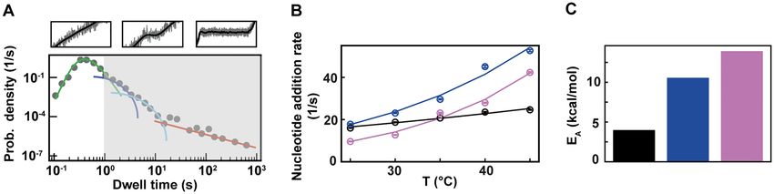
5594 Nucleic Acids Research, 2020, Vol. 48, No. 10

                                                                                                                                                                 Downloaded from https://academic.oup.com/nar/article-abstract/48/10/5591/5819939 by MPI Science of Light user on 10 July 2020
Figure 2. Temperature dependent magnetic tweezers assay show an increased replication rate for human rhinovirus C RdRp at increasing temperatures.
(A) A schematic overview of the experimental assay (not to scale). A M-270 magnetic bead is tethered by a dsRNA that experiences a constant force F. The
RdRp initiates at the 3 -end of the template strand, which ends either with a single-stranded 3 overhang or a short hairpin (see insert on the left). In the
presence of NTPs, the RdRp replicate and unwinds the template strand, creating a ssRNA tether with a difference in extension length L in comparison
to the dsRNA tether at a force F. (B) Low pass filtered (0.5 Hz) HRV-C RdRp activity traces at 25◦ C (blue) (N = 18) and 45◦ C (red) (N = 42), while
applying a 30 pN constant force. (C) Median processivity of 6 (gray), PV (blue) and HRV-C (pink) RdRps. The error bars denote the 68% confidence
interval. (D) The dwell times probability density distribution extracted from at least 35 traces of HRV-C RdRp at either 25◦ C (blue) or 45◦ C (red) acquired
as in (B). The error bars represent the 36% confidence interval obtained from 1000 bootstrapped datasets. (E) Kinetic model of nucleotide incorporation
and misincorporation by RdRps (adapted from Ref. (15)). HFC: high fidelity catalytic state; LFC: low fidelity catalytic state; TMC: terminal mismatch
catalytic state.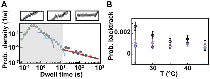
Nucleic Acids Research, 2020, Vol. 48, No. 10 5595
Purification of 6 P2 RdRp camera acquisition rate, at a defined temperature and 30
pN force. The force is defined by the magnet distance to
Recombinant N-terminally histidine-tagged 6 RdRp was
the flow chamber top surface, as described in (35) with a
expressed from plasmid pAA5 in Escherichia coli BL21
relative error of ∼7% (standard deviation). The reaction
(DE3) (38) and purified using Ni-NTA affinity column (Qi-
buffer contains either 1 mM concentration of all NTPs for
agen), HiTrapTM Heparin HP column and HiTrapTM Q HP
PV and HRV-C RdRps or 1 mM ATP/GTP and 0.2 mM
column (GE Healthcare) as previously described (39). The
CTP/UTP for 6 RdRp. The images are analyzed in real-
purified protein was stored in 50% glycerol, 140 mM NaCl,
time using custom-written routines in Labview 2017 and
50 mM Tris–HCl pH 8.0, 0.1 mM EDTA, 0.1% Triton-X
CUDA nVidia to extract the (x, y, z) position of up to
100 at −20◦ C.
Downloaded from https://academic.oup.com/nar/article-abstract/48/10/5591/5819939 by MPI Science of Light user on 10 July 2020
∼800 magnetic beads simultaneously (18). The micrometric
change in tether extension upon RdRp activity (Figure 2A)
Construction of HRV-C and PV RdRp bacterial expression is converted into numbers of transcribed nucleotides using
plasmids the force–extension relationships for dsRNA and ssRNA
The HRV-C RdRp (3D gene) was cloned into the pSUMO constructs (15). RdRp activity traces are low-pass filtered at
bacterial expression plasmid using a similar procedure as 0.5 Hz using a Kaiser–Bessel window, from which the dwell
described for PV RdRp (3D gene) (40). This system allows time were extracted as previously described (14,15,24).
for the production of SUMO fusion proteins containing an
amino-terminal hexahistidine tag fused to SUMO that can Processivity
be purified by Ni-NTA chromatography and subsequently
The processivity was measured from activity traces from
processed by the SUMO protease, Ulp1. Briefly, the HRV-C
which the tether did not detach within 5 min after the ap-
and PV RdRps coding region was amplified using respec-
parent end of the trace. We represented in Figure 2C the
tively the HRV-C15 cDNA (Accession# GU219984.1) and
median processivity of all the traces for a given temperature
the PV type 1 cDNA (Accession# V01149.1) as template, as
and RdRp.
described in (41). The PCR product of HRV-C RdRp was
gel purified and cloned into the pSUMO plasmid using BsaI
and SalI sites. Stochastic-pausing model
There are many kinetic models that are consistent with the
Expression and purification of HRV-C and PV RdRps empirical dwell-time distributions we observe, and we here
HRV-C RdRp was expressed and purified using the same work under the assumption that the probability of pausing
procedure reported for PV RdRp (40,42). Briefly, expres- is low enough that there is only one rate-limiting pause in
sion was performed at 25◦ C by auto-induction, cells har- each dwell-time window. This assumption washes out most
vested, lysed by French Press, subjected to PEI precipitation details of the kinetic scheme that connects pauses and nu-
followed by AMS04 precipitation, Ni- NTA chromatogra- cleotide addition, but allows us to determine the general
phy, cleavage by Ulp1, phosphocellulose chromatography, form of the dwell-time distribution without specifying how
gel filtration and the protein concentrated using Vivaspin the pauses are connected to the nucleotide addition path-
concentrators. Reaction buffer for Φ6 P2. The P2 reaction way
buffer is composed of 50 mM HEPES pH 7.9, 20 mM am- 1
monium acetate, 3% (w/v) polyethylene glycol 4000, 0.1 pdw (t) ∝ pna t; Ndw ,
kna
mM EDTA (pH 8.0), 5 mM MgCl2 , 2 mM MnCl2 , 0.01% ⎛ ⎞
Triton X-100, 5% Superase RNase inhibitor (Life Tech- Nsp
a
+Q (t) ⎝ pn kn e−kn t + ⎠.
bt
nologies), 20 g/ml BSA and 9 nM P2 RdRp. 3/2
(1)
n=1
2(1 + t/1s)
Reaction buffer for PV and HRV-C RdRps In the above expression, the gamma function in the first
The PV/HRV-C RdRp reaction buffer (coined PV reac- term contributes the portion pna of dwell-times that orig-
tion buffer) is composed of 50 mM HEPES pH 7.9, 5 mM inate in the RdRp crossing the dwell-time window of size
MgCl2 , 0.01% Triton X-100, 5% Superase RNase inhibitor Ndw base pairs without pausing; the second term is a sum of
(Life Technologies). To stall PV and HRV-C RdRps on the contributions originating in pause-dominated transitions,
template, 500 nM of RdRp and 1 mM of ATP, CTP and each contributing a fraction pn of dwell-times; the third
GTP in PV reaction buffer were incubated in the flow cham- term captures the asymptotic power-law decay (amplitude
ber for ∼10 min, and the flow cell was subsequently rinsed. abt ) of the probability of dwell-times dominated by a back-
track. The backtracked asymptotic term needs to be regu-
larized for times shorter than the diffusive backtrack step.
Single-molecule RdRp replication activity experiments
We have introduced a regularization at 1 s, but the precise
We select the dsRNA tether in the flow cell in reaction buffer timescale does not matter, as long as it is set within the re-
as previously described (15). To prevent reinitiation on the gion where the exponential pauses dominate over the back-
template-product duplex when PV/HRV-C RdRp falls off, track. From left to right, each term of (Equation 1) is dom-
we first stalled the RdRp and rinsed the flow chamber with inating the distribution for successively longer dwell-times.
400 l of reaction buffer. Subsequently, we flushed in the A cut-off factor Q(t) for short times is introduced to ac-
reaction buffer, and grabbed the data for 30 min at 58 Hz count for the fact that the dwell-time window includes Ndw5596 Nucleic Acids Research, 2020, Vol. 48, No. 10
nucleotide-addition steps, RESULTS
(tkna /Ndw ) Ndw −1 Establishing temperature control of high throughput mag-
Q(t) = −1
. netic tweezers
1 + (tkna /Ndw ) Ndw
To perform a temperature-controlled experiment in a mag-
The fit results dependence on these cut-offs is negligible netic tweezers assay, one needs to maintain the temperature
as long as they are introduced in regions where the corre- setpoint constant in the field of view. To fulfill this condi-
sponding term is sub-dominant. Here the cut is placed un- tion using a magnetic tweezers instrument, it is sufficient to
der the center of the elongation peak, guaranteeing that it only control the temperature at the objective (given that an
Downloaded from https://academic.oup.com/nar/article-abstract/48/10/5591/5819939 by MPI Science of Light user on 10 July 2020
is placed where pausing is sub-dominant. oil immersion objective is used). Indeed, the field of view
is separated by at least one centimeter from the base plate
(Supplementary Figure S1A) and as glass is a poor ther-
Maximum likelihood estimation mal conductor, the field of view remains insulated from heat
transferred from the base plate (30). One may suggest that
The normalized version of (Equation 1) is the dwell-time not maintaining the temperature on the top coverslip of
distribution fit to the experimentally collected dwell-times the flow chamber may result in convection flows that would
ti i by minimizing the likelihood function (43): perturb the measurement. The Rayleigh number (Ra) com-
pares convection over conduction (45). Natural convection
L=− ln pdw (ti ) occurs for Ra ∼2000, which is much larger than our esti-
i mation of Ra in our experimental conditions (for a flow cell
inner thickness ∼ 0.23 mm), i.e. Ra ∼ 4, and therefore con-
with respect to rates and probabilistic weights. We calculate
vection does not occur in our conditions. We have mounted
the errors in our parameter estimates by bootstrapping (44)
a temperature control device derived from Thorlabs parts
the system 100 times, and report the one-sigma confidence
on the objective, meaning a flexible resistive foil heater in-
intervals among the bootstrapped data sets.
cluding a thermistor that is strapped around the oil immer-
sion objective (Supplementary Figure S1B) and a PID con-
troller to adjust the temperature setpoint (Figure 1A, Ma-
Dominating in a dwell-time window versus dominating in one terials and Methods). This system has several advantages in
step comparison with custom ones: it is commercially available,
The fractions pn represent the probability that a particular low price, and it comes with a graphic user interface (GUI)
rate kn dominates the dwell-time. We want to relate this to to adjust the temperature from the data acquisition com-
the probability Pn that a specific exit rate dominates within puter. We first calibrated the temperature measured on the
a 1-nt transcription window. Assuming we have labelled the objective side versus the temperature measured in the flow
pauses so that kn−1 > kn , we can relate the probability of cell. To this end, we used a digital portable thermometer
having rate n dominating in Ndw steps to the probability of (Gochange, Amazon.de, ±1.5% error), whose ∼1 mm3 tem-
having it dominate in one step through perature probe was inserted in the flow cell and immerged in
ultrapure water (Supplementary Figure S1A). In absence of
Ndw Ndw
n
n−1 heating, i.e. at room temperature, the two sensors reported
pn = Pm − Pm , the same temperature. We repeated this measurement sev-
m=0 m=0 eral times at different temperature setpoints assigned on the
GUI to the PID controller, from 25◦ C to 60◦ C, i.e. the max-
p0 = pna = Pna
Ndw
= P0Ndw (2) imum temperature tolerated by the objective. Between each
measurement, we waited at least 30 min for the objective
The first term in (Equation 2) represents the probability temperature to equilibrate, as the focal plane of the objec-
of having no pauses longer than the nth pause in the dwell- tive shifts until equilibrium is reached (Supplementary Fig-
time window, and the second term represents the proba- ure S1C). We observed a linear relationship between the
bility of having no pauses longer than the (n – 1)th pause. temperature at the objective and the temperature in the flow
The difference between the two terms is the probability that chamber, i.e. Tflow cell (Tobj ) = (0.7 ∗ Tobj + 9.23) ◦ C (Figure
the nth pause will dominate. This can be inverted to yield 1B, Supplementary Table S1). Because the probe integrates
a relation between the single-step probabilities (Pn ) and the the temperature over a cubic millimeter volume, the ther-
dwell-time window probabilities ( pn ) mometer measurement may not be representative of the
1/Ndw 1/Ndw temperature within few microns above the bottom cover-
n
n−1
slip surface of the flow cell. To perform an in situ calibra-
Pn = pm − pm ,
tion directly at the surface, we took advantage of the well-
m=0 m=0
characterized temperature dependence of the DNA heli-
P0 = p0
1/Ndw
. cal twist, which decreases by ∼11◦ /◦ C/kb (46–48). Mag-
netic tweezers are an ideal technique to measure the twist
This relationship has been used throughout the of a coilable molecule, such as fully double-stranded DNA,
manuscript to relate our fits over a dwell-time window as they enable a precise control of the torque applied on
to the single-step probabilities. such molecule (Figure 1C). We performed several DNANucleic Acids Research, 2020, Vol. 48, No. 10 5597
rotation-extension experiments at 0.3 pN and at different ssRNA to initiate 6 RdRp (51), or by a small hairpin to
temperatures using a coilable DNA molecule (30,49) (Fig- initiate PV or HRV-C RdRps (14,37) (Figure 2A). Follow-
ure 1D, Materials and Methods). After waiting at least 30 ing successful initiation, the RdRp displaces the template
min for the objective temperature to equilibrate to the set- strand from the tethering strand, converting the tether from
point value (Supplementary Figure S1C) (30), we evaluated dsRNA to ssRNA, which increases linearly the end-to-end
the spatiotemporal resolution of the set up. To this end, we extension of the tether by L at constant force F and is
monitored the z-axis position of a surface attached 3 m subsequently converted from microns to nucleotides (15)
diameter bead, to which another reference bead position (Figure 2A). Of note, the force is not directly applied to
has been subtracted, at either 25◦ C (Supplementary Fig- the RdRp, but on the tethering RNA strand, which low-
Downloaded from https://academic.oup.com/nar/article-abstract/48/10/5591/5819939 by MPI Science of Light user on 10 July 2020
ure S1D) or 35◦ C (Supplementary Figure S1E). The trace ers the energy barrier to the RdRp forward translocation
at each temperature shows little drift, which is further sup- formed by the downstream double-stranded RNA fork. Us-
ported by their respective Allan deviation (35,50) (Supple- ing high throughput magnetic tweezers, dozens of RdRp
mentary Figure S1F). The extension of DNA molecules at activity traces are simultaneously acquired (Figure 2B, Sup-
different supercoiling density shows a typical bell-like shape plementary Figure S2A, C). In this work, we study the in-
distribution, with the maximum extension centered at zero fluence of temperature on the replication kinetics of three
turn (Figure 1D). This shape is well fitted by a Gaussian model RdRps. The direct observation of HRV-C RdRp
function, which enables the evaluation of the zero turn max- traces show that an increase in temperature from 25◦ C
imum extension (Figure 1E). If DNA helical twist decreases, to 45◦ C dramatically increases its average replication rate
the zero turn position shifts towards the negative turns. If (Figure 2B). This is also observable for PV RdRp, and,
DNA helical twist increases, the zero turn position now to a smaller extent, for 6 RdRp (Supplementary Figure
shifts towards the positive turns. Changing the temperature S2A, C). Looking at the processivity of the three RdRps
by steps of 5◦ C from 30◦ C to 50◦ C, and centering at zero for the probed temperature range, we evaluated a constant
turn the maximum extension obtained at 30◦ C, we observed processivity for 6 and HRV-C RdRps and a slight decay
that the rotation-extension traces shift towards the negative for PV RdRp above 40◦ C (Figure 2C). To further quantitate
turns upon temperature increases, as expected from a reduc- the change in replication kinetics, we performed a dwell time
tion in DNA helical twist (Figure 1D, E). Converting the analysis of the 0.5 Hz low pass filtered traces. Specifically,
shift of the peak of the traces into temperature variation us- the traces were scanned with non-overlapping windows of
ing the above described relationship between DNA helical 10 nt to evaluate the duration of ten successive nucleotide
twist and temperature, we extracted almost an one-to-one incorporations, after which the collected dwell times are as-
linear relationship between the temperature measured at the sembled into a probability density distribution (Figure 2D,
surface of the flow chamber and on the side of the objective, Supplementary Figures S2B, D, S3) (14,15,24). The dwell
i.e. Tflow cell (Tobj ) = (0.95 ∗ Tobj + 1.18) ◦ C(Figure 1B, Sup- time analysis is a direct measure of the event(s) that domi-
plementary Table S1). If not indicated otherwise, we will use nated in time over ten successive nucleotide incorporations,
this equation for temperature conversion in the subsequent i.e. either the incorporation of the ten nucleotides only or
sections of the study. As our magnetic tweezers assay has a a pause that interrupted the ten nucleotides addition (Sup-
very large field of view of ∼(0.5 × 0.4) mm, we evaluated plementary Figure S1F). Looking at the dwell times distri-
the homogeneity of the temperature over the whole field of bution extracted from the HRV-C traces acquired at 45◦ C
view. To this end, we performed a rotation extension experi- (Figure 2B) represented in a log-binned histogram, the bins
ment at both at 25◦ C and 45◦ C with the same DNA tether at describe a bell-like shape until ∼0.4 s, then a shoulder from
five different locations in the field of view by moving the flow ∼0.4 s till ∼3 s, and finally a straight line for the longer dwell
cell with the micrometric screws of the stage, and we did not times (Figure 2D, Supplementary Figure S1F). Each region
measure any difference of the rotation-extension traces for of the distribution is the temporal signature of a specific
a given temperature. This result confirms that the tempera- biochemical event in elongating RdRps. To describe these
ture is constant over the whole field of view (Supplementary events, we previously introduced four probability distribu-
Figure S1E) (32). We have now shown that using a simple tion functions: to fit the bell-like shape region, a gamma
and commercially available temperature control system, we distribution that describes ten successive nucleotide addi-
are able to adjust and maintain the temperature in the entire tions cycles without pause; to fit the shoulders, two expo-
field of view of a high throughput magnetic tweezers instru- nentially distributed pauses (Pause 1 and Pause 2); and fi-
ment. This assay is thus suitable to investigate the tempera- nally to fit the dwell times dominated by the longest pauses,
ture dependence of viral RdRps elongation kinetics. a power law distribution (15,16,52) (Supplementary Figure
S1F, Materials and Methods). We have previously showed
(14,15) that RdRp incorporates cognate NTPs fast through
Setting up high throughput magnetic tweezers to study viral
the high fidelity catalytic (HFC) state (Figure 2E, gamma
replication
distribution), and rarely interconverts into another confor-
We have recently developed a high throughput magnetic mation, i.e. the low fidelity catalytic (LFC) state (Figure
tweezers assay to investigate viral RdRp replication kinetics 2E), from which the RdRp slowly incorporates either a cog-
(14–16). As RdRps use an RNA template, we made a non- nate NTP appearing as Pause 1 (diagonal arrow in Figure
coilable dsRNA construct to tether the magnetic beads to 2E) or a non-cognate NTP. In the latter, the nucleotide ad-
the glass coverslip surface of the flow cell as previously de- dition following the mismatch addition is done even slower
scribed (15) (Figure 2A, Materials and Methods). The 3 - than Pause 1 through a state we coined the terminal mis-
end of the template RNA is terminated by either a blunt match catalytic (TMC) state (Figure 2E) and appears in5598 Nucleic Acids Research, 2020, Vol. 48, No. 10
the traces as Pause 2. Furthermore, we also showed that energy landscape of nucleotide addition for PV RdRp (6).
the power law distributed pause relates to RdRp backtrack- Our results demonstrate the large impact of temperature on
ing (16,52). Using a maximum likelihood estimation (MLE) the nucleotide addition rate for human viruses, i.e. PV and
procedure, we extracted the parameters for each distribu- HRV-C RdRp, and the mild impact on the environmental
tion, i.e. exit rates and probabilities (Material and Methods, virus, i.e. the RdRp of Pseudomonas bacteriophage 6.
Supplementary Table S2, Figure S3) (24). Having acquired
and analyzed viral replication traces at several tempera-
Human virus and environmental bacteriophage RdRps elon-
tures, we now describe how the parameters extracted from
gation competent pauses are differently affected by tempera-
the MLE procedure vary for 6, PV and HRV-C RdRps, at
ture
Downloaded from https://academic.oup.com/nar/article-abstract/48/10/5591/5819939 by MPI Science of Light user on 10 July 2020
constant force, i.e. 30 pN, and saturating NTPs concentra-
tion, i.e. 1 mM. We then investigated how Pause 1 and Pause 2 kinetics
and probabilities (Figure 4A and E) are affected by tem-
The nucleotide addition rates of RdRps from an environmen- perature. We previously showed that Pause 1 and Pause 2
tal phage and from two human viruses show different re- are the signature of slow nucleotide addition cycles that
sponses to temperature increase appear as short pauses in the RdRp elongation activ-
ity traces (14,15). For PV and HRV-C RdRps, Pause 1
As noted above, HRV-C and PV RdRps show a dramatic exit rate increases dramatically, i.e. by 3- and 6-fold, re-
increase in average elongation rate when increasing the spectively (Supplementary Table S2, Figure 4BF), which
temperature (Figure 2B, D, Supplementary Figure S2CD), is further highlighted by their respective large Pause 1
while only a mild effect is observable for 6 RdRp (Sup- activation energy, i.e. EA,PV = (11.0 ± 1.1) kcal/mol and
plementary Figure S2A, B). We first look into the pause- EA,HRV-C = (15.2 ± 4.9) kcal/mol (Supplementary Table
free nucleotide addition rate described by the bell-like shape S2, Figure 4B, C), similar to the nucleotide addition ac-
gamma distribution (Figure 3A, Supplementary Figures tivation energy (Figure 3C). Pause 2 exit rate also ex-
S1F, S3). For temperature varying from 25◦ C to 45◦ C, the periences a steep increase, i.e. by 4- and 10-fold for PV
nucleotide addition rate of PV RdRp increases from (17.8 and HRV-C RdRps, respectively, and eventually saturates
± 0.3) nt/s to (52.7 ± 0.5) nt/s, i.e. almost a 3-fold increase, at 35◦ C (Supplementary Table S2, Figure 4F). We de-
and in agreement with previous bulk measurements (27). cided to fit only the values of Pause 2 exit rate below
Similarly, HRV-C RdRp nucleotide addition rate strongly 35◦ C for PV and HRV-C RdRps to fit Equation (3), as
increases, i.e. from (9.6 ± 0.1) nt/s to (42.3 ± 0.3) nt/s, i.e. this equation does not describe the plateau observed at
a 4-fold increase. On the other hand, 6 RdRp nucleotide higher temperatures. Of note, to achieve reasonable fits
addition rate only varies from (16.0 ± 0.1) nt/s to (24.6 ± to the HRV-C dwell time distribution collected at 40◦ C,
0.2) nt/s for the same temperature span, in line with a pre- we were forced to restrict Pause 2 exit rate k2 > 0.2 s−1
vious ensemble measurement (53) (Figure 3A, Supplemen- (Supplementary Figure S3). The large increase of Pause
tary Table S2). Investigating the temperature response of a 2 exit rates is further supported by an activation energy
reaction, one is able to determine whether the reaction is larger than for Pause 1, i.e. EA,PV = (23.2 ± 0.5) kcal/mol
exothermic or endothermic from the Van’t Hoff equation and EA,HRV-C = (34.3 ± 1.8) kcal/mol (Equation 3, Figure
by determining whether the sign of the reaction rate deriva- 4G). Using the temperature calibration from the macro-
tive by the temperature is negative or positive, respectively. scopic thermistor (Figure 1B), we observed a significant
Furthermore, it enables the measurement of the activation variation in activation energies for PV RdRp (Supplemen-
energy E A of the reaction by fitting the reaction rate k, i.e. tary Figure S4), which further supports the utilization of a
nucleotide addition or pause exit, as a function of the tem- calibration in the immediate vicinity of the surface, such as
perature with the Arrhenius equation: DNA rotation extension experiments (Figure 1B). Interest-
EA
ingly, Pause 1 and Pause 2 exit rates for PV and 6 RdRps
k (T) = Pe− RT (3)
increase with assisting tension (14,15), which suggests that
where P is the preexponential factor, T is the abso- forward translocation is the rate limiting step of slow nu-
lute temperature, and R is the universal gas constant cleotide addition through this parallel kinetic pathway (Fig-
(54) (Figure 3B). As the nucleotide addition rate in- ure 2E). The large activation energy of Pause 2 is therefore
creases with temperature, the reaction is endother- consistent with the barrier to RdRp translocation induced
mic. Evaluating the activation energy (±standard by a nucleotide mismatch and is in agreement with previ-
deviation) of the nucleotide addition rate for the ous ensemble measurements (6). The saturation of Pause 2
three RdRps using (Equation 3), we found EA,6 = exit rates >35◦ C suggests that Pause 2 is made of (at least)
(4.1 ± 0.4) kcal/mol, EA,PV = (10.7 ± 1.0) kcal/mol and two successive kinetics states, one being temperature depen-
EA,HRV-C = (14.3 ± 1.2) kcal/mol ((Equation 1)). 6 and dent and rate limiting up to 35◦ C, e.g. the translocation over
PV RdRp nucleotide addition rates are not affected by a nucleotide mismatch, and a second one being not tem-
increasing the mechanical tension on the dsRNA construct perature dependent and rate limiting above 35◦ C, which is
(14,15). Therefore, the nucleotide addition rate is not not determined here. Looking now at 6 RdRp, we ob-
limited by the rate of the polymerase translocating to the served a very different trend. Indeed, Pause 1 and Pause 2
n + 1 position, and the activation energy measured here exit rates are relatively stable, i.e. k1 = (1.7 ± 0.4) s−1 and
relates to another rate limiting process, such as e.g. the k2 = (0.26 ± 0.09) s−1 respectively (mean ± standard devia-
phosphoryl transfer (6). Conclusively, our results confirm tion, Supplementary Table S2, Figure 4BF). Therefore, the
a previous pre-steady state kinetic estimation of the free Arrhenius equation fits poorly to 6 RdRp pause exit ratesNucleic Acids Research, 2020, Vol. 48, No. 10 5599
Downloaded from https://academic.oup.com/nar/article-abstract/48/10/5591/5819939 by MPI Science of Light user on 10 July 2020
Figure 3. 6, PV and HRV-C RdRps show different nucleotide addition rates response to temperature increase. The corresponding data for each RdRp
is represented in black, blue and pink, respectively in (B) and (C). (A) Section (not shaded) of the dwell time distribution that contributes to the MLE
fit (green solid line) of the nucleotide addition (Supplementary Figure S1D). (B) The nucleotide addition rates (circles) as obtained from the maximum
likelihood estimation (MLE) fits. The solid lines represent the respective fit of the Arrhenius equation. The error bars denote the standard deviation from
100 bootstraps of the MLE procedure (Materials and Methods). (C) Activation energy EA extracted from the fits in (B).
Figure 4. Pause 1 and Pause 2 exit rates and probabilities of 6, PV and HRV-C RdRps demonstrate different activation by temperature. The data for
each RdRp is represented in black, blue and pink, respectively. (A) Section (not shaded) of the dwell time distribution that contributes to the MLE fit (dark
blue solid line) of Pause 1. (B) The pause 1 exit rates (circles) extracted from the MLE fits as a function of the temperature. The lines represent the fitted
Arrhenius equation. For 6 RdRp, only the data at 25◦ C, 30◦ C and 35◦ C were considered for the fit. (C) Activation energy for Pause 1 exit rate. (D) The
probabilities to be in Pause 1 state as a function of the temperature. (E) Section (not shaded) of the dwell time distribution that contributes to the MLE
fit (light blue solid line) of Pause 2. (F) The Pause 2 exit rates (circles) extracted from MLE fits. The lines represent the fitted Arrhenius equation. Only
the data at 25◦ C, 30◦ C and 35◦ C were considered for the fit. (G) Activation energy for Pause 2 exit rate. (H) The probabilities to be in Pause 2 state as a
function of the temperature. Error bars in B, D F and H denote the standard deviation extracted from 100 bootstraps of the MLE procedure.
evolution with temperature (Figure 4BF), leading to a poor is largely constant for all RdRps (Figure 4H). As for nu-
estimation of the activation energies of Pause 1 and Pause 2 cleotide addition rate, we observe a very different response
(Figure 4CG, Supplementary Table S2). Off-pathway pause to temperature for the human RNA virus RdRps and the
exit rates have been shown to be temperature insensitive in environmental dsRNA bacteriophage, which could be an
E. coli RNAP (28,29), and could offer an attractive expla- evolutionary advantage for a bacteriophage to not respond
nation for the relative insensitivity of Pause 1 and Pause 2 too strongly on natural environmental temperature varia-
from 6 RdRp. However, 6 RdRp elongation rate also tion in the room temperature range.
increases little with temperature, and therefore we suspect
that the thermal activation of 6 RdRp elongation kinet-
ics occurs at lower temperature than the range we explored. Backtrack related long pause probability mildly decreases
Pause 1 probabilities are largely unaffected by temperature with temperature increase for 6 RdRp, while remaining con-
for 6 and PV RdRps, while it decreases when tempera- stant for PV and HRV-C RdRps
ture increases for HRV-C RdRp (Figure 4D). However, the We next look at the last part of the dwell time distribution,
nucleotide addition rate and Pause 1 regions of the HRV-C which describes the pauses longer larger than 20 s (Figure
RdRp dwell time distributions have a strong overlap at low 5A) and are related to polymerase backtrack, i.e. a back-
temperature (Figure 3B, Supplementary Figure S3), which ward diffusion of the polymerase on its template leading
may bias the probability we measured. Pause 2 probability to an RNA product 3 -end out of register (Supplementary5600 Nucleic Acids Research, 2020, Vol. 48, No. 10
processes of single enzymes, increasing significantly our un-
derstanding of these complex molecular motors (57). In
particular, single molecule biophysics has further comple-
mented the existing wealth of knowledge collected in ensem-
ble measurements with transient, rare and asynchronous ki-
netic events to assemble complete mechanochemical path-
ways. However, while bulk studies are easily performed at
physiological temperature, i.e. 37◦ C for human and E. coli
enzymes, many single molecule studies are performed at
Downloaded from https://academic.oup.com/nar/article-abstract/48/10/5591/5819939 by MPI Science of Light user on 10 July 2020
Figure 5. Temperature has little effect on backtrack probabilities for 6,
room temperature (∼21◦ C) because of the technical difficul-
PV and HRV-C RdRps. (A) Section (not shaded) of the dwell time distri- ties of inserting a temperature control system within home-
bution that contributes to the fit (red solid line) of the backtrack pause. (B) built high-end microscopes. Here, we present a simple tem-
The probabilities for backtrack (circles) as obtained from MLE fits. The er- perature control system with several advantages: low cost,
ror bars denote the standard deviation extracted from 100 bootstraps of the commercially available (Thorlabs), and easily mounted on
MLE procedure. The data for 6, PV and HRV-C RdRps is represented
in black, blue and pink, respectively. an oil immersion objective. We calibrated the temperature
in the field of view of a home-built magnetic tweezers in-
strument by performing rotation-extension experiments on
Figure S5). The polymerase eventually returns in register a supercoiled DNA at several temperatures, using the well-
by hopping from base to base with a forward and back- known relationship describing the decrease in DNA helical
ward rate. This is modeled by a one dimensional diffusion twist as a function of temperature and achieving better per-
process through a periodical energy landscape, and is de- formance than using a macroscopic thermometer probe.
scribed in the dwell time distribution by a power law dis- We have previously investigated the kinetics of PV RdRp
tribution with two parameters, i.e. a –3/2 exponent and a at room temperature, extracting a slow nucleotide addition
characteristic hopping rate (28,52,55) (Materials and Meth- rate of ∼10 nt/s, even at saturating concentration of NTPs
ods). While we have previously observed 6 RdRp back- and high assisting force (14). This result was consistent with
tracking (16), this behavior had remained elusive for PV previous bulk measurements performed at the same tem-
RdRp in absence of the nucleotide analogue T-1106 (14). perature on the same enzyme and using ssRNA templates
We suspected that the absence of long backtrack pauses in (27). Having established a temperature controlled magnetic
our previous study likely originated from an RNase con- tweezers assay, we have presented in this study how tem-
tamination of the PV RdRp stock, which consequently de- perature affects the replication kinetics of RdRps from sev-
creased the tether lifetime and prevented the observation eral different viruses, i.e. 6 (a bacteriophage infecting a
of these rare and long pauses. Having improved PV and plant pathogenic bacteria), PV and HRV-C (two human
HRV-C RdRps purification protocols (Material and Meth- enteroviruses). These RdRps responded very differently to
ods), we are now able to show exemplary traces of deep temperature change. 6 RdRp is only mildly affected in the
backtracks (Supplementary Figure S5) (shallow ones can- temperature range we probed here, which is in line with pre-
not be resolved due to the limited spatiotemporal resolu- vious work (53). Pseudomonas syringae, 6 natural host,
tion of the assay) and characterize the backtrack-related grows at wide range of environmental temperatures (from
pauses for these two enterovirus RdRps (Figure 5, Supple- 0 up to 36◦ C) with an wide optimum around ∼28◦ C (58).
mentary Figure S3). The average hopping rate is hidden in However, the reproduction cycle of 6 is restricted at tem-
the dwell time distribution behind the shoulder of Pause peratures above 30◦ C (59). Consequently, we suggest that
1 and Pause 2, as they statistically dominate, and we have 6 RdRp has evolved to support viral genome replication
therefore decided to fix this parameter at 1 s−1 , a value that under different environmental temperatures, and therefore
has been directly measured for E. coli RNAP (52,56). Us- is not thermally activated >25◦ C. On the other hand, the
ing this fixed hopping rate, we evaluated the probability for kinetics of PV and HRV-C RdRps are strongly affected by
the polymerase to enter a backtrack pause as a function the increase in temperature. The data we present here defi-
of the temperature. For temperatures varying from 25◦ C nitely supports that the temperature is the predominant fac-
to 45◦ C, 6 RdRp probability to enter a backtrack pause tor behind the slow replication we previously observed, as
mildly decreases, i.e. from 0.0027 to 0.0008, while for PV we have now measured nucleotide addition rates similar to
and HRV-C RdRps, this probability is largely constant, i.e. the one measured in bulk at the same temperatures, using
(0.0008 ± 0.0002) and (0.0009 ± 0.0002) (mean ± standard ssRNA templates (27). PV and HRV-C RdRps showed a
deviation), respectively (Figure 5B, Supplementary Table parallel trend, with PV nucleotide addition rate saturating
S2). Though we should be cautious with the extracted prob- at a lower temperature. Interestingly, the activation energy
abilities as we do not know the average hopping rate of a we measured for the nucleotide addition rate and Pause 1,
backtracking RdRp, we observe a different backtracking and the one for Pause 2 are both consistent with the energy
behavior between an environmental bacteriophage RdRp barrier for cognate and non-cognate nucleotide addition, re-
and human virus RdRps. spectively, estimated by pre-steady state kinetic analysis (6).
This result further supports our model where Pause 1 is a
slow nucleotide addition event and Pause 2 is the signature
DISCUSSION
of mismatch nucleotide addition (14,15) (Figure 2E), and
Single molecule biophysics has revolutionized our view of suggests that temperature does not affect nucleotide mis-
molecular biology by enabling the observation of the kinetic incorporation rate (Pause 2 probability is largely constant).Nucleic Acids Research, 2020, Vol. 48, No. 10 5601
Finally, we also show here that the replication traces of non- 4. Bekerman,E. and Einav,S. (2015) Infectious disease. Combating
de novo initiating RdRps, i.e. PV and HRV-C, present long emerging viral threats. Science (New York), N.Y., 348, 282–283.
5. Crotty,S., Maag,D., Arnold,J.J., Zhong,W., Lau,J.Y., Hong,Z.,
pauses related to polymerase backtrack, similarly to de novo Andino,R. and Cameron,C.E. (2000) The broad-spectrum antiviral
initiating 6 RdRp (Figure 5B) (16). We anticipate that this ribonucleoside ribavirin is an RNA virus mutagen. Nat. Med., 6,
behavior is ubiquitous in all RNA virus RdRps and may be 1375–1379.
an important feature for viral replication. On a technical 6. Arnold,J.J. and Cameron,C.E. (2004) Poliovirus RNA-dependent
note, we show here that our analysis framework is particu- RNA polymerase (3DPOL): pre-steady-state kinetic analysis of
ribonucleotide incorporation in the presence of Mg2+. Biochemistry,
larly suitable to detect even very short lived pauses, i.e.5602 Nucleic Acids Research, 2020, Vol. 48, No. 10
26. Potisopon,S., Ferron,F., Fattorini,V., Selisko,B. and Canard,B. (2017) 43. Cowan,G. (1998) In: Statistical Data Analysis. Oxford University
Substrate selectivity of dengue and Zika virus NS5 polymerase Press.
towards 2 -modified nucleotide analogues. Antiviral Res., 140, 25–36. 44. Press,W.H., Flannery,B.P., Teukolsky,S.A. and Vetterling,W.T. (1992)
27. Gong,P., Campagnola,G. and Peersen,O.B. (2009) A quantitative In: Numerical Recipes in C: The Art of Scientific Computing.
stopped-flow fluorescence assay for measuring polymerase elongation Cambridge University Press.
rates. Anal. Biochem., 391, 45–55. 45. Squires,T.M. and Quake,S.R. (2005) Microfluidics: Fluid physics at
28. Mejia,Y.X., Mao,H., Forde,N.R. and Bustamante,C. (2008) Thermal the nanoliter scale. Rev. Mod. Phys., 7, 977–1026.
probing of E. coli RNA polymerase off-pathway mechanisms. J. Mol. 46. Charbonnier,F., Erauso,G., Barbeyron,T., Prieur,D. and Forterre,P.
Biol., 382, 628–637. (1992) Evidence that a plasmid from a hyperthermophilic
29. Abbondanzieri,E.A., Shaevitz,J.W. and Block,S.M. (2005) archaebacterium is relaxed at physiological temperatures. J.
Picocalorimetry of transcription by RNA polymerase. Biophys. J., 89, Bacteriol., 174, 6103–6108.
Downloaded from https://academic.oup.com/nar/article-abstract/48/10/5591/5819939 by MPI Science of Light user on 10 July 2020
L61–L63. 47. Depew,D.E. and Wang,J.C. (1975) Conformational fluctuations of
30. Kriegel,F., Matek,C., Drsata,T., Kulenkampff,K., Tschirpke,S., DNA helix. PNAS, 72, 4275–4279.
Zacharias,M., Lankas,F. and Lipfert,J. (2018) The temperature 48. Duguet,M. (1993) The helical repeat of DNA at high temperature.
dependence of the helical twist of DNA. Nucleic Acids Res., 46, Nucleic Acids Res., 21, 463–468.
7998–8009. 49. Strick,T.R., Croquette,V. and Bensimon,D. (1998) Homologous
31. Gollnick,B., Carrasco,C., Zuttion,F., Gilhooly,N.S., Dillingham,M.S. pairing in stretched supercoiled DNA. PNAS, 95, 10579–10583.
and Moreno-Herrero,F. (2015) Probing DNA helicase kinetics with 50. Lansdorp,B.M. and Saleh,O.A. (2012) Power spectrum and Allan
temperature-controlled magnetic tweezers. Small, 11, 1273–1284. variance methods for calibrating single-molecule video-tracking
32. Galburt,E.A., Tomko,E.J., Stump,W.T. and Ruiz Manzano,A. (2014) instruments. Rev. Sci. Instrum., 83, 025115.
Force-dependent melting of supercoiled DNA at thermophilic 51. van Dijk,A.A., Makeyev,E.V. and Bamford,D.H. (2004) Initiation of
temperatures. Biophys. Chem., 187–188, 23–28. viral RNA-dependent RNA polymerization. J. Gen. Virol., 85,
33. Seidel,R., Bloom,J.G., Dekker,C. and Szczelkun,M.D. (2008) Motor 1077–1093.
step size and ATP coupling efficiency of the dsDNA translocase 52. Depken,M., Galburt,E.A. and Grill,S.W. (2009) The origin of short
EcoR124I. EMBO J., 27, 1388–1398. transcriptional pauses. Biophys. J., 96, 2189–2193.
34. Park,J.S., Lee,K.J., Hong,S.C. and Hyon,J.Y. (2008) Temperature 53. Sarin,L.P., Poranen,M.M., Lehti,N.M., Ravantti,J.J.,
dependence of DNA elasticity and cisplatin activity studied with a Koivunen,M.R., Aalto,A.P., van Dijk,A.A., Stuart,D.I., Grimes,J.M.
temperature-controlled magnetic tweezers system. J. Korean Phys. and Bamford,D.H. (2009) Insights into the pre-initiation events of
Soc., 52, 1927–1931. bacteriophage phi6 RNA-dependent RNA polymerase: towards the
35. Ostrofet,E., Papini,F.S. and Dulin,D. (2018) Correction-free force assembly of a productive binary complex. Nucleic Acids Res., 37,
calibration for magnetic tweezers experiments. Sci. Rep., 8, 15920. 1182–1192.
36. Lipfert,J., Hao,X. and Dekker,N.H. (2009) Quantitative modeling 54. Winzor,D.J. and Jackson,C.M. (2006) Interpretation of the
and optimization of magnetic tweezers. Biophys. J., 96, 5040–5049. temperature dependence of equilibrium and rate constants. J. Mol.
37. Mestas,S.P., Sholders,A.J. and Peersen,O.B. (2007) A fluorescence Recognit., 19, 389–407.
polarization-based screening assay for nucleic acid polymerase 55. Galburt,E.A., Grill,S.W., Wiedmann,A., Lubkowska,L., Choy,J.,
elongation activity. Anal. Biochem., 365, 194–200. Nogales,E., Kashlev,M. and Bustamante,C. (2007) Backtracking
38. Sukhodolets,V.V. (2006) [Unequal crossing-over in Escherichia coli]. determines the force sensitivity of RNAP II in a factor-dependent
Genetika, 42, 1526–1535. manner. Nature, 446, 820–823.
39. Makeyev,E.V. and Bamford,D.H. (2000) Replicase activity of purified 56. Shaevitz,J.W., Abbondanzieri,E.A., Landick,R. and Block,S.M.
recombinant protein P2 of double-stranded RNA bacteriophage (2003) Backtracking by single RNA polymerase molecules observed
phi6. EMBO J., 19, 124–133. at near-base-pair resolution. Nature, 426, 684–687.
40. Arnold,J.J., Bernal,A., Uche,U., Sterner,D.E., Butt,T.R., 57. Ostrofet,E., Papini,F.S., Malinen,A.M. and Dulin,D. (2019) In: Joo,C
Cameron,C.E. and Mattern,M.R. (2006) Small ubiquitin-like and Rueda,D (eds). Biophysics of RNA-protein interactions. Springer,
modifying protein isopeptidase assay based on poliovirus RNA NY., pp. 109–141.
polymerase activity. Anal. Biochem., 350, 214–221. 58. Young,J.M., Luketina,R.C. and Marshall,A.M. (1977) The effects on
41. Gizzi,A.S., Grove,T.L., Arnold,J.J., Jose,J., Jangra,R.K., temperature on growth in vitro of Pseudomonas syringae and and
Garforth,S.J., Du,Q., Cahill,S.M., Dulyaninova,N.G., Love,J.D. et al. Xanthomonas pruni. J. Appl. Bacteriol., 42, 345–354.
(2018) A naturally occurring antiviral ribonucleotide encoded by the 59. Sands,J.A., Cupp,J., Keith,A. and Snipes,W. (1974) Temperature
human genome. Nature, 558, 610–614. sensitivity of the assembly process of the enveloped bacteriophage
42. Gohara,D.W., Ha,C.S., Kumar,S., Ghosh,B., Arnold,J.J., phi6. Biochim. Biophys. Acta, 373, 277–285.
Wisniewski,T.J. and Cameron,C.E. (1999) Production of “authentic” 60. Malik,O., Khamis,H., Rudnizky,S., Marx,A. and Kaplan,A. (2017)
poliovirus RNA-dependent RNA polymerase (3D(pol)) by Pausing kinetics dominates strand-displacement polymerization by
ubiquitin-protease-mediated cleavage in Escherichia coli. Protein reverse transcriptase. Nucleic Acids Res., 45, 10190–10205.
Expr. Purif., 17, 128–138.You can also read