Homozygous mutation in MCM7 causes autosomal recessive primary microcephaly and intellectual disability
←
→
Page content transcription
If your browser does not render page correctly, please read the page content below
Neurogenetics
Original research
J Med Genet: first published as 10.1136/jmedgenet-2020-107518 on 31 May 2021. Downloaded from http://jmg.bmj.com/ on August 24, 2021 by guest. Protected by copyright.
Homozygous mutation in MCM7 causes autosomal
recessive primary microcephaly and
intellectual disability
Ethiraj Ravindran ,1,2,3 Cynthia Gutierrez de Velazco,1,2,3 Ali Ghazanfar,4
Nadine Kraemer,1,2,3 Sami Zaqout,5,6 Abdul Waheed,4 Mohsan Hanif,4 Sadia Mughal,4
Alessandro Prigione,7 Na Li,8 Xiang Fang,8 Hao Hu,8,9,10,11 Angela M Kaindl1,2,3
►► Additional supplemental ABSTRACT replication.2 3 Here, MCM2–7 proteins play an
material is published online Background Minichromosomal maintenance (MCM) important role in the initiation and elongation of
only. To view, please visit
the journal online (http://dx. complex components 2, 4, 5 and 6 have been linked to the replication forks by unwinding double-stranded
doi.org/10.1136/jmedgenet- human disease with phenotypes including microcephaly DNA during S phase.4 After the S phase, the pre-RC
2020-1 07518). and intellectual disability. The MCM complex has activity is negatively regulated by phosphorylation
DNA helicase activity and is thereby important for the of the licensing factors, which in turn prevents
For numbered affiliations see
initiation and elongation of the replication fork and the complex to reassemble.5 When this replica-
end of article.
highly expressed in proliferating neural stem cells. tion process is impaired, neural stem cells (NSCs)
Correspondence to Methods Whole-exome sequencing was applied are unable to proliferate rapidly, resulting in a
Dr Angela M Kaindl, to identify the genetic cause underlying the decreased progenitor pool, a reduction in the final
Institute of Cell Biology and neurodevelopmental disease of the index family. number of neurons in the brain and ultimately in
Neurobiology, Department The expression pattern of Mcm7 was characterised microcephaly.6–8
of Pediatric Neurology,
by performing quantitative real-time PCR, in situ Mutations in various components of the MCM
Center for Chronically Sick
Children (Sozialpädiatrisches hybridisation and immunostaining. To prove the disease- complex and other factors involved in DNA repli-
Zentrum, SPZ), Charité causative nature of identified MCM7, a proof-of-principle cation have been linked to human disease (online
Universitätsmedizin Berlin, experiment was performed. supplemental table S1). Heterozygous MCM2
Berlin, Germany; Results We reported that the homozygous missense missense variants were identified in one pedigree
a ngela.kaindl@c harite.de
variant c.793G>A/p.A265T (g.7:99695841C>T, with eight individuals affected by autosomal domi-
AG and AMK are joint senior NM_005916.4) in MCM7 was associated with nant deafness 70 (MIM#616968).9 Similarly, the
authors. autosomal recessive primary microcephaly (MCPH), deletion of Mcm2 produced a microcephaly pheno-
severe intellectual disability and behavioural type in zebrafish, and MCM2 downregulation
Received 27 October 2020
abnormalities in a consanguineous pedigree with three resulted in short cilia and centriole overduplication
Revised 15 February 2021
Accepted 2 March 2021 affected individuals. We found concordance between in human fibroblasts.10 Biallelic MCM4 variants
the spatiotemporal expression pattern of Mcm7 in mice cause immunodeficiency 54 (MIM#609981) with
and a proliferative state: Mcm7 expression was higher decreased natural killer cells, adrenal insufficiency,
in early mouse developmental stages and in proliferative prenatal and postnatal growth retardations and
zones of the brain. Accordingly, Mcm7/MCM7 levels also microcephaly.11 Moreover, biallelic MCM5
were detectable particularly in undifferentiated mouse variants have been reported to cause Meier-Gorlin
embryonal stem cells and human induced pluripotent syndrome 8 (MIM#617564), a disease charac-
stem cells compared with differentiated neurons. We terised by microcephaly, prenatal and postnatal
further demonstrate that the downregulation of Mcm7 growth retardations, microtia and aplasia/hypo-
in mouse neuroblastoma cells reduces cell viability and plasia of the patellae.12 MCM5 dysfunction caused
proliferation, and, as a proof-of-concept, that this is S-phase progression delay in patient lymphoblastoid
counterbalanced by the overexpression of wild-type but and primary skin fibroblasts, which in turn caused
not mutant MCM7. impaired DNA replication due to hypersensitivity
Conclusion We report mutations of MCM7 as a novel to replicative stress.12 Variants in MCM7 have been
cause of autosomal recessive MCPH and intellectual linked to acute myeloid leukaemia due to increased
disability and highlight the crucial function of MCM7 in proliferation.13 Similarly, mutations in other compo-
© Author(s) (or their
employer(s)) 2021. Re-use nervous system development. nents of the pre-RC have been linked to human
permitted under CC BY-NC. No disease, for example, biallelic ORC1 mutations
commercial re-use. See rights cause Meier- Gorlin syndrome 1 (MIM#224690)
and permissions. Published
by BMJ. and corresponding patient cells showed impaired
INTRODUCTION replication licensing with slower cell cycle progres-
To cite: Ravindran E, The minichromosomal maintenance (MCM) sion and growth restriction.14 Thus, proper DNA
Gutierrez de Velazco C,
complex with its six subunits MCM2–7 is key replication is required for the accurate proliferation
Ghazanfar A, et al.
J Med Genet Epub ahead of for cell proliferation.1 Together with the origin and differentiation, which are necessary to avoid
print: [please include Day recognition complex (ORC) subunits 1–6 and the cortical malformations like microcephaly.15
Month Year]. doi:10.1136/ licensing factors Cdc6 and Cdt1, it assembles in a In this study, we report biallelic mutations of
jmedgenet-2020-107518 prereplicative complex (pre-RC) to initiate DNA MCM7 as a novel cause of a neurodevelopmental
Ravindran E, et al. J Med Genet 2021;0:1–9. doi:10.1136/jmedgenet-2020-107518 1
Neurogenetics
disorder with autosomal recessive primary microcephaly Technologies, Santa Clara, USA) according to manufacturer’s
J Med Genet: first published as 10.1136/jmedgenet-2020-107518 on 31 May 2021. Downloaded from http://jmg.bmj.com/ on August 24, 2021 by guest. Protected by copyright.
(MCPH) and intellectual disability and further elucidate the role protocol using MCM7 Mut forward and reverse primers (online
of MCM7 in brain development. supplemental table S2). The PCR reaction was transformed into
XL-10 Gold cells, and the obtained plasmids were sequenced
SUBJECTS AND METHODS using primers listed in online supplemental table S2 to confirm
Genetic analyses the point mutation.
Detailed genetic methods including whole- exome sequencing
(WES) and bioinformatics analysis are described in the online Generation of WT-MCM7 and Mut-MCM7 overexpressing
supplemental data. stable cell lines
MCM7 overexpressing stable cell lines were generated using the
Prediction of protein structures and mutant (Mut) stability WT-MCM7 and Mut-MCM7 overexpression plasmids (OriGene
Domains of MCM7 were identified based on the National Center Technologies). Plasmids were linearised using PsiI (New England
for Biotechnology Information (NCBI) Conserved Domain Data- Biolabs, Frankfurt am Main, Germany), and DNA band elusion
base (https://www.ncbi.nlm.nih.gov/Structure/cdd/wrpsb.cgi).16 was performed using the NucleoSpin Gel and PCR Clean-up
Multiple sequence alignment of MCM7 was performed using kit (Macherey-Nagel, Düren, Germany) according to the user
the ClustalW program.17 Three-dimensional structural models manual. N2a cells were plated on six-well plates and incubated
of MCM7 (MCM7-WT and MCM7-A265T) were predicted by 48 hours at 37°C. Cells were transfected overnight using Lipo-
the Swiss-model web tool (http://swissmodel.expasy.org/interac- fectamine 2000 transfection reagent (Invitrogen) reduced serum
tive).18 The visual representation of models and structural super- medium (Opti-MEM GlutaMAX supplement, Gibco) and 2 µg
position were generated by software package Visual Molecular of linearised overexpression plasmids without antibiotics. Tryp-
Dynamics.19 The mutant stability change or ΔΔG of variants of sinisation was performed as previously described and cells were
MCM7 was predicted using the I-Mutant server (http://gpcr. plated and incubated overnight at 37°C. Geneticin (600 µg/
biocomp.unibo.it/cgi/predictors/I-Mutant3.0/I-Mutant3.0.cgi).20 mL, G-418 Solution; Roche Diagnostics GmbH, Mannheim,
Germany) was added to the cells, and the medium was changed
Quantitative real-time PCR (qPCR) every 48 hours for 7 days to obtain only cells with the integrated
Established methods reported previously were performed for MCM7 overexpression plasmids.
RNA extraction and cDNA synthesis.21 Sets of primers were
designed using Primer3 online software (www.primer3.ut.ee) in Quantification of cell viability, apoptosis and proliferation
order to detect Mcm7 cDNA and to obtain specific amplification. Cell viability (fluorometric CellTiter- Blue Cell Viability;
Maxima SYBR Green/ROX qPCR Master Mix (Thermo Scien- Promega, Madison, USA), apoptosis (ApoONE Homogeneous
tific, Braunschweig, Germany) according to the manufacturer’s Caspase-3/7, Promega) and proliferation assays (colorimetric
protocol with primers specified in online supplemental table S2 Cell Proliferation BrdU-ELISA, Roche Diagnostics GmbH) were
were used for qPCR experiments, and all experiments were run performed using N2a cells in 96-well plates according to the
in triplicate. Quantification of the qPCR results was performed manufacturer’s protocol, and plates were measured at respec-
as previously described,21 and statistical analysis was performed tive wavelengths using a multiplate reader (SpectraMax iD3;
on GraphPad Prism V.5 Software (GraphPad Software, La Jolla, Molecular Devices, San Jose, California, USA). Trypsinisation of
California, USA). N2a cells was performed as previously described, and cells were
counted using 0.4% Trypan Blue Solution (Sigma Life Sciences,
Cell culture Steinheim am Albuch, Germany) and seeded at a concen-
Mouse embryonal stem cells (mESCs) were cultured and neuronal tration of 1000 cells/well in 100 µL of DMEM with 5% FBS
differentiation was obtained as previously described.22 Human and 1% P/S per well. Transfection of control siRNA (scramble)
induced pluripotent stem cells (iPSCs) were obtained from the (siCo) (AGGUAGUGUAAUCGCCUUG-dTdT) or Mcm7 siRNA
Charité Stem Cell Core Facility. Mouse neuroblastoma (N2a) (siMcm7) (TGCCAAGCGCTACTCAAGA-dTdT) was performed
cells were cultured and maintained in high-glucose Dulbecco’s using Lipofectamine 2000 (Invitrogen) and reduced serum
modified Eagle’s medium (DMEM GlutaMAX supplement medium, and plates were measured after 48 hours. Statistical
pyruvate; Gibco, Paisley, Scotland) with 10% heat-inactivated analysis for all assays was performed on GraphPad Prism V.5
fetal bovine serum (FBS, Gibco) and 1% penicillin streptomycin Software.
(P/S; Gibco, Grand Island, USA). Cells were maintained at 37°C
and passaged at 80% confluence by trypsinisation using 0.05% Western blot
trypsin/0.02% EDTA for 30 s. Protein extraction and Western blot were carried out as previ-
ously reported23 with antibodies listed in online supplemental
Generation of wild-type MCM7 (WT-MCM7) and mutant table S3.
MCM7 (Mut-MCM7) overexpression plasmids
We obtained a human WT-MCM7 plasmid from OriGene Immunohistochemistry
(OriGene Technologies GmbH, Herford, Germany). The lyoph- Poly-L-
lysine-
coated coverslips in 24- well plates or 96-well
ilised plasmid was dissolved and transformed into TOP10 plates were used to plate mESC/iPSC, which were then fixed
cells (Invitrogen, California, USA). The obtained plasmid was in 4% Paraformaldehyde. Cells were incubated in staining
sequenced using the primers listed in online supplemental table buffer (0.2% gelatin, 0.25% Triton X-100, 3% bovine serum
S2 to confirm the intact human WT-MCM7 cDNA. We used the albumin) for 30 min in order to permeabilise and block
WT-MCM7 plasmid as a template to generate a Mut-MCM7 unspecific staining. Primary antibody incubation was done over-
cDNA carrying the same mutation as our index patients night at 4°C, followed by a 2-hour secondary antibody incuba-
(c.793G>A). Site- directed mutagenesis was performed using tion with the antibodies listed in online supplemental table S3.
the QuickChange II XL Site-Directed Mutagenesis kit (Agilent 4’,6-Diamidino-2-phenylindole (DAPI, 1:1000; Sigma-Aldrich)
2 Ravindran E, et al. J Med Genet 2021;0:1–9. doi:10.1136/jmedgenet-2020-107518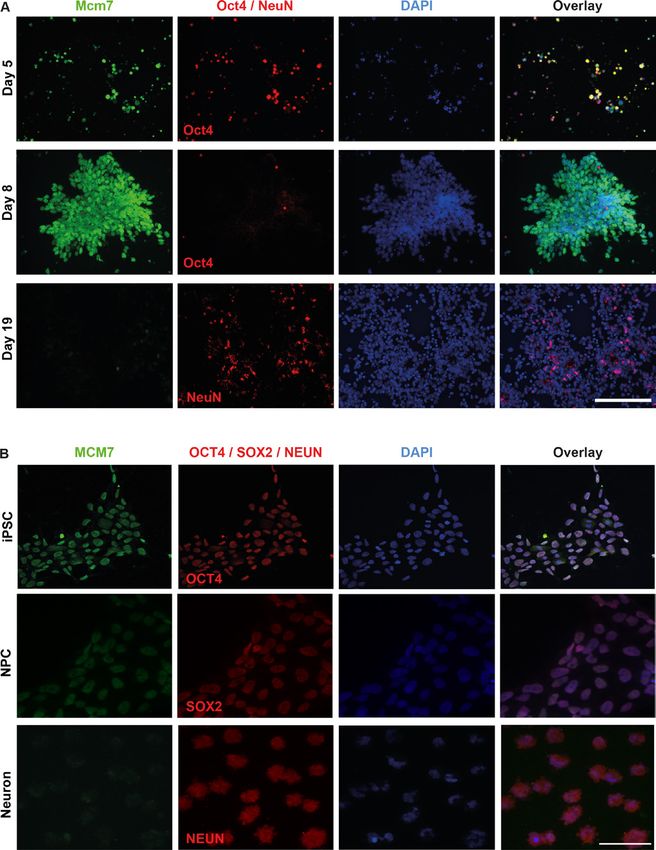
Neurogenetics
was used for labelling the nuclei. A Zeiss Spinning Disk Confocal individuals could speak simple words but lacked the ability to
J Med Genet: first published as 10.1136/jmedgenet-2020-107518 on 31 May 2021. Downloaded from http://jmg.bmj.com/ on August 24, 2021 by guest. Protected by copyright.
Microscope was used to image and analyse the cells labelled with formulate complete, even simple sentences. While they were
fluorescence, and images were processed using ImageJ. able to walk without support, they were unable to climb up
or down the stairs without support. The three subjects were
unable to perform routine activities, lacked the concept of
In situ hybridisation
self-cleaning, and presented with hyperactivity and aggressive
To obtain a 161 bp Mcm7 PCR product, the primers listed in
behaviour. They had no clinical or laboratory findings indic-
online supplemental table S2 were used; the PCR product
ative of malignant disease. Results of ophthalmological and
was then purified and cloned into a p-AL2-T vector (Evrogen,
otorhinolaryngological examinations were normal.
Moscow, Russia). The RNA probe was generated by in vitro
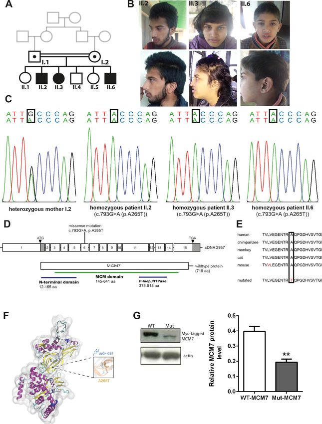
To identify the genetic cause of the disease phenotype, we
transcription using T7 or SP6 polymerase (Roche Diagnostics-
performed WES in the index family. Sequencing data revealed the
GmbH), and in situ hybridisation was performed on 16 µm-thick
homozygous missense variant c.793G>A (g.7:99695841C>T,
brain sections of mice, stages E14 and P0, as well as whole E10.5
NM_005916.4) in MCM7 in all three patients, which was
embryos. In situ hybridisation on E14 and P0 brain sections was
further confirmed by Sanger sequencing (figure 1C,D). The
performed as described previously.24
variant segregates with the phenotype in the index family, and
For whole-mount in situ hybridisation, mouse embryos were
no disease-causative variants in other genes previously linked to
fixed in 4% PFA overnight at 4°C, dehydrated and stored in
neurological diseases were identified. The identified variant in
100% methanol at −20°C. To commence in situ procedure,
MCM7 is located in a highly conserved area and predicted to
embryos were rehydrated in decreasing concentrations of meth-
cause an exchange of a highly conserved hydrophilic nonpolar
anol (75%, 50% and 25%) for 15 min and subsequently washed
alanine by a hydrophilic polar threonine at the protein level
in phosphate buffered saline with 0.15% Tween- 20 (PBT).
(p.A265T, NP_005907.3) (figure 1E). Structural analysis of Mut-
Embryos were then permeabilised at room temperature (RT)
MCM7 protein revealed that the identified mutation (p.A265T)
using Proteinase K solution (10 µg/mL proteinase K in PBT); the
destroyed the intramolecular hydrophobic interactions with
treatment was carried out for 15 min on E10.5 embryos. After
adjacent hydrophobic residues Val271 and Val304 of MCM7
digestion, embryos were postfixed in 0.2% glutaraldehyde/4%
protein, which might impair the local secondary structure and
PFA in PBT for 20 min at RT and washed in PBT. Embryos
molecular functions (figure 1F). The mutation is disease-causative
were incubated in prehybridisation buffer (50% formamide,
in nature, as predicted by Mutation Taster, PolyPhen-2 and
5× Saline-Sodium Citrate (SSC) pH 7.0, 2.5 M EDTA, 0.1%
SIFT (online supplemental table S4) (www.mutationtaster.com,
Tween-20, 0.15% CHAPS, 0.1 mg/mL heparin, 100 µg/mL yeast
http://genetics.bwh.harvard.edu/pph2, https://sift.bii.a-star.edu.
tRNA, 50 µg/mL salmon sperm DNA, 1× Denhardt’s solution)
sg).25–27 Protein immunoblot of N2a cells overexpressing Myc-
at 65°C for 2 hours, after which they were incubated in the
tagged MCM7 carrying the patient mutation p.A265T revealed
desired probe (3 µL probe in 3 mL prehybridisation buffer) at
strongly reduced protein levels when compared with N2a cells
65°C overnight. Washes of 50% formamide (in 20× SSC), 1 M
in which Myc- tagged WT-MCM7 was overexpressed (n=3,
Tris–HCl pH7.5 in 5M NaCl with RNAse A and Tris buffered
unpaired t-test, p=0.0066) (figure 1G).
saline-Tween-20 (NaCl, KCl, 1 M Tris-HCl pH7.5 and Tween-
Because the MCM complex is known to be a major compo-
20) were performed. Embryos were then blocked for 1 hour in
nent in the replication process and proliferation is one of the
20% sheep serum in TBS-T at RT and incubated overnight at
major processes involved in the early stages of brain devel-
4°C in anti-Digoxigenin antibody (1:2000) in 5% sheep serum.
opment,28 29 we analysed the temporospatial expression
Washes with TBS-T were performed, followed by alkaline phos-
of Mcm7 in mice by performing qPCR of Mcm7 mRNA at
phate buffer (5 M NaCl, 1 M Tris–HCl pH 9.5, 1 M MgCl2 and
embryonic (E9, E10, E11 and E14) and postnatal (P0 and
Tween-20) washes. Embryos were placed in chromogenic 5-br
P5) stages, and whole-mount in situ hybridisation of E10.5
omo-4-chloro-3-indolyl-phosphate/nitro bluetetrazolium (NBT/
mouse embryos (figure 2A,B). Mcm7 expression was signifi-
BCIP) (substrate (1:50) in AP buffer until staining developed and
cantly higher during developmental stages corresponding to
reaction was stopped by washes with PBT/1 mM EDTA. Embryos
proliferation and neurogenesis (E9–E14), as opposed to later
were postfixed with 4% PFA in PBS overnight and stored in 80%
postnatal developmental stages (P0 and P5) (figure 2B). In
glycerol in PBS, at 4°C.
situ hybridisation of E10.5 embryos revealed a ubiquitous
Mcm7 expression in the mouse embryo (figure 2A). Based
RESULTS on the interim results, in situ hybridisation was also carried
In this study, we report biallelic MCM7 variants as a novel out on E14 and P0 mouse brain sections to understand the
cause of a neurodevelopmental disorder in three offspring spatiotemporal expression pattern of Mcm7. The results
of healthy consanguineous parents of Kashmiri–Pakistani revealed a high expression of Mcm7 in the ventricular and
descent. All three affected individuals displayed primary subventricular zones of E14 brain sections, consistent with
(congenital) microcephaly, severe intellectual disability, the highly proliferative zones, and reduced expression was
speech and motor impairments and behavioural abnormali- present in the neocortex of P0 brain sections, where mainly
ties (table 1) and (figure 1A). Prenatal, perinatal and neonatal postmitotic cells reside (figure 2C). Immunohistochemistry
medical histories of all the three affected individuals were was then carried out to analyse the localisation of Mcm7
normal. The affected individuals lacked facial dimorphism across cell cycle stages on E14 mouse brain sections. Mcm7
(except for II.6, who displayed frontal bossing) (figure 1B) and protein colocalised with DAPI, a marker of DNA nuclei,
had no visceral malformations. Microcephaly was severe in all throughout the cell cycle but was dispersed throughout the
three individuals aged 8 (II.6), 18 (II.3) and 20 (II.2) years cytosol during mitotic phases when the chromosomes were
at the last examination with head circumference SD ranging condensed (figure 2D).
from −2.07 in the 8-year-old subject (II.6) to −3.31 in the To obtain a better insight into the expression and localisa-
20-year-old subject (II.2). Further anthropomorphic data tion of Mcm7/MCM7 at different stages of mouse and human
also revealed reduced body weight and length (table 1). The cells, we performed immunolocalisation experiments in mESCs
Ravindran E, et al. J Med Genet 2021;0:1–9. doi:10.1136/jmedgenet-2020-107518 3
Neurogenetics
J Med Genet: first published as 10.1136/jmedgenet-2020-107518 on 31 May 2021. Downloaded from http://jmg.bmj.com/ on August 24, 2021 by guest. Protected by copyright.
Table 1 Clinical features of individuals with biallelic variants in MCM7
Pedigree 1
II.2 II.3 II.6
Gender Male Female Male
Age (years) 20 18 8
Gene MCM7 MCM7 MCM7
MCM7 variant g.7:99695841C>T, c.793G>A, p.Ala265Thr g.7:99695841C>T, c.793G>A, p.Ala265Thr g.7:99695841C>T, c.793G>A, p.Ala265Thr
(NM_005916.4)
Head Microcephaly Microcephaly Microcephaly
OFC (cm) 52 (−3.31 SD) 51 (−2.64 SD) 50 (−2.07 SD)
Body
Body weight (kg) 41 (−4.77 SD) 39 (−3.73 SD) 20 (−2.82 SD)
Height (cm) 163.5 (−2.51 SD) 150.5 (−2.36 SD) 112 (−3.96 SD)
Motor development
Motor skills
Walking Mildly impaired Mildly impaired Mildly impaired
Climbing stairs Normal Normal Normal
Severely impaired Severely impaired Severely impaired
Intellectual disability Severe Severe Severe
Speech impairment Severe (unable to form complete sentences) Severe (unable to form complete sentences) Severe (unable to form complete sentences)
Daily activities Cannot be performed Cannot be performed Cannot be performed
Hyperphagia Repetitive finger movements and clapping
Epilepsy No No No
Febrile seizures Yes, at 2 months Yes, at 2 months Yes, at 2 months
Behavioural problems Hyperactivity Hyperactivity Hyperactivity
Aggression Aggression Aggression
No toilet training, no self-cleaning No toilet training, no self-cleaning No toilet training, no self-cleaning
Nocturnal incontinence Nocturnal incontinence
Facial dysmorphism No No Frontal bossing
Eye abnormalities No No No
Visual impairment No No No
Ear abnormalities No No No
Hearing impaiment No No No
Mouth No No No
Teeth abnormalities No No No
OFC, occipitofrontal head circumference.
as well as human iPSCs and differentiated neurons. Undifferen- condition, one-way ANOVA; pNeurogenetics
J Med Genet: first published as 10.1136/jmedgenet-2020-107518 on 31 May 2021. Downloaded from http://jmg.bmj.com/ on August 24, 2021 by guest. Protected by copyright.
Figure 1 Phenotype and genotype of patients with homozygous MCM7 mutation. (A) Pedigree. (B) Pictures of affected individuals. (C) Electropherogram
obtained from Sanger sequencing depicting homozygous point mutation in MCM7 c.793G>A (NM_005916.4) in patients II.2, II.3 and II.6, which is
heterozygous in the healthy mother. (D) Pictogram of exons 1–15 of MCM7 cDNA with localisation of the patient mutation on exon 6 and structure of
the protein with protein domains (N-terminal domain 12–165 aa, MCM domain 145–641 aa, P-loop_NTPase 376–515 aa). (E) The mutation is located
in a highly conserved region of the protein (PhyloP 5.059, PhastCons 1). (F) Three-dimensional structural model of MCM7 and close-up view of structural
superposition of MCM7-WT (white) and MCM7-A265T (orange), which are displayed with transparent new cartoon representation. The Ala265 and Thr265
residues are shown with white and orange licorice representation, respectively. The change of Gibbs free-energy gap and the stability on mutation are also
indicated. (G) Myc-tagged MCM7 protein levels were decreased on a Western blot of N2a cells carrying overexpression of the patients’ mutation (Mut-
MCM7) compared with WT-MCM7 overexpressed N2a cells (MCM7 (79 kDa), actin (43 kDa); n=3, unpaired t-test, **(p=0.0066)). MCM, minichromosomal
maintenance; Mut-MCM7, mutant MCM7; WT-MCM7, wild-type MCM7.
Ravindran E, et al. J Med Genet 2021;0:1–9. doi:10.1136/jmedgenet-2020-107518 5Neurogenetics
J Med Genet: first published as 10.1136/jmedgenet-2020-107518 on 31 May 2021. Downloaded from http://jmg.bmj.com/ on August 24, 2021 by guest. Protected by copyright.
Figure 2 Temporospatial expression of Mcm7 in mouse developmental
stages. (A) Whole-mount in situ hybridisation of Mcm7 on E10.5 wild-
type mouse embryos revealed ubiquitous expression. (B) Mcm7 mRNA
levels analysed by quantitative real-time PCR in mouse embryonal stages Figure 3 Increased Mcm7/MCM7 staining intensity in stem cells
E9–E14 and postnatal stages P0 and P5 (n=6 per group). Mcm7 was compared with differentiated neurons. (A) Costaining of Mcm7 (green) and
highly expressed during embryonal stages corresponding to proliferation Oct4 (red) in day 5 mouse embryonic stem cells and day 8 mouse neural
and neurogenesis with decreased levels seen in postnatal stages. (C) E14 stem cells revealed high expression of Mcm7 in undifferentiated cells,
mouse brain sections (scale bar 500 µm/50 µm) showed high expression whereas differentiated neurons costained with Mcm7 (green) and NeuN
levels of Mcm7 in the ventricular and subventricular zones, and reduced (red) on day 19 showed significantly reduced levels of Mcm7 (scale bar
levels at P0 through in situ hybridisation (scale bar 650 µm/250 µm). 100 µm). (B) Human iPSCs and iPSC-derived NPC costained with MCM7
(D) Immunostaining on E14 mouse brain sections for Mcm7 (green) and and OCT4/SOX2 revealed high levels of MCM7, whereas neurons derived
DAPI (blue) revealed the presence of Mcm7 throughout mitosis (scale from iPSCs and costained with MCM7 (green) and NeuN (red) show greatly
bar 10 µm). Mcm7 colocalised with DAPI-stained nuclei is dispersed reduced intensity of MCM7 (scale bar 50 µm). DAPI, 4’,6-diamidino-2-
throughout the cytosol in mitosis, when the chromosomes are condensed. phenylindole; iPSC, induced pluripotent stem cell; NPC, neural precursor
Arrows indicate the dividing cells across different stages of mitosis. DAPI, cell.
4’,6-d iamidino-2-phenylindole.
function during embryonic neurogenesis.31 A defect in the
condition, one-way ANOVA, pNeurogenetics
J Med Genet: first published as 10.1136/jmedgenet-2020-107518 on 31 May 2021. Downloaded from http://jmg.bmj.com/ on August 24, 2021 by guest. Protected by copyright.
Figure 4 Downregulation of Mcm7 reduces cell viability and proliferation and rescued by WT-M CM7 but not Mut MCM7. (A) Cell viability in live N2a cells
was significantly impaired when transfected with siMcm7 (one-way ANOVA, ****(pNeurogenetics
7
University Children’s Hospital, Department of General Pediatrics, Heinrich-Heine- REFERENCES
J Med Genet: first published as 10.1136/jmedgenet-2020-107518 on 31 May 2021. Downloaded from http://jmg.bmj.com/ on August 24, 2021 by guest. Protected by copyright.
Universitat Dusseldorf, Düsseldorf, Germany 1 Li Z, Xu X. Post-Translational modifications of the mini-chromosome maintenance
8
Laboratory of Medical Systems Biology, Guangzhou Women and Children’s Medical proteins in DNA replication. Genes 2019;10. doi:10.3390/genes10050331. [Epub
Center, Guangzhou Medical University, Guangzhou, China ahead of print: 30 Apr 2019].
9
Guangdong Provincial Key Laboratory of Research in Structural Birth Defect Disease, 2 Champeris Tsaniras S, Kanellakis N, Symeonidou IE, Nikolopoulou P, Lygerou Z,
Guangzhou Women and Children’s Medical Center, Guangzhou Medical University, Taraviras S. Licensing of DNA replication, cancer, pluripotency and differentiation: an
Guangzhou, China interlinked world? Semin Cell Dev Biol 2014;30:174–80.
10
Third Affiliated Hospital of Zhengzhou University, Zhengzhou, China 3 Fragkos M, Ganier O, Coulombe P, Méchali M. DNA replication origin activation in
11
School of Medicine, South China University of Technology, Guangzhou, China space and time. Nat Rev Mol Cell Biol 2015;16:360–74.
4 Blow JJ, Dutta A. Preventing re-replication of chromosomal DNA. Nat Rev Mol Cell
Correction notice This article has been corrected since it was published Online Biol 2005;6:476–86.
First. The list of authors and affiliations has been amended to include Sami Zaqout. 5 Siddiqui K, On KF, Diffley JFX. Regulating DNA replication in eukarya. Cold Spring
The Collaborators and Acknowledgement statements have also been updated Harb Perspect Biol 2013;5. doi:10.1101/cshperspect.a012930. [Epub ahead of print:
accordingly. 01 Sep 2013].
6 Burrage LC, Charng W-L, Eldomery MK, Willer JR, Davis EE, Lugtenberg D, Zhu W,
Acknowledgements We thank our patients and their families for the contribution.
Leduc MS, Akdemir ZC, Azamian M, Zapata G, Hernandez PP, Schoots J, de Munnik
We thank Jessica Fassbender, Lena-Luise Becker, Kathrin Blaesius, Bianca Hartmann,
SA, Roepman R, Pearring JN, Jhangiani S, Katsanis N, Vissers LELM, Brunner HG,
Paraskevi Bessa, Sebastian Rademacher, Britta Eickholt, Aleksandra Rusanova,
Beaudet AL, Rosenfeld JA, Muzny DM, Gibbs RA, Eng CM, Xia F, Lalani SR, Lupski
Mateusz Ambrozkiewicz, Victor Tarabykin, Annika Zink, Judit Küchler, Amjad Shehzad
JR, Bongers EMHF, Yang Y. De novo GMNN mutations cause autosomal-dominant
for the technical help and discussion. We thank the Berlin Institute of Health stem
cell core facility (Harald Stachelscheid, Judit Küchler) for provision of human iPSC primordial dwarfism associated with Meier-Gorlin syndrome. Am J Hum Genet
and support. 2015;97:904–13.
7 Rakic P. Specification of cerebral cortical areas. Science 1988;241:170–6.
Contributors AMK was responsible for project conception. AG, AW, MH, SM 8 Zeman MK, Cimprich KA. Causes and consequences of replication stress. Nat Cell Biol
contributed clinical samples by recruiting subjects, gathering patient history, clinical 2014;16:2–9.
information and written informed consents. HH, NL, and XF performed WES and 9 Gao J, Wang Q, Dong C, Chen S, Qi Y, Liu Y. Whole exome sequencing identified Mcm2
bioinformatics data analysis, NK performed Sanger sequencing and segregation as a novel causative gene for autosomal dominant nonsyndromic deafness in a
analysis, SZ performed immunostaining, CG and ER performed all other experiments Chinese family. PLoS One 2015;10:e0133522.
mentioned in this manuscript. CG, ER and AMK drafted the manuscript that was 10 Casar Tena T, Maerz LD, Szafranski K, Groth M, Blätte TJ, Donow C, Matysik S,
revised and accepted by all coauthors. Walther P, Jeggo PA, Burkhalter MD, Philipp M. Resting cells rely on the DNA helicase
Funding The study was funded by the German Research Foundation (DFG, component MCM2 to build cilia. Nucleic Acids Res 2019;47:134–51.
SFB1315, FOR3005), the Berlin Institute of Health (BIH), the Charité, the Major 11 Gineau L, Cognet C, Kara N, Lach FP, Dunne J, Veturi U, Picard C, Trouillet C,
Medical Collaboration and Innovation Program of Guangzhou Science Technology Eidenschenk C, Aoufouchi S, Alcaïs A, Smith O, Geissmann F, Feighery C, Abel L,
and Innovation Commission (201604020020), the National Natural Science Smogorzewska A, Stillman B, Vivier E, Casanova J-L, Jouanguy E. Partial MCM4
Foundation of China (81671067, 81974163, and 81701451), the Key-Area Research deficiency in patients with growth retardation, adrenal insufficiency, and natural killer
and Development Program of Guangdong Province (2019B020227001) and the cell deficiency. J Clin Invest 2012;122:821–32.
Higher Education Commission (HEC) of Pakistan. 12 Vetro A, Savasta S, Russo Raucci A, Cerqua C, Sartori G, Limongelli I, Forlino A,
Maruelli S, Perucca P, Vergani D, Mazzini G, Mattevi A, Stivala LA, Salviati L, Zuffardi O.
Competing interests None declared.
MCM5: a new actor in the link between DNA replication and Meier-Gorlin syndrome.
Patient consent for publication Parental/guardian consent obtained. Eur J Hum Genet 2017;25:646–50.
Ethics approval The human study was approved by the local ethics committee of 13 Tripon F, Iancu M, Trifa A, Crauciuc GA, Boglis A, Dima D, Lazar E, Bănescu C.
the Charité (approval no. EA1/212/08) and University of Azad Jammu and Kashmir, Modelling the effects of MCM7 variants, somatic mutations, and clinical features on
Muzaffarabad, Pakistan and written informed consent was obtained from the acute myeloid leukemia susceptibility and prognosis. J Clin Med 2020;9. doi:10.3390/
parents of the patients for the molecular genetic analysis, the publication of clinical jcm9010158. [Epub ahead of print: 08 Jan 2020].
data, and photos. All animal work was carried out in accordance to the national 14 Bicknell LS, Walker S, Klingseisen A, Stiff T, Leitch A, Kerzendorfer C, Martin C-A,
ethic principles and approved by the local committee (T0344/12). Yeyati P, Al Sanna N, Bober M, Johnson D, Wise C, Jackson AP, O’Driscoll M, Jeggo PA.
Mutations in ORC1, encoding the largest subunit of the origin recognition complex,
Provenance and peer review Not commissioned; externally peer reviewed. cause microcephalic primordial dwarfism resembling Meier-Gorlin syndrome. Nat
Data availability statement Data are available upon reasonable request. Data Genet 2011;43:350–5.
may be obtained from a third party and are not publicly available. All data relevant 15 Kalogeropoulou A, Lygerou Z, Taraviras S, Development C. Cortical development and
to the study are included in the article or uploaded as supplemental information. brain malformations: insights from the differential regulation of early events of DNA
Patient data relevant to the study are included in the article. Further experimental replication. Front Cell Dev Biol 2019;7.
data are available from Ethiraj Ravindran (ethiraj.ravindran@charite.de) and genetic 16 Marchler-Bauer A, Bo Y, Han L, He J, Lanczycki CJ, Lu S, Chitsaz F, Derbyshire MK, Geer
data are available from Hao Hu (huh@cougarlab.org) upon reasonable request. RC, Gonzales NR, Gwadz M, Hurwitz DI, Lu F, Marchler GH, Song JS, Thanki N, Wang
Z, Yamashita RA, Zhang D, Zheng C, Geer LY, Bryant SH. CDD/SPARCLE: functional
Supplemental material This content has been supplied by the author(s). It
classification of proteins via subfamily domain architectures. Nucleic Acids Res
has not been vetted by BMJ Publishing Group Limited (BMJ) and may not have
2017;45:D200–3.
been peer-reviewed. Any opinions or recommendations discussed are solely those
17 Thompson JD, Gibson TJ, Higgins DG. Multiple sequence alignment using ClustalW
of the author(s) and are not endorsed by BMJ. BMJ disclaims all liability and
and ClustalX. Curr Protoc Bioinformatics 2002;Chapter 2. Unit 2 3.
responsibility arising from any reliance placed on the content. Where the content
18 Waterhouse A, Bertoni M, Bienert S, Studer G, Tauriello G, Gumienny R, Heer
includes any translated material, BMJ does not warrant the accuracy and reliability
FT, de Beer TAP, Rempfer C, Bordoli L, Lepore R, Schwede T. SWISS-MODEL:
of the translations (including but not limited to local regulations, clinical guidelines,
homology modelling of protein structures and complexes. Nucleic Acids Res
terminology, drug names and drug dosages), and is not responsible for any error
2018;46:W296–303.
and/or omissions arising from translation and adaptation or otherwise.
19 Humphrey W, Dalke A, Schulten K. VMD: visual molecular dynamics. J Mol Graph
Open access This is an open access article distributed in accordance with the 1996;14:33–8. 27-8.
Creative Commons Attribution Non Commercial (CC BY-NC 4.0) license, which 20 Capriotti E, Calabrese R, Casadio R. Predicting the insurgence of human genetic
permits others to distribute, remix, adapt, build upon this work non-commercially, diseases associated to single point protein mutations with support vector machines
and license their derivative works on different terms, provided the original work is and evolutionary information. Bioinformatics 2006;22:2729–34.
properly cited, appropriate credit is given, any changes made indicated, and the use 21 Kraemer N, Neubert G, Issa L, Ninnemann O, Seiler AEM, Kaindl AM. Reference genes
is non-commercial. See: http://creativecommons.org/licenses/by-nc/4.0/. in the developing murine brain and in differentiating embryonic stem cells. Neurol Res
2012;34:664–8.
ORCID iD 22 Kraemer N, Ravindran E, Zaqout S, Neubert G, Schindler D, Ninnemann O, Gräf R,
Ethiraj Ravindran http://orcid.org/0000-0002-0 095-116X Seiler AEM, Kaindl AM. Loss of CDK5RAP2 affects neural but not non-neural mESC
differentiation into cardiomyocytes. Cell Cycle 2015;14:2044–57.
23 Issa L, Kraemer N, Rickert CH, Sifringer M, Ninnemann O, Stoltenburg-Didinger G,
Kaindl AM. CDK5RAP2 expression during murine and human brain development
correlates with pathology in primary autosomal recessive microcephaly. Cereb Cortex
2013;23:2245–60.
8 Ravindran E, et al. J Med Genet 2021;0:1–9. doi:10.1136/jmedgenet-2020-107518Neurogenetics
24 Ravindran E, Hu H, Yuzwa SA, Hernandez-Miranda LR, Kraemer N, Ninnemann 34 Mazouzi A, Velimezi G, Loizou JI. DNA replication stress: causes, resolution and
J Med Genet: first published as 10.1136/jmedgenet-2020-107518 on 31 May 2021. Downloaded from http://jmg.bmj.com/ on August 24, 2021 by guest. Protected by copyright.
O, Musante L, Boltshauser E, Schindler D, Hübner A, Reinecker H-C, Ropers H-H, disease. Exp Cell Res 2014;329:85–93.
Birchmeier C, Miller FD, Wienker TF, Hübner C, Kaindl AM. Homozygous ARHGEF2 35 Khetarpal P, Das S, Panigrahi I, Munshi A. Primordial dwarfism: overview of clinical
mutation causes intellectual disability and midbrain-hindbrain malformation. PLoS and genetic aspects. Mol Genet Genomics 2016;291:1–15.
Genet 2017;13:e1006746. 36 Fujii-Yamamoto H, Kim JM, Arai K-ichi, Masai H. Cell cycle and developmental
25 Schwarz JM, Cooper DN, Schuelke M, Seelow D. MutationTaster2: mutation prediction regulations of replication factors in mouse embryonic stem cells. J Biol Chem
for the deep-sequencing age. Nat Methods 2014;11:361–2. 2005;280:12976–87.
26 Adzhubei I, Jordan DM, Sunyaev SR. Predicting functional effect of human missense 37 Ballabeni A, Park I-H, Zhao R, Wang W, Lerou PH, Daley GQ, Kirschner MW. Cell cycle
mutations using PolyPhen-2. Curr Protoc Hum Genet 2013;Chapter 7. Unit7 20. adaptations of embryonic stem cells. Proc Natl Acad Sci U S A 2011;108:19252–7.
27 Sim N-L, Kumar P, Hu J, Henikoff S, Schneider G, Ng PC. SIFT web server: predicting 38 Shultz RW, Lee T-J, Allen GC, Thompson WF, Hanley-Bowdoin L. Dynamic localization
effects of amino acid substitutions on proteins. Nucleic Acids Res 2012;40:W452–7. of the DNA replication proteins MCM5 and MCM7 in plants. Plant Physiol
28 Stiles J, Jernigan TL. The basics of brain development. Neuropsychol Rev 2009;150:658–69.
2010;20:327–48. 39 Zaqout S, Ravindran E, Stoltenburg-Didinger G, Kaindl AM. Congenital
29 Tau GZ, Peterson BS. Normal development of brain circuits. Neuropsychopharmacology microcephaly-linked CDK5RAP2 affects eye development. Ann Hum Genet
2010;35:147–68. 2020;84:87–91.
30 Wang Y-J, Zhou X-K, Xu D. [Update on autosomal recessive primary microcephaly 40 Kong L, Yin H, Yuan L. Centrosomal MCM7 strengthens the Cep68-VHL interaction
(MCPH)-associated proteins]. Yi Chuan 2019;41:905–18. and excessive MCM7 leads to centrosome splitting resulting from increase in
31 Zaqout S, Morris-Rosendahl D, Kaindl AM. Autosomal recessive primary microcephaly Cep68 ubiquitination and proteasomal degradation. Biochem Biophys Res Commun
(MCPH): an update. Neuropediatrics 2017;48:135–42. 2017;489:497–502.
32 Bond J, Woods CG. Cytoskeletal genes regulating brain size. Curr Opin Cell Biol 41 Pagan JK, Marzio A, Jones MJK, Saraf A, Jallepalli PV, Florens L, Washburn MP,
2006;18:95–101. Pagano M. Degradation of Cep68 and PCNT cleavage mediate Cep215 removal from
33 Cox J, Jackson AP, Bond J, Woods CG. What primary microcephaly can tell us about the PCM to allow centriole separation, disengagement and licensing. Nat Cell Biol
brain growth. Trends Mol Med 2006;12:358–66. 2015;17:31–43.
Ravindran E, et al. J Med Genet 2021;0:1–9. doi:10.1136/jmedgenet-2020-107518 9You can also read