ATAC-seq identifies thousands of extrachromosomal circular DNA in cancer and cell lines
←
→
Page content transcription
If your browser does not render page correctly, please read the page content below
SCIENCE ADVANCES | RESEARCH ARTICLE
CANCER Copyright © 2020
The Authors, some
ATAC-seq identifies thousands of extrachromosomal rights reserved;
exclusive licensee
circular DNA in cancer and cell lines American Association
for the Advancement
of Science. No claim to
Pankaj Kumar, Shashi Kiran, Shekhar Saha, Zhangli Su, Teressa Paulsen, Ajay Chatrath, original U.S. Government
Yoshiyuki Shibata, Etsuko Shibata, Anindya Dutta* Works. Distributed
under a Creative
Extrachromosomal circular DNAs (eccDNAs) are somatically mosaic and contribute to intercellular hetero Commons Attribution
geneity in normal and tumor cells. Because short eccDNAs are poorly chromatinized, we hypothe- NonCommercial
sized that they are sequenced by tagmentation in ATAC-seq experiments without any enrichment of License 4.0 (CC BY-NC).
circular DNA. Indeed, ATAC-seq identified thousands of eccDNAs in cell lines that were validated by in-
verse PCR and by metaphase FISH. ATAC-seq in gliomas and glioblastomas identify hundreds of
eccDNAs, including one containing the well-known EGFR gene amplicon from chr7. More than 18,000
eccDNAs, many carrying known cancer driver genes, are identified in a pan-cancer analysis of ATAC-seq
libraries from 23 tumor types. Somatically mosaic eccDNAs are identified by ATAC-seq even before am-
plification is recognized by genome-wide copy number variation measurements. Thus, ATAC-seq is a
sensitive method to detect eccDNA present in a tumor at the pre-amplification stage and can be used to
Downloaded from http://advances.sciencemag.org/ on March 24, 2021
predict resistance to therapy.
INTRODUCTION factor receptor (EGFR) gene, which is amplified through the for-
ATAC-seq (assay for transposase-accessible chromatin using se- mation of eccDNA in GBM. Last, we analyzed ATAC-seq data from
quencing) identifies open chromatin regions all across the genome GBM and low-grade glioma (LGG) generated by The Cancer
(1). The method uses the hyperactive transposase Tn5 to cut the acces- Genome Atlas (TCGA) consortium to identify hundreds of eccDNAs
sible chromatin with simultaneous ligation of adapters at cut sites even before their amplification was apparent as a copy number
(1). To reduce the contamination of mitochondrial DNA in library variation (CNV) by hybridization to single-nucleotide polymorphism
preparation, the nuclear pellets are isolated first from cells or tissues arrays. Genes involved in pathways related to nucleosomal events
before the tagmentation step (2). were significantly enriched in these loci.
We previously reported the presence of tens of thousands of ex-
trachromosomal circular DNA (eccDNA) in the nuclei of human
and mouse cell lines as well as normal tissues and cancers using RESULTS
paired-end high-throughput sequencing of circular DNA–enriched Principle of circular DNA identification
preparations (3–5). Several other groups have also used similar ap- by tagmentation method
proaches to describe the presence of eccDNAs in various eukaryotes eccDNAs are known to have chromosomal origin. A linear DNA frag-
ranging from yeasts to humans (6–11). More recently, it has been ment is generated either by the chromosome breakage due to ad-
shown that circular DNA promotes the expression of oncogenes (12). joining DNA breaks, e.g., in chromothripsis (15), or by DNA synthesis
Not only the oncogenes but also the regulatory regions associated related to DNA replication or repair. The two ends of a linear DNA are
with genes are also amplified as eccDNA (13). Since isolated nuclei ligated to make a circular DNA (Fig. 1A), creating a specific junc-
as a whole are subjected to the transposition reaction in ATAC-seq, tional sequence that is not present in the normal reference genome.
we hypothesized that the transposase will also cleave DNA from We have developed a very simple method to identify eccDNAs by
eccDNAs, so the ATAC-seq libraries will contain fragments of DNA collecting all the read pairs where one read of a pair maps uniquely
from eccDNA. to the genome in a contiguous manner [≤5–base pair (bp) insertions
To test our hypothesis, we first prepared ATAC-seq libraries us- and/or deletions and/or substitutions] and the other read maps as a
ing C4-2B (prostate cancer) and OVCAR8 (ovarian cancer) cell split read (noncontiguous segments that could be as far apart as a few
lines and identified hundreds of eccDNAs using our newly devel- megabases but usually are much closer) flanking the mapped read
oped computational pipeline. Inverse polymerase chain reaction (Fig. 1B). The split read maps to the circular DNA ligation junction,
(PCR) on exonuclease-resistant eccDNA (highly enriched in cir- and the other (contiguously mapped read) maps to the body of the
cular DNA) and fluorescence in situ hybridization (FISH) on meta- putative eccDNA. The start of the first split read and the end of the
phase spreads confirmed the presence of the identified somatically second read are annotated as the start and end of the eccDNA.
mosaic eccDNA. To provide additional evidence of the success of Tandem duplication of DNA in the genome will also create a similar
ATAC-seq in identifying eccDNA, we analyzed an ATAC-seq li- junctional sequence, but for the purpose of identifying incipient gene
brary generated from patient-derived glioblastoma (GBM) cell amplification, an eccDNA or a tandem duplication of a chromosomal
lines (14) and identified the eccDNA harboring epidermal growth segment is equally important. However, if the aim is to exclusively
and comprehensively identify eccDNA, then the ATAC-seq library
should be prepared from eccDNA-enriched samples where linear DNA
Department of Biochemistry and Molecular Genetics, University of Virginia School
of Medicine, Charlottesville, VA 22908, USA. has been removed by exonuclease digestion. The complete pipeline to
*Corresponding author. Email: ad8q@virginia.edu identify eccDNA coming from one locus (nonchimeric eccDNA) of
Kumar et al., Sci. Adv. 2020; 6 : eaba2489 15 May 2020 1 of 11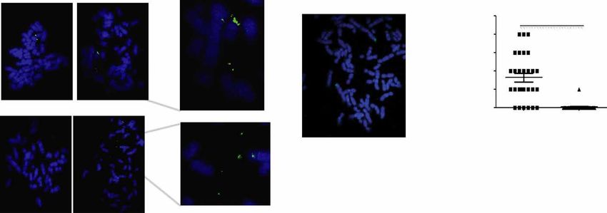
SCIENCE ADVANCES | RESEARCH ARTICLE

                           A                                                                               B                Circular DNA identification
                                 Circular DNA should get cut by Tn5, and adaptors should get
                                 ligated to newly generated free ends.
                           Linear DNA

                                                     Ligation junction
                           Circular DNA                                        Tn5 loaded with adaptors

                                                                         Amplification
                                                                                                                                   Mapped reads

                                                                                                               Break point (junction of B & E) always flanks mapped read.

                             C
                                             Read pairs mapping as contiguous or split reads.

                                                                                                                                                                                      Downloaded from http://advances.sciencemag.org/ on March 24, 2021
                                             Keep read pairs that map to three sites in the genome.

                                             Identify split reads that map uniquely to the same chromosome in the same
                                             orientation.

                                             Paired ends where one end maps as a split read and the other end maps contiguously.

                                             Paired reads where the contiguous read maps between the split reads and on
                                             opposite strands.

Fig. 1. A schematic to show that a circle could be part of an ATAC-seq library. (A) If circular DNA has open chromatin structure near or around the ligation point, then
the library preparation method will cut and attach an adaptor into a DNA fragment from eccDNA. (B) One end of paired-end read mapping on the body of a circular DNA
with read from the other end mapping on the ligation junction. (C) Detailed steps from mapping to identification of the new Circle_finder pipeline.

any length is available through our GitHub page (https://github.                                          Validation of eccDNA identified in C4-2 and OVCAR8 cells
com/pk7zuva/Circle_finder and https://github.com/pk7zuva/Circle_                                          by inverse PCR
finder/blob/master/circle_finder-pipeline-bwa-mem-samblaster.sh).                                         To confirm that the identified junctions are genuinely from eccDNA
The steps to find a circular DNA from any paired-end high-through-                                        and not from tandem genome duplications, we isolated circular
put sequencing library are detailed in Fig. 1 (B and C) and Materials                                     DNA by our previously described method that relies on column chro-
and Methods.                                                                                              matography and exonuclease digestion to remove all linear DNA
                                                                                                          and enrich eccDNA (Fig. 3A; see Materials and Methods for more
Application of ATAC-seq to identify circular DNA in OVCAR8                                                details) (5). Inverse PCR was performed with primers designed to
and C4-2B cell lines                                                                                      amplify across the junctions of eccDNAs from C4-2B and OVCAR8
We prepared ATAC-seq libraries from C4-2B prostate cancer and                                             cell lines (Fig. 3B). Eleven eccDNAs from OVCAR8 and six from
OVCAR8 ovarian cancer cell lines. The sequencing and mapping statistics                                   C4-2B were tested. Nine of the 11 targets from OVCAR8 and 2 of
are given in table S1. Less than 90% of the reads were mapped to human                                    the 6 from C4-2B gave amplicons of expected sizes (Fig. 3, B and C).
genome, and the computational pipeline identified hundreds of cir-                                        Sanger sequencing of the amplicons confirmed the junctional sequences
cular DNA. The length distribution of eccDNA is shown in Fig. 2A:                                         identified by ATAC-seq (Fig. 3D). A fraction of the primers (two in
Around 68% in C4-2B and 37% in OVCAR8 of eccDNA are 1 kb, including eccDNAs long enough                                                survive column chromatography and exonuclease digestion.
to encode gene segments or even complete genes. The eccDNA are
derived from all the chromosomes (Fig. 2B). As a positive control,                                        Validation of eccDNA by metaphase FISH in OVCAR8 cells
we identified hundreds of junctional sequences from the circular                                          An independent method for ascertaining whether a locus identified
mitochondrial genome contaminating the nuclear preparations.                                              in this study is in an extrachromosomal DNA is to carry out FISH on

Kumar et al., Sci. Adv. 2020; 6 : eaba2489     15 May 2020                                                                                                                  2 of 11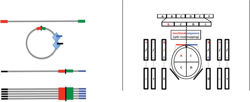
SCIENCE ADVANCES | RESEARCH ARTICLE

                                     A

                                                      C4-2

                          Fraction of circles

                                                0.8
                                                      OVCAR8

                                                0.4
                                                0.0
                                                               2                 4                        6                   8

                                                                                Circle length in bp (log10)

                                     B
                                             chr1
                                             chr2
                                             chr3
                                             chr4
                                             chr5
                                             chr6

                                                                                                                                                                         Downloaded from http://advances.sciencemag.org/ on March 24, 2021
                                             chr7
                                             chr8
                                             chr9
                                            chr10
                                                                                                                                   C42
                                            chr11
                                            chr12                                                                                  OVCAR8
                                            chr13
                                            chr14
                                            chr15
                                            chr16
                                            chr17
                                            chr18
                                            chr19
                                            chr20
                                            chr21
                                            chr22
                                            chrX
                                            chrY

                                                                    eccDNA distribution on various chromosomes
Fig. 2. eccDNA in C4-2 and OVCAR8 cell lines. (A) Length distribution of identified eccDNA in C4-2 and OVCAR8 cell lines. (B) Karyotype plot showing chromosomal
distribution of C4-2 and OVCAR8 cell lines.

metaphase spreads. We performed this analysis with two loci that                           is present in 43% of patients with GBM (17). Recent studies have provided
were predicted to be present as either an eccDNA or a gene dupli-                          further evidence that this oncogenic amplification occurs on eccDNA
cation in OVCAR8 cells but not in C4-2 cells: chr2:238136071-                              (9, 11, 18). To check whether we can detect the eccDNA in ATAC-seq data
238170279 and chr10:103457331-103528085. Both were confirmed                               generated from GBM cell lines, we turned to six ATAC-seq libraries generat-
by inverse PCR in Fig. 3 (C1 and C7). Signal was detected off the main                     ed from GBM cell lines developed from a single patient with GBM (14).
chromosomes in some of the metaphase spreads, but not others                               We ran the Circle_finder pipeline combining all the six libraries
(Fig. 4A), consistent with the hypothesis that the junctional sequences                    (GSM3318539,GSM3318540,GSM3318541, GSM3318542, GSM3318543,
identify somatically mosaic eccDNA in this cell line. For negative con-                    and GSM3318544) and found 58 eccDNAs varying in size from few
trol C4-2B (Fig. 4B), the spreads do not show an extrachromosomal                          hundred bases to few megabases. The length distribution and chromo-
DNA signal. The 71-kb eccDNA in OVCAR8 (n = 28) and C4-2B                                  somal distribution of identified eccDNAs are shown in fig. S1 (A and B).
(n = 24) (negative control) metaphase spreads were quantified for lo-                      eccDNA harboring the EGFR gene was the most abundant eccDNA.
cus chr10:103457331-103528085 and shown in the graph (Fig. 4C).                            The top five most abundant eccDNAs (or tandem gene duplications)
                                                                                           identified were chr4:118591708-119454712 (METTL14, SEC24D, SYNPO2,
Identification of eccDNA from ATAC-seq data for                                            MYOZ2, USP53, C4orf3, and FABP2), chr7:54590796-55256528 (SEC61G
GBM cell lines                                                                             and EGFR), chr7:54771165-54782815(no protein-coding genes), chr7:65038261-
EGFR was one of the first oncogenes identified in brain cancer and is                      65873269 (transcribed unprocessed pseudogenes), and chr7:65038264-
massively amplified in some patients with GBM (16). This somatic CNV                       65873256 (transcribed unprocessed pseudogenes).

Kumar et al., Sci. Adv. 2020; 6 : eaba2489            15 May 2020                                                                                              3 of 11
SCIENCE ADVANCES | RESEARCH ARTICLE
A B
C10
C3
C11
C1
C4
C5
C6
C7
C2
C8
C9
M M M M M M
OVCAR8 C4-2B
DNA markers (M): 1, 500 bp; 2, 1000bp; 3, 1600 bp; 4, 4000 bp
D
C
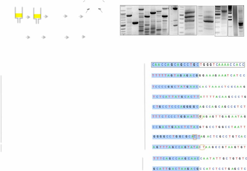
eccDNA Chr Start End Size (bp) #JT Genes present
C1 2 238136072 238170279 34208 4 KLHL30 (all exons 8/8)
ERFE (all exons 8/8)
C2 21 41641805 41645953 4147 26 None
C3 11 96046281 96049856 3580 3 MAML2 (intron 2)
C4 8 132926653 132931284 4625 21 Thyroglobulin (exon 23)
Downloaded from http://advances.sciencemag.org/ on March 24, 2021
C5 9 124475742 124513870 38128 7 NR5A1 (all exons 7/7)
USP54
MYOZ1 (all exons 6/6)
AGAP5 (all exons 8/8)
C6 10 73493290 73902830 409541 23 SEC24C (all exons 23/23)
NDST2 (all exons 13/13)
CAMK2G (all exons 21/21)
ZSWIM8 (all exons 26/26)
CALHM3 (all exons 3/3)
C7 10 103457332 103528085 70754 4 CALHM1 (exon 1 )
NEURL1 (exon 1)
C8 1 169109067 169116581 7515 3 ATP1B1 (exon 3)
C9 6 54059860 54063911 4052 14 MLIP (intron 1)
C10 7 70855001 70900090 45090 2 None
C11 16 75206880 75212516 5637 2 CTRB2 (exon 1)
Fig. 3. Experimental validation of randomly selected eccDNA identified by ATAC-seq in C4-2B and OVCAR8 cells. (A) Schematic for isolation and detection of
eccDNA. See Materials and Methods for details. (B) PCR detection of eccDNA. DNA bands marked with blue boxes were gel-purified and sequenced. (C) Description of
eccDNAs validated in (B) on the basis of analysis of ATAC-seq data from OVCAR8 and C4-2B. (D) Junctional tags obtained after sequencing of PCR products in (B). Shaded
(blue) and unshaded sequences depict 15 bases on either side of junctions. Numbers indicate chromosomal location on respective chromosomes. Note the match be-
tween numbers for each circle in (C) and (D). Some of the junction sequence identified by Sanger sequencing differ by few bases because of multiple species of eccDNA
present in the given cell lines. Oval circles represent insertion, and boxed sequences represent mismatches. *Sequence obtained from the bottom strand.
Application to GBM and LGG TCGA ATAC-seq data We see a higher number of eccDNA/duplication events in WGS
Having demonstrated above that ATAC-seq data can be repurposed compared to ATAC-seq, but 21 and 44 eccDNAs were common be-
to identify eccDNA, we turned our attention to ATAC-seq data tween ATAC-seq and WGS in TCGA-DU-5870-02A and TCGA-DU-
generated by TCGA consortium (2) with a primary focus on two 6407-02B libraries, respectively (table S2, A and B). The lack of more
LGGs for which we have whole-genome sequencing (WGS) data and overlap between the eccDNAs identified by ATAC-seq and WGS from
ATAC-seq data. In the TCGA-DU-5870-02A ATAC-seq library, we even the same tumor is most likely due to somatic mosaicism (i) be-
found 21 eccDNAs (junctional tag ≥2; 13, >1 kb and 7, >50 kb). In cause different sections are used for the two libraries and (ii) because
the TCGA-DU-5870-02A WGS library, we found 637 eccDNAs (junc- of insufficient depth of sequencing in either library.
tional tag ≥2; 361, >1 kb and 105, > 50 kb). We further compared As mentioned earlier, the Circle_finder algorithm cannot distin-
the eccDNAs identified in ATAC-seq and WGS libraries and found guish between an extrachromosomal circle and chromosomal segmen-
21 common eccDNAs (junctional tag ≥1; table S2A). tal tandem duplication without experimentally purifying the circles
In the ATAC-seq library from TCGA-DU-6407-02B, we found before library preparation, so we will refer to these loci as eccDNA/
64 eccDNAs (junctional tag ≥2; 21, >1 kb and 15, >50 kb), and in duplication. The signal for the eccDNA/duplication detected from
WGS libraries from the same tumor, we found 455 eccDNAs (junctional WGS data was strong in the two tumors and was also evident in a
tag ≥2; 307, >1 kb and 131, >50 kb). Forty-four common eccDNAs targeted copy number analysis from the WGS data (not a genome-
were identified in both libraries (junctional tag ≥1; table 2B). Many wide analysis). The median sequencing read coverage at the eccDNA/
of the common eccDNAs had a high number of junctional tags in duplication loci was 1.5-fold higher compared to equivalent up-
the WGS library, perhaps a surrogate marker of their abundance. stream or downstream regions, suggesting that at least a twofold
Kumar et al., Sci. Adv. 2020; 6 : eaba2489 15 May 2020 4 of 11SCIENCE ADVANCES | RESEARCH ARTICLE
A B C
chr10:103457331-103528085
-
5
eccDNA/metaphase
**
4
spread
3
2
C4-2
1
0
OVCAR8 C4-2
D Median = 1.5-fold
4
3
Fold (log2)
2
1
0
−1
−2
DU-6407-WGS DU-6407-ATAC DU-5870-WGS DU-5870-ATAC
E
LGG
Fraction of circles
0.8
GBM
Downloaded from http://advances.sciencemag.org/ on March 24, 2021
0.4
0.0
2 4 6 8
Circle length in bp (log10)
F chr1
chr2
chr3
chr4
chr5
chr6
chr7
chr8
chr9
chr10
GBM
chr11
chr12 LGG
chr13
chr14
chr15
chr16
chr17
chr18
chr19
chr20
chr21
chr22
chrX
chrY
Fig. 4. eccDNA in cell lines and LGG or GBM tumors. (A) Detection of eccDNA in OVCAR8 cell line by FISH: Metaphase spread of chromosome (blue) from OVCAR8 cells
were stained with the probe (green) against the eccDNA locus chr2:238136071-238170279 (top row) or chr10:103457331-103528085 (bottom row). The spreads on the
left do not have an extrachromosomal signal, while the spreads on the right have extrachromosomal signals that are better seen in the magnified insets on the extreme
right. White arrows mark the eccDNA signals. (B) For the negative control cell lines, C4-2, the spread does not have an extrachromosomal DNA signal. (C) The eccDNA
signals in OVCAR8 (n = 28) and C4-2 (n = 24) (negative control) were quantified for locus chr10:103457331-103528085 and shown in the graph. P values were calculated
using Student’s t test; **P < 0.01. (D) eccDNA/duplication loci identified in whole-genome sequencing (WGS) libraries show genomic amplification (median, 1.5-fold),
suggesting at least one allele is duplicated in all the cells. eccDNA loci identified in ATAC-seq libraries do not show genomic amplification (median close to zero), suggest-
ing that the eccDNA are apparent before a CNV can be detected at the locus. The value of copy number amplification (CNA) in the y axis is in log2. (E) Length distribution
of eccDNA identified in LGG and GBM TCGA ATAC-seq data. (F) Karyotype plot showing chromosomal distribution of eccDNA identified in LGG and GBM from TCGA
ATAC-seq data.
amplification of one allele occurred in at least 50% of the cells. Un- seq are somatically mosaic in the GBM cell lines and are detected
expectedly, the eccDNA/duplication events detected by ATAC-seq even before a CNV is apparent from WGS of a large population of
did not show corresponding amplification in WGS (Fig. 4D). tumor cells.
This result suggests that as with eccDNAs detected by rolling circle We next analyzed 10 LGG and 8 GBM ATAC-seq libraries and found
amplification, the eccDNA/duplication events identified by ATAC- a total of 2152 and 3147 eccDNA/duplication events in LGG and GBM
Kumar et al., Sci. Adv. 2020; 6 : eaba2489 15 May 2020 5 of 11SCIENCE ADVANCES | RESEARCH ARTICLE samples, respectively. The length distribution of eccDNA/duplications Cumulative analysis of all small eccDNA (microDNA) is shown in Fig. 4E. Fifty-eight percent of the loci are
SCIENCE ADVANCES | RESEARCH ARTICLE

also similar to our previous reports. Last, around 15% of the small                  the circle. The cancer driver genes (19) amplified as eccDNA/duplica-
eccDNAs reported here appear to have used flanking sequences of                      tion in individual tumor type are shown in Table 1. Gene ontology
2- to 15-base microhomology (Fig. 5D) to promote the ligation that                   analysis of all the genes carried on the eccDNA/duplication loci shows
gives rise to the circle.                                                            that pathways related to nucleosomal events are significantly enriched
                                                                                     in these loci (Fig. 5E).
Pan-cancer analysis of eccDNA in TCGA ATAC-seq data
Last, we analyzed 360 ATAC-seq libraries from 23 tumor types gen-
erated by TCGA consortium (see the Supplementary Materials) (2).                     DISCUSSION
We found a total of 18,143 eccDNAs/duplications of which 86%                         We demonstrate that the application of Circle_finder to ATAC-seq
were
SCIENCE ADVANCES | RESEARCH ARTICLE
cells showed the presence of these eccDNA loci as a signal of the length, 90%) of eccDNA were shorter than 2 kb (3, 5). Turner et al. (11)
find several cancer driver genes located in such loci (Table 1). These and deCarvalho et al. (9) have identified long circles of DNA in cancers,
results suggest that deeper sequencing of tumors by ATAC-seq with called ecDNA. We believe that the long circles identified in tumors
longer paired-end reads will identify many more clinically important by ATAC-seq, e.g., the one containing the EGFR gene, belong to this
sites involved in eccDNA/duplication in these tumors. latter class of circles. The consistent properties of the small circles
Chromosome ends are protected by telomeres. Once the chro- suggest that common mechanisms are involved in their generation
mosome suffers a catastrophic fragmentation, as in chromothripsis, in cell lines and in tumors, although it is unclear whether exactly the
some parts of the chromosome may be protected from degradation same mechanisms are involved in producing the longer circles seen in
by eccDNA formation. eccDNA can also be generated from extra ecDNAs that give rise to clinically significant gene amplifications.
Downloaded from http://advances.sciencemag.org/ on March 24, 2021
linear DNA produced by some kind of copying mechanism as a
byproduct of DNA replication or repair. Either way, our results suggest
that eccDNAs are very prevalent in cancer cell lines and tumors and MATERIALS AND METHODS
that ATAC-seq is an easy method to identify such eccDNAs. ATAC-seq library preparation
It has been reported that eccDNA longer than a few kilobases may ATAC-seq for cell lines was performed as per the Omni-ATAC-seq
have origins of replication and may get amplified independent of the protocol (21). Briefly, C4-2 and OVCAR8 cells were grown in RPMI
main chromosome. Thus, if an eccDNA harbors an oncogene, then 1640 (Corning, no.10-040) supplemented with 10% fetal bovine serum
amplification of such eccDNA in tumor cells will increase the fitness (FBS) to ~80% confluence. Fifty thousand viable cells were lysed in
of the tumor cell. In addition, since a centromere is absent in the 10 mM tris-HCl (pH 7.4), 10 mM NaCl, 3 mM MgCl2, and 0.1%
eccDNA (11), eccDNA may segregate unevenly between daughter Tween 20. Nuclear pellet was then subjected to transposition reaction
cells and result in tumor heterogeneity (9). Both these mechanisms using Nextera DNA Sample Preparation kit (Illumina, no. FC-121-
will increase the likelihood that if a particular type of therapy inhibits 1030) in the presence of 0.01% digitonin and 0.1% Tween 20 at 37°C
a gene resident on a preexisting eccDNA, then the tumor is likely to for 30 min and cleaned up with DNA Clean and Concentrator-5 Kit
acquire resistance through the selective amplification of that eccDNA. (Zymo, no. D4014). For quantitative PCR (qPCR), three to six addi-
In this context, it is particularly exciting that circle (or gene dupli- tional cycles of PCR amplification was performed using NEBNext
cation) at an important locus in a subset of the tumor cells is identi- High-Fidelity 2X PCR Master Mix (NEB, no. M0541L) and Nextera
fied by ATAC-seq even before the amplification is apparent by a CNV Index Kit (Illumina, no. 15055289). Cleaned up libraries were quan-
analysis of the whole tumor (Fig. 4D). To estimate whether ATAC-seq tified and pooled for sequencing by Novogene.
can identify loci in early, somatically mosaic states of amplification
as eccDNA/segmental duplication, we analyzed the amplicons identi- Identification of eccDNA from ATAC-seq and WGS libraries
fied by TCGA from gene array hybridization. It is apparent that an Paired-end reads were mapped to the hg38 genome build using
amplicon has to be at least around 1.5 Mb long to be detected as a bwa-mem (22) with default setting. The split reads (reads not mapped
single-copy amplification by gene microarrays (three copies per cell) in contiguous manner) were collected using the tool samblaster (23).
(see Materials and Methods and fig. S2). In contrast, we could detect If one tag of a paired read is mapped contiguously (one entry in
somatically mosaic increase in the copy number of loci far smaller mapped file) and the other tag is mapped in a split manner (two
than that length by use of Circle_finder on ATAC-seq or WGS data. entries in mapped file), then the particular read ID will have three
For example, Circle_finder on ATAC-seq data identifies sites of entries in alignment file. We therefore collected all the read pair IDs
incipient amplification of the EGFR gene in a subset of tumor cells that mapped to three unique sites in the genome from the align-
even before such amplification is detected by copy number mea- ment file. Next, we collected the split reads that mapped uniquely at
surements, predicting that even if the tumor responds to anti-EGF two positions on the same chromosome and in the same orientation.
therapy, it is likely to recur because of amplification of the EGFR gene. Returning to the list of paired-end IDs that mapped uniquely to
Many of the abundant eccDNA loci intersect with unprocessed three sites in the genome, we identified paired-end IDs where the
pseudogenes, which are known to have introns and regulatory se- contiguously mapped read is between the two split reads and on the
quences, but are crippled by stop codons in the open reading frames opposite strand. From this list, we annotate a circle if we find at least
(20). Since eccDNA evolve and pick up substitution, insertion, and one junctional sequence. For karyotype and box plot, we consid-
deletion mutations (11, 18), it is tempting to speculate that amplifi- ered at least two junctional reads.
cation of unprocessed pseudogenes on eccDNA and their evolution
may make these genes translationally competent to give an unknown Copy number amplification analysis
advantage during tumorigenesis. For each identified eccDNA (JTGE2), an upstream and downstream
Last, we note that a large fraction of eccDNA identified by ATAC- genomic interval of equivalent length was created. Next, we counted
seq have properties similar to the microDNA that we reported earlier: the number of reads that mapped to each of the three intervals
Kumar et al., Sci. Adv. 2020; 6 : eaba2489 15 May 2020 8 of 11SCIENCE ADVANCES | RESEARCH ARTICLE
(upstream, eccDNA, and downstream). Last, copy number amplifica- Fidelity DNA Polymerase (NEB) according to the manufacturer’s
tion (CNA) was computed by counting the number of mapped read instructions. Purified circular DNA (3 ng) was used as template. Unless
in eccDNA interval divided by the mean of the number of reads in otherwise stated, all the computation and plots were made of eccDNA
upstream and downstream intervals. A CNA value more than 1 would present on chr1-22, chrX, and chrY.
suggest the amplification of the locus defined by the eccDNA.
Metaphase FISH
eccDNA isolation OVCAR8 cells were cultured in RPMI medium supplemented with
eccDNA for Fig. 3 was prepared from the human cancer cell lines. 10% FBS and 1% penicillin-streptomycin in the presence of 5% CO2
The cells were grown on 150-mm plates until reaching confluence. in a humidified incubator at 37°C. Cells were treated with 2 mM
Approximately 4 × 10⁷ cells were isolated per sample. The cells were thymidine for 16 hours and released for 9 hours in a regular medium,
trypsinized and then spun down at 300g. The cells were washed followed by another block with 2 mM thymidine to arrest the cells
with phosphate-buffered saline (PBS), spun down at 300g, and at G1-S boundary. The cells were released from the double-thymidine
resuspended in 6 ml of resuspension buffer (P1) of the Qiagen HiSpeed block for 3 hours in regular medium and 9 hours in colcemid
Plasmid Midi Kit (catalog no. 12643). Lysis buffer (6 ml) (P2) was (0.1 g/ml). Mitotic cells were shaken off, washed twice with 1× PBS,
added according to the manufacturer’s instructions. The cells were and resuspended in 75 mM KCl for 30 min at 370°C. The cells
lysed for 5 min before adding the neutralization buffer (P3) and in- were centrifuged at 300g for 5 min, fixed with Carnoy’s fixative (3:1
cubated at room temperature for 10 min. The cell lysate was passed methanol:glacial acetic acid, v/v) on ice for 30 min, and washed twice
through the QIAfilter cartridge. Equilibration buffer (4 ml) (QBT) with fixative, and metaphase spreads were prepared.
Downloaded from http://advances.sciencemag.org/ on March 24, 2021
was added to the HiSpeed Tip and allowed to pass through the resin. The glass slides containing metaphase spreads were immersed in
The lysate was added to the HiSpeed Tip, and then, the HiSpeed Tip prewarmed denaturation buffer [70% formamide and 2× SSC (pH 7.0)]
was washed with 20 ml of washing buffer (QC). Then, the DNA was at 73°C for 5 min, and slides were serially dehydrated with ethanol
eluted from the HiSpeed Tip with 5 ml of elution buffer (QF), pre- (70, 85, and 100%) for 2 min each and dried at room temperature
cipitated with 3.5 ml of isopropanol, and incubated at room tem- until all the ethanol evaporated. The FISH probes (Empire Genomics)
perature for 5 min. The DNA was passed through a QIAprecipitator, were denatured with hybridization buffer at 730°C for 5 min and
which was washed with 2 ml of 70% ethanol. The excess ethanol was immediately chilled on ice for 2 min. The probe mixture was added
removed by passing air through the QIAprecipitator five times. The onto the slide, and coverslips were applied onto the slide, sealed with
DNA was precipitated from the QIAprecipitator by 1 ml of TE buf- rubber cement, and incubated at 370°C for overnight in a humidified
fer and quantified using a NanoDrop spectrophotometer. The DNA chamber. The coverslips were removed, and slides were washed with
eluted from the QIAprecipitator was then precipitated again by the prewarmed 0.4× SSC containing 0.3% NP-40 at 73°C for 2 min, fol-
addition of 2 ml of ethanol and 1 g of glycogen and centrifuged at lowed by washing with 2× SSC buffer containing 0.1% NP-40 at room
15,000g. The supernatant was removed; the DNA was air-dried for temperature for 5 min. The slides were dried at room temperature
5 min, resuspended in 20 l of TE, and warmed to 37°C for 5 min. and mounted with VECTASHIELD 4′,6-diamidino-2-phenylindole
Then, the DNA was digested with the Lucigen adenosine triphosphate medium.
(ATP)–dependent Plasmid-Safe deoxyribonuclease (catalog no. E3101K).
The 10× buffer and ATP were added according to the manufacturer’s List of TCGA IDs that were used for LGG and GBM
recommendations. In addition, ribonuclease A was added to the data analysis
solution to digest RNA concurrently with the linear DNA. The sample The TCGA IDs used for LGG and GBM data analysis are as follows:
was digested overnight and then purified using a Zymo PCR purifi- LGG: TCGA-P5-A77X-01A, TCGA-DU-5870-02A, TCGA-DB-A75K-
cation kit (catalog no. D4003). Briefly, the DNA binding buffer was 01A, TCGA-W9-A837-01A, TCGA-F6-A8O3-01A, TCGA-FG-A4MY-
added to the DNA solution in a 5:1 ratio. The mixture was then added 01A, TCGA-E1-A7YI-01A, TCGA-P5-A735-01A, and TCGA-DU-
to a Zymo-Spin column in a collection tube. The sample was centri- 6407-02B and GBM: TCGA-06-A7TK-01A, TCGA-4W-AA9S-01A,
fuged for 30 s at 10,000g. Then, the column was washed with 200 l TCGA-OX-A56R-01A, TCGA-76-6656-01A, TCGA-RR-A6KB-01A,
of DNA wash buffer and centrifuged for 30 s at 10,000g. The wash TCGA-06-A6S1-01A, TCGA-06-A5U0-01A, and TCGA-06-A7TL-01A.
step was repeated. The DNA was eluted by adding 50 l of DNA elution
buffer and centrifuged for 30 s at 10,000g. The DNA was quantified Testing the limit of detection of gene amplification
using a NanoDrop spectrophotometer to ensure digestion of the by CNV measurements
linear DNA, and then, the DNA digestion, purification, and quanti- We tested whether the detection of eccDNAs from ATAC-seq data
fication steps were repeated until the DNA concentration no longer can identify somatically mosaic amplifications before they can be
decreased after digestion. Together, this process helped ensure that detected by CNV analyses from genotyping array data. To determine
the digestion of linear DNA was complete. the sensitivity of detection of an amplicon by genotyping arrays, we
These methods are comparable to the methods previously used downloaded the previously released CNV results generated by the
where we validated the loss of linear DNA by quantifying the loss of TCGA research network. The algorithm used by the TCGA research
linear DNA with qPCR compared to circular DNA. Electron micro- network segments the chromosomes into smaller sections where an
scope imaging in those experiments showed that the linear DNA amplification or deletion is detected. Empirically, the resulting lengths
was no longer present in the samples (3, 5). of segments with CNV determined by the algorithm are the result of
(i) the true length of the amplified or deleted segment and (ii) the
Outward-directed PCRs (inverse PCR) for detection of eccDNA extent to which the segment was amplified or deleted. While we cannot
Outward-directed primers were designed across the junctional tags know whether or not a reported CNV segment should have been
identified from ATAC-seq analysis. PCR was done with Phusion High- further segmented, we hypothesized that if we analyzed ten segments
Kumar et al., Sci. Adv. 2020; 6 : eaba2489 15 May 2020 9 of 11SCIENCE ADVANCES | RESEARCH ARTICLE
with a similar level of amplification, then the smallest length among DNA elements of chromosomal origin are common in healthy human somatic tissue.
Nat. Commun. 9, 1069 (2018).
them approximates the smallest length that can be detected by the
9. A. C. deCarvalho, H. Kim, L. M. Poisson, M. E. Winn, C. Mueller, D. Cherba, J. Koeman,
algorithm at that level of amplification since the power to detect CNV S. Seth, A. Protopopov, M. Felicella, S. Zheng, A. Multani, Y. Jiang, J. Zhang, D. H. Nam,
changes increases as the extent of amplification increases. E. F. Petricoin, L. Chin, T. Mikkelsen, R. G. W. Verhaak, Discordant inheritance
The TCGA research network reported amplifications as segment of chromosomal and extrachromosomal DNA elements contributes to dynamic disease
mean > 0, where segment mean is ln(copy number/2). All segments evolution in glioblastoma. Nat. Genet. 50, 708–717 (2018).
10. M. J. Shoura, I. Gabdank, L. Hansen, J. Merker, J. Gotlib, S. D. Levene, A. Z. Fire, Intricate
with segment mean > 0.1 were ordered by reported segment mean and cell type-specific populations of endogenous circular DNA (eccDNA)
values. Bins of ten segments were analyzed for the smallest segment in Caenorhabditis elegans and Homo sapiens. G3 7, 3295–3303 (2017).
in each bin. The median segment mean value of each bin (extent of 11. K. M. Turner, V. Deshpande, D. Beyter, T. Koga, J. Rusert, C. Lee, B. Li, K. Arden, B. Ren,
amplification) is plotted against the log-transformed smallest seg- D. A. Nathanson, H. I. Kornblum, M. D. Taylor, S. Kaushal, W. K. Cavenee, R. Wechsler-Reya,
F. B. Furnari, S. R. Vandenberg, P. N. Rao, G. M. Wahl, V. Bafna, P. S. Mischel,
ment length in that bin (fig. S2).
Extrachromosomal oncogene amplification drives tumour evolution and genetic
The correlation between the segment length and segment am- heterogeneity. Nature 543, 122–125 (2017).
plification can be modeled as a linear function with the following 12. S. Wu, K. M. Turner, N. Nguyen, R. Raviram, M. Erb, J. Santini, J. Luebeck, U. Rajkumar,
formula: ln(minimum segment length) = 15.8304 − 2.7475 × median Y. Diao, B. Li, W. Zhang, N. Jameson, M. R. Corces, J. M. Granja, X. Chen, C. Coruh,
segment mean. This relatively simple model captured the relation- A. Abnousi, J. Houston, Z. Ye, R. Hu, M. Yu, H. Kim, J. A. Law, R. G. W. Verhaak,
M. Hu, F. B. Furnari, H. Y. Chang, B. Ren, V. Bafna, P. S. Mischel, Circular ecDNA
ship between the minimum segment length and the extent of am- promotes accessible chromatin and high oncogene expression. Nature 575,
plification as measured by segment mean (adjusted R2 = 0.5442; 699–703 (2019).
P < 2.2×10⁻¹⁶). 13. A. R. Morton, N. Dogan-Artun, Z. J. Faber, G. MacLeod, C. F. Bartels, M. S. Piazza,
Downloaded from http://advances.sciencemag.org/ on March 24, 2021
If one extra copy of an amplicon is present in every single cell of K. C. Allan, S. C. Mack, X. Wang, R. C. Gimple, Q. Wu, B. P. Rubin, S. Shetty, S. Angers,
P. B. Dirks, R. C. Sallari, M. Lupien, J. N. Rich, P. C. Scacheri, Functional enhancers
the sample, then the segment mean value is 0.585 [log2(3/2)]. From
shape extrachromosomal oncogene amplifications. Cell 179, 1330–1341.e13
the linear model in fig. S2, the minimum segment length detectable (2019).
at this segment mean value is 1.5 Mb. Therefore, most of the somat- 14. Q. Xie, T. P. Wu, R. C. Gimple, Z. Li, B. C. Prager, Q. Wu, Y. Yu, P. Wang, Y. Wang,
ically mosaic amplifications driven by most of the eccDNAs in our D. U. Gorkin, C. Zhang, A. V. Dowiak, K. Lin, C. Zeng, Y. Sui, L. J. Y. Kim, T. E. Miller, L. Jiang,
study (median length, ~2 kb) will not be captured using genotyping C. H. Lee, Z. Huang, X. Fang, K. Zhai, S. C. Mack, M. Sander, S. Bao, A. E. Kerstetter-Fogle,
A. E. Sloan, A. Z. Xiao, J. N. Rich, N6-methyladenine DNA modification in glioblastoma. Cell
arrays. 175, 1228–1243.e20 (2018).
The number of ATAC-seq libraries analyzed in this study for various 15. C. A. Maher, R. K. Wilson, Chromothripsis and human disease: Piecing together
tumor type is as follows: ACC, 8; BLCA, 10; BRCA, 70; CESC, 3; the shattering process. Cell 148, 29–32 (2012).
CHOL, 1; COAD, 38; ESCA, 17; GBM, 8; HNSC, 9; KIRC, 15; KIRP, 16. T. A. Libermann, H. R. Nusbaum, N. Razon, R. Kris, I. Lax, H. Soreq, N. Whittle,
M. D. Waterfield, A. Ullrich, J. Schlessinger, Amplification, enhanced expression
29; LGG, 10; LIHC, 15; LUAD, 21; LUSC, 12; MESO, 5; PCPG, 9;
and possible rearrangement of EGF receptor gene in primary human brain tumours
PRAD, 21; SKCM, 9; STAD, 19; TGCT, 8; THCA, 12; and UCEC, 10. of glial origin. Nature 313, 144–147 (1985).
17. C. L. Maire, K. L. Ligon, Molecular pathologic diagnosis of epidermal growth factor
receptor. Neuro-Oncology 16 (Suppl 8), viii1–6 (2014).
SUPPLEMENTARY MATERIALS 18. K. Xu, L. Ding, T. C. Chang, Y. Shao, J. Chiang, H. Mulder, S. Wang, T. I. Shaw, J. Wen,
Supplementary material for this article is available at http://advances.sciencemag.org/cgi/
L. Hover, C. McLeod, Y. D. Wang, J. Easton, M. Rusch, J. Dalton, J. R. Downing, D. W. Ellison,
content/full/6/20/eaba2489/DC1
J. Zhang, S. J. Baker, G. Wu, Structure and evolution of double minutes in diagnosis
View/request a protocol for this paper from Bio-protocol. and relapse brain tumors. Acta Neuropathol. 137, 123–137 (2019).
19. M. H. Bailey, C. Tokheim, E. Porta-Pardo, S. Sengupta, D. Bertrand, A. Weerasinghe,
A. Colaprico, M. C. Wendl, J. Kim, B. Reardon, P. Kwok-Shing Ng, K. J. Jeong, S. Cao,
REFERENCES AND NOTES Z. Wang, J. Gao, Q. Gao, F. Wang, E. M. Liu, L. Mularoni, C. Rubio-Perez, N. Nagarajan,
1. J. D. Buenrostro, B. Wu, H. Y. Chang, W. J. Greenleaf, ATAC-seq: A method for assaying I. Cortés-Ciriano, D. C. Zhou, W. W. Liang, J. M. Hess, V. D. Yellapantula, D. Tamborero,
chromatin accessibility genome-wide. Curr. Protoc. Mol. Biol. 109, 21.29.1–21.29.9 A. Gonzalez-Perez, C. Suphavilai, J. Y. Ko, E. Khurana, P. J. Park, E. M. Van Allen, H. Liang;
(2015). Group M. C. Working; Network Cancer Genome Atlas Research, M. S. Lawrence, A. Godzik,
2. M. R. Corces, J. M. Granja, S. Shams, B. H. Louie, J. A. Seoane, W. Zhou, T. C. Silva, N. Lopez-Bigas, J. Stuart, D. Wheeler, G. Getz, K. Chen, A. J. Lazar, G. B. Mills, R. Karchin,
C. Groeneveld, C. K. Wong, S. W. Cho, A. T. Satpathy, M. R. Mumbach, K. A. Hoadley, L. Ding, Comprehensive characterization of cancer driver genes and mutations. Cell 174,
A. G. Robertson, N. C. Sheffield, I. Felau, M. A. A. Castro, B. P. Berman, L. M. Staudt, 1034–1035 (2018).
J. C. Zenklusen, P. W. Laird, C. Curtis; Cancer Genome Atlas Analysis Network, 20. Y. Tutar, Pseudogenes. Comp. Funct. Genomics 2012, 424526 (2012).
W. J. Greenleaf, H. Y. Chang, The chromatin accessibility landscape of primary human 21. M. R. Corces, A. E. Trevino, E. G. Hamilton, P. G. Greenside, N. A. Sinnott-Armstrong,
cancers. Science 362, eaav1898 (2018). S. Vesuna, A. T. Satpathy, A. J. Rubin, K. S. Montine, B. Wu, A. Kathiria, S. W. Cho,
3. L. W. Dillon, P. Kumar, Y. Shibata, Y. H. Wang, S. Willcox, J. D. Griffith, Y. Pommier, M. R. Mumbach, A. C. Carter, M. Kasowski, L. A. Orloff, V. I. Risca, A. Kundaje, P. A. Khavari,
S. Takeda, A. Dutta, Production of extrachromosomal microDNAs is linked to mismatch T. J. Montine, W. J. Greenleaf, H. Y. Chang, An improved ATAC-seq protocol reduces
repair pathways and transcriptional activity. Cell Rep. 11, 1749–1759 (2015). background and enables interrogation of frozen tissues. Nat. Methods 14, 959–962
4. P. Kumar, L. W. Dillon, Y. Shibata, A. A. Jazaeri, D. R. Jones, A. Dutta, Normal and cancerous (2017).
tissues release extrachromosomal circular DNA (eccDNA) into the circulation. Mol. Cancer Res. 22. H. Li, R. Durbin, Fast and accurate short read alignment with Burrows-Wheeler transform.
15, 1197–1205 (2017). Bioinformatics 25, 1754–1760 (2009).
5. Y. Shibata, P. Kumar, R. Layer, S. Willcox, J. R. Gagan, J. D. Griffith, A. Dutta, 23. G. G. Faust, I. M. Hall, SAMBLASTER: Fast duplicate marking and structural variant read
Extrachromosomal microDNAs and chromosomal microdeletions in normal tissues. extraction. Bioinformatics 30, 2503–2505 (2014).
Science 336, 82–86 (2012).
6. H. D. Moller, C. E. Larsen, L. Parsons, A. J. Hansen, B. Regenberg, T. Mourier, Formation Acknowledgments: We would like to thank the Dutta laboratory members for thoughtful
of extrachromosomal circular DNA from long terminal repeats of retrotransposons discussion on this paper. We thank the High Performance Computing team at the University
in saccharomyces cerevisiae. G3 6, 453–462 (2015). of Virginia for providing all the support with computation. We thank the dbGAP and the
7. H. D. Moller, L. Parsons, T. S. Jorgensen, D. Botstein, B. Regenberg, Extrachromosomal TCGA data management teams for data access. We would also like to thank the patients for
circular DNA is common in yeast. Proc. Natl. Acad. Sci. U.S.A. 112, E3114–E3122 their participation in TCGA and R. Corces from H. Y. Chang’s group (Howard Hughes Medical
(2015). Institute, Stanford University, Stanford) for alerting us about raw sequencing data
8. H. D. Møller, M. Mohiyuddin, I. Prada-Luengo, M. R. Sailani, J. F. Halling, P. Plomgaard, availability through TCGA. We would also like to thank the SOM core facility. Funding: This
L. Maretty, A. J. Hansen, M. P. Snyder, H. Pilegaard, H. Y. K. Lam, B. Regenberg, Circular work was supported by grants from NIH R01 CA60499 and the Owens Foundation to A.D., a
Kumar et al., Sci. Adv. 2020; 6 : eaba2489 15 May 2020 10 of 11SCIENCE ADVANCES | RESEARCH ARTICLE
fellowship to S.K. from the UVA Cancer Center, and T32 GM007267 grant to A.C. from the ATAC-seq data generated for OVCAR8 and C4-2B cell lines are deposited to GEO with
NIH. Author contribution: P.K. and A.D. conceived and designed the study and wrote the accession number GSE145409.
paper. P.K. collected the data and did most of the analysis. S.K. did the inverse PCR
experiment. T.P. isolated circular DNA. S.S. and E.S. performed the FISH experiment. Z.S. Submitted 17 November 2019
prepared the OVCAR8 and C4-2 ATAC-seq library. A.C. performed the cancer driver gene Accepted 6 March 2020
analysis. Y.S. helped P.K. in testing and improving the Circle_finder algorithm. Competing Published 15 May 2020
interests: P.K. and A.D. are inventors on a U.S. provisional patent application related to 10.1126/sciadv.aba2489
this work filed by University of Virginia (no. 62/832,443, filed 11 April 2019). The authors
declare no other competing interests. Data and materials availability: All data needed to Citation: P. Kumar, S. Kiran, S. Saha, Z. Su, T. Paulsen, A. Chatrath, Y. Shibata, E. Shibata, A. Dutta,
evaluate the conclusions in the paper are present in the paper and/or the Supplementary ATAC-seq identifies thousands of extrachromosomal circular DNA in cancer and cell lines. Sci. Adv.
Materials. Additional data related to this paper may be requested from the authors. The 6, eaba2489 (2020).
Downloaded from http://advances.sciencemag.org/ on March 24, 2021
Kumar et al., Sci. Adv. 2020; 6 : eaba2489 15 May 2020 11 of 11ATAC-seq identifies thousands of extrachromosomal circular DNA in cancer and cell lines
Pankaj Kumar, Shashi Kiran, Shekhar Saha, Zhangli Su, Teressa Paulsen, Ajay Chatrath, Yoshiyuki Shibata, Etsuko Shibata
and Anindya Dutta
Sci Adv 6 (20), eaba2489.
DOI: 10.1126/sciadv.aba2489
Downloaded from http://advances.sciencemag.org/ on March 24, 2021
ARTICLE TOOLS http://advances.sciencemag.org/content/6/20/eaba2489
SUPPLEMENTARY http://advances.sciencemag.org/content/suppl/2020/05/11/6.20.eaba2489.DC1
MATERIALS
REFERENCES This article cites 23 articles, 5 of which you can access for free
http://advances.sciencemag.org/content/6/20/eaba2489#BIBL
PERMISSIONS http://www.sciencemag.org/help/reprints-and-permissions
Use of this article is subject to the Terms of Service
Science Advances (ISSN 2375-2548) is published by the American Association for the Advancement of Science, 1200 New
York Avenue NW, Washington, DC 20005. The title Science Advances is a registered trademark of AAAS.
Copyright © 2020 The Authors, some rights reserved; exclusive licensee American Association for the Advancement of
Science. No claim to original U.S. Government Works. Distributed under a Creative Commons Attribution NonCommercial
License 4.0 (CC BY-NC).You can also read


